histomorphological and morphometrical changes of placental terminal villi of normotensive and...
TRANSCRIPT

5
This article can be downloaded from http://www.ijmspr.com/currentissue.php
Int. J. of Med. Sc. & Pharm. Res., 2015 Madhu L et al., 2015
HISTOMORPHOLOGICAL AND MORPHOMETRICAL
CHANGES OF PLACENTAL TERMINAL VILLI OF
NORMOTENSIVE AND HYPERTENSIVE MOTHERS
Madhu L1*, Karthavya S L1 and Lepakshi B G1
Research Paper
Background & Objectives: Placental examination has clinical value in preeclampsia (PE) and
IUGR. The luminal diameter of the uterine spiral arterioles in women with PE is narrowed leading
to placental ischemia thus causing fetal hypoxia and pathological changes in placenta. The
main objective of the present study is to compare morphological and histomorphometrical
changes in placentas of preeclamptic and normotensive mothers. Methods: 50 placentas from
both vaginal and LSCS delivery were collected at Dept. of OBG in a tertiary care center, half of
them from normotensive pregnancies and the rest from preeclamptic mothers. An inclusion
criterion for control was normal blood pressure and no proteinuria. Exclusion criteria for both
control and study group was DM, obesity, severe anemia or any systemic disorders. Placental
thickness, weight, diameter and surface area were recorded. Histopathological sections stained
with H&E were observed for surface area and diameter of TV. Results: The mean placental
weight in PE was 430 g. The placental diameter was decreased in PE (16 cm) compared to
controls (19 cm). Neonatal weight followed the same trend. Histologically, the changes in the TV
and blood vessels was significant; there was decrease in the diameter of villi in PE cases(0.01
μm) when compared to controls (0.05 μm). There was significant decrease in the diameter of
blood vessels in PE (0.0049 μm) than in controls (0.01 mm). Conclusion: This study has revealed
that there are significant changes in the placenta in cases of PE both morphologically and
histologically. There is also a need for further studies to prove the molecular and genetic factors
involved in preeclampsia.
Keywords: Histology, Hypertension, Morphometry, Placenta, Preeclampsia, Terminal villi
*Corresponding Author: Madhu L � [email protected]
INTRODUCTION
The placenta is an ephemeral organ interposed
between the mother and fetus and is vital for the
survival of the fetus (Mardi, 2003). Fetal growth
ISSN 2394-8973 www.ijmspr.com
Vol. 1, No. 3, August 2015
© 2015 IJMSPR. All Rights Reserved
Int. J. of Med. Sc. & Pharm. Res., 2015
1 Department of Obstetrics and Gynecology, Shimoga Institute of Medical Sciences, Shimoga-577201, Karnataka, India.
depends on the proper development and function
of the placenta, which serves to maintain
maternofetal interference for the exchange of
blood gases, nutrients and waste (Vogel, 2005).

6
This article can be downloaded from http://www.ijmspr.com/currentissue.php
Int. J. of Med. Sc. & Pharm. Res., 2015 Madhu L et al., 2015
The placenta, at term, is almost a circular disc
with a diameter of 15-20 cm and thickness of
about 3 cm at centre. It thins off towards edges.
The architecture of the placenta is altered in many
maternal diseases such as diabetes mellitus,
hypertension, preeclampsia (PE), and eclampsia.
Although the placenta is a vital organ, its systemic
study has been neglected; however, in recent
times, it has evoked great interest, and much work
is being conducted to understand the unique
biological status of this complex organ. Placental
examination has clinical value in cases of PE and
Intrauterine Growth Retardation (IUGR), both of
which are associated with high perinatal morbidity
and mortality accompanied with gross
pathological changes in the placenta.
PE is unique pregnancy-related disease that
affects 5-7% pregnancies worldwide (Bdolah,
2005). It is associated with hypertension and
proteinuria. The primary cause of PE is the
widespread apoptosis of cytotrophoblast cells but
the disease has a multifocal nature of
pathogenesis. The mean luminal diameter of
uterine spiral arterioles in women with PE is less
than one-third of the diameter of similar vessels
from uncomplicated pregnancies (Catwright,
2010). Consequently, uteroplacental perfusion
reduces, and the placenta becomes ischemic as
gestation progresses. This causes fetal hypoxia
as well as morphological and histological
changes in the placenta, leading to PE, which
contributes to premature delivery and fetal death.
It is important to know the pathologic changes in
PE to understand the prognosis of disease. This
study aims at finding the histological and
morphological changes in the placenta
complicated by PE.
LITERATURE REVIEW
Hypertension is one of the common medical
complications of pregnancy and contributes
significantly to maternal and perinatal morbidity
and mortality. It is a sign of an underlying pathology
which may be preexisting or appears for the first
time during pregnancy. The identification of this
clinical entity and effective management play a
significant role in the outcome of pregnancy both
for mother and the baby. Hypertension in
pregnancy includes gestational hypertension,
preeclampsia and eclampsia. This study
emphasizes on pregnancies complicated by
preeclampsia.
Preeclampsia is a multisystem disorder of
varied etiology characterized by development of
hypertension to the extent of 140/90 mm Hg or
more with proteinuria after the 20th week in a
previously normotensive and non proteinuric
woman. The preeclamptic features may appear
even before 20th week as in case of hydatidiform
mole and acute polyhydramnios.
AMERICAN CONGRESS OF
OBSTETRICS AND
GYNAECOLOGY (ACOG)
CRITERIA FOR DIAGNOSIS
OF SEVERE PRECLAMPSIA
(ACOG, 2002)
Preeclampsia is considered severe if one or more
of the following criteria are present,
• Blood pressure of 160mm of Hg, systolic or
higher or 110 mm of Hg diastolic or higher, on
two occasions at least six hours apart while
the patient is on bed rest
• Oliguria of less than 500 mL in 24 h
• Cerebral or visual disturbance
7
This article can be downloaded from http://www.ijmspr.com/currentissue.php
Int. J. of Med. Sc. & Pharm. Res., 2015 Madhu L et al., 2015
• Pulmonary edema or cyanosis
• Epigastric or right upper quadrant pain
• Impaired liver function
• Thrombocytopenia
• Fetal growth restriction.
Numerous invitro and animal models have
been used to study aspects of preeclampsia, the
most common being models of placental oxygen
dysregulation, abnormal trophoblast invasion,
inappropriate maternal vascular damage, and
anomalous maternal-fetal immune interactions.
Investigations into pathophysiology and treatment
of preeclampsia continue to move the field
forward, albeit at a frustratingly slow pace. There
remains a pressing need for novel approaches,
new disease models and innovative investigators
to effectively tackle this complex and devastating
disorder.
The multiple criteria for the diagnosis of severe
preeclampsia illustrate the multifocal nature of the
disease. There are multiple theories and little
argument about the cause of preeclampsia. The
disease is characterized by disruption of vascular
remodeling, a systemic antiangiogenic response,
oxygen dysregulation, immune changes.
In normal pregnancy cytotrophoblast cells
originating in anchoring villi of fetal portion of
placenta attach to and invade the maternal
endometrium. A subset of these extravillous
trophoblast cells acquire endothelial
characteristics and invade maternal spiral
arteries and plug the arteries maintaining a
hypoxic uterine environment in turn replacing
some of the endothelial cells of vessel wall and
leads to alteration of vessel compliance causing
filling up intervillous space of placenta (Kaufmann,
2003).
Hunkapiller and Fisher study also supports this
theory of trophoblast invasion where invitro
models of trophoblast invasion, including cultured
placental explants, primary trophoblast cells,
human embryonic stem cells and human
choriocarcinoma cells.
These cells are cultured in typical conditions
using invasion chambers and thus supporting the
theory of invasion. These trophoblast invasion
leads to histological changes in terminal villi and
blood vessels (Hunka, 2008).
The placental samples examined at term as
well as from Doppler ultrasound study of
placental perfusion show that the remodeling of
spiral arteries is incomplete in patients with
preeclampsia (Khong, 1986). Thus poor
trophoblast invasion is a vital event in disease
progression although it has not been determined
whether it is the cause of preeclampsia or a result
of another underlying problem. It is hypothesized
that without proper remodeling the placenta is
deprived of oxygen and that the resulting hypoxia
triggers the symptom of preeclampsia and the
histomorphological and morphometrical changes
in the placenta (Roberts, 2009).
A study was conducted by Wu et al. (2010)
showing strong evidence that changes in
circulating levels of regulators of angiogenesis
cause many of the clinically significant symptoms
of preeclampsia. Members of the vascular
endothelial growth factor family, VEGF-A, VEGF-
B and Placental Growth Factor (PLGF) act
through membrane receptors to regulate
angiogenesis. Binding of VEGF-A to VEGFR-2 or
PLGF to VEGFR-1 promotes angiogenesis
where as soluble form of FLT-1 (sFLT-1: Fms-
like tyrosine kinase-1) inhibits angiogenesis.
8
This article can be downloaded from http://www.ijmspr.com/currentissue.php
Int. J. of Med. Sc. & Pharm. Res., 2015 Madhu L et al., 2015
Clinical studies conducted by Levine et al.
(2004) demonstrated an increase in circulatinglevels of sFLT-1 and a significant increase in theratio of sFLT-1 to PLGF in both early and late onsetpreeclampsia.
Similarly studies done by Venkatesha et al.(2006) demonstrated increased placentalexpression and circulating concentration ofsoluble endoglin, an inhibitor of capillary formationare associated with preeclampsia and arepositively correlated with disease severity.
There are other studies showing models ofoxygen dysregulation in the pathophysiology ofpreeclampsia. Studies conducted by Abitbol(1982) and Makris et al. (2007) used ReducedUterine Perfusion Pressure (RUPP) model todemonstrate hypoxic stimulus for the changes inpreeclampsia. These models used rat forexhibiting RUPP model. A related hypothesisstates that hypoxia is not only a result ofinsufficient trophoblast invasion but a cause forit. These models have been used to examine theeffect of hypoxia and hypoxia reperfusion injuryon extravillous trophoblast differentiation andinvasion (Hunka, 2008).
The studies done on a transcription factor i.e.hypoxia- inducible factor 1α (HIF-1α) assess therole of hypoxia in preeclampsia. These includeHIF-1α over expressing transgenic mice (Tal,2010), knockdown of HIF-1α inhibitor CITED 2(Withington, 2006) knockout of the COMT enzymewhich produces HIF-1α inhibitor 2-methoxyestradiol (Kanasaki, 2008). Thesemodels show incomplete remodeling of maternalspiral arteries, fetal and placental growthrestriction, hypertension and proteinuria. Thesehypoxic events lead to changes in the numericaldensity of blood vessels due to chronic stimulation
for angiogenesis.
There are several lines of evidence supporting
a role for maternal immune response in the
development of preeclampsia. First, several
immune associated risk factors increase the
probability that a woman will develop
preeclampsia including pre existing auto immune
disease (Duckitt, 2005; Trogstad, 2011). Second,
primiparity, a change of partner and a short initial
coitus-to-conception interval are all risk factors
for preeclampsia suggesting that the response
to paternal antigens play a role (Trogstad, 2011,
Basso, 2001). Finally, concentrations of
inflammatory cytokines are significantly
increased and placental production of anti-
inflammatory cytokines IL-10 is decreased in
woman with preeclampsia (Kumferminc, 1994;
Vince, 1995; Makris, 2006).
A study conducted by Shankar et al. revealed
that there in increased presence of
vasculosyncytial knots in cases of preeclampsia
(Sankar, 2012). There are other studies which
show there is presence of histological changes
in the placenta of mothers whose pregnancies
are complicated by preeclampsia. The study done
by Devi Shankar et al. (2013) concludes there
are numerous pathological findings and terminal
villi changes in the placentas of PE.
Thus the above studies show conclusively that
preeclampsia has a multi factorial nature of
pathophysiology leading to both histological and
morphological changes in placenta mainly due
to ineffective trophoblast invasion, hypoxia and
immunological changes.
AIMS AND OBJECTIVES
The main objective of the present study is to
compare morphological and histomorphometrical
changes in placentas from mothers with
hypertension (mainly preeclamptic) with those in

9
This article can be downloaded from http://www.ijmspr.com/currentissue.php
Int. J. of Med. Sc. & Pharm. Res., 2015 Madhu L et al., 2015
placentas from normotensive mothers, in relation
to the surface area and diameter of the terminal
villi (TV) and blood vessels.
The study also throws a light upon other
pathological changes in the placenta of
preeclamptic mothers.
MATERIALS AND METHODS
The study was conducted in a tertiary care center
in the months of June and August 2014. 50
placentas were collected at the Department of
Obstetrics and Gynecology. Informed consent
was obtained from each mother (consent form
enclosed) and a clearance from the Institutional
ethical Committee was obtained in an appropriate
manner.
Half the number of placentas was collected
from normotensive pregnant patients (Controls)
and the remaining was obtained from patients
whose pregnancy was complicated by
preeclampsia. Preeclampsia was defined as a
blood pressure more than 140/90 mm of hg with
protein values more than 300 mg in the 24 h urine
or a protein concentration of 1 g/L on two
occasions at least 6 h apart.
Placentas were collected from both vaginal
delivery as well as LSCS (Lower Segment Caesarian
Section) irrespective of the gestational age.
Inclusion criteria for controls were normal blood
pressure and no proteinuria. Exclusion criteria for
both the control and PE groups was diabetes
mellitus, obesity, severe anemia (Hemoglobin <6
g %) and any other systemic or endocrine
disorders.
Immediately after the delivery, the umbilical
cord was clamped close to the placental insertion
point; membranes were trimmed. The placental
weight, thickness (thickness at center) diameter
and surface area were recorded. For histological
studies, full-depth tissue samples were placed
in 10% formol-saline solution for 24-48 h and were
subsequently embedded in paraffin. The 5 μm
thick sections were stained with hematoxylin and
eosin. Terminal villi were observed micro-
scopically with a 40x objective. TV was those
which had the smallest villi containing capillary
loops without any histological artifacts. In addition
to TV other pathological findings of placenta were
noted.
The following parameters in histomor-
phometrical analysis were estimated in TV of the
control and PE groups.
• Diameter of TV and blood vessels were
measured using ocular reticule micrometers.
• The surface area of TV in the control and the
PE groups were measured.
Table 1: Macroscopic Findings of Maternal, Neonatal and Placental Parametersin Control and Preeclampsia [Values are presented as mean]
Parameter Control(n=25) PE(n=25)
Maternal age (y) 25 28
Gestational age (w) 37 33
Neonatal weight (g) 2,650 2,100
Placental weight (g) 510 430
Placental thickness (cm) 4.15 2.64
Placental diameter (cm) 19 16
Placental surface area (cm2) 283.38 200.96
10
This article can be downloaded from http://www.ijmspr.com/currentissue.php
Int. J. of Med. Sc. & Pharm. Res., 2015 Madhu L et al., 2015
The observations were analyzed using SPSS
software and the results are tabulated.
OBSERVATIONS AND
RESULTS
The placentas collected from both the control and
PE groups showed the following results.
The mean maternal age was 25 years in
control group and 28 years in the PE group. The
mean gestational age in control group was 37
weeks and in PE it was 33 weeks. Infants born to
the PE group had statistically and significantly
lower gestational age and birth weight than those
in the control group. The gross morphological
findings of placenta are shown in Table 1.
On gross placental morphometrical study, it
revealed that the placental parameters like
placental weight, thickness, diameter and surface
area were significantly reduced in PE group
compared to the control group.
HISTOLOGICAL FINDINGS
The stem villi of the PE placentas showed
numerous arteriosclerotic blood vessels with
endothelial degeneration presenting progressive
fibrosis and subsequent lumen obliteration. These
villi had smooth muscle hypertrophy with greatly
multiplied numbers of muscle layers in the tunica
intima. Stem villi thrombosis seen as
atheromatous plaques was observed in the PE
placentas.
Perivillous fibrin and intervillous fibrin
deposition, which also extended to the intervillous
bridges, were observed in the PE cases. The
number and structure of TV specifically varied in
PE. The total numbers of TV were significantly
lesser, indicating distal villous hypoplasia. The
paucity of TV was probably because the capillary,
which initiates villous sprouting in the placental
core, had not been established.
Numerous avascular TV surrounded the
arteriosclerotic stem villi, possibly reflecting failure
of vascular organization (villitis). TV
syncytiotrophoblasts invariably developed
clusters and sprouts to form syncytial knots; the
PE group had significantly more knots than the
control group.
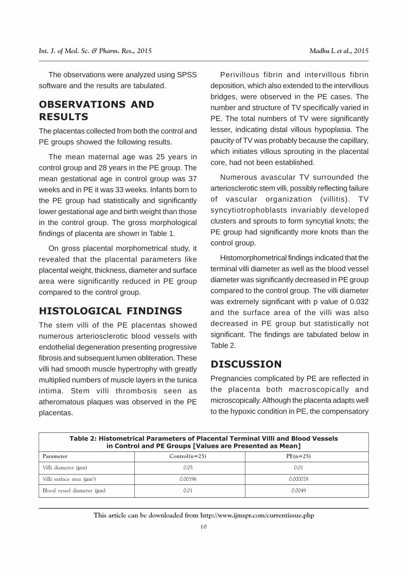
Histomorphometrical findings indicated that the
terminal villi diameter as well as the blood vessel
diameter was significantly decreased in PE group
compared to the control group. The villi diameter
was extremely significant with p value of 0.032
and the surface area of the villi was also
decreased in PE group but statistically not
significant. The findings are tabulated below in
Table 2.
DISCUSSION
Pregnancies complicated by PE are reflected in
the placenta both macroscopically and
microscopically. Although the placenta adapts well
to the hypoxic condition in PE, the compensatory
Table 2: Histometrical Parameters of Placental Terminal Villi and Blood Vesselsin Control and PE Groups [Values are Presented as Mean]
Parameter Control(n=25) PE(n=25)
Villi diameter (μm) 0.05 0.01
Villi surface area (μm2) 0.00196 0.000078
Blood vessel diameter (μm) 0.01 0.0049
11
This article can be downloaded from http://www.ijmspr.com/currentissue.php
Int. J. of Med. Sc. & Pharm. Res., 2015 Madhu L et al., 2015
changes that occur are insufficient. These
compensatory changes cause maldevelopment
and inadequate placental mass, causing placental
dysfunction that leads to oxidative stress and
chronic fetal hypoxemia (Myatt, 2002).
In the present study comparing preeclamptic
placentas to control placentas, the mean
placental weight, thickness, diameter, and surface
area were decreased and were found to be more
significant. The macroscopic changes found in
the study are analogous to the findings of other
PE cases in the literature. The gross reduction
of the PE placenta impedes normal placentation
and pathologically results in massive microscopic
changes in the placenta.
In contrast to the histomorphometrical findings
in the normal placentas, changes in the PE
placentas cause functional disturbance, which is
the result of the oxidative stress/hypoxic
conditions due to PE. The surface areas, villi
diameters, and blood vessel diameters of the
placentas in PE cases were lesser than those of
controls in the present study.
In normal placentation, during the first and early
second trimesters, the villous growth and
arborization are regulated, which are necessary
for fetal well being.
The cytotrophoblast cells invade into the
uterine spiral arteries and transform them from
small-caliber resistance vessels into high-caliber
capacitance vessels capable of providing
enhanced placental perfusion adequate for the
growing fetus. For this transformation, a certain
amount of hypoxia is needed to stimulate
placental blood vessel formation. Until
approximately 10 weeks of gestation, the embryo
exists in a hypoxic environment with nutrients
provided by the endometrial glands. However,
prolonged durations of hypoxia or oxidative stress
leads to poor placental perfusion, which is the
underlying pathogenesis of PE.
In PE, invasion of the uterine spiral arteries is
limited to the proximal decidua, and 30-50% of
the spiral arteries of the placental bed escape
endovascular trophoblast remodeling.
Persistence of muscular and elastic tissues of
the media of spiral arteries, fail to dilate and
remain responsive to vasomotor influences that
lead to high resistance low flow choriodecidual
circulation.
The reduction in the vascular dimensions is
constantly accompanied by a significant impact
on the lumen of the arteriole with changes in its
muscular wall. Thus, the average diameter of the
blood vessels, which normally expands to 4 times
their original size, is greatly decreased in PE.
In the present study there is significant
decrease in the diameter of blood vessels. This
decrease in lumen ultimately fails to replicate and
establish a network into TV. This results in the
complete absence of capillaries in the TV in most
vicinities of the placenta, leading to the formation
of avascular villitis. Consequently, the resultant
decreased perfusion causes oxidative stress.
There are studies which show that due to
chronicity of hypoxic stimulation, the density of
blood vessels and villi are increased and it is
believed that the increased villi density and
decreased villi diameter in PE cases may have
occurred because of the continuous sprouting of
the intermediate villi into TV in order to
compensate for the placental maldevelopment
and dysfunction.
Placental architecture is altered in many
maternal diseases such as PE and eclampsia.
12
This article can be downloaded from http://www.ijmspr.com/currentissue.php
Int. J. of Med. Sc. & Pharm. Res., 2015 Madhu L et al., 2015
Placental weight in women with PE is directly
proportional to neonatal birth weight. The present
study showed that both morphological and
histological changes in the PE placenta such as
decreased weight, thickness, and diameter of villi
and vessels which are said to be the
pathogenesis involved in maternal and fetal
morbidity and mortality in women with PE.
CONCLUSION
This study has revealed that there are both
histological and morphometrical changes in the
placenta of mothers whose pregnancy is
complicated by preeclampsia. There is significant
decrease in the placental morphological features
like weight, thickness, surface area etc.
compared to the placenta of normal pregnancies.
The histological findings are also significant in
cases of preeclampsia. There is decrease in the
diameter of villi and blood vessels. Placental
changes have a direct effect on outcome of
pregnancy like intrauterine growth retardation.
Therefore the changes in PE have direct adverse
affect on neonatal weight.
It can be concluded that preeclampsia has a
multifocal nature of origin and has significant
changes in placenta. This study brings out both
the morphological and histological changes
present in cases of PE by comparing with the
normal placentas.
REFERENCES
1. Abitbol M M (1982), “Simplified technique to
produce toxemia in the rat: considerations
on cause of toxemia”, Clin. Exp. Hypertens.,
Vol. B1, pp. 93-103 [PubMed:7184666]
2. ACOG (2002), “American College of
Obstetricians and Gynecologists practice
bulletin. Diagnosis and management of
preeclampsia and eclampsia”, Int. J.
Gynaecol. Obstet., Vol. 77, pp. 67-75
[PubMed: 12094777].
3. Basso O, Christensen K and Olsen J
(2001), “Higher risk of preeclampsia after
change of partner. An effect of longer
interpregnancy intervals?”, Epidemiology,
Vol. 12, pp. 624-629 [PubMed:11679788]
4. Bdolah Y, Karumanchi S A and Sachs B P
(2005), “Recent advances in understanding
of preeclampsia”, Croat Med J., Vol. 46, pp.
728-36.
5. Cartwright J E, Fraser R, Leslie K, Wallace
A E, James J L (2010), “Remodeling at the
maternal- fetal interface: relevance to
human pregnancy disorders”,
Reproduction, Vol. 140, pp. 803-13.
6. Duckitt K and Harrington D (2005), “Risk
factors for preeclampsia at antenatal
booking: Systematic review of controlled
studies”, BMJ, Vol. 330, pp. 565.
[PMCID:PMC554027] [PubMed:15743856]
7. Kaufmann P, Black S and Huppertz B
(2003), “Endovascular trophoblast invasion:
implications for the pathogenesis of
intrauterine growth retardation and
preeclampsia”, Biol. Reprod., Vol. 69, pp.
1-7 [PubMed: 12620937].
8. Hunka Piller N M and Fisher S J (2008),
Chapter 12, “Placental remodeling of the
uterine vasculature”, Methods Enzymol.,
Vol. 445, pp. 281-302 [PMCID:
PMC2857511] [PubMed : 19022064]
9. Kanasaki K, Palmsten K, Sugimoto H,
Ahmad S, Hamano Y, Xie L, Parry S,
Augustin H G, Gattone V H, Folkman J et al.
(2008), “Deficiency in catechol-O-
13
This article can be downloaded from http://www.ijmspr.com/currentissue.php
Int. J. of Med. Sc. & Pharm. Res., 2015 Madhu L et al., 2015
methyltransferase and 2-methoxyestradiolis associated with preeclampsia”, Nature,Vol. 453, pp. 1117-1121 [PubMed:18469803]
10. Khong T Y, De Wolf F, Robertson W B,Brosens eye (1986). “Inadequate maternalvascular response to placentations inpregnancies complicated by preeclampsiaand by small-for-gestational age infants”, Br.J. Obstet. Gynaecol., Vol. 93, pp. 1049-105[PubMed: 3790464]
11. Kupferminc M J, Peaceman A M, Wigton TR, Rehnberg K A, Socol M L (1994), “Tumornecrosis factor-alpha is elevated in plasmaand amniotic fluid of patients with severepreeclampsia”, Am. J. Obstet. Gynaecol.,Vol. 170, pp. 1752-1757; discussion 1757-1759. [PubMed:8203436]
12. Levine R J, Maynard S E, Qian C, Lim K H,England L J, Yu K F, Schisterman E F,Thadhani R, Sachs B P, Epstein F H et al.(2004), “Circulating angiogenic factors andthe risk of preeclampsia”, N. Engl. J. Med.,Vol. 350, pp. 672-683 [PubMed: 14764923].
13. Makris A, Thornton C, Thompson J,Thomson S, Martin R, Ogle R, Waugh R,McKenzie P, Kirwan P, Hennessy A (2007),“Uteroplacental ischemia results inproteinuric hypertension and elevated sFLT-1”, Kidney Int., Vol. 71, pp. 977-984[PubMed:17377512]
14. Makris A, Xu B, Yu B, Thornton C, HennessyA (2006), “Placental deficiency of interleukin-10 (IL-10). In preeclampsia and itsrelationship to an IL10 promoterpolymorphism”, Placenta, Vol. 27, pp. 445-451 [PubMed: 16026832]
15. Mardi K and Sharma J (2003),
“Histopathological evaluation of placentas in
IUGR pregnancies”, Indian J Pathol
Microbiol., Vol. 46, pp. 551- 4.
16. Myatt L (2002), “Role of placenta in
preeclampsia”, Endocrine, Vol. 19, pp. 103-
11.
17. Roberts J M and Hubel C A (2009), “The two
stage model of preeclampsia; variations on
the theme”, Placenta, 30 Suppl. A, S32-S37
[PMCID: PMC2680383] [PubMed:19070896]
18. Sankar K D, Bhanu P S, Kiran S,
Ramakrishna B A, Shanthi V (2012),
“Vasculosyncytial membrane in relation to
syncytial knots complicates the placenta in
preeclampsia: a histomorphometrical
study”, Anat Cell Biol., Vol. 45, pp. 86-91.
19. Sankar Devi K, Sharmila Bhanu P,
Ramalingam K, Sujatha Kiran B A and
Ramakrishna (2013), “Histomorphological
and morphometrical changes of placental
terminal villi of normotensive and
preeclamptic mothers”, Anat Cell Biol., Vol.
46, pp. 285-290.
20. Tal R, Shaish A, Barshack I, Polak-Charcon
S, Afek A, Volkov A, Feldman B, Avivi C,
Harats D (2010), “Effects of hypoxia
indivisible factor 1alpha overexpression in
pregnant mice: possible implications for
preeclampsia and intrauterine growth
restriction”, Am. J. Pathol., Vol. 177, pp.
2950-2962. [PMCID:PMC2993274]
[PubMed:20952590]
21. Vogel P (2005), “The current molecular
phylogeny of eutherian mammals
challenges previous interpretations of
placental evolutions”, Placenta, Vol. 26, pp.
591-6.
14
This article can be downloaded from http://www.ijmspr.com/currentissue.php
Int. J. of Med. Sc. & Pharm. Res., 2015 Madhu L et al., 2015
22. Venkatesha S, Toporsian M, Lam C, Hanai
J, Mammoto T, Kim Y M, Bdolah Y, Lim K H,
Yuan H T, Liebermann T A et al. (2006),
“Soluble endoglin contributes to the
pathogenesis of preeclampsia”, Nat. Med.,
Vol. 12, pp. 642-649 [PubMed:16751767]
23. Wu F T, Stefanani M O, Mac Gabhann F,
Kontos C D, Annex B H, Popel A S (2010),
“A system’s biology perspective on
sVEGFR1: its biological function, Pathogenic
role and therapeutic use”, J. Cell. Mol. Med.,
Vol. 14, pp. 528-552 [PMCID:PMC3039304]
[PubMed: 19840194]
24. Withington S L, Scott A N, Saunders D N,
Lopes Floro K, Priest J I, Michalicek J,
Maclean K, Sparrow D B, Barbera J P,
Dunwoodie S L (2006), “Loss of CITED2,
affects trophoblast formation and
vascularization of the mouse placenta”, Dev.
Biol., Vol. 294, pp. 67-82. [PubMed:16579983]
25. Trogstad L, Magnus P and Stoltenberg C
(2011), “Preeclampsia: Risk factors and
causal models”, Best Pract. Res. Clin.
Obstet. Gynaecol., Vol. 25, pp. 329-342
[PubMed:21349772]
26. Vince G S, Starkey P M, Austgulen R,
Kwiatkowski D, Redman C W (1995),
“Interleuikin-6, tumor necrosis factor and
soluble tumor necrosis factor receptors in
women with preeclampsia”, Br.J.Obstet.
Gynaecol., Vol. 102, pp. 20-25 [PubMed:
7833306]