high resolution x-ray structures of different metal-substituted forms of phosphotriesterase from...
TRANSCRIPT
High Resolution X-ray Structures of Different Metal-Substituted Forms ofPhosphotriesterase fromPseudomonas diminuta†,‡
Matthew M. Benning,⊥ Hyunbo Shim,§ Frank M. Raushel,§ and Hazel M. Holden*,⊥
Department of Biochemistry, UniVersity of Wisconsin, Madison, Wisconsin 53706, and Department of Chemistry,P. O. Box 30012, Texas A&M UniVersity, College Station, TX 77842-3012
ReceiVed NoVember 20, 2000; ReVised Manuscript ReceiVed January 4, 2001
ABSTRACT: Phosphotriesterase, isolated from the soil-dwelling bacteriumPseudomonas diminuta, catalyzesthe detoxification of organophosphate-based insecticides and chemical warfare agents. The enzyme hasattracted significant research attention in light of its possible employment as a bioremediation tool. Asnaturally isolated, the enzyme is dimeric. Each subunit contains a binuclear zinc center that is situated atthe C-terminal portion of a “TIM” barrel motif. The two zincs are separated by∼3.4 Å and coordinatedto the protein via the side chains of His 55, His 57, His 201, His 230, Asp 301, and a carboxylated Lys169. Both Lys 169 and a water molecule (or hydroxide ion) serve to bridge the two zinc ions together.Interestingly, these metals can be replaced with cadmium or manganese ions without loss of enzymaticactivity. Here we describe the three-dimensional structures of the Zn2+/Zn2+-, Zn2+/Cd2+-, Cd2+/Cd2+-,and Mn2+/Mn2+-substituted forms of phosphotriesterase determined and refined to a nominal resolutionof 1.3 Å. In each case, the more buried metal ion, referred to as theR-metal, is surrounded by ligands ina trigonal bipyramidal ligation sphere. For the more solvent-exposed orâ-metal ion, however, the observedcoordination spheres are either octahedral (in the Cd2+/Cd2+-, Mn2+/Mn2+-, and the mixed Zn2+/Cd2+-species) or trigonal bipyramidal (in the Zn2+/Zn2+-protein). By measuring the anomalous X-ray data fromcrystals of the Zn2+/Cd2+-species, it has been possible to determine that theR-metal ion is zinc and theâ-site is occupied by cadmium.
Phosphotriesterase, isolated from the soil-dwelling bacte-rium Pseudomonas diminuta, catalyzes the hydrolysis oforganophosphate nerve agents (1-2). The reaction, illustratedwith the insecticide paraoxon, is depicted in Scheme 1. Inaddition to the commonly employed organophosphorus-basedpesticides, the enzyme has also been shown to catalyze thedetoxification of chemical warfare agents such as sarin (GB),soman (GD), and VX (3).
Both EPR and NMR data have demonstrated that eachsubunit of the dimeric enzyme contains two divalent metalions which, in the naturally occurring protein, are zinc ions(4-5). Interestingly, the two zincs can be replaced with Cd2+,Ni2+, Co2+, and Mn2+ ions without loss of enzymatic activity(6). Specifically, using paraoxon as the substrate, the valueof kcat for the Co2+/Co2+-containing enzyme is the fastest,followed by the Ni2+/ Ni2+-, Cd2+/ Cd2+-, Zn2+/Zn 2+-, andMn2+/Mn 2+ -substituted proteins (6).
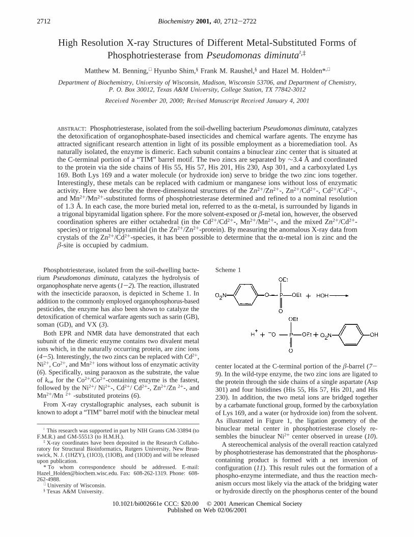
From X-ray crystallographic analyses, each subunit isknown to adopt a “TIM” barrel motif with the binuclear metal
center located at the C-terminal portion of theâ-barrel (7-9). In the wild-type enzyme, the two zinc ions are ligated tothe protein through the side chains of a single aspartate (Asp301) and four histidines (His 55, His 57, His 201, and His230). In addition, the two metal ions are bridged togetherby a carbamate functional group, formed by the carboxylationof Lys 169, and a water (or hydroxide ion) from the solvent.As illustrated in Figure 1, the ligation geometry of thebinuclear metal center in phosphotriesterase closely re-sembles the binuclear Ni2+ center observed in urease (10).
A stereochemical analysis of the overall reaction catalyzedby phosphotriesterase has demonstrated that the phosphorus-containing product is formed with a net inversion ofconfiguration (11). This result rules out the formation of aphospho-enzyme intermediate, and thus the reaction mech-anism occurs most likely via the attack of the bridging wateror hydroxide directly on the phosphorus center of the bound
† This research was supported in part by NIH Grants GM-33894 (toF.M.R.) and GM-55513 (to H.M.H.).
‡ X-ray coordinates have been deposited in the Research Collabo-ratory for Structural Bioinformatics, Rutgers University, New Brun-swick, N. J. (1HZY), (1IO3), (1IOB), and (1IOD) and will be releasedupon publication.
* To whom correspondence should be addressed. E-mail:[email protected]. Fax: 608-262-1319. Phone: 608-262-4988.
⊥ University of Wisconsin.§ Texas A&M University.
Scheme 1
2712 Biochemistry2001,40, 2712-2722
10.1021/bi002661e CCC: $20.00 © 2001 American Chemical SocietyPublished on Web 02/06/2001
substrate. The precise roles that each of the two divalentcations play in the catalytic process are not entirely clear.These two metal ions may function singly or in concert tolower the pKa of the bridging solvent molecule. The divalentcations may also polarize the phosphoryl oxygen bond ofthe substrate to make the reaction center more electrophilic.Additionally, one or both of these metal ions may serve toneutralize the distribution of negative charge on the leavinggroup as the reaction progresses.
The differential roles in binding and catalysis, played bythe two divalent cations within the binuclear metal centerof phosphotriesterase, have been probed by X-ray diffractionstudies and the preparation of a unique mixed-metal hybridenzyme. Recent X-ray crystallographic studies of phospho-triesterase have indicated that the more solvent-exposed orâ-metal ion is within 2.5 Å of the phosphoryl oxygen of thenon-hydrolyzable inhibitor, diisopropyl methyl phosphonate,bound to the active site (12). This metal ion is thus poisedto polarize the PdO bond of a true substrate for nucleophilicattack. The mixed-metal Zn2+/Cd2+-species of phosphotri-esterase has been prepared, and the catalytic properties ofthis unique hybrid most closely resemble those of the Zn2+/Zn 2+-phosphotriesterase (5). Therefore, it appears that oneof the two metal ions may dominate the catalytic steps thatgovern the magnitude ofkcat andkcat/Km during the hydrolysisof paraoxon.113Cd-NMR spectroscopy of the Zn2+/Cd2+-phosphotriesterase hybrid has demonstrated that only one ofthe two possible hybrid complexes is formed, but theassignment of a specific metal ion to an individual bindingsite has not been possible thus far (5).
Here we report the high-resolution X-ray structures of theZn2+/Zn2+-, Cd2+/Cd2+-, Mn2+/Mn2+-, and Zn2+/Cd2+-substituted forms of phosphotriesterase crystallized in theabsence of substrate analogues. These X-ray crystal structureswere solved to a nominal resolution of 1.3 Å to providegreater molecular insight into the perturbations imposed on
the binuclear metal center and the surrounding active sitethrough the occupancy of metal ions of various radii andcharge densities. Additionally, by measuring the anomalousX-ray data from crystals of the Zn2+/Cd2+-substituted species,it has been possible to assign the individual divalent cationsto the two specificR- andâ-metal binding sites.
MATERIALS AND METHODS
Crystallization and X-ray Data Collection.Crystals of thevarious substituted forms of phosphotriesterase were grownaccording to previously published procedures with the onlymodification being the replacement of 1% (v/v) diethyl4-methylbenzylphosphonate with 1% (v/v) 2-phenylethanol(8). All of the crystals belonged to the space groupC2 withtypical unit cell dimensions ofa ) 129.5 Å,b ) 91.4 Å,c) 69.4 Å,â ) 91.9° and two subunits per asymmetric unit.
For X-ray data collection, the crystals were first seriallytransferred to cryoprotectant solutions of a synthetic motherliquor containing 6, 12, 18, and 23% ethylene glycol. Thesynthetic mother liquor was composed of 14% (w/v) poly-(ethylene glycol) 8000, 200 mM NaCl, 0.5% (v/v) 2-phen-ylethanol, and 50 mM CHES (pH 9.0).1 Each crystal wasthen flash-cooled to-150 °C in a stream of nitrogen gasand subsequently stored under liquid nitrogen until synchro-tron beam time became available.
Each X-ray data set was collected on a 3× 3 tiled “SBC2”CCD detector at the Structural Biology Center 19-IDBeamline (Advanced Photon Source, Argonne NationalLaboratory). The X-ray data were processed with DENZOand scaled with SCALEPACK (13). Relevant X-ray datacollection statistics are presented in Table 1.
Structural Analyses.All of the structures presented herewere solved by Difference Fourier techniques. Each model
1 Abbreviations: bicine,N,N-bis(2-hydroxyethyl)glycine; CHES,2-(cyclohexylamino)ethanesulfonic acid.
FIGURE 1: Close-up view of the binuclear metal center located in the active site of phosphotriesterase. This figure, and Figures 4-7 wereprepared with the software package, MOLSCRIPT (21). X-ray coordinates utilized for this figure were determined in this laboratory andcan be obtained from the Protein Data Bank (1DPM). The two zinc ions are shown as large gray spheres with the position of the bridgingwater or hydroxide ion indicated by the large red sphere. The structure presented here was solved in the presence of a substrate analogue,diethyl 4-methylbenzyl phosphonate (not shown), and as such theâ-metal is tetrahedrally ligated. Coordinate covalent bonds between themetals and the ligands are indicated by the black lines.
Structure of Phosphotriesterase Biochemistry, Vol. 40, No. 9, 20012713
was subjected to alternate cycles of least-squares refinementat 1.3 Å resolution with the software package TNT (14) andmanual adjustment with the graphics program TURBO (15).Relevant refinement statistics are given in Table 2. Note thatthe R-factors listed in Table 2 are based on all measuredX-ray data with no sigma cutoffs applied. In each structuralanalysis, the electron density corresponding to subunit II wasslightly better ordered than that for subunit I. Consequently,the following discussion of the various metal-substitutedforms of phosphotriesterase will refer only to subunits II inthe X-ray coordinate files unless otherwise indicated. Sodium
ions were identified on the basis of both the octahedralcoordination geometry of the ligands surrounding them andthe bond distances.
Determination of the Metal Binding Positions for the Zn2+/Cd2+-Substituted Enzyme.To determine the exact locationof the zinc and cadmium ions in the mixed metal hybridspecies of phosphotriesterase, two separate X-ray experimentswere conducted. In the first study, an X-ray data set, designedto measure the anomalous scattering from a crystal of themixed hybrid species, was collected to 1.3 Å resolution atthe Advanced Photon Source, Structural Biology Centerbeamline. This X-ray data set was processed with DENZOand scaled with SCALEPACK (13). Protein phases, calcu-lated from the phosphotriesterase model with the metals andsolvents removed from the coordinate file, were employedto calculate an anomalous difference electron density map.At an X-ray wavelength of 0.70087, the∆f′′ for zinc is 1.431and for cadmium is 1.202 (International Tables, Vol. IV).Consequently, the larger peak in the difference anomalouselectron density map should correspond to zinc. Integratedpeak heights were obtained from MAPMAN (16). In subunitI of the asymmetric unit, the integrated intensities for the
Table 1: X-ray Data Collection Statistics
Zn2+/Zn2+ Cd2+/Cd2+ Mn2+/Mn2+ Zn2+/Cd2+
resolution range (Å) 30.0-1.3 30.0-1.3 30.0-1.3 30.0-1.3integrated reflect. 1 448 033 1 231 444 1 367 510 1 626 798independent reflect. 186 115 187 426 184 739 190 056data completeness
(%)97 (97) 98 (99) 96 (99) 99 (99)
avgI/avgσ(I)a 25 (4.0) 32 (8.0) 21.6 (4.3) 30.3 (2.8)Rsym (%)a 9.6 (17.3) 6.9 (18.4) 9.2 (23.3) 5.8 (31.0)
a Rsym ) (∑|I - Ih|/∑ I) × 100. The numbers in the parenthesescorrespond to the resolution range of 1.35 to 1.30 Å.
Table 2: Least-Squares Refinement Statistics
Zn2+/Zn2+ Cd2+/Cd2+ Mn2+/Mn2+ Zn2+/Cd2+
resolution limits (Å) 30.0-1.3 30.0-1.3 30.0-1.3 30.0-1.3R-factor (overall) %/no. of rflnsa 19.6 (186 115) 20.4 (187 426) 22.5 (184 739) 19.3 (190 056)R-factor (working) %/no. of rflns 19.6 (176 950) 20.2 (178 183) 22.2 (175 396) 19.1 (180 701)R-factor (free) %/no. of rflns 22.8 (9165) 23.6 (9243) 26.7 (9343) 22.4 (9355)no. of protein atoms 5044 5044 5044 5044multiple conformations 17 18 17 18
weighted root-mean-square deviations from idealitybond lengths (Å) 0.012 0.011 0.011 0.011bond angles (Å) 2.2 2.1 2.3 2.2trigonal planes (Å) 0.007 0.006 0.007 0.007general planes (Å) 0.009 0.008 0.009 0.009torsional angles (deg)b 14.6 14.6 15.0 14.6
solventswaters 692 738 710 737Na+ ions 2 2 2 2ethylene glycols 20 27 26 152-phenylethanols 2 2 2 2
multiple conformations
Zn2+/Zn2+ Cd2+/Cd2+ Mn2+/Mn2+ Zn2+/Cd2+
110 VAL 110 VAL 110 VAL 110 VAL111 SER 111 SER 111 SER 111 SER159 GLU 196 VAL 196 VAL 156 TYR196 VAL 222 SER 222 SER 196 VAL222 SER 295 GLN 238 SER 222 SER238 SER 298 VAL 241 THR 238 SER241 THR 359 SER 295 GLN 241 THR295 GLN 510 VAL 359 SER 295 GLN359 SER 518 ARG 510 VAL 359 SER510 VALc 573 THR 511 SER 510 VAL518 ARG 596 VAL 518 ARG 511 SER596 VAL 669 SER 622 SER 518 ARG622 SER 685 LYS 685 LYS 622 SER669 SER 695 GLN 698 VAL 685 LYS685 LYS 698 VAL 759 SER 698 VAL698 VAL 759 SER 810 EG 759 SER759 SER 810 EGd 811 EG 810 EG
852 PEAe 811 EGa R-factor) (Σ|Fo - Fc|/Σ|Fo|) × 100 whereFo is the observed structure-factor amplitude andFc is the calculated structure-factor amplitude. For
calculation ofR-free, 5% of the X-ray data were removed from the reflection file.b The torsional angles were not restrained during the refinement.c Amino acid residues with numbers higher than 400 correspond to those found in subunit II (beginning at residue 434).d Ethylene glycol is abbreviatedas EG.e 2-phenylethanol is abbreviated as PEA.
2714 Biochemistry, Vol. 40, No. 9, 2001 Benning et al.
peaks at theR- andâ-sites were 30.3 and 25.6, respectively.Likewise, in subunit II, integrated intensities of 35.2 and 29.7were observed for theR- and â-sites, respectively. Thesedata indicate that theR-metal site is occupied by zinc, whilecadmium binds to theâ-site.
As further evidence to the identity of the metals occupyingtheR- andâ-sites, a second X-ray data set was collected to1.9 Å resolution with Cu KR radiation. The X-ray data weremeasured with a Proteum/R rotating anode generator/CCDdetector system, processed with SaintPlus, and scaled withProScale (Bruker AXS, Inc., Madison, WI 53711 USA).Again Friedel pairs were measured. At a wavelength of1.54178 Å, the∆f ′′ for zinc is 0.678 and for cadmium is4.653. In this case, the larger integrated peak intensity shouldcorrespond to the cadmium ion. In subunit I of the asym-metric unit, the integrated intensity for theR-site was 31.8and for theâ-site was 34.5, while for subunit II the intensitieswere 36.1 and 41.0, respectively, for theR- andâ-sites. Againthese data are indicative of a mixed hybrid species wherebythe zinc ion occupies theR-site while the cadmium ionlocates to theâ-site. A portion of the electron density mapnear the Zn2+/Cd2+-binuclear metal center is displayed inFigure 2.
RESULTS
Structure of the Zn2+/Zn2+-Containing Phosphotriesterase.A Ramachandran plot of all non-glycinylφ, ψ angles forthe Zn2+/Zn2+-containing enzyme is given in Figure 3. Ascan be seen, the quality of the refined model is outstandingwith the only significant outliers being Ser 61, Trp 131, Glu159, and Asn 312 in both subunits of the dimer. In eachcase, the electron densities corresponding to the above-mentioned amino acid residues are unambiguous. Ser 61 islocated at the end of aâ-strand near the C-terminal portionof the “TIM” barrel, while the indole ring of Trp 131 islocated within∼5 Å of the binuclear metal center. Both Glu159 and Asn 312 are located in reverse turns, (type II′ andtype II, respectively), which are distant from the active siteof the enzyme.
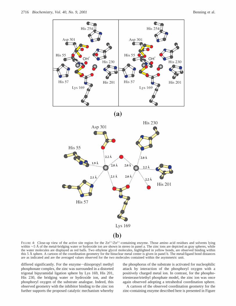
Shown in Figure 4, panel a, is a close-up view of the activesite for the Zn2+/Zn2+-containing phosphotriesterase. Onlythose residues that are located within∼5 Å of the metal-bridging water or hydroxide ion are displayed. All of the
crystals employed for these high-resolution X-ray analysesdescribed here were grown in the absence of substrateanalogues or inhibitors. However, each crystal was trans-ferred to a cryoprotectant solution containing ethylene glycoland, indeed, several ethylene glycols are observed bindingin the active site region of the zinc-substituted protein asindicated in Figure 4, panel a. Interestingly, these ethyleneglycols occupy similar positions to those observed for theethoxy groups of triethyl phosphate when this inhibitor isbound to phosphotriesterase (12).
The first X-ray structure of the Zn2+/Zn2+-form of phos-photriesterase was solved in the presence of the inertinhibitor, diethyl 4-methylbenzylphosphonate (9). In thatstructure, the zinc ion in theR-site was ligated by His 55,His 57, Lys 169, Asp 301, and the metal-bridging water orhydroxide in a trigonal bipyramidal arrangement. Theâ-sitemetal ion, however, was coordinated in a distorted tetrahedralenvironment via Lys 169, His 201, His 230, and the metal-bridging water or hydroxide ion. Following this X-raycrystallographic analysis, the structure of the zinc-containingenzyme was determined in the presence of either diisopropylmethyl phosphonate (a sarin mimic) or triethyl phosphate(12). In both complexes, theR-metal ions adopted similartrigonal bipyramidal coordination spheres. With respect tothe zincs occupying theâ-sites, however, the two models
FIGURE 2: Representative portion of the electron density map calculated for the Zn2+/Cd2+-hybrid phosphotriesterase. The electron densitydisplayed was contoured at 1σ and calculated with coefficients of the form (2Fo-Fc), whereFo was the native structure factor amplitudeandFc was the calculated structure factor amplitude. The positions of the zinc and cadmium ions are indicated by the light and dark grayspheres, respectively.
FIGURE 3: Ramachandran plot of all non-glycinyl main chainφ, ψangles. Those dihedral angles that are fully or partially allowedare enclosed by the solid or dashed lines, respectively. All of themodels for phosphotriesterase presented here adhere tightly to theallowed regions of the Ramachandran plot.
Structure of Phosphotriesterase Biochemistry, Vol. 40, No. 9, 20012715
differed significantly. For the enzyme-diisopropyl methylphosphonate complex, the zinc was surrounded in a distortedtrigonal bipyramidal ligation sphere by Lys 169, His 201,His 230, the bridging water or hydroxide ion, and thephosphoryl oxygen of the substrate analogue. Indeed, thisobserved geometry with the inhibitor binding to the zinc ionfurther supports the proposed catalytic mechanism whereby
the phosphorus of the substrate is activated for nucleophilicattack by interaction of the phosphoryl oxygen with apositively charged metal ion. In contrast, for the phospho-triesterase/triethyl phosphate model, the zinc ion was onceagain observed adopting a tetrahedral coordination sphere.
A cartoon of the observed coordination geometry for thezinc-containing enzyme described here is presented in Figure
FIGURE 4: Close-up view of the active site region for the Zn2+/Zn2+-containing enzyme. Those amino acid residues and solvents lyingwithin ∼5 Å of the metal-bridging water or hydroxide ion are shown in stereo in panel a. The zinc ions are depicted as gray spheres, whilethe water molecules are displayed as red balls. Two ethylene glycol molecules, highlighted in yellow bonds, are observed binding withinthis 5 Å sphere. A cartoon of the coordination geometry for the binuclear metal center is given in panel b. The metal/ligand bond distancesare as indicated and are the averaged values observed for the two molecules contained within the asymmetric unit.
2716 Biochemistry, Vol. 40, No. 9, 2001 Benning et al.
4, panel b. Again, theR-site zinc is ligated by His 55, His57, Asp 301, Lys 169, and the bridging water or hydroxideion in a distorted trigonal bipyramidal geometry withcoordinate covalent bonds ranging in length from 1.9 to 2.2Å. The axial ligands are provided by the side chain oxygensof Asp 301 and Lys 169, while the equatorial bonds areformed between the metal and His 55, His 57, and thebridging water or hydroxide. The angles observed betweenan axial ligand, the metal, and an equatorial ligand range insize from 86.0 to 100.2° with an average angle of 90.3°(subunit II). The angles observed between the equatorialligands and the metal are 114.2° (His 55-Zn2+-His 57),111.6° (His 55-Zn2+-hydroxide), and 134.2° (His 57-Zn2+-hydroxide). Contrary to that observed for phospho-triesterase solved in the presence of either the bound inhibitordiethyl 4-methylbenzyl phosphonate or triethyl phosphate,the â-site zinc ion in the structure presented here issurrounded in a distorted trigonal bipyramidal arrangementwith the axial ligands being His 201 and the metal-bridgingwater or hydroxide ion and the equatorial ligands providedby Lys 169, His 230, and a water molecule. In this case, theangles observed between an axial ligand, the metal, and anequatorial ligand range in size from 77.1 to 99.2° with anaverage angle of 90.9° (subunit II). The angles observedbetween the equatorial ligands and the metal are 110.0° (Lys169-Zn2+-water), 111.3° (Lys 169-Zn2+-His 230), and138.7° (His 230-Zn2+-water). There is one water moleculelocated at∼3.3 Å that if it were positioned somewhat closercould coordinate to the zinc to complete an octahedralligation sphere. Note that the Zn2+ ions, with ionic radii of0.74 Å, are separated by 3.5 Å (subunit II).
One question that immediately arises is why theâ-site zincis tetrahedrally ligated when either triethyl phosphate ordiethyl 4-methylbenzyl phosphonate is bound in the activesite pocket but is surrounded in a trigonal bipyramidalcoordination sphere in the current model. A superpositionof the various zinc-containing protein models demonstratesthat the phosphoryl oxygens of the inhibitors (triethylphosphate or diethyl 4-methylbenzylphosphonate) lie within∼1 Å of where the water molecule (shown in Figure 4, panelb) is situated in the structure presented here. Consequently,these two specific inhibitors effectively exclude water fromthe zinc ligation sphere thereby reducing the number ofcoordinate covalent bonds to the metal from five to four.
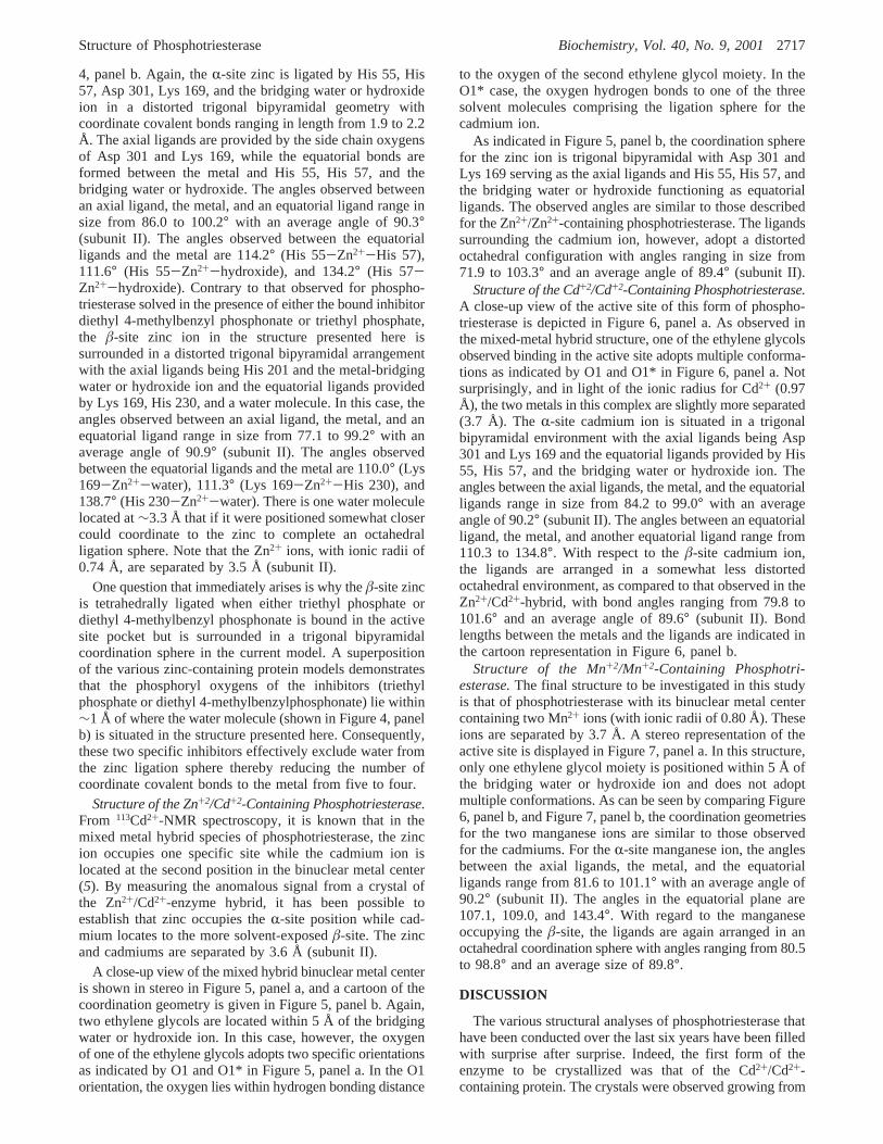
Structure of the Zn+2/Cd+2-Containing Phosphotriesterase.From 113Cd2+-NMR spectroscopy, it is known that in themixed metal hybrid species of phosphotriesterase, the zincion occupies one specific site while the cadmium ion islocated at the second position in the binuclear metal center(5). By measuring the anomalous signal from a crystal ofthe Zn2+/Cd2+-enzyme hybrid, it has been possible toestablish that zinc occupies theR-site position while cad-mium locates to the more solvent-exposedâ-site. The zincand cadmiums are separated by 3.6 Å (subunit II).
A close-up view of the mixed hybrid binuclear metal centeris shown in stereo in Figure 5, panel a, and a cartoon of thecoordination geometry is given in Figure 5, panel b. Again,two ethylene glycols are located within 5 Å of thebridgingwater or hydroxide ion. In this case, however, the oxygenof one of the ethylene glycols adopts two specific orientationsas indicated by O1 and O1* in Figure 5, panel a. In the O1orientation, the oxygen lies within hydrogen bonding distance
to the oxygen of the second ethylene glycol moiety. In theO1* case, the oxygen hydrogen bonds to one of the threesolvent molecules comprising the ligation sphere for thecadmium ion.
As indicated in Figure 5, panel b, the coordination spherefor the zinc ion is trigonal bipyramidal with Asp 301 andLys 169 serving as the axial ligands and His 55, His 57, andthe bridging water or hydroxide functioning as equatorialligands. The observed angles are similar to those describedfor the Zn2+/Zn2+-containing phosphotriesterase. The ligandssurrounding the cadmium ion, however, adopt a distortedoctahedral configuration with angles ranging in size from71.9 to 103.3° and an average angle of 89.4° (subunit II).
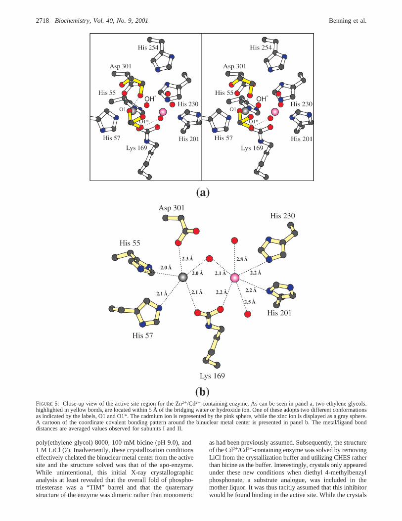
Structure of the Cd+2/Cd+2-Containing Phosphotriesterase.A close-up view of the active site of this form of phospho-triesterase is depicted in Figure 6, panel a. As observed inthe mixed-metal hybrid structure, one of the ethylene glycolsobserved binding in the active site adopts multiple conforma-tions as indicated by O1 and O1* in Figure 6, panel a. Notsurprisingly, and in light of the ionic radius for Cd2+ (0.97Å), the two metals in this complex are slightly more separated(3.7 Å). TheR-site cadmium ion is situated in a trigonalbipyramidal environment with the axial ligands being Asp301 and Lys 169 and the equatorial ligands provided by His55, His 57, and the bridging water or hydroxide ion. Theangles between the axial ligands, the metal, and the equatorialligands range in size from 84.2 to 99.0° with an averageangle of 90.2° (subunit II). The angles between an equatorialligand, the metal, and another equatorial ligand range from110.3 to 134.8°. With respect to theâ-site cadmium ion,the ligands are arranged in a somewhat less distortedoctahedral environment, as compared to that observed in theZn2+/Cd2+-hybrid, with bond angles ranging from 79.8 to101.6° and an average angle of 89.6° (subunit II). Bondlengths between the metals and the ligands are indicated inthe cartoon representation in Figure 6, panel b.
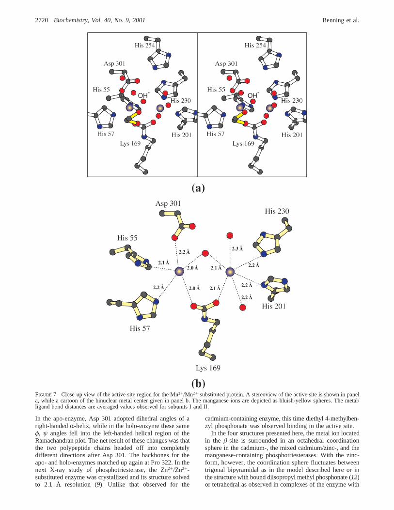
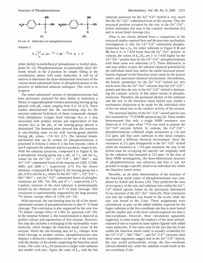
Structure of the Mn+2/Mn+2-Containing Phosphotri-esterase.The final structure to be investigated in this studyis that of phosphotriesterase with its binuclear metal centercontaining two Mn2+ ions (with ionic radii of 0.80 Å). Theseions are separated by 3.7 Å. A stereo representation of theactive site is displayed in Figure 7, panel a. In this structure,only one ethylene glycol moiety is positioned within 5 Å ofthe bridging water or hydroxide ion and does not adoptmultiple conformations. As can be seen by comparing Figure6, panel b, and Figure 7, panel b, the coordination geometriesfor the two manganese ions are similar to those observedfor the cadmiums. For theR-site manganese ion, the anglesbetween the axial ligands, the metal, and the equatorialligands range from 81.6 to 101.1° with an average angle of90.2° (subunit II). The angles in the equatorial plane are107.1, 109.0, and 143.4°. With regard to the manganeseoccupying theâ-site, the ligands are again arranged in anoctahedral coordination sphere with angles ranging from 80.5to 98.8° and an average size of 89.8°.
DISCUSSION
The various structural analyses of phosphotriesterase thathave been conducted over the last six years have been filledwith surprise after surprise. Indeed, the first form of theenzyme to be crystallized was that of the Cd2+/Cd2+-containing protein. The crystals were observed growing from
Structure of Phosphotriesterase Biochemistry, Vol. 40, No. 9, 20012717
poly(ethylene glycol) 8000, 100 mM bicine (pH 9.0), and1 M LiCl (7). Inadvertently, these crystallization conditionseffectively chelated the binuclear metal center from the activesite and the structure solved was that of the apo-enzyme.While unintentional, this initial X-ray crystallographicanalysis at least revealed that the overall fold of phospho-triesterase was a “TIM” barrel and that the quaternarystructure of the enzyme was dimeric rather than monomeric
as had been previously assumed. Subsequently, the structureof the Cd2+/Cd2+-containing enzyme was solved by removingLiCl from the crystallization buffer and utilizing CHES ratherthan bicine as the buffer. Interestingly, crystals only appearedunder these new conditions when diethyl 4-methylbenzylphosphonate, a substrate analogue, was included in themother liquor. It was thus tacitly assumed that this inhibitorwould be found binding in the active site. While the crystals
FIGURE 5: Close-up view of the active site region for the Zn2+/Cd2+-containing enzyme. As can be seen in panel a, two ethylene glycols,highlighted in yellow bonds, are located within 5 Å of thebridging water or hydroxide ion. One of these adopts two different conformationsas indicated by the labels, O1 and O1*. The cadmium ion is represented by the pink sphere, while the zinc ion is displayed as a gray sphere.A cartoon of the coordinate covalent bonding pattern around the binuclear metal center is presented in panel b. The metal/ligand bonddistances are averaged values observed for subunits I and II.
2718 Biochemistry, Vol. 40, No. 9, 2001 Benning et al.
grown under these new conditions did, indeed, allow for astructural determination of the holo-enzyme with an intactbinuclear metal center, the diethyl 4-methylbenzyl phospho-nate was observed binding between two symmetry-relatedmolecules in the crystalline lattice and not in the active site(8). Two additional surprises unfolded in this second X-raystructural analysis of phosphotriesterase. First, one of thebridging ligands to the cadmiums was, quite unexpectedly,
a carboxylated lysine residue (Lys 169). This residue wasnot covalently modified in the apo-enzyme structure. Second,the conformations of the polypeptide chains for the apo- andholo-enzymes were quite different such that theirR-carbonssuperimposed with a root-mean-square deviation of 3.4 Å.The changes in backbone conformations between the twoforms of the protein were limited to several regions, however,with the most striking difference occurring near Asp 301.
FIGURE 6: Close-up view of the active site region for the Cd2+/Cd2+-containing phosphotriesterase. The two cadmium ions of the binuclearmetal center are colored in pink. Like that observed in the zinc/cadmium hybrid, one of the ethylene glycols adopts two specific conformations,O1 and O1*, as indicated in panel a. A schematic of the binuclear metal center is depicted in panel b. The metal/ligand bond distances areaveraged values observed for subunits I and II.
Structure of Phosphotriesterase Biochemistry, Vol. 40, No. 9, 20012719
In the apo-enzyme, Asp 301 adopted dihedral angles of aright-handedR-helix, while in the holo-enzyme these sameφ, ψ angles fell into the left-handed helical region of theRamachandran plot. The net result of these changes was thatthe two polypeptide chains headed off into completelydifferent directions after Asp 301. The backbones for theapo- and holo-enzymes matched up again at Pro 322. In thenext X-ray study of phosphotriesterase, the Zn2+/Zn2+-substituted enzyme was crystallized and its structure solvedto 2.1 Å resolution (9). Unlike that observed for the
cadmium-containing enzyme, this time diethyl 4-methylben-zyl phosphonate was observed binding in the active site.
In the four structures presented here, the metal ion locatedin the â-site is surrounded in an octahedral coordinationsphere in the cadmium-, the mixed cadmium/zinc-, and themanganese-containing phosphotriesterases. With the zinc-form, however, the coordination sphere fluctuates betweentrigonal bipyramidal as in the model described here or inthe structure with bound diisopropyl methyl phosphonate (12)or tetrahedral as observed in complexes of the enzyme with
FIGURE 7: Close-up view of the active site region for the Mn2+/Mn2+-substituted protein. A stereoview of the active site is shown in panela, while a cartoon of the binuclear metal center given in panel b. The manganese ions are depicted as bluish-yellow spheres. The metal/ligand bond distances are averaged values observed for subunits I and II.
2720 Biochemistry, Vol. 40, No. 9, 2001 Benning et al.
either diethyl 4-methylbenzyl phosphonate or triethyl phos-phate (9, 12). Phosphotriesterase accommodates these dif-ferent metals in theâ-position by simply filling in thecoordination sphere with water molecules. It will be ofinterest to determine the three-dimensional structures of thevarious metal-substituted forms of phosphotriesterase in thepresence of additional substrate analogues. This work is inprogress.
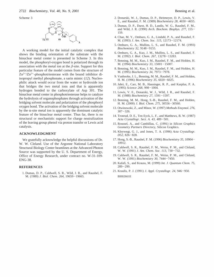
The metal-substituted variants of phosphotriesterase hadbeen previously analyzed for their ability to hydrolyze alibrary of organophosphate triesters possessing leaving-groupphenols with pKa values ranging from 4 to 10 (17). Thesestudies demonstrated that the rate-limiting step for theenzyme-catalyzed hydrolysis of these compounds changesfrom phosphorus-oxygen bond cleavage (k3) to a stepassociated with product release and regeneration of freeenzyme (k5) as the pKa of the leaving-group phenol isdiminished. The Brønsted plots showed that this transitionin rate-limiting steps occurs with leaving-group phenolshaving pKa values ∼7.8-8.0 (17). The minimal kineticmechanism that can be written for phosphotriesterase ispresented in Scheme 2 where E is the free enzyme, while Aand P represent the substrate and two products, respectively.With the substrate paraoxon, compoundI in Figure 8, thep-nitrophenol leaving-group has a pKa of 7.1, and thekcat
values for the Zn2+/Zn2+-, Cd2+/Cd2+-, Mn2+/Mn2+-, andZn2+/Cd2+-substituted forms of the enzyme are 2200, 12 900,4400, and 2600 s-1, respectively (17). For the slowersubstrate, compoundII in Figure 8, the leaving group has apKa of 8.6, and thekcat values for the Zn2+/Zn2+-, Cd2+/Cd2+-,Mn2+/Mn2+-, and Zn2+/Cd2+-substituted forms of phospho-triesterase are 100, 730, 300, and 47 s-1, respectively (17).Catalytic turnover of the slow substrate is predominantlylimited by the chemical rate of P-O bond cleavage. Thisconclusion is experimentally supported by the large primary18O kinetic isotope effect of 1.036 (18-19).
With paraoxon, the rate-limiting step for all of the metal-substituted variants of phosphotriesterase is after P-O bondcleavage. This conclusion is also supported by the very low18O kinetic isotope effect of 1.002 for paraoxon (18-19).In the minimal Scheme 2, this transformation is depicted asproduct release and regeneration of free enzyme. However,this step also includes a recharging of the nucleophilic watermolecule, which bridges the binuclear metal center of theenzyme. When the rate-limiting step forkcat changes frombond cleavage to product release, phosphotriesterase alsodisplays a distinctive dependence on the rate of the reactionwith the identity of the metals comprising the binuclear metalcenter. The value ofkcat for paraoxon is larger with cadmiumand smaller with zinc. Again, the value ofkcat with the fast
substrate paraoxon for the Zn2+/Cd2+-hybrid is very muchlike the Zn2+/Zn2+-substituted form of the enzyme. Thus thestructural position occupied by the zinc in the Zn2+/Cd2+-hybrid dominates this step in the catalytic mechanism (k5)and in actual bond cleavage (k3).
What is not clearly defined from a comparison of thestructural studies reported here and the previous mechanisticinvestigations is why the Cd2+/Cd2+-substituted phospho-triesterase has akcat for either substrate in Figure 8 (I andII ) that is 6- to 7-fold faster than the Zn2+/Zn2+-protein. Incontrast, the values ofkcat/Km are 3- to 7-fold higher for theZn2+/Zn2+-protein than for the Cd2+/Cd2+-phosphotriesterasewith these same two substrates (17). These differences inrate may reflect, in part, the inherent chemical potentials bythe individual metal ions and/or the subtle structural pertur-bations imposed on the binuclear metal center by the proteinmatrix and associated chemical environment. Nevertheless,the kinetic parameters for the Zn2+/Cd2+-hybrid are verymuch more like the Zn2+/Zn2+-enzyme than the Cd2+/Cd2+-protein and thus the zinc in the Zn2+/Cd2+-hybrid is dominat-ing the catalytic activity of this metal variant of phospho-triesterase. Therefore, the positional identity of the cadmiumand the zinc in the binuclear metal hybrid may enable amechanistic distinction to be made for the individual rolesof the two metal ions in the catalytic activity of the enzyme.
The structural identity of the Zn2+/Cd2+-mixed species wasfirst examined by113Cd NMR spectroscopy (5). These studiesdemonstrated that only a single NMR resonance wasobserved at 115 ppm when113Cd was used to prepare theZn2+/Cd2+-enzyme hybrid. In contrast, the Cd2+/Cd2+-phosphotriesterase exhibited single resonances at 116 and212 ppm, and thus each cadmium in this metal complexexperienced a different chemical environment. Since theresonance at 212 ppm disappeared in the Zn2+/Cd2+-hybridwhile the resonance at∼116 ppm remained, the zinc in thehybrid must be occupying the specific site originally heldby the cadmium that resonated at 212 ppm. At the time ofthese NMR investigations, the three-dimensional structureof phosphotriesterase was unknown and thus it was notpossible to assign a specific metal to an individual site withinthe binuclear metal center.
Recently, an ab initio determination of the structure ofthe binuclear metal center of phosphotriesterase was com-pleted by Kafafi and Krauss (20). They predicted the sitesof occupancy of the zinc and cadmium ions within the Zn2+/Cd2+-hybrid species based on the previously determinedX-ray structure of the Zn2+/Zn2+-form (9). Their conclusionwas that the cadmium was bound to theR-site while thezinc was bound to theâ-site. These assignments wererationalized, in part, on the added stability expected for thelarger cadmium at the five-coordinate site that is more buriedand the smaller zinc at the more solvent-exposed site that isfour-coordinate. However, these calculations apparentlyneglected, to some extent, the tendency of the more solvent-exposedâ-site to expand its inner-sphere ligands with addedwater molecules. If one takes note of the fact that theâ-sitewithin the binuclear metal center is actually octahedral forthe Cd2+/Cd2+-, Mn2+/Mn2+-, and Zn2+/Cd2+-enzymes, thenthese same arguments suggest that in the Zn2+/Cd2+-hybrid,the zinc would preferentially occupy the five-coordinatesolvent-shielded site, while the cadmium would reside in thesix-coordinateâ-site.
FIGURE 8: Substrates for phosphotriesterase.
Scheme 2
Structure of Phosphotriesterase Biochemistry, Vol. 40, No. 9, 20012721
A working model for the initial catalytic complex thatshows the binding orientation of the substrate with thebinuclear metal center is presented in Scheme 3. In thismodel, the phosphoryl-oxygen bond is polarized through itsassociation with the metal ion at theâ-site. Support for thisparticular feature of the model comes from the structure ofZn2+/Zn2+-phosphotriesterase with the bound inhibitor di-isopropyl methyl phosphonate, a sarin mimic (12). Nucleo-philic attack would occur from the water or hydroxide ionthat bridges the two metal ions and that is apparentlyhydrogen bonded to the carboxylate of Asp 301. Thebinuclear metal center in phosphotriesterase helps to catalyzethe hydrolysis of organophosphates through activation of thebridging solvent molecule and polarization of the phosphoryloxygen bond. The activation of the bridging solvent moleculeby theR-site metal ion is apparently the dominant catalyticfeature of the binuclear metal center. Thus far, there is nostructural or mechanistic support for charge neutralizationof the leaving group phenol via proton transfer or Lewis acidcatalysis.
ACKNOWLEDGMENT
We gratefully acknowledge the helpful discussions of Dr.W. W. Cleland. Use of the Argonne National LaboratoryStructural Biology Center beamlines at the Advanced PhotonSource was supported by the U. S. Department of Energy,Office of Energy Research, under contract no. W-31-109-ENG-38.
REFERENCES
1. Dumas, D. P., Caldwell, S. R., Wild, J. R., and Raushel, F.M. (1989)J. Biol. Chem. 264, 19659-19665.
2. Donarski, W. J., Dumas, D. P., Heitmeyer, D. P., Lewis, V.E., and Raushel, F. M. (1989)Biochemistry 28, 4650-4655.
3. Dumas, D. P., Durst, H. D., Landis, W. G., Raushel, F. M.,and Wild, J. R. (1990)Arch. Biochem. Biophys. 277, 155-159.
4. Chae, M. Y., Omburo, G. A., Lindahl, P. A., and Raushel, F.M. (1993)J. Am. Chem. Soc. 115, 12173-12174.
5. Omburo, G. A., Mullins, L. S., and Raushel, F. M. (1993)Biochemistry 32, 9148-9155.
6. Omburo, G. A., Kuo, J. M., Mullins, L. S., and Raushel, F.M. (1992)J. Biol. Chem. 267, 13278-13283.
7. Benning, M. M., Kuo, J. M., Raushel, F. M., and Holden, H.M. (1994)Biochemistry 33, 15001-15007.
8. Benning, M. M., Kuo, J. M., Raushel, F. M., and Holden, H.M. (1995)Biochemistry 34, 7973-7978.
9. Vanhooke, J. L., Benning, M. M., Raushel, F. M., and Holden,H. M. (1996)Biochemistry 35, 6020-6025.
10. Jabri, E., Carr, M. B., Hausinger, R. P., and Karplus, P. A.(1995)Science 268, 998-1004.
11. Lewis, V. E., Donarski, W. J., Wild, J. R., and Raushel, F.M. (1988)Biochemistry 27, 1591-1597.
12. Benning, M. M., Hong, S.-B., Raushel, F. M., and Holden,H. M. (2000)J. Biol. Chem. 275, 30556-30560.
13. Otwinowski, Z., and Minor, W. (1997)Methods Enzymol. 276,307-326.
14. Tronrud, D. E., Ten Eyck, L. F., and Matthews, B. W. (1987)Acta Crystallogr. Sect. A. 43, 489-501.
15. Roussel, A., and Cambillau, C. (1991) inSilicon GraphicsGeometry Partners Directory, Silicon Graphics.
16. Kleywegt, G. J., and Jones, T. A. (1996)Acta Crystallogr.D52, 826-828.
17. Hong, S.-B., Raushel, F. M. (1996)Biochemistry 35, 10904-10912.
18. Caldwell, S. R., Raushel, F. M., Weiss, P. M., and Cleland,W. W. (1991)J. Am. Chem. Soc. 113, 730-732.
19. Caldwell, S. R., Raushel, F. M., Weiss, P. M., and Cleland,W. W. (1991)Biochemistry 30, 7444-7450.
20. Kafafi, S., and Krauss, M. (1999)Int. J. Quantum Chem. 75,289-299.
21. Kraulis, P. J. (1991)J. Appl. Crystallogr. 24, 946-950.
BI002661E
Scheme 3
2722 Biochemistry, Vol. 40, No. 9, 2001 Benning et al.