her2 kinase domain mutation results in constitutive phosphorylation and activation of her2 and egfr...
TRANSCRIPT

A R T I C L E
HER2 kinase domain mutation results in constitutivephosphorylation and activation of HER2 and EGFRand resistance to EGFR tyrosine kinase inhibitors
Shizhen Emily Wang,1 Archana Narasanna,2 Marianela Perez-Torres,1 Bin Xiang,5 Frederick Y. Wu,2
Seungchan Yang,2 Graham Carpenter,3 Adi F. Gazdar,6 Senthil K. Muthuswamy,5
and Carlos L. Arteaga1,2,4,*
1 Department of Cancer Biology, Vanderbilt University School of Medicine, Nashville, Tennessee 372322 Department of Medicine, Vanderbilt University School of Medicine, Nashville, Tennessee 372323 Department of Biochemistry, Vanderbilt University School of Medicine, Nashville, Tennessee 372324 Breast Cancer Research Program, Vanderbilt-Ingram Comprehensive Cancer Center, Vanderbilt University School of Medicine,
Nashville, Tennessee 372325 Cold Spring Harbor Laboratory, Cold Spring Harbor, New York 117246 Hamon Center for Therapeutic Oncology Research, University of Texas Southwest Medical Center, Dallas, Texas 75390*Correspondence: [email protected]
Summary
HER2/Neu gene mutations have been identified in lung cancer. Expression of a HER2 mutant containing a G776YVMA insertionin exon 20 was more potent than wild-type HER2 in associating with and activating signal transducers, phosphorylatingEGFR, and inducing survival, invasiveness, and tumorigenicity. HER2YVMA transphosphorylated kinase-dead EGFRK721R
and EGFRWT in the presence of EGFR tyrosine kinase inhibitors (TKIs). Knockdown of mutant HER2 in H1781 lung cancercells increased apoptosis and restored sensitivity to EGFR TKIs. The HER2 inhibitors lapatinib, trastuzumab, and CI-1033inhibited growth of H1781 cells and cells expressing exogenous HER2YVMA. These data suggest that (1) HER2YVMA activatescellular substrates more potently than HER2WT; and (2) cancer cells expressing this mutation remain sensitive to HER2-targeted therapies but insensitive to EGFR TKIs.
Introduction
HER2/Neu (ErbB2) is a member of the ErbB family of transmem-brane receptor tyrosine kinases, which also includes the epider-mal growth factor receptor (EGFR, ErbB1), HER3 (ErbB3), andHER4 (ErbB4). Binding of ligands to the ectodomain of EGFR,ErbB3, and ErbB4 results in the formation of catalytically activehomo- and heterodimers to which HER2 is recruited as a pre-ferred partner (Yarden and Sliwkowski, 2001). Although HER2cannot bind any of the ErbB ligands directly, its catalytic activitycan potently amplify signaling by ErbB-containing heterodimersvia increasing ligand binding affinity and/or receptor recyclingand stability (Graus-Porta et al., 1997; Pinkas-Kramarski et al.,1996; Wang et al., 1998; Worthylake et al., 1999). Activationof the ErbB network leads to receptor autophosphorylationin C-terminal tyrosines and the recruitment to these sites of
CANCER CELL 10, 25–38, JULY 2006 ª2006 ELSEVIER INC. DOI 10.101
cytoplasmic signal transducers that regulate cellular processessuch as proliferation, differentiation, motility, adhesion, protec-tion from apoptosis, and transformation. Cytoplasmic signaltransducers that are activated by this network include PLC-g1,Ras-Raf-MEK-MAPKs, phosphatidylinositol-3 kinase (PI3K)-Akt-ribosomal S6 kinase, Src, the stress-activated protein ki-nases (SAPKs), PAK-JNKK-JNK, and the signal transducersand activators of transcription (STATs) (Yarden and Sliwkowski,2001).
HER2 gene amplification has been reported in approximately20% of metastatic breast cancers, where it is associated withpoor patient outcome (Slamon et al., 1989). Trastuzumab, a hu-manized monoclonal IgG1 that binds the extracellular domain ofHER2, has been shown to induce clinical responses in HER2-overexpressing breast cancers and prolong patient survival(Slamon et al., 2001; Vogel et al., 2002). HER2 is overexpressed
S I G N I F I C A N C E
Mutations in the kinase domain of the HER2/Neu proto-oncogene were identified recently. Expression of a HER2 mutant containinga G776YVMA insertion in exon 20 was more potent than wild-type HER2 in activating postreceptor signal transducers and in transformingmammary and bronchial epithelial cells. HER2YVMA activated the EGFR in the presence of EGFR tyrosine kinase inhibitors (TKIs). Knock-down of mutant HER2 in H1781 lung cancer cells induced cell death and sensitized these cells to EGFR TKIs. The HER2 inhibitors lapatinib,trastuzumab, and CI-1033 inhibited growth of cells expressing mutant HER2. These data suggest that (1) HER2YVMA signals more potentlythan HER2WT; and (2) cancer cells harboring exon 20 insertions in HER2 remain sensitive to HER2-targeted therapies but insensitive toEGFR TKIs.
6/j.ccr.2006.05.023 25

A R T I C L E
in a cohort of non-small cell lung cancers (NSCLCs), and in-creased HER2 gene copy number has been associated withtherapeutic response to EGFR tyrosine kinase inhibitors (TKIs)(Cappuzzo et al., 2005; Hirsch et al., 2002). Recently, intragenicsomatic mutations in the HER2 gene were reported in <5% ofNSCLCs. These involve in-frame duplications/insertions in asmall stretch within exon 20 that correspond to the identicalnine codon region in exon 20 of the EGFR gene, where duplica-tions/insertions have also been reported (Stephens et al., 2004).Interestingly, mutations in both receptor genes do not overlapand occur predominantly in patients of Asian ethnicity, non-smokers, females, and lung adenocarcinomas (Shigematsuet al., 2005). Because of the location of these insertions at theC-terminal end of the aC helix in the tyrosine kinase domain, ithas been postulated that they result in a conformational changeand shift in the helical axis, thus narrowing the ATP binding cleftand increasing kinase activity over that in wild-type receptors(Gazdar et al., 2004).
To study the potential gain-of-function effects of HER2 muta-tions, we generated a retroviral vector encoding HER2 with anin-frame YVMA insertion at residue 776, which is the most com-mon abnormality detected in a recent survey in 96 unselectedNSCLC specimens (Shigematsu et al., 2005). Stable expressionof HER2YVMA in BEAS2B bronchial and MCF10A mammaryepithelial cells resulted in enhanced transformation comparedto cells transduced with wild-type HER2. HER2YVMA was po-tently autophosphorylated and induced transphosphorylationof kinase-dead EGFR and HER3 as well as higher associationwith signal transducers that activate proliferative and antiapop-totic pathways compared to HER2WT. RNA interference of mu-tant but not wild-type HER2 in a lung cancer cell line containinga VC insertion at G776 in exon 20 inhibited antiapoptotic signalsand induced tumor cell death. Cells expressing HER2YVMA orHER2VC exhibited resistance to the EGFR TKIs such as erlotiniband gefitinib but remained sensitive to direct HER2 inhibitorssuch as the receptor antibody trastuzumab and the small mole-cule dual kinase inhibitors lapatinib and CI-1033.
Results
Transient and stable expression of HER2YVMA mutantWe generated a retroviral vector encoding Myc-tagged full-length HER2YVMA (Figure S1A in the Supplemental Data availablewith this article online). Vectors expressing wild-type HER2(HER2WT) and HER2YVMA were transiently transfected into 293human embryonic kidney cells. Cells expressing HER2YVMA con-tained higher levels of phosphorylated MAPK and Akt comparedto vector- or HER2WT-transfected cells (Figure S1B, lanes 1–3).In immunoblots of Myc pull-downs, HER2YVMA showed highertyrosine phosphorylation and stronger associations with theadaptor proteins Shc and p85 compared to HER2WT (Figure S1B,lanes 4–6).
Human immortalized BEAS2B bronchial and MCF10A mam-mary epithelial cells were then stably transduced with HER2YVMA
or HER2WT retroviral vectors. After single colony selection, trans-gene expression was detected by Myc and HER2 immunoblot. Inboth lines, HER2YVMA-transfected clones exhibited higher levelsof a tyrosine phosphorylated protein that comigrated with theHER2 and Myc bands compared to clones expressing HER2WT
(Figures S1C and S1D). BEAS2B and MCF10A clones
26
expressing equal levels of HER2WT and HER2YVMA were selectedfor subsequent functional studies.
HER2YVMA is more transforming than HER2WT
in bronchial epithelial cellsWe next determined the transforming effects of HER2YVMA onbronchial epithelial cells. BEAS2B cells expressing HER2YVMA
proliferated faster than cells expressing HER2WT or empty vectorin both serum-containing and serum-free medium (Figure 1A).Cell survival was assessed by Apo-BrdU assay. After serum star-vation for 3 days, 16%–18% of the BEAS2B cells expressingHER2WT or vector exhibited evidence of apoptosis as measuredby the incorporation of Br-dUTP into double-stranded DNA.However, expression of HER2YVMA protected the cells from ap-optosis induced by serum starvation (Figure 1B). Similar resultswere obtained in anchorage-independent colony-formingassays. Ten days after seeding in soft agar, cells expressingHER2YVMA formed 40% more and significantly larger coloniesthan HER2WT-expressing cells (Figure 1C). To determine if in-creased focus formation correlated with tumorigenicity in vivo,cells were injected in athymic nude mice. Ten days after s.c. in-oculation, four out of six mice injected with cells expressingHER2YVMA exhibited established tumors, whereas no tumorwas detected in animals injected with cells expressing HER2WT
or vector alone (Figure 1D). Finally, we examined the effect ofthe HER2 insertion mutation on cell motility. HER2YVMA-express-ing cells migrated into the wounded area and closed the woundwithin 24 hr, whereas in BEAS2B/HER2WT cells the wound re-mained open. Similar results were obtained in transwell assays,where cells expressing the insertion mutant exhibited 3-foldincreased migration through 5 mm pore size filters (Figure 1E).
HER2YVMA is a more potent suppressor of apoptosisthan HER2WT in mammary epithelial cellsThe transforming effects of ectopic HER2WT have been wellcharacterized in MCF10A human mammary epithelial cells(Debnath and Brugge, 2005). In these cells, endogenous HER2protein levels are undetectable (Figure S1D). Compared to themodest proliferative effect of transfected HER2WT over vectorcontrols, stable expression of HER2YVMA resulted in acceleratedanchorage-dependent growth (Figure 2A). MCF10A cells formpolarized, quiescent acini-like spheroids in three-dimensional(3D) basement membrane gels. Activation of chimeric ErbB2in these cells reinitiates proliferation, disrupts tight junctionsand apical polarity, and induces acinar expansion but without in-vasion into the surrounding matrix. MCF10A cells expressingHER2WT or HER2YVMA were grown on Matrigel containing base-ment membrane components. As early as day 4, HER2YVMA-expressing cells formed multiacinar structures that by day 14exhibited invading protrusions into the surrounding matrix.This process was more delayed with MCF10A/HER2WT cells,which developed into smaller multiacinar structures with a hol-low lumen (Figure 2B, days 14–16, arrows). Acini were trypsi-nized and cell numbers were determined at regular intervals.MCF10A/HER2YVMA cells continued to proliferate up to 16days, whereas the slower proliferation of HER2WT cells reacheda plateau after day 10 (Figure 2C). The large multiacinar struc-tures formed by HER2YVMA-expressing cells suggested a moreinvasive phenotype. Therefore, we examined the ability of thecells to invade through Matrigel-coated chambers. Cells wereseeded on top of a Matrigel invasion chamber and allowed to
CANCER CELL JULY 2006

A R T I C L E
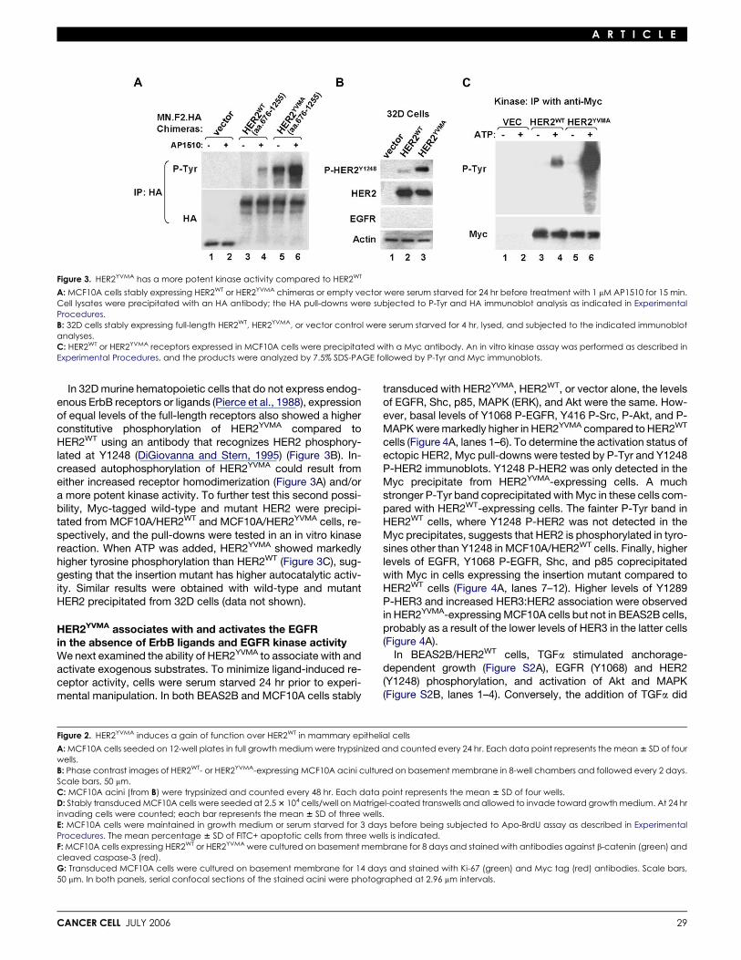
Figure 1. HER2YVMA is potently transforming in bronchial epithelial cells
A: BEAS2B cells stably expressing HER2WT, HER2YVMA, or vector were seeded on 12-well plates and allowed to grow in full growth medium or serum-free medium.Cells were trypsinized and counted every 24 hr. Each data point represents the mean 6 SD of four wells.B: Stably transduced BEAS2B cells were maintained in growth medium or serum starved for 3 days before being subjected to Apo-BrdU assay as described inExperimental Procedures. The percentage of FITC+ (apoptotic) cells is indicated. Quantitative data represent the mean 6 SD of three experiments.C: Stably transduced BEAS2B cells were seeded in colony-forming assay as described in Experimental Procedures. Colonies were photographed at day 10.D: The indicated BEAS2B cells (2 3 106) were injected s.c. in athymic nude mice. Detectable tumors of R3 mm in minimal diameter are shown (n = 6 per group).E: Left panel: close-to-confluent monolayers of BEAS2B cells in serum-free medium were wounded with a pipette tip; wound closure was monitored at theindicated times. Right panel: transwell assays. Cells that had migrated to the underside of transwell filters are shown after fixation. Quantitative data aredepicted below, with each bar representing the mean 6 SD of six fields from three independent experiments.
invade into it for 24 hr. Compared to controls, HER2WT andHER2YVMA cells invaded 3-fold and 10-fold more the otherside of the Matrigel chamber, respectively (Figure 2D).
In cell survival assays, both MCF10A/HER2WT and vectorcontrol cells exhibited 58%–62% apoptosis after 3 days of se-rum starvation, whereas cells expressing the insertion mutantwere protected from apoptosis (Figure 2E). Cells in the centerof MCF10A polarized acini grown in 3D have been shown to un-dergo apoptosis resulting in the formation of a central lumen.Apoptosis and lumen formation have been shown to be pre-vented by the expression of activated oncogenes (Debnathet al., 2002). Thus, we subjected MCF10A acini to immunofluo-rescence staining of cleaved (active) caspase-3, an apoptosismarker, and Ki-67, a proliferation marker. In day 8 HER2WT acini,the centrally located cells underwent apoptosis, as indicated bycaspase-3 staining. However, caspase-3 was undetectable inHER2YVMA acini (Figure 2F). In day 14 HER2WT acini, Ki-67 stain-ing was mostly located at the edges, whereas in HER2YVMA-ex-pressing acini, both peripheral and centrally located cells wereproliferating (Ki-67+; Figure 2G).
CANCER CELL JULY 2006
HER2YVMA kinase has stronger catalyticactivity than HER2WT
ErbB receptor chimeras containing the ligand binding domainof FK506 binding protein (FKBP) can be induced to homodi-merize by the bivalent ligand AP1510 and, therefore, providea method to elucidate the activity of ectopic ErbB homo-and heterodimers without interference from the endogenousErbB receptors (Muthuswamy et al., 1999). Thus, we con-structed HER2 chimeras containing the extracellular andtransmembrane domains of low-affinity NGF receptor (p75),the kinase domain (aa 676–1255) of HER2YVMA or HER2WT,and two copies of FKBP with an HA tag at the carboxyl termi-nus. MCF10A cells stably expressing equal levels of thetwo chimeras were treated with AP1510 to induce receptorhomodimerization. In cells expressing the wild-type chimera,HER2WT phosphorylation was modestly induced by AP1510.However, in cells expressing the mutant chimera, HER2YVMA
phosphorylation was high in the absence of AP1510 andinduced further by the addition of the dimerizing ligand(Figure 3A).
27

A R T I C L E
28 CANCER CELL JULY 2006
A R T I C L E
Figure 3. HER2YVMA has a more potent kinase activity compared to HER2WT
A: MCF10A cells stably expressing HER2WT or HER2YVMA chimeras or empty vector were serum starved for 24 hr before treatment with 1 mM AP1510 for 15 min.Cell lysates were precipitated with an HA antibody; the HA pull-downs were subjected to P-Tyr and HA immunoblot analysis as indicated in ExperimentalProcedures.B: 32D cells stably expressing full-length HER2WT, HER2YVMA, or vector control were serum starved for 4 hr, lysed, and subjected to the indicated immunoblotanalyses.C: HER2WT or HER2YVMA receptors expressed in MCF10A cells were precipitated with a Myc antibody. An in vitro kinase assay was performed as described inExperimental Procedures, and the products were analyzed by 7.5% SDS-PAGE followed by P-Tyr and Myc immunoblots.
In 32D murine hematopoietic cells that do not express endog-enous ErbB receptors or ligands (Pierce et al., 1988), expressionof equal levels of the full-length receptors also showed a higherconstitutive phosphorylation of HER2YVMA compared toHER2WT using an antibody that recognizes HER2 phosphory-lated at Y1248 (DiGiovanna and Stern, 1995) (Figure 3B). In-creased autophosphorylation of HER2YVMA could result fromeither increased receptor homodimerization (Figure 3A) and/ora more potent kinase activity. To further test this second possi-bility, Myc-tagged wild-type and mutant HER2 were precipi-tated from MCF10A/HER2WT and MCF10A/HER2YVMA cells, re-spectively, and the pull-downs were tested in an in vitro kinasereaction. When ATP was added, HER2YVMA showed markedlyhigher tyrosine phosphorylation than HER2WT (Figure 3C), sug-gesting that the insertion mutant has higher autocatalytic activ-ity. Similar results were obtained with wild-type and mutantHER2 precipitated from 32D cells (data not shown).
HER2YVMA associates with and activates the EGFRin the absence of ErbB ligands and EGFR kinase activityWe next examined the ability of HER2YVMA to associate with andactivate exogenous substrates. To minimize ligand-induced re-ceptor activity, cells were serum starved 24 hr prior to experi-mental manipulation. In both BEAS2B and MCF10A cells stably
CANCER CELL JULY 2006
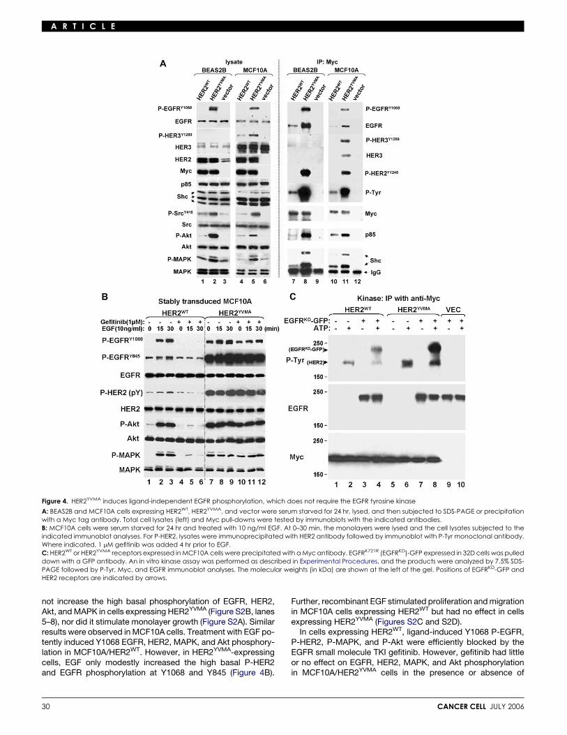
transduced with HER2YVMA, HER2WT, or vector alone, the levelsof EGFR, Shc, p85, MAPK (ERK), and Akt were the same. How-ever, basal levels of Y1068 P-EGFR, Y416 P-Src, P-Akt, and P-MAPK were markedly higher in HER2YVMA compared to HER2WT
cells (Figure 4A, lanes 1–6). To determine the activation status ofectopic HER2, Myc pull-downs were tested by P-Tyr and Y1248P-HER2 immunoblots. Y1248 P-HER2 was only detected in theMyc precipitate from HER2YVMA-expressing cells. A muchstronger P-Tyr band coprecipitated with Myc in these cells com-pared with HER2WT-expressing cells. The fainter P-Tyr band inHER2WT cells, where Y1248 P-HER2 was not detected in theMyc precipitates, suggests that HER2 is phosphorylated in tyro-sines other than Y1248 in MCF10A/HER2WT cells. Finally, higherlevels of EGFR, Y1068 P-EGFR, Shc, and p85 coprecipitatedwith Myc in cells expressing the insertion mutant compared toHER2WT cells (Figure 4A, lanes 7–12). Higher levels of Y1289P-HER3 and increased HER3:HER2 association were observedin HER2YVMA-expressing MCF10A cells but not in BEAS2B cells,probably as a result of the lower levels of HER3 in the latter cells(Figure 4A).
In BEAS2B/HER2WT cells, TGFa stimulated anchorage-dependent growth (Figure S2A), EGFR (Y1068) and HER2(Y1248) phosphorylation, and activation of Akt and MAPK(Figure S2B, lanes 1–4). Conversely, the addition of TGFa did
Figure 2. HER2YVMA induces a gain of function over HER2WT in mammary epithelial cells
A: MCF10A cells seeded on 12-well plates in full growth medium were trypsinized and counted every 24 hr. Each data point represents the mean 6 SD of fourwells.B: Phase contrast images of HER2WT- or HER2YVMA-expressing MCF10A acini cultured on basement membrane in 8-well chambers and followed every 2 days.Scale bars, 50 mm.C: MCF10A acini (from B) were trypsinized and counted every 48 hr. Each data point represents the mean 6 SD of four wells.D: Stably transduced MCF10A cells were seeded at 2.5 3 104 cells/well on Matrigel-coated transwells and allowed to invade toward growth medium. At 24 hrinvading cells were counted; each bar represents the mean 6 SD of three wells.E: MCF10A cells were maintained in growth medium or serum starved for 3 days before being subjected to Apo-BrdU assay as described in ExperimentalProcedures. The mean percentage 6 SD of FITC+ apoptotic cells from three wells is indicated.F: MCF10A cells expressing HER2WT or HER2YVMA were cultured on basement membrane for 8 days and stained with antibodies against b-catenin (green) andcleaved caspase-3 (red).G: Transduced MCF10A cells were cultured on basement membrane for 14 days and stained with Ki-67 (green) and Myc tag (red) antibodies. Scale bars,50 mm. In both panels, serial confocal sections of the stained acini were photographed at 2.96 mm intervals.
29
A R T I C L E
Figure 4. HER2YVMA induces ligand-independent EGFR phosphorylation, which does not require the EGFR tyrosine kinase
A: BEAS2B and MCF10A cells expressing HER2WT, HER2YVMA, and vector were serum starved for 24 hr, lysed, and then subjected to SDS-PAGE or precipitationwith a Myc tag antibody. Total cell lysates (left) and Myc pull-downs were tested by immunoblots with the indicated antibodies.B: MCF10A cells were serum starved for 24 hr and treated with 10 ng/ml EGF. At 0–30 min, the monolayers were lysed and the cell lysates subjected to theindicated immunoblot analyses. For P-HER2, lysates were immunoprecipitated with HER2 antibody followed by immunoblot with P-Tyr monoclonal antibody.Where indicated, 1 mM gefitinib was added 4 hr prior to EGF.C: HER2WT or HER2YVMA receptors expressed in MCF10A cells were precipitated with a Myc antibody. EGFRK721R (EGFRKD)-GFP expressed in 32D cells was pulleddown with a GFP antibody. An in vitro kinase assay was performed as described in Experimental Procedures, and the products were analyzed by 7.5% SDS-PAGE followed by P-Tyr, Myc, and EGFR immunoblot analyses. The molecular weights (in kDa) are shown at the left of the gel. Positions of EGFRKD-GFP andHER2 receptors are indicated by arrows.
not increase the high basal phosphorylation of EGFR, HER2,Akt, and MAPK in cells expressing HER2YVMA (Figure S2B, lanes5–8), nor did it stimulate monolayer growth (Figure S2A). Similarresults were observed in MCF10A cells. Treatment with EGF po-tently induced Y1068 EGFR, HER2, MAPK, and Akt phosphory-lation in MCF10A/HER2WT. However, in HER2YVMA-expressingcells, EGF only modestly increased the high basal P-HER2and EGFR phosphorylation at Y1068 and Y845 (Figure 4B).
30
Further, recombinant EGF stimulated proliferation and migrationin MCF10A cells expressing HER2WT but had no effect in cellsexpressing HER2YVMA (Figures S2C and S2D).
In cells expressing HER2WT, ligand-induced Y1068 P-EGFR,P-HER2, P-MAPK, and P-Akt were efficiently blocked by theEGFR small molecule TKI gefitinib. However, gefitinib had littleor no effect on EGFR, HER2, MAPK, and Akt phosphorylationin MCF10A/HER2YVMA cells in the presence or absence of
CANCER CELL JULY 2006

A R T I C L E
added ligand. In both cell types, gefitinib did not block EGFRphosphorylation at Y845 (Figure 4B). Phosphorylation of thisresidue does not depend on EGFR tyrosine kinase activity(Stover et al., 1995) and has been shown to be induced byc-Src (Biscardi et al., 1999), consistent with the higher levels ofP-Src in cells expressing HER2YVMA (Figure 4A). The resultswith gefitinib suggested that the EGFR/HER2 association andEGFR phosphorylation in cells expressing HER2YVMA did not re-quire EGFR’s catalytic activity. Therefore, we performed anin vitro kinase assay using GFP-fused K721R (kinase-dead)EGFR (Ewald et al., 2003) (EGFRKD) as substrate. Myc-taggedwild-type and mutant HER2 receptors were precipitated fromtransfected MCF10A cells and incubated with EGFRKD-GFP inthe presence or absence of ATP. In the absence of ATP, no phos-phorylation of EGFRKD-GFP was detected. Both HER2WT andHER2YVMA phosphorylated EGFRKD-GFP in vitro, but this effectwas at least 5-fold higher with the insertion mutant (Figure 4C).
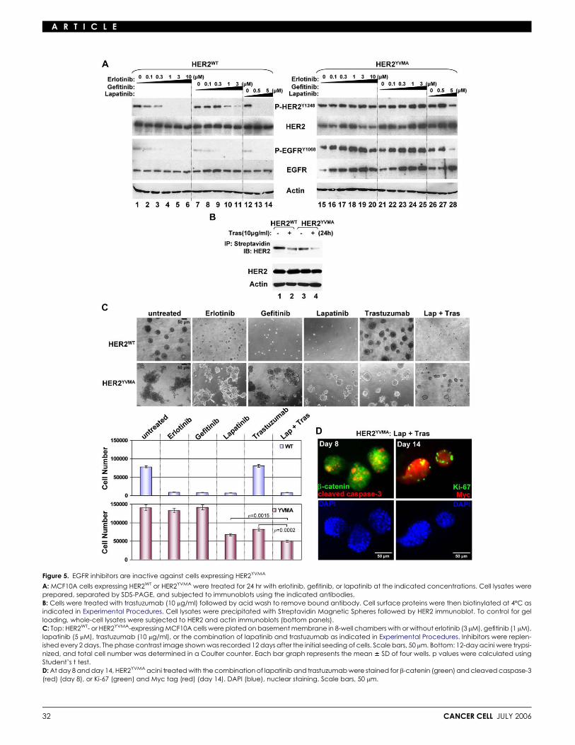
Cells expressing HER2YVMA are resistant to EGFRinhibitors but still sensitive to HER2 inhibitorsThe EGFR TKIs erlotinib and gefitinib inhibited the weak basalEGFR phosphorylation in MCF10A/HER2WT cells (Figure 5A,lanes 1–11). Although not direct inhibitors of HER2, it has beenshown that (EGFR-specific) low concentrations of erlotinib orgefitinib inhibit HER2 phosphorylation and growth of HER2-de-pendent tumor cells, presumably by blocking the EGFR tyrosinekinase and, thus, interfering with EGFR-HER2 crosstalk andheterodimerization (Hernan et al., 2003; Moasser et al., 2001;Moulder et al., 2001). Consistent with this, erlotinib and gefitinibsuppressed HER2WT cell proliferation in a dose-dependentmanner (Figure S3, top) and inhibited the 3D growth of these ac-ini in Matrigel devoid of added EGFR ligands (Figure 5C, top). Onthe other hand, the ability of HER2YVMA to transactivate theEGFR in an EGFR kinase-independent manner anticipatedlack of an inhibitory effect of EGFR TKIs against cells expressingthe insertion mutant. Indeed, erlotinib and gefitinib had no effecton EGFR and HER2 phosphorylation in MCF10A/HER2YVMA
cells (Figure 5A, lanes 15–25) nor inhibited their growth in mono-layer (Figure S3, bottom) or in 3D in Matrigel (Figure 5C, bottom).
We next examined the effect of direct inhibitors of HER2 onthe insertion mutant. Treatment with the HER2 antibody trastu-zumab downregulated both wild-type and mutant receptorsfrom the cell surface, as indicated by HER2 immunoblot ofstreptavidin precipitates of surface-biotinylated cells (Figure 5B).However, treatment with trastuzumab inhibited growth and inva-siveness of HER2YVMA but not HER2WT acini in 3D. This lack of aneffect of trastuzumab on cells overexpressing HER2WT is consis-tent with the reported inability of the IgG1 to interfere with EGF,TGFa, and heregulin-induced growth and/or EGFR/HER2 heter-odimerization (Agus et al., 2002; Moulder et al., 2001; Ye et al.,1999). The EGFR/HER2 dual TKI lapatinib (Rusnak et al., 2001)inhibited EGFR and HER2 phosphorylation in MCF10A/HER2WT
cells (Figure 5A, lanes 12–14). Lapatinib also inhibited HER2YVMA
and EGFR phosphorylation in HER2YVMA cells but at higher con-centrations (Figure 5A, lanes 26–28). Trastuzumab alone or incombination with lapatinib also suppressed Y1068 P-EGFR(Figure S4). The growth in 3D of HER2YVMA-expressing aciniwas also suppressed by lapatinib, although not as much aswith MCF10A/HER2WT acini (Figure 5C). The combination oflapatinib and trastuzumab was the most potent in suppressinggrowth of HER2YVMA acini (Figure 5C). Addition of erlotinib or
CANCER CELL JULY 2006
gefitinib did not enhance the effect of trastuzumab against thesecells (data not shown). At day 8 and day 14, HER2YVMA acinitreated with both drugs exhibited apoptotic cells in the center,similar to those exhibited by HER2WT acini (Figure 5D, comparedto Figures 2F and 2G). These data suggest that HER2YVMA is stillsensitive to direct inhibitors of HER2.
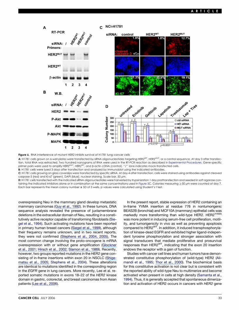
Mutant HER2 in H1781 cells is required for proliferationand survivalNCI-H1781 lung cancer cells express wild-type EGFR and con-tain a VC insertion at G776 in exon 20 of the HER2 gene (Shige-matsu et al., 2005) (Figure S1A). Small interfering RNA oligonu-cleotides targeting HER2WT, HER2MUT, or a control sequencewere transfected into H1781 cells. Three days posttransfection,we used a PCR primer set specific for mutant HER2 mRNA andobserved efficient knockdown of HER2MUT in cells expressingsiRNA targeting HER2MUT but not HER2WT. The HER2 mutationin H1781 cells has been reported to be homozygous (Shige-matsu et al., 2005), explaining the lack of HER2WT mRNA ex-pression by RT-PCR (Figure 6A). By immunoblot analysis,RNAi resulted in 90% reduction of mutant HER2 protein aswell as basal P-MAPK and P-Akt (Figure 6B). These cells ex-hibited markedly increased apoptosis (by cleaved caspase-3staining) and decreased proliferation (by Ki-67 staining), com-pared to cells transfected with siRNA targeting HER2WT or thecontrol sequence (Figure 6C). Similar to MCF10A/HER2YVMA
cells, trastuzumab, lapatinib, and the combination, but not erlo-tinib and gefitinib, inhibited H1781 colony formation (Figure 6D).In cells expressing siRNA targeting HER2MUT but not in controlcells, erlotinib and gefitinib reduced H1781 colony formation(Figure 6D), suggesting that the insertion mutant counteractsthe inhibitory effect of the EGFR TKIs.
The irreversible ErbB2 inhibitor CI-1033 is a potentinhibitor of the mutant HER2It has been shown that a mutant EGFR with an exon 20 insertionis resistant to gefitinib and erlotinib but highly sensitive to the ir-reversible covalent EGFR/HER2 inhibitor CL-387,785 (Greulichet al., 2005). Therefore, we tested the effect of a similar irrevers-ible inhibitor, the 4-anilinoquinazoline CI-1033, which is pre-dicted to covalently modify Cys773 within the ATP binding siteof the HER2 kinase (Citri et al., 2002; Smaill et al., 2000). Indose-dependent fashion, submicromolar concentrations ofCI-1033 inhibited tyrosine phosphorylation of wild-type and mu-tant HER2 and EGFR in transfected MCF10A and H1781 cells(Figure 7A). The same range of concentrations (0.1–3 mM) mark-edly inhibited growth on Matrigel of both MCF10A/HER2WT andMCF10A/HER2YVMA cells, but the dose response was steeperagainst cells expressing the mutant (Figure 7B). Similar concen-trations of CI-1033 also inhibited H1781 colony formation in softagar with a 25% and 90% reduction in colony number at 0.1 and1 mM, respectively (Figure 7C).
Discussion
The orphan HER2/Neu (ErbB2) receptor plays a critical role inthe cellular responses mediated by ligand-dependent activationof ErbB coreceptors (Yarden and Sliwkowski, 2001). Indeed,overexpression of Neu alone or in combination with EGFR orHER3 can transform mammary epithelial cells and fibroblasts(Pierce et al., 1991; Zhang et al., 1996). Transgenic mice
31
A R T I C L E
Figure 5. EGFR inhibitors are inactive against cells expressing HER2YVMA
A: MCF10A cells expressing HER2WT or HER2YVMA were treated for 24 hr with erlotinib, gefitinib, or lapatinib at the indicated concentrations. Cell lysates wereprepared, separated by SDS-PAGE, and subjected to immunoblots using the indicated antibodies.B: Cells were treated with trastuzumab (10 mg/ml) followed by acid wash to remove bound antibody. Cell surface proteins were then biotinylated at 4ºC asindicated in Experimental Procedures. Cell lysates were precipitated with Streptavidin Magnetic Spheres followed by HER2 immunoblot. To control for gelloading, whole-cell lysates were subjected to HER2 and actin immunoblots (bottom panels).C: Top: HER2WT- or HER2YVMA-expressing MCF10A cells were plated on basement membrane in 8-well chambers with or without erlotinib (3 mM), gefitinib (1 mM),lapatinib (5 mM), trastuzumab (10 mg/ml), or the combination of lapatinib and trastuzumab as indicated in Experimental Procedures. Inhibitors were replen-ished every 2 days. The phase contrast image shown was recorded 12 days after the initial seeding of cells. Scale bars, 50 mm. Bottom: 12-day acini were trypsi-nized, and total cell number was determined in a Coulter counter. Each bar graph represents the mean 6 SD of four wells. p values were calculated usingStudent’s t test.D: At day 8 and day 14, HER2YVMA acini treated with the combination of lapatinib and trastuzumab were stained for b-catenin (green) and cleaved caspase-3(red) (day 8), or Ki-67 (green) and Myc tag (red) (day 14). DAPI (blue), nuclear staining. Scale bars, 50 mm.
32 CANCER CELL JULY 2006
A R T I C L E
Figure 6. RNA interference of mutant HER2 inhibits survival of H1781 lung cancer cells
A: H1781 cells grown on 6-well plates were transfected by siRNA oligonucleotides targeting HER2WT, HER2MUT, or a control sequence. At day 3 after transfec-tion, total RNA was extracted. Two hundred nanograms of RNA were used in the RT-PCR reaction as described in Experimental Procedures. Gene-specificprimer pairs were used to amplify HER2MUT, HER2WT, and b-actin cDNA (control). ‘‘/’’ lane indicates mock-transfected cells.B: H1781 cells were lysed 3 days after transfection and analyzed by immunoblot using the indicated antibodies.C: H1781 cells growing on glass coverslips were transfected by specific siRNA. At day 4 after transfection, cells were stained using antibodies against cleavedcaspase-3 (red) and Ki-67 (green). DAPI (blue), nuclear staining. Scale bar, 50 mm.D: H1781 cells transfected with the indicated siRNA oligonucleotides were harvested by trypsinization 1 day posttransfection and seeded in soft agarose con-taining the indicated inhibitors alone or in combination at the same concentrations used in Figure 5C. Colonies measuring R50 mm were counted at day 7.Each bar represents the mean colony number 6 SD of 3 wells. p values were calculated using Student’s t test.
overexpressing Neu in the mammary gland develop metastaticmammary carcinomas (Guy et al., 1992). In these tumors, DNAsequence analysis revealed the presence of juxtamembranedeletions in the extracellular domain of Neu, resulting in a consti-tutively active receptor capable of transforming fibroblasts (Sie-gel et al., 1994). Such activating mutations have been reportedin primary human breast cancers (Siegel et al., 1999), althoughtheir frequency remains unknown, and in two recent reports,they were not confirmed (Stephens et al., 2004, 2005). Themost common change involving the proto-oncogene is mRNAoverexpression with or without gene amplification (Glockneret al., 2001; Hirsch et al., 2002; Slamon et al., 1989). Recently,however, two groups reported mutations in the HER2 gene con-sisting of in-frame insertions within exon 20 in NSCLC (Shige-matsu et al., 2005; Stephens et al., 2004). These alterationsare identical to mutations identified in the corresponding regionin the EGFR gene in lung cancers. More recently, Lee et al. re-ported somatic mutations in exons 18–22 of the HER2 kinasedomain in gastric, colorectal, and breast carcinomas from Asianpatients (Lee et al., 2006).
CANCER CELL JULY 2006
In the present report, stable expression of HER2 containing anin-frame YVMA insertion at residue 776 in nontumorigenicBEAS2B (bronchial) and MCF10A (mammary) epithelial cells wasmarkedly more transforming than wild-type HER2. HER2YVMA
was more potent in inducing serum-free cell proliferation, motil-ity, and tumorigenicity in vivo as well as preventing apoptosiscompared to HER2WT. In addition, it induced transphosphoryla-tion of kinase-dead EGFR and exhibited higher ligand-indepen-dent tyrosine phosphorylation and stronger association withsignal transducers that mediate proliferative and prosurvivalresponses than HER2WT, indicating that the exon 20 insertionendows the receptor with a gain of function.
Studies with cancer cell lines and human tumors have demon-strated constitutive phosphorylation of (wild-type) HER2 (Ali-mandi et al., 1995; Thor et al., 2000). The biochemical basisfor this constitutive activation is not clear but is consistent withthe reported ability of wild-type Neu to multimerize and becomeactivated when present in cells at high density (Samanta et al.,1994). Thus, it is generally accepted that spontaneous dimeriza-tion and activation of HER2 occurs in cancers with HER2 gene
33

A R T I C L E
Figure 7. CI-1033 inhibits mutant HER2-driven growth and signaling
A: MCF10A cells expressing HER2WT or HER2YVMA and H1781 cells were treated for 16 hr with CI-1033 at the indicated concentrations. Cell lysates were pre-pared, separated by SDS-PAGE, and subjected to immunoblots using the indicated antibodies.B: Top: HER2WT- or HER2YVMA-expressing MCF10A cells were plated on basement membrane in 8-well chambers with or without CI-1033. Inhibitors were replen-ished every 2 days. Phase contrast image shown was recorded 12 days after the initial seeding of cells. Scale bars, 50 mm. Bottom: 12-day acini were trypsi-nized, and total cell number was determined in a Coulter counter. Each bar graph represents the mean 6 SD of four wells.C: H1781 cells were seeded in soft agarose containing the indicated concentration of CI-1033. Colonies measuring R50 mm were counted at day 7. Each barrepresents the mean colony number 6 SD of three wells.
amplification. Another possible mechanism for activation ofthe HER2 kinase is transactivation by ligand bound EGFR orHER3/4. Indeed, coexpression of the EGFR ligand TGFa with Neuin the mammary gland of transgenic mice markedly acceleratestumor onset and progression compared to mice expressingthe Neu or TGFa transgenes alone. In this study, TGFa/Neu bi-genic mice exhibited increased tyrosine phosphorylation of bothEGFR and Neu (Muller et al., 1996), providing evidence of EGFR-Neu crosstalk and of the ability of HER2/Neu to amplify ligand-activated EGFR signals.
On the other hand, EGFR function has been shown to be re-quired for Neu-induced transformation. For example, EGFRTKIs can markedly delay Neu-induced tumors in transgenicmice (Lenferink et al., 2000; Lu et al., 2003). Further, MMTV/Neu mice carrying a catalytically impaired EGFR (waved-2mice) developed breast tumors only after a prolonged latency,and the tumors that developed were fewer in number (Gillgrasset al., 2003). In MCF10A/HER2WT cells, EGFR inhibition withgefitinib blocks the gain-of-function effects of transfectedwild-type HER2 (Figure 5 and Ueda et al. [2004]). HER2-overex-pressing cancers that also overexpress EGFR exhibit a worseoutcome compared to tumors with high HER2 but undetectableEGFR (DiGiovanna et al., 2005). Finally, EGFR-positive NSCLCsthat also exhibit increased (wild-type) HER2 gene copy numberrespond clinically to EGFR TKIs (Cappuzzo et al., 2005; Hirsch
34
et al., 2002), further suggesting that the EGFR is required for sig-naling by wild-type HER2.
In BEAS2B and MCF10A cells, HER2YVMA potently inducedEGFR phosphorylation. This transactivation did not require theEGFR tyrosine kinase in that it was also observed with catalyti-cally dead K721R EGFR and was not blocked by the EGFR TKIserlotinib and gefitinib. These results suggest that in hetero-dimers containing EGFR and exon 20 HER2 mutants, theEGFR tyrosine kinase activity is dispensable. EGFR functionhas been shown to be critical for the antiapoptotic signals gen-erated by other oncogenes such as SOS (Sibilia et al., 2000).Therefore, it is conceivable that transactivation of the EGFR isalso critical for the potent antiapoptotic effect of HER2YVMA. Ifso, HER2 mutations would overlap functionally with EGFR acti-vating mutations, perhaps explaining the reported lack of coex-istence of these alterations in primary human tumors (Lee et al.,2006; Shigematsu et al., 2005; Stephens et al., 2004).
The receptor-specific effects of EGFR and HER2 on transfor-mation have been characterized in MCF10A mammary epithelialcells expressing chimeric EGFR or HER2 that can be homodi-merized by bivalent synthetic ligands (Muthuswamy et al.,1999). In this system, activation of HER2, but not the EGFR,has been shown to reinitiate proliferation and generate 3D multi-acinar MCF10A structures with a filled lumen where apoptosisis abrogated. In the study herein, HER2YVMA but not HER2WT
CANCER CELL JULY 2006

A R T I C L E
Figure 8. Schema of HER2YVMA signaling outputand potential response to ErbB inhibitors
Signaling by EGFR homodimers is brief and lim-ited because of receptor endocytosis and deg-radation following ligand-mediated activation(Yarden and Sliwkowski, 2001) (left). Ligand-in-duced heterodimers of EGFR and wild-typeHER2 are more stable and recruit additionalcytoplasmic transducers, thus signaling with in-creased potency compared to EGFR homo-dimers (Yarden and Sliwkowski, 2001). Probablybecause of EGFR/HER2 transactivation, cellsdriven by these heterodimers are sensitive toEGFR TKIs (middle). In cells expressing the HER2exon 20 insertion mutant, HER2 dimerizes consti-tutively and associates with and transactivatesthe EGFR. This HER2/EGFR transactivationdoes not require the EGFR tyrosine kinase, ex-plaining their resistance to EGFR TKIs. Althoughat higher concentrations than those required toinhibit EGFR and wild-type HER2 phosphoryla-tion, lapatinib still inhibits HER2YVMA phosphoryla-tion. Irreversible ErbB TKI CI-1033 inhibits bothHER2WT and HER2YVMA phosphorylation. LikeHER2WT, the mutant receptor is partially downre-gulated from the cell surface by trastuzumab(right), which also inhibits cells expressing thismutation.
behaved similarly to ligand-activated chimeric ErbB2 reportedby Muthuswamy et al. (2001) in the absence of AP1510, support-ing the ability of the insertion mutant to potently engage antia-poptotic signals in a ligand-independent fashion. AlthoughHER2WT was also phosphorylated, it associated with Shc andp85 with lower efficiency and activated MAPK and Akt weaklycompared to HER2YVMA, thus potentially not reaching a thresh-old required for the generation of sustained survival signals.
Inhibition of mutant HER2 with RNA interference in H1781lung cancer cells, which contain a VC insertion at G776 inexon 20 of the HER2 gene, inhibited P-MAPK and P-Akt, re-duced proliferation, and induced cell death. These cells havehighly phosphorylated wild-type EGFR (Tracy et al., 2004) andare insensitive to gefitinib and erlotinib (Figure 6). Of note, a sim-ilar insertion is exon 20 of the EGFR is also resistant to theseEGFR TKIs (Greulich et al., 2005). However, direct inhibitors ofHER2 such as trastuzumab, lapatinib, and CI-1033 reducedcolony formation by MCF10A/HER2YVMA and H1781 cells. InMCF10/HER2YVMA cells, treatment with trastuzumab downre-gulated mutant HER2 from the cell surface, while lapatinib andCI-1033 inhibited HER2 phosphorylation, further suggestingthat cancers expressing this mutant remain HER2 dependentand thus sensitive to HER2-targeted therapies. Clinical data tosupport this possibility are not available, since there are nopublished trials with single-agent trastuzumab or lapatinib inNSCLC, where a low frequency of HER2 mutations has beenreported. Thus, the therapeutic efficacy of anti-HER2 strategiesin this patient population requires prospective investigation.
Finally, and based on the results with MCF10A and H1781cells treated with erlotinib and gefitinib, we propose, first, thatEGFR inhibitors would be effective against tumors driven by het-erodimers of EGFR and wild-type HER2. In these cancers, HER2amplifies signals by EGFR/HER2 heterodimers, which remaindependent on the EGFR kinase (Figure 8). This possibility is sup-ported by a recent report in which EGFR-positive NSCLCs withwild-type HER2 gene amplification responded clinically to EGFR
CANCER CELL JULY 2006
TKI gefitinib (Cappuzzo et al., 2005). In a recent randomizedsurvival study of erlotinib versus best supportive care in NSCLCpatients (Shepherd et al., 2005), this correlation with clinicalresponse remains to be investigated. Second, we predict thatcancers expressing mutation/insertions in exon 20 of HER2would be insensitive to small molecule inhibitors of the EGFR ty-rosine kinase. Indeed, in a very recent study from Korea, all fourpatients with lung adenocarcinoma containing HER2 mutationsprogressed on gefitinib. Three of the four contained mutations inexon 20 (Han et al., 2006). Third, in these cancers, a combinationof trastuzumab and lapatinib or the irreversible HER2 inhibitorCI-1033 may have clinical activity and is worthy of prospectiveinvestigation.
Experimental procedures
Cell lines, plasmids, and viruses
All cells were from the American Type Culture Collection (ATCC). Cell culture,
plasmid cloning, and retroviral transduction were carried out according to
standard procedures (details provided in Supplemental Data). The following
reagents were used: human TGFa and EGF (Calbiochem), puromycin (Cal-
biochem), G418 (Research Products International Corp.), erlotinib (from
Mark Sliwkowski, Genentech), gefitinib (from Alan Wakeling, AstraZeneca
Pharmaceuticals), lapatinib (from Tona Gilmer, GlaxoSmithKline), and CI-
1033 (from David Fry, Parke-Davis). Trastuzumab was purchased at the Van-
derbilt University Medical Center Pharmacy. The EGFRK721R-GFP plasmid
was kindly provided by Brent Polk (Vanderbilt University).
Immunoprecipitation and immunoblot analysis
Immunoprecipitation and immunoblot analysis were performed using stan-
dard protocols (details provided in Supplemental Data).
Cell growth, apoptosis, and motility assays
Equal numbers of cells were seeded on 12-well plates and allowed to grow in
full medium. Cells were harvested by trypsinization every 24 hr, and cell num-
ber was determined in a Coulter counter. Cells growing on 100 mm dishes
were serum starved for 3 days and collected for apoptosis assay using an
Apo-BrdU kit (Phoenix Flow Systems, Inc.) according to the manufacturer’s
35
A R T I C L E
protocol. FITC-positive apoptotic cells were quantitated in a FACS/Calibur
Flow Cytometer (BD Biosciences).
For wound closure assays, cells were allowed to reach confluence on a 6-
well plate and then serum starved for 24 hr. The monolayers were scraped
with a plastic pipette tip and replenished with fresh serum-free medium.
Phase contrast images were photographed at 0, 10, and 24 hr after wound-
ing. Transwell motility assays were performed utilizing 5 mm pore, 6.5 mm
polycarbonate transwell filters (Corning Costar Corp.). Cells (1.5 3 105 per
well) were seeded in serum-free medium onto the upper surface of the filters
and allowed to migrate. After 24 hr, the cells on the upper surface of the filters
were wiped off with a cotton swab. Cells that had migrated to the filter under-
side were fixed, stained with Diff-Quik stain set (Dade Behring, Inc.), and
counted by bright field microscopy.
Soft agarose colony-forming assay
Base layers consisting of growth medium containing 0.8% low-melting point
agarose (Gibco) and 10 mM HEPES (pH 7.5) were poured onto 6-well plates
and allowed to solidify. Cells (3 3 104 per well) were plated in triplicate in top
layers consisting of growth medium containing 0.4% agarose and 5 mM
HEPES. Colonies measuring R50 mm were photographed after 7–10 days
and counted manually.
Xenograft studies
Exponentially growing cells on 150 mm dishes were scraped off and resus-
pended in serum-free medium; 2 3 106 cells in 0.3 ml were then injected sub-
cutaneously via a 22-gauge needle into each of 4-week-old female athymic
nude mice (Harlan Sprague-Dawley). Tumor formation was monitored by pal-
pation twice a week. Volume of tumors measuring R3 mm in diameter was
calculated by the formula: volume = width2 3 length/2. These experiments
were approved by the Vanderbilt Institutional Animal Care Committee and
performed under institutional guidelines in accordance with approved regu-
latory standards.
Three-dimensional morphogenesis and indirect
immunofluorescence
MCF10A cells expressing HER2WT, HER2YVMA, or vector were seeded on
Growth Factor Reduced Matrigel (BD Biosciences) in 8-well chamber slides
following the protocol described by Debnath et al. (2003) Morphogenesis of
acini was photographed every 2 days. For cell number counting, cultures
growing on Matrigel were trypsinized and cell numbers were measured in
a Coulter counter. Invasion assay was performed in BD BioCoat Growth
Factor Reduced Matrigel invasion chambers (BD Biosciences) according
to the manufacturer’s protocol. Immunofluorescence staining of 3D acini
was performed as described by Debnath et al. (2003) using antibodies
against b-catenin (BD Biosciences), cleaved caspase-3 (Cell Signaling),
Ki-67 (Calbiochem), and Myc tag. Confocal analyses were performed with
Zeiss inverted LSM510 confocal microscopy system. Indirect immunofluo-
rescence assay (IFA) was performed as described previously (Wang et al.,
2005). Fluorescent images were captured using a Princeton Instruments
cooled CCD digital camera from a Zeiss Axiophot upright microscope. Pri-
mary antibodies include Ki-67 and cleaved caspase-3. The fluorescent
antibodies are Oregon green-a-mouse IgG and Texas red-a-rabbit IgG
(Molecular Probes).
Transfection of DNA and siRNA, total RNA extraction, and RT-PCR
Cell transfection, RNA extraction, and RT-PCR were performed using stan-
dard protocols (details provided in Supplemental Data).
Cell surface biotinylationCells plated in 100 mm dishes were treated or not with trastuzumab (10 mg/ml)
for 24 hr at 37ºC and washed with PBS. Bound antibody was removed by in-
cubating cells for 6 min on ice with cold acid wash buffer (0.5 M NaCl, 0.2 M
sodium acetate [pH 3.0]). After three washes with cold PBS (pH 8.0), cells
were incubated with freshly prepared Sulfo-NHS-Biotin reagent (2 mM;
Pierce) for 30 min at 4ºC. The reaction was quenched with 100 mM glycine
in PBS, and the cells were lysed in NP-40 buffer. Equal amounts of protein
extracts (500 mg) were subjected to precipitation using Streptavidin Magnetic
Spheres (Promega) followed by SDS-PAGE and HER2 immunoblot.
36
In vitro kinase assays
Five hundred micrograms of total protein extracted from MCF10A/HER2WT
or MCF10A/HER2YVMA cells were immunoprecipitated with a Myc tag anti-
body. The precipitates were washed twice in NP-40 lysis buffer, twice in
kinase buffer (20 mM HEPES [pH 7.5], 10 mM MgCl2, 10 mM MnCl2, 1 mM
dithiothreitol, 0.1 mM Na3VO4), and then aliquoted on ice into two equal por-
tions, each brought up to a final volume of 40 ml by adding kinase buffer. ATP
was added to one aliquot (final concentration 0.1 mM) but not to the other
aliquot. The kinase reaction was allowed to proceed for 5 min at 30ºC and
then terminated by adding 53 loading buffer and boiling for 3 min before
separation by 7.5% SDS-PAGE followed by immunoblot analysis. In the re-
action using kinase-dead EGFR as substrate, the EGFRK721R-GFP plasmid
was transiently expressed by 32D cells followed by precipitation using
a GFP antibody. Precipitated EGFRK721R-GFP was incubated with HER2WT,
HER2YVMA receptors, or control precipitates in the absence or presence of
0.1 mM ATP. A kinase reaction was carried out under the same conditions
as above, and its products were resolved by 7.5% SDS-PAGE and visualized
by P-Tyr, EGFR, and Myc immunoblot analyses.
Supplemental data
The Supplemental Data include Supplemental Experimental Procedures and
four supplemental figures and can be found with this article online at http://
www.cancercell.org/cgi/content/full/10/1/25/DC1/.
Acknowledgments
This work was supported by NCI R01 CA62212 (C.L.A.), R01 CA80195
(C.L.A.), Breast Cancer Specialized Program of Research Excellence
(SPORE) grant P50 CA98131, Lung Cancer SPORE grant P50 CA90949,
and Vanderbilt-Ingram Comprehensive Cancer Center Support Grant P30
CA68485. M.P.-T. is supported by NCI T32 CA78136.
Received: April 13, 2006Revised: May 13, 2006Accepted: May 31, 2006Published: July 17, 2006
References
Agus, D.B., Akita, R.W., Fox, W.D., Lewis, G.D., Higgins, B., Pisacane, P.I.,Lofgren, J.A., Tindell, C., Evans, D.P., Maiese, K., et al. (2002). Targetingligand-activated ErbB2 signaling inhibits breast and prostate tumor growth.Cancer Cell 2, 127–137.
Alimandi, M., Romano, A., Curia, M.C., Muraro, R., Fedi, P., Aaronson, S.A.,Di Fiore, P.P., and Kraus, M.H. (1995). Cooperative signaling of ErbB3 andErbB2 in neoplastic transformation and human mammary carcinomas.Oncogene 10, 1813–1821.
Biscardi, J.S., Maa, M.C., Tice, D.A., Cox, M.E., Leu, T.H., and Parsons, S.J.(1999). c-Src-mediated phosphorylation of the epidermal growth factorreceptor on Tyr845 and Tyr1101 is associated with modulation of receptorfunction. J. Biol. Chem. 274, 8335–8343.
Cappuzzo, F., Varella-Garcia, M., Shigematsu, H., Domenichini, I., Bartolini,S., Ceresoli, G.L., Rossi, E., Ludovini, V., Gregorc, V., Toschi, L., et al. (2005).Increased HER2 gene copy number is associated with response to gefitinibtherapy in epidermal growth factor receptor-positive non-small-cell lung can-cer patients. J. Clin. Oncol. 23, 5007–5018.
Citri, A., Alroy, I., Lavi, S., Rubin, C., Xu, W., Grammatikakis, N., Patterson,C., Neckers, L., Fry, D.W., and Yarden, Y. (2002). Drug-induced ubiquitylationand degradation of ErbB receptor tyrosine kinases: implications for cancertherapy. EMBO J. 21, 2407–2417.
Debnath, J., and Brugge, J.S. (2005). Modelling glandular epithelial cancersin three-dimensional cultures. Nat. Rev. Cancer 5, 675–688.
Debnath, J., Mills, K.R., Collins, N.L., Reginato, M.J., Muthuswamy, S.K., andBrugge, J.S. (2002). The role of apoptosis in creating and maintaining luminalspace within normal and oncogene-expressing mammary acini. Cell 111,29–40.
CANCER CELL JULY 2006

A R T I C L E
Debnath, J., Muthuswamy, S.K., and Brugge, J.S. (2003). Morphogenesisand oncogenesis of MCF-10A mammary epithelial acini grown in three-dimensional basement membrane cultures. Methods 30, 256–268.
DiGiovanna, M.P., and Stern, D.F. (1995). Activation state-specific monoclo-nal antibody detects tyrosine phosphorylated p185neu/erbB-2 in a subset ofhuman breast tumors overexpressing this receptor. Cancer Res. 55, 1946–1955.
DiGiovanna, M.P., Stern, D.F., Edgerton, S.M., Whalen, S.G., Moore, D., II,and Thor, A.D. (2005). Relationship of epidermal growth factor receptor ex-pression to ErbB-2 signaling activity and prognosis in breast cancer patients.J. Clin. Oncol. 23, 1152–1160.
Ewald, J.A., Wilkinson, J.C., Guyer, C.A., and Staros, J.V. (2003). Ligand- andkinase activity-independent cell survival mediated by the epidermal growthfactor receptor expressed in 32D cells. Exp. Cell Res. 282, 121–131.
Gazdar, A.F., Shigematsu, H., Herz, J., and Minna, J.D. (2004). Mutations andaddiction to EGFR: the Achilles ‘heal’ of lung cancers? Trends Mol. Med. 10,481–486.
Gillgrass, A., Cardiff, R.D., Sharan, N., Kannan, S., and Muller, W.J. (2003).Epidermal growth factor receptor-dependent activation of Gab1 is involvedin ErbB-2-mediated mammary tumor progression. Oncogene 22, 9151–9155.
Glockner, S., Lehmann, U., Wilke, N., Kleeberger, W., Langer, F., and Kreipe,H. (2001). Amplification of growth regulatory genes in intraductal breastcancer is associated with higher nuclear grade but not with the progressionto invasiveness. Lab. Invest. 81, 565–571.
Graus-Porta, D., Beerli, R.R., Daly, J.M., and Hynes, N.E. (1997). ErbB-2, thepreferred heterodimerization partner of all ErbB receptors, is a mediator oflateral signaling. EMBO J. 16, 1647–1655.
Greulich, H., Chen, T.H., Feng, W., Janne, P.A., Alvarez, J.V., Zappaterra, M.,Bulmer, S.E., Frank, D.A., Hahn, W.C., Sellers, W.R., and Meyerson, M.(2005). Oncogenic transformation by inhibitor-sensitive and -resistantEGFR mutants. PLoS Med 2, e313. 10.1371/journal.pmed.0020313.
Guy, C.T., Webster, M.A., Schaller, M., Parsons, T.J., Cardiff, R.D., andMuller, W.J. (1992). Expression of the neu protooncogene in the mammaryepithelium of transgenic mice induces metastatic disease. Proc. Natl.Acad. Sci. USA 89, 10578–10582.
Han, S.W., Kim, T.Y., Jeon, Y.K., Hwang, P.G., Im, S.A., Lee, K.H., Kim, J.H.,Kim, D.W., Heo, D.S., Kim, N.K., et al. (2006). Optimization of patient selec-tion for gefitinib in non-small cell lung cancer by combined analysis of epider-mal growth factor receptor mutation, K-ras mutation, and Akt phosphoryla-tion. Clin. Cancer Res. 12, 2538–2544.
Hernan, R., Fasheh, R., Calabrese, C., Frank, A.J., Maclean, K.H., Allard, D.,Barraclough, R., and Gilbertson, R.J. (2003). ERBB2 up-regulates S100A4and several other prometastatic genes in medulloblastoma. Cancer Res.63, 140–148.
Hirsch, F.R., Varella-Garcia, M., Franklin, W.A., Veve, R., Chen, L., Helfrich,B., Zeng, C., Baron, A., and Bunn, P.A., Jr. (2002). Evaluation of HER-2/neu gene amplification and protein expression in non-small cell lung carcino-mas. Br. J. Cancer 86, 1449–1456.
Lee, J.W., Soung, Y.H., Seo, S.H., Kim, S.Y., Park, C.H., Wang, Y.P., Park, K.,Nam, S.W., Park, W.S., Kim, S.H., et al. (2006). Somatic mutations of ERBB2kinase domain in gastric, colorectal, and breast carcinomas. Clin. CancerRes. 12, 57–61.
Lenferink, A.E., Simpson, J.F., Shawver, L.K., Coffey, R.J., Forbes, J.T., andArteaga, C.L. (2000). Blockade of the epidermal growth factor receptor tyro-sine kinase suppresses tumorigenesis in MMTV/Neu + MMTV/TGF-a bigenicmice. Proc. Natl. Acad. Sci. USA 97, 9609–9614.
Lu, C., Speers, C., Zhang, Y., Xu, X., Hill, J., Steinbis, E., Celestino, J., Shen,Q., Kim, H., Hilsenbeck, S., et al. (2003). Effect of epidermal growth factorreceptor inhibitor on development of estrogen receptor-negative mammarytumors. J. Natl. Cancer Inst. 95, 1825–1833.
Moasser, M.M., Basso, A., Averbuch, S.D., and Rosen, N. (2001). The tyro-sine kinase inhibitor ZD1839 (‘‘Iressa’’) inhibits HER2-driven signaling andsuppresses the growth of HER2-overexpressing tumor cells. Cancer Res.61, 7184–7188.
CANCER CELL JULY 2006
Moulder, S.L., Yakes, F.M., Muthuswamy, S.K., Bianco, R., Simpson, J.F.,and Arteaga, C.L. (2001). Epidermal growth factor receptor (HER1) tyrosinekinase inhibitor (Iressa) inhibits HER2/neu (erbB2)-overexpressing breastcancer cells in vitro and in vivo. Cancer Res. 61, 8887–8895.
Muller, W.J., Arteaga, C.L., Muthuswamy, S.K., Siegel, P.M., Webster, M.A.,Cardiff, R.D., Meise, K.S., Li, F., Halter, S.A., and Coffey, R.J. (1996). Syner-gistic interaction of the Neu proto-oncogene product and transforminggrowth factor a in the mammary epithelium of transgenic mice. Mol. Cell.Biol. 16, 5726–5736.
Muthuswamy, S.K., Gilman, M., and Brugge, J.S. (1999). Controlled dimer-ization of ErbB receptors provides evidence for differential signaling byhomo- and heterodimers. Mol. Cell. Biol. 19, 6845–6857.
Muthuswamy, S.K., Li, D., Lelievre, S., Bissell, M.J., and Brugge, J.S. (2001).ErbB2, but not ErbB1, reinitiates proliferation and induces luminal repopula-tion in epithelial acini. Nat. Cell Biol. 3, 785–792.
Pierce, J.H., Ruggiero, M., Fleming, T.P., Di Fiore, P.P., Greenberger, J.S.,Varticovski, L., Schlessinger, J., Rovera, G., and Aaronson, S.A. (1988). Sig-nal transduction through the EGF receptor transfected in IL-3-dependenthematopoietic cells. Science 239, 628–631.
Pierce, J.H., Arnstein, P., DiMarco, E., Artrip, J., Kraus, M.H., Lonardo, F., DiFiore, P.P., and Aaronson, S.A. (1991). Oncogenic potential of erbB-2 in hu-man mammary epithelial cells. Oncogene 6, 1189–1194.
Pinkas-Kramarski, R., Soussan, L., Waterman, H., Levkowitz, G., Alroy, I.,Klapper, L., Lavi, S., Seger, R., Ratzkin, B.J., Sela, M., and Yarden, Y.(1996). Diversification of Neu differentiation factor and epidermal growth fac-tor signaling by combinatorial receptor interactions. EMBO J. 15, 2452–2467.
Rusnak, D.W., Affleck, K., Cockerill, S.G., Stubberfield, C., Harris, R., Page,M., Smith, K.J., Guntrip, S.B., Carter, M.C., Shaw, R.J., et al. (2001). Thecharacterization of novel, dual ErbB-2/EGFR, tyrosine kinase inhibitors: po-tential therapy for cancer. Cancer Res. 61, 7196–7203.
Samanta, A., LeVea, C.M., Dougall, W.C., Qian, X., and Greene, M.I. (1994).Ligand and p185c-neu density govern receptor interactions and tyrosine ki-nase activation. Proc. Natl. Acad. Sci. USA 91, 1711–1715.
Shepherd, F.A., Rodrigues Pereira, J., Ciuleanu, T., Tan, E.H., Hirsh, V.,Thongprasert, S., Campos, D., Maoleekoonpiroj, S., Smylie, M., Martins,R., et al. (2005). Erlotinib in previously treated non-small-cell lung cancer.N. Engl. J. Med. 353, 123–132.
Shigematsu, H., Takahashi, T., Nomura, M., Majmudar, K., Suzuki, M., Lee,H., Wistuba, I.I., Fong, K.M., Toyooka, S., Shimizu, N., et al. (2005). Somaticmutations of the HER2 kinase domain in lung adenocarcinomas. Cancer Res.65, 1642–1646.
Sibilia, M., Fleischmann, A., Behrens, A., Stingl, L., Carroll, J., Watt, F.M.,Schlessinger, J., and Wagner, E.F. (2000). The EGF receptor provides an es-sential survival signal for SOS-dependent skin tumor development. Cell 102,211–220.
Siegel, P.M., Dankort, D.L., Hardy, W.R., and Muller, W.J. (1994). Novel ac-tivating mutations in the neu proto-oncogene involved in induction of mam-mary tumors. Mol. Cell. Biol. 14, 7068–7077.
Siegel, P.M., Ryan, E.D., Cardiff, R.D., and Muller, W.J. (1999). Elevated ex-pression of activated forms of Neu/ErbB-2 and ErbB-3 are involved in theinduction of mammary tumors in transgenic mice: implications for humanbreast cancer. EMBO J. 18, 2149–2164.
Slamon, D.J., Godolphin, W., Jones, L.A., Holt, J.A., Wong, S.G., Keith, D.E.,Levin, W.J., Stuart, S.G., Udove, J., Ullrich, A., et al. (1989). Studies of theHER-2/neu proto-oncogene in human breast and ovarian cancer. Science244, 707–712.
Slamon, D.J., Leyland-Jones, B., Shak, S., Fuchs, H., Paton, V., Bajamonde,A., Fleming, T., Eiermann, W., Wolter, J., Pegram, M., et al. (2001). Use ofchemotherapy plus a monoclonal antibody against HER2 for metastaticbreast cancer that overexpresses HER2. N. Engl. J. Med. 344, 783–792.
Smaill, J.B., Rewcastle, G.W., Loo, J.A., Greis, K.D., Chan, O.H., Reyner,E.L., Lipka, E., Showalter, H.D., Vincent, P.W., Elliott, W.L., and Denny,W.A. (2000). Tyrosine kinase inhibitors. 17. Irreversible inhibitors of the epi-dermal growth factor receptor: 4-(phenylamino)quinazoline- and 4-(phenyla-mino)pyrido. J. Med. Chem. 43, 1380–1397.
37
A R T I C L E
Stephens, P., Hunter, C., Bignell, G., Edkins, S., Davies, H., Teague, J., Ste-vens, C., O’Meara, S., Smith, R., Parker, A., et al. (2004). Lung cancer: intra-genic ERBB2 kinase mutations in tumours. Nature 431, 525–526.
Stephens, P., Edkins, S., Davies, H., Greenman, C., Cox, C., Hunter, C.,Bignell, G., Teague, J., Smith, R., Stevens, C., et al. (2005). A screen of thecomplete protein kinase gene family identifies diverse patterns of somaticmutations in human breast cancer. Nat. Genet. 37, 590–592.
Stover, D.R., Becker, M., Liebetanz, J., and Lydon, N.B. (1995). Src phos-phorylation of the epidermal growth factor receptor at novel sites mediatesreceptor interaction with Src and P85 a. J. Biol. Chem. 270, 15591–15597.
Thor, A.D., Liu, S., Edgerton, S., Moore, D., 2nd, Kasowitz, K.M., Benz, C.C.,Stern, D.F., and DiGiovanna, M.P. (2000). Activation (tyrosine phosphoryla-tion) of ErbB-2 (HER-2/neu): a study of incidence and correlation with out-come in breast cancer. J. Clin. Oncol. 18, 3230–3239.
Tracy, S., Mukohara, T., Hansen, M., Meyerson, M., Johnson, B.E., andJanne, P.A. (2004). Gefitinib induces apoptosis in the EGFRL858R non-small-cell lung cancer cell line H3255. Cancer Res. 64, 7241–7244.
Ueda, Y., Wang, S., Dumont, N., Yi, J.Y., Koh, Y., and Arteaga, C.L. (2004).Overexpression of HER2 (erbB2) in human breast epithelial cells unmaskstransforming growth factor b-induced cell motility. J. Biol. Chem. 279,24505–24513.
Vogel, C.L., Cobleigh, M.A., Tripathy, D., Gutheil, J.C., Harris, L.N., Fehren-bacher, L., Slamon, D.J., Murphy, M., Novotny, W.F., Burchmore, M., et al.
38
(2002). Efficacy and safety of trastuzumab as a single agent in first-line treat-ment of HER2-overexpressing metastatic breast cancer. J. Clin. Oncol. 20,719–726.
Wang, L.M., Kuo, A., Alimandi, M., Veri, M.C., Lee, C.C., Kapoor, V., Ellmore,N., Chen, X.H., and Pierce, J.H. (1998). ErbB2 expression increases thespectrum and potency of ligand-mediated signal transduction throughErbB4. Proc. Natl. Acad. Sci. USA 95, 6809–6814.
Wang, S.E., Wu, F.Y., Shin, I., Qu, S., and Arteaga, C.L. (2005). Transforminggrowth factor b (TGF-b)-Smad target gene protein tyrosine phosphatasereceptor type kappa is required for TGF-b function. Mol. Cell. Biol. 25,4703–4715.
Worthylake, R., Opresko, L.K., and Wiley, H.S. (1999). ErbB-2 amplificationinhibits down-regulation and induces constitutive activation of both ErbB-2and epidermal growth factor receptors. J. Biol. Chem. 274, 8865–8874.
Yarden, Y., and Sliwkowski, M.X. (2001). Untangling the ErbB signalling net-work. Nat. Rev. Mol. Cell Biol. 2, 127–137.
Ye, D., Mendelsohn, J., and Fan, Z. (1999). Augmentation of a humanizedanti-HER2 mAb 4D5 induced growth inhibition by a human-mouse chimericanti-EGF receptor mAb C225. Oncogene 18, 731–738.
Zhang, K., Sun, J., Liu, N., Wen, D., Chang, D., Thomason, A., and Yoshi-naga, S.K. (1996). Transformation of NIH 3T3 cells by HER3 or HER4 recep-tors requires the presence of HER1 or HER2. J. Biol. Chem. 271, 3884–3890.
CANCER CELL JULY 2006