hepatocyte-specific activation of nf-κb does not aggravate chemical hepatocarcinogenesis in...
TRANSCRIPT

TitleHepatocyte-specific activation of NF-kB does notaggravate chemical hepatocarcinogenesis in transgenicmice
Author(s) Yau, TO; Chan, CF; Lam, SGS; Cheung, OF; Ching, YP;Jin, DY; Sham, MH; Ng, IOL
Citation Journal of Pathology, 2009, v. 217 n. 3, p. 353-361
Issue Date 2009
URL http://hdl.handle.net/10722/129101
Rights Journal of Pathology. Copyright © John Wiley & Sons.

For Peer ReviewHepatocyte-specific activation of NF- B does not aggravate chemical
hepatocarcinogenesis in transgenic mice
Journal: Journal of Pathology
Manuscript ID: 08-185.R1
Wiley - Manuscript type: Original Research Article
Date Submitted by the Author:
n/a
Complete List of Authors: Yau, Tai-on; The University of Hong Kong, Pathology Chan, Chan-fung; The University of Hong Kong, Department of Pathology Lam, Sandra; The University of Hong Kong, Department of Pathology Oi-Fung, Cheung; The University of Hong Kong, Department of Pathology Yick-Pang, Ching; The University of Hong Kong, Department of Pathology Dong-Yang, Jin; The University of Hong Kong, Department of Biochemistry Mai-Har, Sham; The University of Hong Kong, Department of Biochemistry Ng, Irene; The University of Hong Kong, Pathology
Key Words : NFķB, IKK, chemical carcinogenesis, DEN
Tissue: Liver
Pathology: Neoplasia
Technique: In vivo models
http://mc.manuscriptcentral.com/jpath
The Journal of Pathology

For Peer Review
1
Hepatocyte-specific activation of NF-κB does not aggravate chemical
hepatocarcinogenesis in transgenic mice
Tai-On Yau1, Chun-Fung Chan1, Sandra Gee-San Lam1, Oi-Fung Cheung1, Yick-Pang
Ching1, Dong-Yan Jin2, Mai-Har Sham2, and Irene Oi-Lin Ng1
1Liver Cancer and Hepatitis Research Laboratory and SH Ho Foundation Research
Laboratories, Department of Pathology, and 2Department of Biochemistry, The University of
Hong Kong, Hong Kong
To whom correspondence should be addressed:
Prof. Irene O.L. Ng, Room 127B, University Pathology Building, Department of Pathology,
The University of Hong Kong, Queen Mary Hospital, Pokfulam, Hong Kong.
Tel: (852) 2855-3967; Fax: (852) 2872-5197; E-mail: [email protected]
Keywords: Chemical hepatocarcinogenesis, diethylnitrosamine, IκB, IKK
List of abbreviations: DEN, diethylnitrosamine; IκB, inhibitor of nuclear factor kappa B;
IKKα, IKKβ and IKKγ, α, β and γ subunits of IκB kinase complex; MEF, murine embryonic
fibroblast; NF-κB, nuclear factor kappa B; TTR, transthyretin.
Financial support: This study was supported by a Hong Kong Research Grants Council
General Research Fund (HKU 7329/02M) and RGC Collaborative Research Fund (HKU
1/06C). I.O.L. Ng is Loke Yew Professor in Pathology.
Page 1 of 23
http://mc.manuscriptcentral.com/jpath
The Journal of Pathology
123456789101112131415161718192021222324252627282930313233343536373839404142434445464748495051525354555657585960

For Peer Review
2
Abstract
The NF-κB signaling pathway plays important roles in liver organogenesis and
carcinogenesis. Mouse embryos deficient in IKKβ die in mid-gestation due to excessive
apoptosis of hepatoblasts. Although activation of the NF-κB signaling pathway has been
demonstrated in human hepatocellular carcinoma, the direct effect of NF-κB in
hepatocarcinogenesis is controversial. In this study, we have generated transgenic mice in
which a constitutively active form of IKKβ was expressed in a hepatocyte-specific manner.
Using electrophoretic mobility shift assay, we documented increased NF-κB activities and
upregulated levels of NF-κB downstream target genes, Bcl-xL and STAT5, in the transgenic
mouse livers. These results confirmed that the NF-κB pathway was activated in the livers of
the transgenic mice. However, there was no significant difference in tumor formation
between transgenic and wild-type mice up to an age of 50 weeks. When we treated the
transgenic mice with the chemical carcinogen diethylnitrosamine (DEN), we observed no
significant differences in the incidence and size of liver tumors formed in these mice with and
without DEN treatment at 35 weeks of age, suggesting that activated NF-κB pathway in the
livers of the transgenic mice did not enhance hepatocarcinogenesis. Interestingly, some of the
transient transgenic embryos (E12.5) had abnormal excessive accumulation of nucleated red
blood cells in their developing livers. In summary, NF-κB activation in hepatocytes did not
significantly affect chemical hepatocarcinogenesis. In addition, the TTR/IKKCA transgenic
mice may serve as a useful model for studying the role of NF-κB activation in
hepatocarcinogenesis as well as inflammatory and metabolic diseases.
Page 2 of 23
http://mc.manuscriptcentral.com/jpath
The Journal of Pathology
123456789101112131415161718192021222324252627282930313233343536373839404142434445464748495051525354555657585960

For Peer Review
3
The nuclear factor κB (NF-κB) plays important roles in cell survival, inflammation,
and innate immune response. In unstimulated cells, NF-κB is kept in the cytoplasm by the
inhibitor of NF-κB (IκB), which shields the nuclear localization signal (NLS). Upon
stimulation, the IκB kinase (IKK) complex, consisting of IKKα, IKKβ and IKKγ/NEMO
subunits, phosphorylates IκB and the phosphorylated IκB is then degraded through the
ubiquitin-proteosome pathway. Once the inhibitor is degraded, the NLS is exposed. NF-κB is
then translocated into the nucleus and activates expression of downstream target genes.
IKKα and IKKβ share a high degree of homology and are catalytic subunits, whereas
IKKγ are regulatory subunit. IKKα and IKKβ, however, do not respond to proinflammatory
stimuli such as tumor necrosis factor (TNF-α), interleukin 1 (IL-1), or lipopolysaccharide
(LPS) in the same manner. It was demonstrated that IKKα was not required for the activation
of NF-κB pathway by proinflammatory stimuli in IKKα-deficient murine embryonic
fibroblast [1]. This finding implicates that the other catalytic subunit IKKβ may play a major
role in inflammatory response. This is strongly supported by gene targeting experiments to
disrupt the mouse IKKβ locus [2-4]. In these studies, loss of IKKβ led to embryonic lethality.
Embryos deficient in IKKβ could not survive for more than 13.5 days. Excessive apoptosis of
hepatoblasts was demonstrated in the embryonic livers. This phenotype was similar to that of
mice deficient in RelA (p65), a subunit of NF-κB [5]. Also, marked reduction of NF-κB
activity was observed in IKKβ-deficient MEF treated with proinflammatory stimuli [2-4].
However, IKKβ-deficient hepatocytes did not show reduced level of NF-κB activation or
increased level of apoptosis upon treatment with TNF-α [6]. The different phenotypes
observed in constitutive and hepatocyte-specific IKKβ-deficient mice implicate that IKKβ
may play different roles in development and adult organs [2-4].
Page 3 of 23
http://mc.manuscriptcentral.com/jpath
The Journal of Pathology
123456789101112131415161718192021222324252627282930313233343536373839404142434445464748495051525354555657585960

For Peer Review
4
It has been shown that NF-κB is activated in hepatocellular carcinoma (HCC) [7, 8].
Pikarsky et al. [9] have demonstrated that NF-κB promoted hepatocarcinogenesis in the Mdr2
knockout mice. The above studies strongly suggest that NF-κB is a tumor promoting factor.
However, in a recent study from another group using diethylnitrosamine (DEN) to treat
hepatocyte-specific IKKβ-knockout mice, the mice showed an increased incidence of
hepatocarcinogenesis [10]. Also, it was shown in the same study that mice lacking IKKβ in
both hepatocytes and hematopoiesis-derived Kupffer cells exhibited a decreased incidence of
hepatocarcinogenesis when treated with DEN. The authors suggested that activation of NF-
κB in Kupffer cells promoted hepatocarcinogenesis. Nevertheless, the role of hepatocyte-
specific NF-κB activation in hepatocarcinogenesis remains unclear. In this study, we
generated transgenic mice with liver-specific expression of constitutively active IKKβ,
designated TTR/IKKCA, to examine if activated NF-κB pathway had influence on
hepatocarcinogenesis. We also treated the TTR/IKKCA transgenic mice with
diethylnitrosamine (DEN) to induce hepatocarcinogenesis. Here we show that NF-κB
activation in hepatocytes did not significantly affect hepatocarcinogenesis and chemical
hepatocarcinogenesis. We also generated transient transgenic embryos to examine liver
organogenesis.
Materials and Methods
Transgene constructs. We generated the transgenic mice, designated TTR/IKKCA,
and transient transgenic embryos. It has been demonstrated that serine residues 177 and 181
of IKKβ are important phosphorylation sites of the subunit [11]. Phosphorylation of these
sites activates the complex and leads to subsequent activation of the NF-κB pathway.
Replacement of these serine residues with alanine (S177A/S181A) abolishes IKK activation
Page 4 of 23
http://mc.manuscriptcentral.com/jpath
The Journal of Pathology
123456789101112131415161718192021222324252627282930313233343536373839404142434445464748495051525354555657585960

For Peer Review
5
by proinflammatory stimuli. In contrast, replacement of these serine residues with glutamate
(S177E/S181E) renders the complex constitutively active. In both, the constitutively active
form (S177E/S181E) of IKKβ was expressed in a hepatocyte-specific manner using the
transthyretin (TTR) promoter [12]. The TTR promoter has been extensively used to mediate
hepatocyte-specific expression in transgenic mice [13-16]. Serine residues 177 and 181 of a
mouse IKKβ expression vector [17] were mutated to glutamate by PCR site-directed
mutagenesis. The construct was designated as IKKβ-CA since this mutation renders the
kinase subunit constitutively active [11]. Oligonucleotides encoding two FLAG epitopes (5’
to IKKβ-CA) and the fragment containing the IKKβ-CA fragment were then subcloned into
pCR2, an expression vector with CMV promoter, and TTR1 exV3 vector (kind gift of T. van
Dyke). TTR1 exV3 is a hepatocyte-specific expression vector derived from pTTR1 [12]. The
hepatocyte-specific expression is mediated by the transthyretin (TTR) promoter. The
resulting CMV/FLAG-IKKβ-CA and TTR/FLAG-IKKβ-CA, respectively, (Figure 1a) were
used for transfection and microinjection to generate transgenics.
Generation of transgenic mice and mouse embryos, PCR genotyping, and RT-
PCR for the detection of IKKβ-CA mRNA. The animal experiments complied with the
guidelines prepared by the Committee on the Use of Live Animals in Teaching and Research
of the University of Hong Kong. Transgenic mice and mouse embryos were generated as
described previously [18]. Briefly, fertilized oocytes obtained from FVB/N mice were used
for DNA microinjection. Transgenic mice and mouse embryos were genotyped by PCR using
DNA extracted from tails and yolk sacs. Primers for genotyping were: forward primer, 5′-
GCCACCATGGATTACAAGGA; reverse primer, 5′-CACCAGCGGTTTCTGTTCTT. For
the detection of IKKβ-CA mRNA with RT-PCR, the following primers were used; forward
Page 5 of 23
http://mc.manuscriptcentral.com/jpath
The Journal of Pathology
123456789101112131415161718192021222324252627282930313233343536373839404142434445464748495051525354555657585960

For Peer Review
6
primer, 5′-GCTGGACTGGTATTTGTGTCTG; reverse primer, 5′-
GAATTCCTTATCGTCGTCATCCTTG (Figure 1a).
Immunohistochemistry. Embryos were fixed with 4% paraformaldehye and then
embedded in paraffin. Four-micron-thick sections were cut and detected with anti-FLAG
antibody (M2 mouse monoclonal antibody, Sigma, USA; 1:1000 dilution) using DAKO
EnVision™ + System (DAKO, California, USA). For the detection of p65 nuclear
localization on cryostat sections, anti-p65 antibody (Cat. #RB-1683, NeoMarker, Fremont,
USA; dilution 1:200) was used.
Western blotting analysis. Mouse livers were harvested and homogenized in RIPA
buffer containing 1 x CompleteTM protease inhibitors (Roche Diagnostics GmbH, Mannheim,
Germany). The protein concentrations were determined by Bradford assay (Bio-Rad
Laboratories, Hercules, CA, USA). Proteins (20 mg) were separated on a 10% SDS–PAGE
gel and transferred onto membranes. The following antibodies were used: rabbit polyclonal
anti-p65, (sc-372, Santa Cruz, CA, USA, 1:500 dilution), rabbit polyclonal anti-IkBα (sc-371,
Santa Cruz, CA, USA, 1:500 dilution), rabbit polyclonal anti-STAT5 (sc-835, Santa Cruz,
CA, USA, 1:500 dilution), and mouse monoclonal anti-Bcl-x (Transduction Lab., 1:500
dilution). Relative protein expression levels were determined with densitometry using
AlphaEaseFC software (Alpha Innotech Corporation, San Leandro, USA).
Preparation of nuclear extracts. Nuclear extracts were prepared as described
previously [19] with some modifications. Briefly, frozen tissue samples were homogenized in
300 µl cold hypotonic buffer A [10 mM HEPES (pH 7.9), 10 mM KCl, 1 mM EDTA, 1 mM
dithiothreitol (DTT), 1 x CompleteTM]. Cells were placed and swollen on ice for 15 min, then
Page 6 of 23
http://mc.manuscriptcentral.com/jpath
The Journal of Pathology
123456789101112131415161718192021222324252627282930313233343536373839404142434445464748495051525354555657585960

For Peer Review
7
centrifuged at 1500 rpm for 5 min at 4oC. The cell pellets were resuspended with 200 µl
buffer A with 0.5% Nonidet NP-40 (Sigma, St Louis, MO) and incubated on ice for an hour
to lyse the cytoplasmic membranes. After incubation the suspensions were centrifuged at
11,000 rpm for 5 min at 4oC. The nuclear pellets were resuspended in 50 µl of ice cold buffer
B [20 mM HEPES (pH 7.9), 0.4 M NaCl, 1mM EDTA, 1 mM DTT, 1 x CompleteTM] and
incubated on ice for 30 min. Nuclear extracts were obtained by centrifugation at 11,000 rpm
for 5 min at 4oC.
Electrophoretic mobility assay (EMSA). The NF-κB DNA-binding activity was
analyzed by EMSA as described previously [7]. Each binding reaction (20 µl) containing 5
µg of nuclear extracts, 10 mM HEPES (pH 7.9), 50 mM NaCl, 0.5 mM EDTA, 5 mM MgCl2,
1 mM DTT, 10% glycerol, 1 µg of poly(deoxyinosinic-deoxycytidylic acid), and 1 pmol 32P-
5'-endlabeled probe (5 x 10⁴ cpm) containing NF-κB-binding site sequence (Santa Cruz, CA,
USA) was incubated for 30 min at room temperature. For supershift EMSA, rabbit anti-
p65(sc-372, Santa Cruz, CA, USA, 1:50 dilution) or anti-p50 (sc-114, Santa Cruz, CA, USA,
1:50 dilution) were added to binding reactions, without labeled oligonucleotide, and
incubated for 30 min at 4°C. After incubation, labeled oligonucleotides were added and
incubated at room temperature. DNA-protein complexes were resolved in 5% polyacrylamide
gels. Gels were dried and autoradiographed at -80°C. Relative NF-κB activities were
determined by densitometry using AlphaEaseFC software (Alpha Innotech Corporation, San
Leandro, USA).
Diethylnitrosamine (DEN) treatment of mice. The hepatocarcinogenesis protocol
was as follows. The 15-day-old male TTR/IKKCA transgenic mice and non-transgenic little
mates were injected intraperitoneally with 25 mg/kg DEN in sterile PBS. At 21 days of age,
Page 7 of 23
http://mc.manuscriptcentral.com/jpath
The Journal of Pathology
123456789101112131415161718192021222324252627282930313233343536373839404142434445464748495051525354555657585960
For Peer Review
8
mice were separated and genotyped. At 35 weeks of age, mice were sacrificed and their livers
were harvested and examined. The livers were then fixed in 4% formaldehyde in PBS,
embedded in paraffin and cut into 4-µm sections for histological examination. All
experiments were approved by the Committee on the Use of Live Animals in Teaching and
Research of the University of Hong Kong, and performed according to the institutional
guidelines for animal care.
Results
Activation of NF-κB in cultured cells and in transgenic mouse livers. To test the
utility of plasmid CMV/FLAG-IKKβ-CA for expression of FLAG-IKKβ-CA and activation
of the NF-κB pathway, the construct was transfected into HepG2 hepatoblastoma cells. As
shown by immunofluorescence staining, we observed nuclear localization of p65 in HepG2
cells expressing FLAG-IKKβ-CA (Figure 1b). As expected, transgenic mice generated with
the TTR-FLAG/IKKβ-CA construct (Figure 1a) showed liver-specific expression of IKKβ-
CA mRNA as detected by RT-PCR (Figure 1c). Also, p65 was found to be upregulated in the
livers of these transgenic mice (Figure 1e), and enhanced p65 nuclear localization was
detected with immunohistochemistry in the hepatocytes of transgenic mouse livers (Figure
1d). In addition, the level of IκBα was found to be lower than that in the non-transgenic
mouse livers (Figure 1e), suggesting that the exogenous FLAG-IKKβ-CA phosphorylated
and triggered the degradation of IκBα. With electrophoretic mobility shift assay (EMSA), we
showed that NF-κB activities were upregulated, at about 3-fold, in the transgenic mouse
livers (Figure 2). Furthermore, the downstream target genes of NF-κB, Bcl-xL [20] and
STAT5 [21], were upregulated in the livers of the transgenic mice (Figure 1e). Taken
Page 8 of 23
http://mc.manuscriptcentral.com/jpath
The Journal of Pathology
123456789101112131415161718192021222324252627282930313233343536373839404142434445464748495051525354555657585960

For Peer Review
9
together, these results confirmed that the NF-κB pathway was activated in the transgenic
mouse livers at least within 2 to 50 weeks of age being studied.
Hepatocarcinogenesis in transgenic mice. To address the controversy as to whether
liver-specific NF-κB activation or inactivation promotes hepatocarcinogenesis, we compared
male TTR/IKKCA mice (n = 7) and male non-transgenic littermates (n = 8) up to an age of
50 weeks. None of these mice developed liver tumors.
Chemical hepatocarcinogenesis in transgenic mice. To address the controversy as
to whether liver-specific NF-κB activation or inactivation promotes chemical
hepatocarcinogenesis, we treated the 15-day old male TTR/IKKCA mice and non-transgenic
littermates with DEN. When the mice reached 35 weeks of age, they were dissected and their
livers were examined for tumor formation. We observed that liver tumors developed in all of
the mice treated with DEN. In addition, the numbers of tumors formed and the maximal
tumor diameters were similar in both the TTR/IKKCA transgenic mice and non-transgenic
littermates (P = .67 and .80, respectively) (Figures 3a - 3d). Also, we did not observe any
significant histological differences between the transgenic and non-transgenic mouse livers
(Figures 3c- 3l). These findings suggest that activation of the NF-κB pathway in hepatocytes
alone may not exert significant effects on chemical hepatocarcinogenesis.
Liver organogenesis in embryos with activation of NF-κB pathway. The NF-κB
pathway has been documented to be essential for the development of embryonic livers and
prevent uncontrolled apoptosis in livers [5, 10]. We therefore investigated whether activation
of the NF-κB pathway had any effects on liver organogenesis. Since it has been shown that
IKKβ-deficient mice died at about 13.5 dpc, we performed a series of microinjections and
Page 9 of 23
http://mc.manuscriptcentral.com/jpath
The Journal of Pathology
123456789101112131415161718192021222324252627282930313233343536373839404142434445464748495051525354555657585960

For Peer Review
10
harvested embryos at different embryonic stages from 11.5 to 13.5 dpc. These transient
transgenic embryos from 11.5 dpc to 13.5 dpc during embryogenesis were examined. Most of
the transient transgenic embryos did not have any observable phenotypes (Table 2). However,
the livers of four transient transgenic embryos at 12.5 dpc were intensely red as compared
with those of the others (Figures 4a and 4b). These four embryos had remarkably numerous
nucleated red blood cells accumulated in their livers (Figures 4c-4f). Also, there appeared to
be altered vasculature with enlarged sinusoids. With immunohistochemical staining with anti-
FLAG antibody, we were able to demonstrate that the FLAG-IKKβ-CA was expressed in the
hepatocytes of these embryos (Figures 4g and 4h). However, FLAG-IKKβ-CA expression
was much lower or undetectable in other transgenic embryos without the above phenotype
(data not shown). This may explain why only a small fraction (4 out of 25) of transgenic
embryos had the above phenotype.
Discussion
We successfully generated a transgenic mouse model with hepatocyte-specific
expression of constitutively active IKKβ. We also confirmed that NF-κB was significantly
activated in the livers of this transgenic mouse model. In the transgenic mouse livers, NF-κB
activities and the levels of NF-κB downstream targets, Bcl-xL and STAT5, were upregulated.
Bcl-xL, an anti-apoptotic member of the Bcl-2 family, has been shown to be elevated in
human HCCs [22, 23] and demonstrated to be anti-apoptotic in HepG2 cells [23]. STAT5 is
implicated in cancer cell survival [24] and tumor progression [25] in prostate cancer, and in
tumor invasiveness in human hepatocellular carcinomas [26]. STAT5 is also a direct activator
of Bcl-xL [27].
Page 10 of 23
http://mc.manuscriptcentral.com/jpath
The Journal of Pathology
123456789101112131415161718192021222324252627282930313233343536373839404142434445464748495051525354555657585960
For Peer Review
11
However, our results showed that activation of the NF-κB pathway in hepatocytes did not
have significant effects on the incidence of formation of HCC, up to an age of 50 weeks.
Furthermore, such specific activation of the NF-κB pathway in hepatocytes did not affect the
incidence of formation of HCC after DEN treatment at 35 weeks of age.
In recent study, Pikarsky et al. [9] investigated the role of the NF-κB pathway in the
development of HCC with a hepatocyte-specific controllable IκB-super-repressor system in
Mdr2-knockout mice. Mdr2-knockout mice develop HCC in a similar manner as the
development of human HCC, which develops progressively through inflammation, dysplasia,
dysplastic nodules, carcinoma and metastasis. The authors found that the super-repressor
could suppress tumor progression through inactivation of the NF-κB pathway and switching
off the super-repressor enhanced tumor progression. They therefore suggested that NF-κB
promoted hepatocarcinogenesis in the Mdr2-knockout mice. Our results were different from
those observed by Pikarsky et al. We showed that activation of NF-κB did not promote
chemical-induced hepatocarcinogenesis. These observations suggest that the underlying
mechanisms of hepatocarcinogenesis in these mouse models may be different, and hence the
NF-κB pathway may not affect hepatocarcinogenesis in these models in a similar manner.
Another recent report from Maeda et al. [10] showed that hepatocyte-specific deletion of
IKKβ promoted DEN-induced hepatocarcinogenesis in the IKKβ∆hep mice. This finding was
also supported by a recent report that hepatocyte-specific IKKγ/NEMO deficient mice, in
which the NF-κB pathway was inhibited in hepatocytes, were prone to develop steatohepatitis
and HCC [28]. Maeda et al. [10] also suggested that Kupffer cells, rather than hepatocytes
themselves, played a more important role in DEN-induced hepatocarcinogenesis. They
proposed a model that DEN-treated hepatocytes caused Kupffer cells to produce growth
Page 11 of 23
http://mc.manuscriptcentral.com/jpath
The Journal of Pathology
123456789101112131415161718192021222324252627282930313233343536373839404142434445464748495051525354555657585960

For Peer Review
12
factors, such as IL-6, TNFα and HGF, which in turn promoted the survival of hepatocytes
accumulating DEN-induced mutations by stimulating compensatory proliferation. Sakuri et al.
[29] further supported this compensatory proliferation model of hepatocarcinogenesis in the
IKKβ∆hep mice by showing that loss of JNK1 reversed the increased susceptibility to
hepatocarcinogenesis in these mice. Our findings that activation of the NF-κB pathway in
hepatocytes had no significant effects on chemical hepatocarcinogenesis did not contradict
and might even be in accordance with this proposed model.
Some of these transient transgenic embryos at 12.5 dpc had abnormal accumulation of
nucleated red blood cells (RBCs) in their developing livers. Normally, nucleated RBCs are
generated in primitive erythropoiesis in embryonal and fetal stages and will be replaced by
anucleated RBCs in erythropoiesis after birth. The accumulation of nucleated red blood cells
in the livers of our transgenic mice might be due to activation of NF-κB mediated by the
constitutively active form of IKKβ in the hepatoblasts of these embryos. We hypothesize that
during embryonic liver development, NF-κB in hepatoblasts plays an important role in
controlling erythropoiesis. This is supported by the following lines of evidence. Koibuchi et
al. (2004) has shown that hepatocyte growth factor (HGF) regulates primitive hematopoiesis.
Although the authors did not investigate NF-κB signaling in their study, HGF has been
shown to cause rapid nuclear translocation of the p65 (RelA) subunit of NF-κB in epithelial
cells by another group [30]. This suggests that NF-κB activation in hepatoblasts may be
essential for erythropoiesis in developing embryos.
Cai et al. [31] have generated a mouse line, designated as LIKK, in which a low-level
activation of NF-κB was observed in the livers of these mice. The low-level activation of NF-
κB resulted in subacute inflammation, as demonstrated by increased level of transcription of
Page 12 of 23
http://mc.manuscriptcentral.com/jpath
The Journal of Pathology
123456789101112131415161718192021222324252627282930313233343536373839404142434445464748495051525354555657585960

For Peer Review
13
proinflammatory cytokine genes such as IL-1β, IL-6, and TNFα in the livers of LIKK mice. It
was suggested that cytokines secreted by hepatocytes could induce local and systemic insulin
resistance. The design of our TTR/IKKCA mice was similar to that of the LIKK mice, except
that the hepatocyte-specific expression in our mice was mediated by the mouse TTR
promoter, whereas that in the LIKK mice was mediated by the mouse albumin promoter.
Also, the murine form of IKKβ was used in our mice and the human counterpart was used in
the LIKK mice. Thus, our TTR/IKKCA mice will also be a good model for studying the link
among inflammation, obesity, insulin resistance and type 2 diabetes.
Page 13 of 23
http://mc.manuscriptcentral.com/jpath
The Journal of Pathology
123456789101112131415161718192021222324252627282930313233343536373839404142434445464748495051525354555657585960

For Peer Review
14
Legend to figures
Figure 1. Activation of NF-κB pathway in HepG2 cells and livers of transgenic mice
expressing constitutively active IKKβ (IKKβCA). (a) Schematic diagrams of constitutively
active IKKβ constructs. Arrows represent primers used for RT-PCR to detect IKK-CA
mRNA. Black boxes represent the exons of transthyretin vector; grey boxes represent FLAG
epitopes; white boxes represent IKKbeta-CA coding sequence. (b) Confocal images showing
nuclear localization of p65 in HepG2 cells expressing FLAG-tagged IKKβCA. (c) Liver-
specific expression of IKK-CA mRNA in transgenic mice (6 weeks old) by RT-PCR. Sk.
Muscle, skeletal muscle. (d) Enhanced nuclear localization of p65 in livers of transgenic mice
(6 weeks old) detected by immunohistochemistry on cryostat sections. Arrows indicate p65
nuclear localization in hepatocytes. (e) Western Blotting showing increased levels of p65
subunit of NF-κB and NF-κB downstream targets, Bcl-xL and STAT5, and decreased level of
IκB in livers of transgenic mice (from 2 to 50 weeks old). NTg, non-transgenic; Tg,
Transgenic; NS, non-specific. Numbers indicate the relative levels determined by
densitometry (normalized with actin level).
Figure 2. Electrophoretic mobility shift assays (EMSA) showing increased NF-κB activities
in nuclear extracts from livers of transgenic mice. (a) Increased level of nuclear factors,
which bound to radioactive-labeled probes containing NF-κB-binding site sequence, in
nuclear extracts from livers of transgenic mice (6 weeks old). Lane 1, negative control, no
nuclear extract added; Lanes 2 - 3, nuclear extracts from non-transgenic mouse livers; Lanes
4 - 6, nuclear extracts from transgenic mouse livers. Numbers indicate the relative NF-κB
activities determined by densitometry (normalized with lamin B level). Arrow indicates
mobility shift. Western blotting with anti-lamin B as loading control. (b) EMSA with nuclear
extracts from transgenic mouse livers showing the nuclear factors specifically bound to the
Page 14 of 23
http://mc.manuscriptcentral.com/jpath
The Journal of Pathology
123456789101112131415161718192021222324252627282930313233343536373839404142434445464748495051525354555657585960

For Peer Review
15
labeled probes containing NF-κB-binding site sequence. Lane 1, no competitor added; Lane 2,
50X unlabeled specific competitor (SC) added; 50X unlabeled non-specific competitor (NSC)
added. (c) Super-shift assay showing the nuclear factors which bound to the labeled probed
were p50/p50 homodimer and p50/65 heterdimer of NF-κB. Lane 1, no antibody added; Lane
2, anit-p65 added; Lane 3, p-50 added; Lane 4, Control IgG added. Arrowheads indicate
“super-shift”.
Figure 3. NF-κB activation in hepatocytes did not affect chemical hepatocarcinogenesis. (a)
The numbers of liver tumor and (b) maximum tumor sizes (diameters in mm) of DEN-treated
mice at the age of 35 weeks. Mean ± standard deviations are shown at the top of the graphs
and the statistical analysis was done using the student t-test. Livers of DEN-treated (c)
TTR/IKKCA mouse and (d) wild type mouse at the age of 35 weeks. (e – h), Haematoxylin
and Eosin (H&E) stained liver sections of mice without DEN treatment. (i – l), H&E stained
liver sections of mice with DEN treatment.
Figure 4. Nucleated red blood cells accumulated in livers of transgenic embryos expressing
constitutively active IKKβ. (a) and (b), Representative non-transgenic and transgenic
embryos at 12.5 dpc. (c) and (d), Embryonic liver sections stained with H & E. (e) and (f),
High power magnification of E and F respectively. (g) and (h), Embryonic liver sections
stained with anti-FLAG antibody.
Page 15 of 23
http://mc.manuscriptcentral.com/jpath
The Journal of Pathology
123456789101112131415161718192021222324252627282930313233343536373839404142434445464748495051525354555657585960

For Peer Review
16
References:
1. Takeda K, Takeuchi O, Tsujimura T, Itami S, Adachi O, Kawai T, et al. Limb and skin abnormalities in mice lacking IKKalpha. Science 1999; 284: 313-6.
2. Li Q, Van Antwerp D, Mercurio F, Lee KF, Verma IM. Severe liver degeneration in mice lacking the IkappaB kinase 2 gene. Science 1999; 284: 321-5.
3. Li ZW, Chu W, Hu Y, Delhase M, Deerinck T, Ellisman M, et al. The IKKbeta subunit of IkappaB kinase (IKK) is essential for nuclear factor kappaB activation and prevention of apoptosis. J Exp Med 1999; 189: 1839-45.
4. Tanaka M, Fuentes ME, Yamaguchi K, Durnin MH, Dalrymple SA, Hardy KL, et al. Embryonic lethality, liver degeneration, and impaired NF-kappa B activation in IKK-beta-deficient mice. Immunity 1999; 10: 421-9.
5. Beg AA, Baltimore D. An essential role for NF-kappaB in preventing TNF-alpha-induced cell death. Science 1996; 274: 782-4.
6. Luedde T, Assmus U, Wustefeld T, Meyer zu Vilsendorf A, Roskams T, Schmidt-Supprian M, et al. Deletion of IKK2 in hepatocytes does not sensitize these cells to TNF-induced apoptosis but protects from ischemia/reperfusion injury. J Clin Invest 2005; 115: 849-59.
7. Chan CF, Yau TO, Jin DY, Wong CM, Fan ST, Ng IO. Evaluation of nuclear factor-kappaB, urokinase-type plasminogen activator, and HBx and their clinicopathological significance in hepatocellular carcinoma. Clin Cancer Res 2004; 10: 4140-9.
8. Tai DI, Tsai SL, Chen YM, Chuang YL, Peng CY, Sheen IS, et al. Activation of nuclear factor kappaB in hepatitis C virus infection: implications for pathogenesis and hepatocarcinogenesis. Hepatology 2000; 31: 656-64.
9. Pikarsky E, Porat RM, Stein I, Abramovitch R, Amit S, Kasem S, et al. NF-kappaB functions as a tumour promoter in inflammation-associated cancer. Nature 2004; 431:461-6.
10. Maeda S, Kamata H, Luo JL, Leffert H, Karin M. IKKbeta couples hepatocyte death to cytokine-driven compensatory proliferation that promotes chemical hepatocarcinogenesis. Cell 2005; 121: 977-90.
11. Mercurio F, Zhu H, Murray BW, Shevchenko A, Bennett BL, Li J, et al. IKK-1 and IKK-2: cytokine-activated IkappaB kinases essential for NF-kappaB activation. Science 1997; 278: 860-6.
12. Yan C, Costa RH, Darnell JE, Jr., Chen JD, Van Dyke TA. Distinct positive and negative elements control the limited hepatocyte and choroid plexus expression of transthyretin in transgenic mice. EMBO J 1990; 9: 869-78.
13. Figge A, Lammert F, Paigen B, Henkel A, Matern S, Korstanje R, et al. Hepatic overexpression of murine Abcb11 increases hepatobiliary lipid secretion and reduces hepatic steatosis. J Biol Chem 2004; 279: 2790-9.
14. Horb ME, Shen CN, Tosh D, Slack JM. Experimental conversion of liver to pancreas. Curr Biol 2003; 13: 105-15.
15. Hughes DE, Stolz DB, Yu S, Tan Y, Reddy JK, Watkins SC, et al. Elevated hepatocyte levels of the Forkhead box A2 (HNF-3beta) transcription factor cause postnatal steatosis and mitochondrial damage. Hepatology 2003; 37: 1414-24.
16. Nicolas G, Bennoun M, Porteu A, Mativet S, Beaumont C, Grandchamp B, et al. Severe iron deficiency anemia in transgenic mice expressing liver hepcidin. Proc Natl Acad Sci U S A 2002; 99: 4596-601.
17. Nakano H, Shindo M, Sakon S, Nishinaka S, Mihara M, Yagita H, et al. Differential regulation of IkappaB kinase alpha and beta by two upstream kinases, NF-kappaB-
Page 16 of 23
http://mc.manuscriptcentral.com/jpath
The Journal of Pathology
123456789101112131415161718192021222324252627282930313233343536373839404142434445464748495051525354555657585960
For Peer Review
17
inducing kinase and mitogen-activated protein kinase/ERK kinase kinase-1. Proc Natl Acad Sci U S A 1998; 95: 3537-42.
18. Yau TO, Kwan CT, Jakt LM, Stallwood N, Cordes S, Sham MH. Auto/cross-regulation of Hoxb3 expression in posterior hindbrain and spinal cord. Dev Biol 2002; 252: 287-300.
19. Tai DI, Tsai SL, Chang YH, Huang SN, Chen TC, Chang KS, et al. Constitutive activation of nuclear factor kappaB in hepatocellular carcinoma. Cancer 2000; 89:2274-81.
20. Chen C, Edelstein LC, Gelinas C. The Rel/NF-kappaB family directly activates expression of the apoptosis inhibitor Bcl-x(L). Mol Cell Biol 2000; 20: 2687-95.
21. Hinz M, Lemke P, Anagnostopoulos I, Hacker C, Krappmann D, Mathas S, et al. Nuclear factor kappaB-dependent gene expression profiling of Hodgkin's disease tumor cells, pathogenetic significance, and link to constitutive signal transducer and activator of transcription 5a activity. J Exp Med 2002; 196: 605-17.
22. Watanabe J, Kushihata F, Honda K, Mominoki K, Matsuda S, Kobayashi N. Bcl-xL overexpression in human hepatocellular carcinoma. Int J Oncol 2002; 21: 515-9.
23. Takehara T, Liu X, Fujimoto J, Friedman SL, Takahashi H. Expression and role of Bcl-xL in human hepatocellular carcinomas. Hepatology 2001; 34: 55-61.
24. Li H, Ahonen TJ, Alanen K, Xie J, LeBaron MJ, Pretlow TG, et al. Activation of signal transducer and activator of transcription 5 in human prostate cancer is associated with high histological grade. Cancer Res 2004; 64: 4774-82.
25. Li H, Zhang Y, Glass A, Zellweger T, Gehan E, Bubendorf L, et al. Activation of signal transducer and activator of transcription-5 in prostate cancer predicts early recurrence. Clin Cancer Res 2005; 11: 5863-8.
26. Lee TK, Man K, Poon RT, Lo CM, Yuen AP, Ng IO, et al. Signal transducers and activators of transcription 5b activation enhances hepatocellular carcinoma aggressiveness through induction of epithelial-mesenchymal transition. Cancer Res 2006; 66: 9948-56.
27. Kirito K, Watanabe T, Sawada K, Endo H, Ozawa K, Komatsu N. Thrombopoietin regulates Bcl-xL gene expression through Stat5 and phosphatidylinositol 3-kinase activation pathways. J Biol Chem 2002; 277: 8329-37.
28. Luedde T, Beraza N, Kotsikoris V, van Loo G, Nenci A, De Vos R, et al. Deletion of NEMO/IKKgamma in liver parenchymal cells causes steatohepatitis and hepatocellular carcinoma. Cancer Cell 2007; 11: 119-32.
29. Sakurai T, Maeda S, Chang L, Karin M. Loss of hepatic NF-kappa B activity enhances chemical hepatocarcinogenesis through sustained c-Jun N-terminal kinase 1 activation. Proc Natl Acad Sci U S A 2006; 103: 10544-51.
30. Fan S, Gao M, Meng Q, Laterra JJ, Symons MH, Coniglio S, et al. Role of NF-kappaB signaling in hepatocyte growth factor/scatter factor-mediated cell protection. Oncogene 2005; 24: 1749-66.
31. Cai D, Frantz JD, Tawa NE, Jr., Melendez PA, Oh BC, Lidov HG, et al. IKKbeta/NF-kappaB activation causes severe muscle wasting in mice. Cell 2004; 119: 285-98.
Page 17 of 23
http://mc.manuscriptcentral.com/jpath
The Journal of Pathology
123456789101112131415161718192021222324252627282930313233343536373839404142434445464748495051525354555657585960
For Peer Review
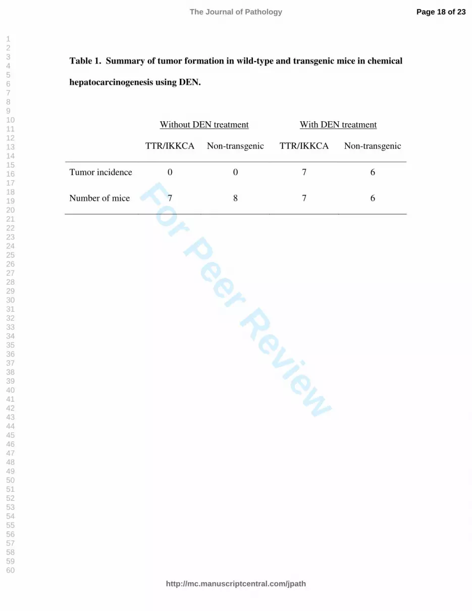
Table 1. Summary of tumor formation in wild-type and transgenic mice in chemical
hepatocarcinogenesis using DEN.
Without DEN treatment With DEN treatment
TTR/IKKCA Non-transgenic TTR/IKKCA Non-transgenic
Tumor incidence 0 0 7 6
Number of mice 7 8 7 6
Page 18 of 23
http://mc.manuscriptcentral.com/jpath
The Journal of Pathology
123456789101112131415161718192021222324252627282930313233343536373839404142434445464748495051525354555657585960

For Peer Review
Table 2. Summary of the transient embryos carrying the FLAG-IKKβ-CA
transgene.
Embryonic stage Number of transgenic embryos Number of embryos
11.5 36 106
12.0 9 14
12.25 11 15
12.5 24 (4*) 49
13.5 8 15
* Number of transgenic embryos showing accumulation of nucleated RBCs in their livers.
Page 19 of 23
http://mc.manuscriptcentral.com/jpath
The Journal of Pathology
123456789101112131415161718192021222324252627282930313233343536373839404142434445464748495051525354555657585960

For Peer Review
Activation of NF-kappaB pathway in HepG2 cells and livers of transgenic mice expressing constitutively active IKKbeta.
Page 20 of 23
http://mc.manuscriptcentral.com/jpath
The Journal of Pathology
123456789101112131415161718192021222324252627282930313233343536373839404142434445464748495051525354555657585960

For Peer Review
Electrophoretic mobility shift assays (EMSA) showing increased NF-kappaB activities in nuclear extracts from livers of transgenic mice.
Page 21 of 23
http://mc.manuscriptcentral.com/jpath
The Journal of Pathology
123456789101112131415161718192021222324252627282930313233343536373839404142434445464748495051525354555657585960

For Peer Review
NF-kappaB activation in hepatocytes did not affect chemical hepatocarcinogenesis.
Page 22 of 23
http://mc.manuscriptcentral.com/jpath
The Journal of Pathology
123456789101112131415161718192021222324252627282930313233343536373839404142434445464748495051525354555657585960

For Peer Review
Nucleated red blood cells accumulated in livers of transgenic embryos expressing constitutively active IKKbeta
Page 23 of 23
http://mc.manuscriptcentral.com/jpath
The Journal of Pathology
123456789101112131415161718192021222324252627282930313233343536373839404142434445464748495051525354555657585960