heme oxygenase-1 gene delivery by sleeping beauty inhibits vascular stasis in a murine model of...
TRANSCRIPT
ORIGINAL ARTICLE
Heme oxygenase-1 gene delivery by Sleeping Beauty inhibitsvascular stasis in a murine model of sickle cell disease
John D. Belcher & Julie V. Vineyard & Carol M. Bruzzone & Chunsheng Chen &
Joan D. Beckman & Julia Nguyen & Clifford J. Steer & Gregory M. Vercellotti
Received: 1 December 2009 /Revised: 12 February 2010 /Accepted: 8 March 2010 /Published online: 23 March 2010# Springer-Verlag 2010
Abstract Increases in heme oxygenase-1 (HO-1) andadministration of heme degradation products CO andbiliverdin inhibit vascular inflammation and vasoocclusionin mouse models of sickle cell disease (SCD). In this study,an albumin (alb) promoter-driven Sleeping Beauty (SB)transposase plasmid with a wild-type rat hmox-1 (wt-HO-1)transposable element was delivered by hydrodynamic tail veininjections to SCD mice. Eight weeks after injection, SCDmice had three- to five-fold increases in HO-1 activity andprotein expression in liver, similar to hemin-treated mice.Immunohistochemistry demonstrated increased perinuclearHO-1 staining in hepatocytes. Messenger RNA transcriptionof the hmox-1 transgene in liver was confirmed byquantitative real-time polymerase chain reaction restrictionfragment length polymorphism (qRT-PCR RFLP) with nodetectible transgene expression in other organs. The livers ofall HO-1 overexpressing mice had activation of nuclearphospho-p38 mitogen-activated protein kinase (MAPK) and
phospho-Akt, decreased nuclear expression of nuclear factor-kappa B (NF-κB) p65, and decreased soluble vascular celladhesion molecule-1 (sVCAM-1) in serum. Hypoxia-induced stasis, a characteristic of SCD, but not normal mice,was inhibited in dorsal skin fold chambers in wt-HO-1 SCDmice despite the absence of hmox-1 transgene expression inthe skin suggesting distal effects of HO activity on thevasculature. No protective effects were seen in SCD miceinjected with nonsense (ns-) rat hmox-1 that encodescarboxy-truncated HO-1 with little or no enzyme activity.We speculate that HO-1 gene delivery to the liver isbeneficial in SCD mice by degrading pro-oxidative heme,releasing anti-inflammatory heme degradation products COand biliverdin/bilirubin into circulation, activating cytopro-tective pathways and inhibiting vascular stasis at sites distalto transgene expression.
Keywords Gene therapy . Heme oxygenase .
Sickle cell disease . Sleeping Beauty
Introduction
Sickle cell disease (SCD) is an unrelenting hemolyticdisease caused by a single base pair mutation in the β-globin chain of hemoglobin (Hb). SCD is characterized byrecurring episodes of painful vasoocclusion, leading toischemia–reperfusion injury and organ damage. Polymeri-zation of hemoglobin-S (HbS) in the deoxy conformationshortens the lifespan of sickle erythrocytes and promotesintravascular hemolysis. When erythrocytes are lysed,extracellular hemoglobin is released and easily oxidizedfrom ferrous (Fe2+) to ferric (Fe3+) hemoglobin (methemo-globin), which in turn, readily releases heme into thevasculature [1].
J. D. Belcher : J. V. Vineyard :C. M. Bruzzone : C. Chen :J. D. Beckman : J. Nguyen :G. M. Vercellotti (*)Division of Hematology, Oncology and Transplantation,Department of Medicine, University of Minnesota,420 Delaware St SE, MMC 480,Minneapolis, MN 55455, USAe-mail: [email protected]
J. D. Belcher : J. V. Vineyard :C. M. Bruzzone : C. Chen :J. D. Beckman : J. Nguyen :G. M. VercellottiVascular Biology Center, University of Minnesota,Minneapolis, MN 55455, USA
C. M. Bruzzone : C. J. SteerDivision of Gastroenterology, Department of Medicine,and Department of Genetics, Cell Biology and Development,University of Minnesota,420 Delaware St SE, MMC 36,Minneapolis, MN 55455, USA
J Mol Med (2010) 88:665–675DOI 10.1007/s00109-010-0613-6
Heme is a potent inducer of vascular inflammation [2].In SCD mice, the combination of heme-induced oxidativestress, inflammation, and the adhesion of circulating bloodcells to vascular endothelium is a key driver of theproinflammatory and prothrombogenic vasculature thatpromotes sludging and stasis of blood flow in thepostcapillary venules [3, 4]. Cells defend or adapt toheme-mediated oxidative stress by inducing HO-1 todegrade heme. In animal models, increased expression ofHO-1 has been shown to protect tissues and cells againstischemia–reperfusion injury, oxidative stress, inflammation,transplant rejection, apoptosis, and cell proliferation [5, 6].Conversely, HO-1 gene null mice (hmox-1−/−) and humanpatients deficient in HO-1 are especially prone to oxidativestress and inflammation [2, 7, 8]. The central importance ofthis protective system was recently highlighted by a childdiagnosed with HO-1 deficiency, who exhibited extensiveendothelial damage [8].
Not surprisingly, SCD patients and mice have elevatedHO-1 in response to chronic hemolysis [2, 9, 10]. HO-1expression is increased four- to sixfold in the organs ofsickle mice compared to normal C57BL6 mice [2, 9, 10].However, the level of HO-1 expression in SCD patients andmice may be inadequate. Vascular defenses against heme,including HO-1, can be overwhelmed during a hemolyticcrisis resulting in heme-induced oxidative stress, inflam-mation, and vasoocclusion. Transgenic sickle mice express-ing βS hemoglobin have activated vascular endotheliumthat exhibits enhanced expression of NF-κB and adhesionmolecules that promote vascular stasis in sickle, but notnormal mice in response to hypoxia [3, 10, 11]. However,further induction of HO-1 in sickle mice with daily hemininjections for 3 days or administration of HO-1-adenovirusinhibits NF-κB activation, adhesion molecule expression,leukocyte–endothelium interactions, and hypoxia-inducedvascular stasis [10]. Remarkably, administration of HO-1products, CO or biliverdin to SCD mice, mimics the effectsof HO-1 overexpression. Conversely, inhibition of HO-1activity by tin protoporphyrin exacerbates hypoxia-inducedstasis in sickle mice. Thus, HO enzyme activity plays avital role in the inhibition of vasoocclusion in sickle mice.
In this study, we treated the S+S-Antilles sickle mousemodel with hmox-1 gene therapy targeted to the liver andevaluated the effects on cytoprotective pathways andhypoxia-induced vasoocclusion in a dorsal skin foldchamber model. To transfer the rat HO-1 gene into mice,we cloned the rat hmox-1 gene into a cis Sleeping Beautytransposase (SB-Tn) system with the transposase driven byan albumin promoter [12]. The SB-Tn system wasdeveloped by resurrecting nonfunctional remnants of anancient vertebrate transposable element from salmonidfish [13]. SB-Tn has features that make it particularlyattractive as a vector for gene therapy. It can accommodate
a much larger transgene than viral vectors, it is nonviral, itdoes not induce an immune inflammatory response inrodent models, and it mediates efficient stable transgeneintegration that exhibits persistent expression. Here, wereport that SB-Tn-mediated HO-1 gene delivery to thelivers of S+S-Antilles sickle mice leads to sustained(8 weeks+) HO-1 transgene expression, an increase inHO enzyme activity and cytoprotective signaling path-ways in the liver, and distal inhibition of hypoxia-inducedvascular stasis in the skin.
Methods
Mice All animal experiments were approved by theUniversityof Minnesota’s Institutional Animal Care and Use Committee.We utilized male and female S+S-Antilles [14] transgenicsickle mice as a model for human SCD. The S+S-Antillesmice are on a C57BL/6 genetic background, are homozygousfor deletion of the mouse βmajor globin locus, and expresshuman α, βS, and βS-Antilles globin transgenes. βS-Antilles
globins contain, in addition to the βS mutation at β6, asecond mutation at β23 (Val→Ile). βS-Antilles has low oxygenaffinity and decreased solubility under deoxygenated con-ditions, resulting in a more severe form of SCD. In the S+S-Antilles mice, approximately 42% of the β globinsexpressed are βS, and 36% are βS-Antilles. These animalshave moderately severe organ pathology [15].
The mice used in these studies were 17–23 weeks ofage at sacrifice. The mice weighed 25–35 g and werehoused in specific pathogen-free housing to preventcommon murine infections that could cause an inflam-matory response. All the mice were maintained on astandard chow diet.
Rat HO-1 plasmids For in vivo experiments in mice, a rathmox-1 pBluescript plasmid containing an SV40 enhancer,a Friend’s Spleen Focus-Forming Virus LTR promoter, theentire coding region of the wild-type (wt) rat hmox-1 gene,and SV40 polyadenylation [16] was blunt cloned betweentwo direct repeat (DR) binding sites within two invertedrepeat (IR) sites (IR/DR) into a 2-kb, albumin (Alb)-driven, SB10 transposase plasmid [12]. The IR/DRelements act as essential binding sites for the transposase[17]. To prepare this construct, the hmox-1 plasmid wasdigested with HindIII and SalI, and the resulting enhancer,promoter, coding region, and polyadenylation segment(1,600 bp) were separated from the vector backbone byagarose gel electrophoresis and purified using a Qiagengel purification kit. The purified hmox-1 construct wastreated with Klenow to generate blunt ends. A secondconstruct containing an Alb-driven SB10 transposase(4,193 bp), inserted outside a pair of IR/DRs (Alb/SB10/
666 J Mol Med (2010) 88:665–675
IRDR), was prepared from a pCMV SB10 plasmid usingEcoRI and XhoI [13] and purified using a Qiagen™plasmid isolation kit. The purified Alb/SB10/IRDR plas-mid was opened at a unique EcoRI site between the IR/DRs and phosphatase treated. The prepared SB10 vectorand hmox-1 constructs were ligated overnight at 16°C withthe hmox-1 DNA inserted between the IR/DRs of the SB10vector. Clones were screened by restriction digest map-ping and sequencing followed by protein expression intissue culture, HO-1 western blot analysis, and HOenzyme activity. The resultant clone (SB-wt-HO-1,
5,793 bp) expressed wt rat HO-1 (wt-HO-1) as previouslydescribed [12].
A nonsense rat hmox-1 (ns-HO-1) control vector wasconstructed by inserting 4 bp containing an early stopcodon into the hmox-1 sequence of the SB-wt-HO-1plasmid. To prepare this plasmid, SB-wt-HO-1 was digestedat the unique type two restriction endonuclease siterecognized by NheI (GCTAGC) occurring at bp723. Thelinearized plasmid was blunt filled with Klenow enzymeand religated with T4 ligase to create a four-nucleotideinsertion shown in bold in the sequence below.
Translation of that region in the wild-type clone includesamino acids (AA):
Ser Gln Thr Glu Phe Leu Arg Gln Arg Pro Ala Ser LeuVal Gln Asp Thr Thr Ser…
Translation is conserved for the Ser due to redundantcoding but terminated by the 4-bp insertion immediatelyafter the serine to create a ns codon:
Ser Gln Thr Glu Phe Leu Arg Gln Arg Pro Ala Ser C-termThe wt protein translates a 289-AA protein while the ns
transcript translates a 241-AA carboxy-truncated protein.The SB-ns-HO-1 construct was screened as described abovefor SB-wt-HO-1. The resultant ns-HO-1 construct (SB-ns-HO-1, 5,797 bp) had no demonstrable enzymatic activity.
Gene transfer of rat hmox-1 into sickle mice An SB-wt-HO-1 (n=4) or SB-ns-HO-1 (n=6) plasmid was delivered toS+S-Antilles sickle mice by hydrodynamic tail veininjection of 25 μg of plasmid DNA in 10% of the mouse’sbody weight (up to 2.5 ml) of sterile lactated Ringer’ssolution (LRS) [18]. Vehicle control sickle mice (n=5) wereinjected with LRS without any DNA. Another group ofcontrol sickle mice were not injected with anything (n=7).Positive HO-1 control sickle mice (n=5) were injectedintraperitoneally (ip) with hemin chloride (Calbiochem) at adose of 40 μmol/kg/day) for 3 days. Eighteen hours afterthe third hemin injection, hypoxia-induced stasis wasmeasured. Stock hemin chloride (10 mM) was preparedfresh daily by dissolving hemin chloride in 0.1 M NaOH inthe dark. The stock hematin was diluted 1:10 (v/v) in LRSbefore ip injections into sickle mice.
Measurement of vascular stasis Hypoxia-induced stasis ofvenular blood flow in the subcutaneous skin was measured inmice with an implanted dorsal skin fold chamber (DSFC)using intravital microscopy as previously described [3, 19].All measurements of blood flow parameters in the DSFCwere made 3 days after DSFC implantation. Hypoxia-inducedstasis of venular blood flow was measured 7–8 weeks aftergene transfer. At baseline, with the mice in ambient air,flowing venules were selected at random and their relativelocations were noted on a map of the microscopic field andrecorded. After baseline selection of flowing venules inambient air, the mice were subjected to 1 h of hypoxia (7%O2/93% N2), followed by reoxygenation in room air. After1 h of reoxygenation, the same venules were re-examined forblood flow. Venules with no observable flow were counted asstatic. The percentage of static vessels was calculated bydividing the number of static venules by the total number offlowing venules selected at baseline. A minimum of 26subcutaneous venules were examined in each mouse. Weequated vascular stasis with vasoocclusion for experimentalpurposes. Certainly prolonged vasoocclusion seen clinicallyis associated with activation of coagulation and thrombosisleading to organ infarction.
Mouse tissue collection After 4 h of reoxygenation, the micein ambient air were sacrificed and tissues harvested aspreviously described [3]. Mice were asphyxiated in a CO2
chamber for approximately 2 min. Blood was collected bycardiac puncture for isolation of serum. Organs wereremoved and divided for homogenate preparation, immuno-
J Mol Med (2010) 88:665–675 667
histochemistry, and RNA isolation; wrapped in aluminumfoil; frozen in liquid nitrogen; and later stored at −85°C.Liver sections from a subset of the mice were collected forimmunohistochemistry and placed in 10% buffered formalinprior to embedding in paraffin blocks and sectioning. Dorsalskin was collected from mice for RNA isolation.
Homogenization of mouse livers and purification of livernuclear extracts and microsomes All liver processing andpurifications were done on ice or at 4°C. The liver tissue,frozen in liquid nitrogen, was broken into small pieces witha hammer between layers of aluminum foil, then transferredto a mortar and reduced to a fine powder in liquid nitrogen.The thawed powder was homogenized on ice in 2 ml ofbuffer A containing 0.1% Triton X-100 (Sigma-Aldrich),300 mM NaCl, 1.5 mM MgCl2, 20 μM EDTA, 25 mMHEPES (N-2-hydroxyethylpiperazine-N′-2-ethanesulfonicacid) pH7.6 in a 5-ml Dounce tissue homogenizer(Wheaton) with 20 strokes of the tight-fitting pestle B. Celldebris was removed by centrifuging the crude homogenateat 500×g for 30 s at 4°C. Nuclei were pelleted bycentrifuging aliquots of liver homogenates at 15,000×g for5 min. Nuclear extracts were prepared by adding buffer B(Panomics, Nuclear Extraction Kit) to the nuclear pellet,sonicating for 10 s (Misonix) and incubating on ice for 2 hwith gentle shaking. Samples were centrifuged at 15,000×gfor 30 min, and nuclear extract supernatants were collectedand stored at −85°C until use. Buffers A and B containedprotease and phosphatase inhibitors at these final concen-trations: 5 mM dithiothreitol, 0.1 mM orthovanadate,20 mM β-glycerophosphate, 20 μg/ml leupeptin, 1 mMphenylmethane-sulfonylfluoride, and mammalian proteaseinhibitor cocktail (1:50, v/v; Sigma-Aldrich). Liver micro-somes were prepared by centrifuging the 15,000×g super-natants at 105,000×g for 1 h. Microsomal pellets weresuspended in 2 mM MgCl2, 0.1 M K2HPO4 buffer, pH7.4.Protein concentrations were measured in all samples usinga Bio-Rad protein assay kit.
Measurement of HO enzyme activity in liver microsomes HOactivity was measured as previously described [20] infreshly isolated liver microsomes sonicated once for 10 s.Microsomes (2 mg) in 2 mM MgCl2, 0.1 M K2HPO4
buffer, pH7.4, were added to the reaction mixture (400 μl,final) containing 2.5 μg of recombinant biliverdin reductase(Assay Designs), 2 mM glucose-6-phosphate, 0.2 Uglucose-6-phosphate dehydrogenase, 50 μM hemin chlo-ride, and 0.8 mM NADPH (Calbiochem) for 1 h in thedark. The bilirubin formed was extracted into chloroform,and the delta OD 464–530 nm was measured (extinctioncoefficient, 40 mM−1cm−1 for bilirubin). HO activity isexpressed as picomole of bilirubin formed per milligrammicrosomal protein per hour.
Western blots of liver HO-1, p38 mitogen-activated proteinkinase (MAPK), Akt, and NF-κB p65 An equal amount ofliver microsomal (HO-1) or nuclear extract (p38, Akt, andp65) protein per lane was loaded in SDS buffer and subjectedto electrophoresis on 10% or 15% polyacrylamide Tris–HClgels (Bio-Rad). Afterward, the samples were transferredelectrophoretically to polyvinylidene fluoride membranes(Millipore), and the membranes were probed with mousemonoclonal anti-HO-1 (Assay Designs), rabbit anti-phospho-p38 MAPK (residue Thr180/Tyr182), rabbit anti-phospho-Akt (residue Ser473; Cell Signaling Technology), or antinu-clear factor-kappa B p65 (Santa Cruz Biotechnology). Sites ofprimary antibody binding were visualized with alkalinephosphatase-conjugated goat antimouse or goat antirabbitIgG (Santa Cruz Biotechnology). The final detection ofimmunoreactive bands was performed using an ECF™substrate (GEHealthcare) and visualized on a Storm™Reader(GE Healthcare). All membranes were stripped using RestoreStripping Buffer (Thermo Scientific) and reprobed with rabbitanti-GAPDH (Sigma-Aldrich), rabbit anti-p38 MAPK, orrabbit anti-Akt (Cell Signaling Technology). Bands werequantitated using Image J software (NIH).
RNA analysis Total RNA was isolated from frozen organsections, and HO-1 mRNA was quantitated by quantitativereal-time polymerase chain reaction (qRT-PCR) as previ-ously described [12]. The presence or absence of the wt- orns-rat HO-1 transgene was confirmed by restrictionfragment length polymorphism (RFLP) analysis of HO-1cDNA using ApaI restriction enzyme digestion as previ-ously described [12].
Immunohistochemistry Formalin-fixed liver sections wereprocessed routinely, embedded in paraffin, sectioned, andstained with mouse monoclonal anti-HO-1 (Assay Designs).Sites of primary antibody binding were visualized withhorseradish peroxidase (HRP)-conjugated goat antimouseIgG (Biocare Medical). The final detection of HO-1 immu-noconjugates was performed with the HRP substrates dia-minobenzidine and H2O2. Nuclei were counterstained withhematoxylin.
Enzyme-linked immunosorbent assay for soluble vascularcell adhesion molecule-1 in serum Serum levels of solublevascular cell adhesion molecule-1 (sVCAM-1) were mea-sured by enzyme-linked immunosorbent assay (ELISA)according to the manufacturer’s protocol (R&D Systems,Minneapolis, MN).
Statistics All statistical analyses were performed withSigmaStat 2.0 for Windows (SPSS Inc, Chicago, IL).Comparisons of multiple treatment groups (control, wt-HO-1, ns-HO-1, and hemin) to LRS-treated mice were
668 J Mol Med (2010) 88:665–675
made using one-way analysis of variance (ANOVA). Theproportions of venules exhibiting stasis in each treatmentgroup were compared using a Z test.
Results
S+S-Antilles sickle mice were injected hydrodynamicallywith an alb promoter-driven cis SB-Tn vector with either awt rat hmox-1 gene (wt-HO-1) or a ns-HO-1 containing anearly stop codon. The two negative controls for this studyincluded sickle mice that were not injected (control) andsickle mice hydrodynamically injected with LRS. Sicklemice injected three consecutive days with hemin served aspositive controls.
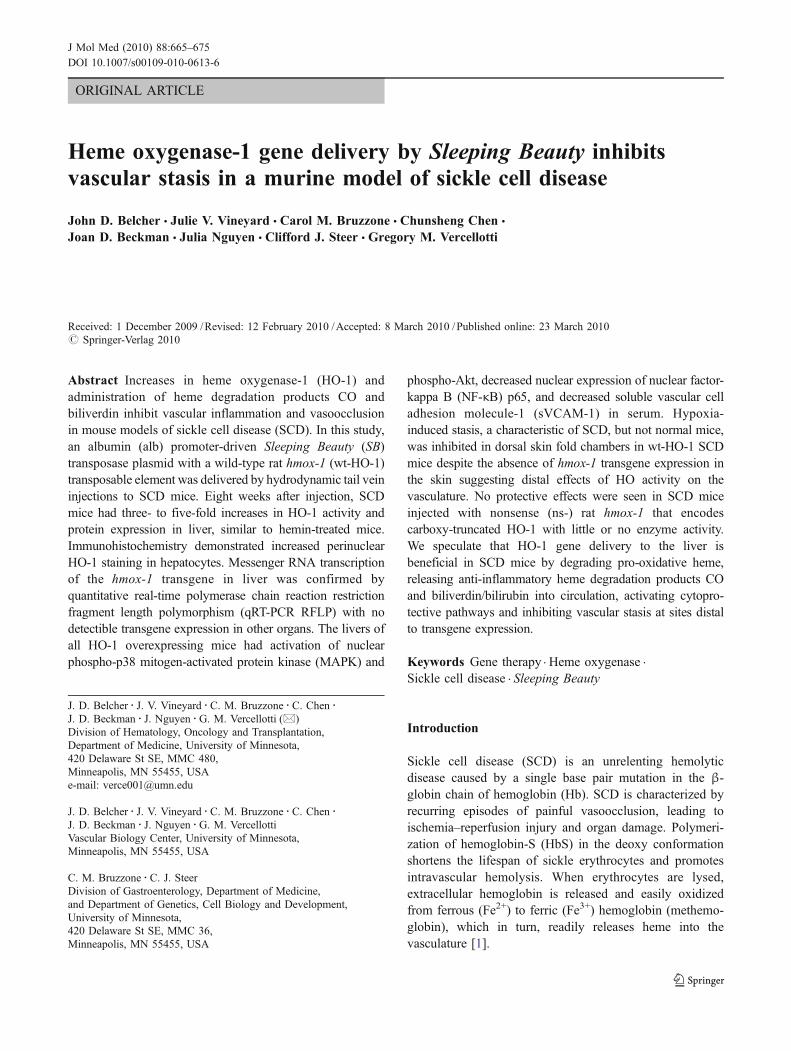
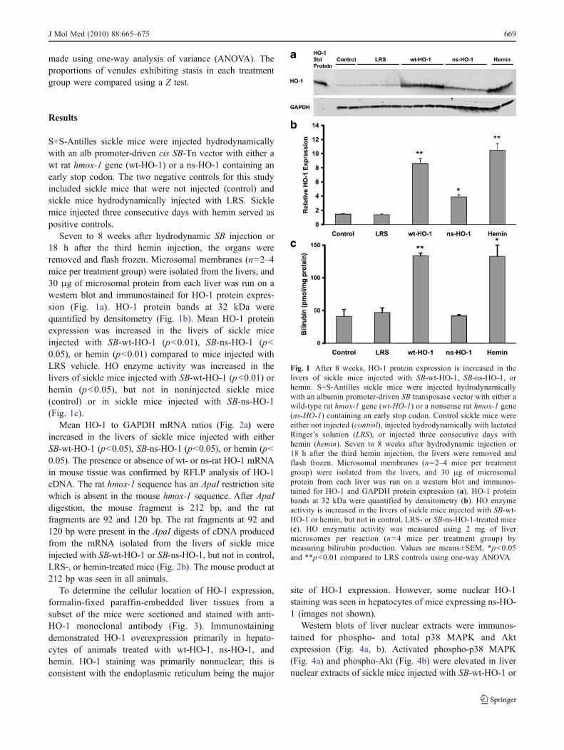
Seven to 8 weeks after hydrodynamic SB injection or18 h after the third hemin injection, the organs wereremoved and flash frozen. Microsomal membranes (n=2–4mice per treatment group) were isolated from the livers, and30 μg of microsomal protein from each liver was run on awestern blot and immunostained for HO-1 protein expres-sion (Fig. 1a). HO-1 protein bands at 32 kDa werequantified by densitometry (Fig. 1b). Mean HO-1 proteinexpression was increased in the livers of sickle miceinjected with SB-wt-HO-1 (p<0.01), SB-ns-HO-1 (p<0.05), or hemin (p<0.01) compared to mice injected withLRS vehicle. HO enzyme activity was increased in thelivers of sickle mice injected with SB-wt-HO-1 (p<0.01) orhemin (p<0.05), but not in noninjected sickle mice(control) or in sickle mice injected with SB-ns-HO-1(Fig. 1c).
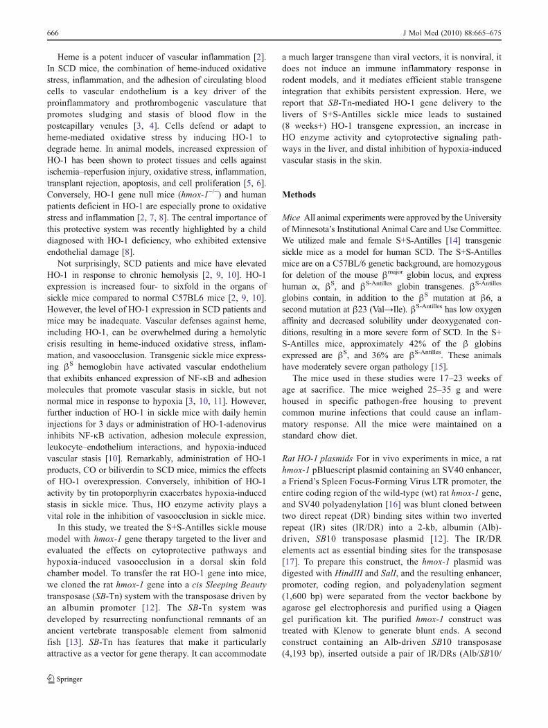
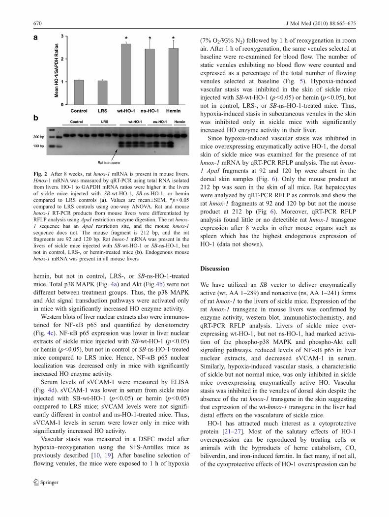
Mean HO-1 to GAPDH mRNA ratios (Fig. 2a) wereincreased in the livers of sickle mice injected with eitherSB-wt-HO-1 (p<0.05), SB-ns-HO-1 (p<0.05), or hemin (p<0.05). The presence or absence of wt- or ns-rat HO-1 mRNAin mouse tissue was confirmed by RFLP analysis of HO-1cDNA. The rat hmox-1 sequence has an ApaI restriction sitewhich is absent in the mouse hmox-1 sequence. After ApaIdigestion, the mouse fragment is 212 bp, and the ratfragments are 92 and 120 bp. The rat fragments at 92 and120 bp were present in the ApaI digests of cDNA producedfrom the mRNA isolated from the livers of sickle miceinjected with SB-wt-HO-1 or SB-ns-HO-1, but not in control,LRS-, or hemin-treated mice (Fig. 2b). The mouse product at212 bp was seen in all animals.
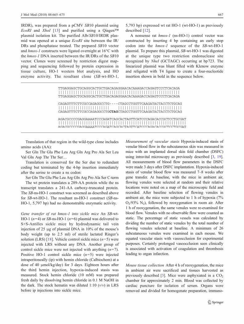
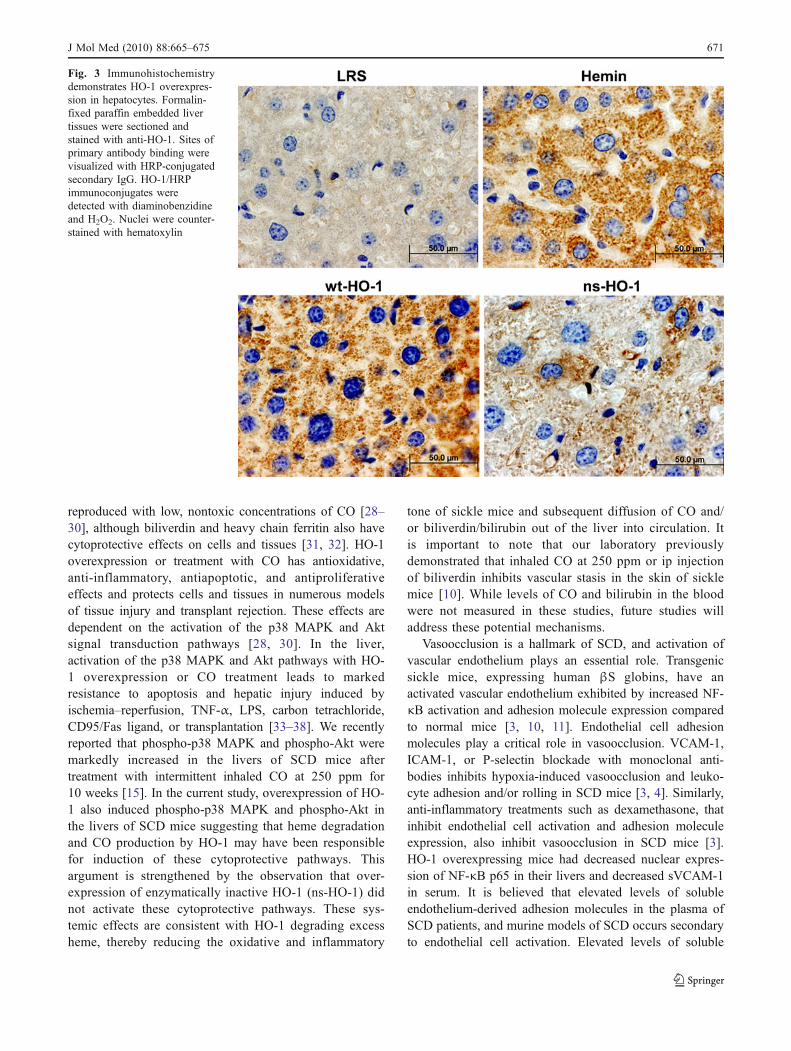
To determine the cellular location of HO-1 expression,formalin-fixed paraffin-embedded liver tissues from asubset of the mice were sectioned and stained with anti-HO-1 monoclonal antibody (Fig. 3). Immunostainingdemonstrated HO-1 overexpression primarily in hepato-cytes of animals treated with wt-HO-1, ns-HO-1, andhemin. HO-1 staining was primarily nonnuclear; this isconsistent with the endoplasmic reticulum being the major
site of HO-1 expression. However, some nuclear HO-1staining was seen in hepatocytes of mice expressing ns-HO-1 (images not shown).
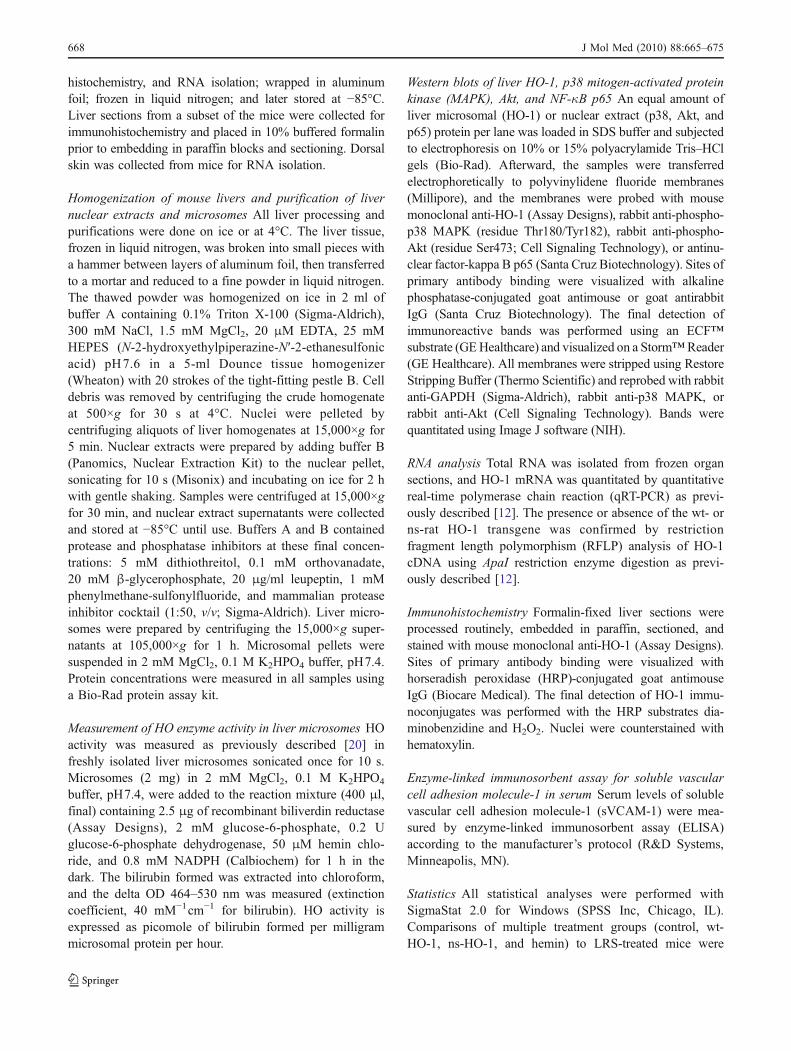
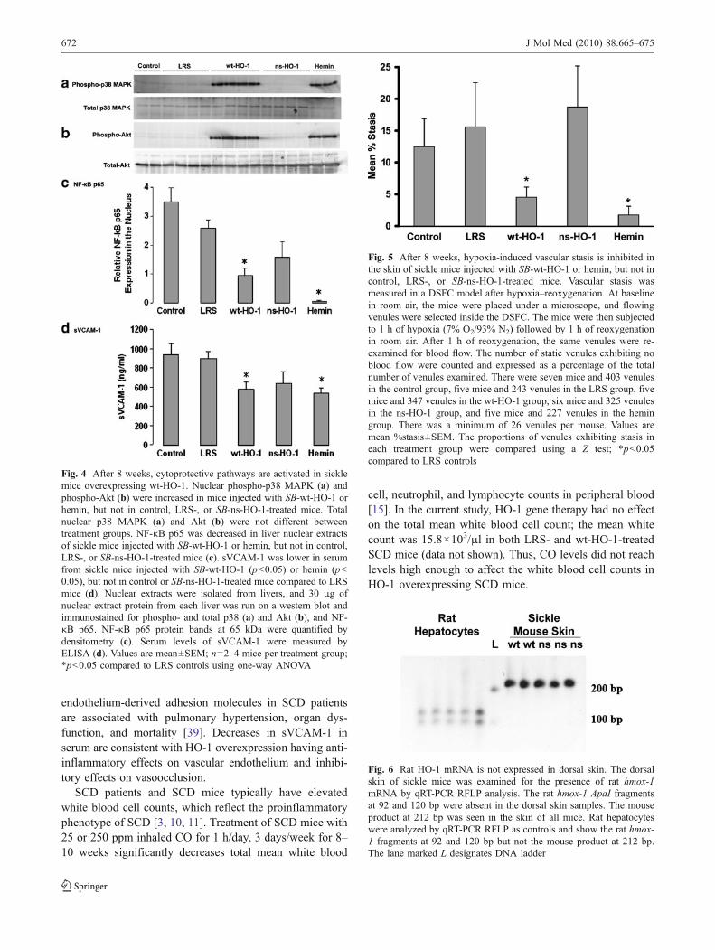
Western blots of liver nuclear extracts were immunos-tained for phospho- and total p38 MAPK and Aktexpression (Fig. 4a, b). Activated phospho-p38 MAPK(Fig. 4a) and phospho-Akt (Fig. 4b) were elevated in livernuclear extracts of sickle mice injected with SB-wt-HO-1 or
Fig. 1 After 8 weeks, HO-1 protein expression is increased in thelivers of sickle mice injected with SB-wt-HO-1, SB-ns-HO-1, orhemin. S+S-Antilles sickle mice were injected hydrodynamicallywith an albumin promoter-driven SB transposase vector with either awild-type rat hmox-1 gene (wt-HO-1) or a nonsense rat hmox-1 gene(ns-HO-1) containing an early stop codon. Control sickle mice wereeither not injected (control), injected hydrodynamically with lactatedRinger’s solution (LRS), or injected three consecutive days withhemin (hemin). Seven to 8 weeks after hydrodynamic injection or18 h after the third hemin injection, the livers were removed andflash frozen. Microsomal membranes (n=2–4 mice per treatmentgroup) were isolated from the livers, and 30 μg of microsomalprotein from each liver was run on a western blot and immunos-tained for HO-1 and GAPDH protein expression (a). HO-1 proteinbands at 32 kDa were quantified by densitometry (b). HO enzymeactivity is increased in the livers of sickle mice injected with SB-wt-HO-1 or hemin, but not in control, LRS- or SB-ns-HO-1-treated mice(c). HO enzymatic activity was measured using 2 mg of livermicrosomes per reaction (n=4 mice per treatment group) bymeasuring bilirubin production. Values are means±SEM, *p<0.05and **p<0.01 compared to LRS controls using one-way ANOVA
J Mol Med (2010) 88:665–675 669
hemin, but not in control, LRS-, or SB-ns-HO-1-treatedmice. Total p38 MAPK (Fig. 4a) and Akt (Fig 4b) were notdifferent between treatment groups. Thus, the p38 MAPKand Akt signal transduction pathways were activated onlyin mice with significantly increased HO enzyme activity.
Western blots of liver nuclear extracts also were immunos-tained for NF-κB p65 and quantified by densitometry(Fig. 4c). NF-κB p65 expression was lower in liver nuclearextracts of sickle mice injected with SB-wt-HO-1 (p<0.05)or hemin (p<0.05), but not in control or SB-ns-HO-1-treatedmice compared to LRS mice. Hence, NF-κB p65 nuclearlocalization was decreased only in mice with significantlyincreased HO enzyme activity.
Serum levels of sVCAM-1 were measured by ELISA(Fig. 4d). sVCAM-1 was lower in serum from sickle miceinjected with SB-wt-HO-1 (p<0.05) or hemin (p<0.05)compared to LRS mice; sVCAM levels were not signifi-cantly different in control and ns-HO-1-treated mice. Thus,sVCAM-1 levels in serum were lower only in mice withsignificantly increased HO activity.
Vascular stasis was measured in a DSFC model afterhypoxia–reoxygenation using the S+S-Antilles mice aspreviously described [10, 19]. After baseline selection offlowing venules, the mice were exposed to 1 h of hypoxia
(7% O2/93% N2) followed by 1 h of reoxygenation in roomair. After 1 h of reoxygenation, the same venules selected atbaseline were re-examined for blood flow. The number ofstatic venules exhibiting no blood flow were counted andexpressed as a percentage of the total number of flowingvenules selected at baseline (Fig. 5). Hypoxia-inducedvascular stasis was inhibited in the skin of sickle miceinjected with SB-wt-HO-1 (p<0.05) or hemin (p<0.05), butnot in control, LRS-, or SB-ns-HO-1-treated mice. Thus,hypoxia-induced stasis in subcutaneous venules in the skinwas inhibited only in sickle mice with significantlyincreased HO enzyme activity in their liver.
Since hypoxia-induced vascular stasis was inhibited inmice overexpressing enzymatically active HO-1, the dorsalskin of sickle mice was examined for the presence of rathmox-1 mRNA by qRT-PCR RFLP analysis. The rat hmox-1 ApaI fragments at 92 and 120 bp were absent in thedorsal skin samples (Fig. 6). Only the mouse product at212 bp was seen in the skin of all mice. Rat hepatocyteswere analyzed by qRT-PCR RFLP as controls and show therat hmox-1 fragments at 92 and 120 bp but not the mouseproduct at 212 bp (Fig 6). Moreover, qRT-PCR RFLPanalysis found little or no detectible rat hmox-1 transgeneexpression after 8 weeks in other mouse organs such asspleen which has the highest endogenous expression ofHO-1 (data not shown).
Discussion
We have utilized an SB vector to deliver enzymaticallyactive (wt, AA 1–289) and nonactive (ns, AA 1–241) formsof rat hmox-1 to the livers of sickle mice. Expression of therat hmox-1 transgene in mouse livers was confirmed byenzyme activity, western blot, immunohistochemistry, andqRT-PCR RFLP analysis. Livers of sickle mice over-expressing wt-HO-1, but not ns-HO-1, had marked activa-tion of the phospho-p38 MAPK and phospho-Akt cellsignaling pathways, reduced levels of NF-κB p65 in livernuclear extracts, and decreased sVCAM-1 in serum.Similarly, hypoxia-induced vascular stasis, a characteristicof sickle but not normal mice, was only inhibited in sicklemice overexpressing enzymatically active HO. Vascularstasis was inhibited in the venules of dorsal skin despite theabsence of the rat hmox-1 transgene in the skin suggestingthat expression of the wt-hmox-1 transgene in the liver haddistal effects on the vasculature of sickle mice.
HO-1 has attracted much interest as a cytoprotectiveprotein [21–27]. Most of the salutary effects of HO-1overexpression can be reproduced by treating cells oranimals with the byproducts of heme catabolism, CO,biliverdin, and iron-induced ferritin. In fact many, if not all,of the cytoprotective effects of HO-1 overexpression can be
Fig. 2 After 8 weeks, rat hmox-1 mRNA is present in mouse livers.Hmox-1 mRNA was measured by qRT-PCR using total RNA isolatedfrom livers. HO-1 to GAPDH mRNA ratios were higher in the liversof sickle mice injected with SB-wt-HO-1, SB-ns-HO-1, or hemincompared to LRS controls (a). Values are mean±SEM, *p<0.05compared to LRS controls using one-way ANOVA. Rat and mousehmox-1 RT-PCR products from mouse livers were differentiated byRFLP analysis using ApaI restriction enzyme digestion. The rat hmox-1 sequence has an ApaI restriction site, and the mouse hmox-1sequence does not. The mouse fragment is 212 bp, and the ratfragments are 92 and 120 bp. Rat hmox-1 mRNA was present in thelivers of sickle mice injected with SB-wt-HO-1 or SB-ns-HO-1, butnot in control, LRS-, or hemin-treated mice (b). Endogenous mousehmox-1 mRNA was present in all mouse livers
670 J Mol Med (2010) 88:665–675
reproduced with low, nontoxic concentrations of CO [28–30], although biliverdin and heavy chain ferritin also havecytoprotective effects on cells and tissues [31, 32]. HO-1overexpression or treatment with CO has antioxidative,anti-inflammatory, antiapoptotic, and antiproliferativeeffects and protects cells and tissues in numerous modelsof tissue injury and transplant rejection. These effects aredependent on the activation of the p38 MAPK and Aktsignal transduction pathways [28, 30]. In the liver,activation of the p38 MAPK and Akt pathways with HO-1 overexpression or CO treatment leads to markedresistance to apoptosis and hepatic injury induced byischemia–reperfusion, TNF-α, LPS, carbon tetrachloride,CD95/Fas ligand, or transplantation [33–38]. We recentlyreported that phospho-p38 MAPK and phospho-Akt weremarkedly increased in the livers of SCD mice aftertreatment with intermittent inhaled CO at 250 ppm for10 weeks [15]. In the current study, overexpression of HO-1 also induced phospho-p38 MAPK and phospho-Akt inthe livers of SCD mice suggesting that heme degradationand CO production by HO-1 may have been responsiblefor induction of these cytoprotective pathways. Thisargument is strengthened by the observation that over-expression of enzymatically inactive HO-1 (ns-HO-1) didnot activate these cytoprotective pathways. These sys-temic effects are consistent with HO-1 degrading excessheme, thereby reducing the oxidative and inflammatory
tone of sickle mice and subsequent diffusion of CO and/or biliverdin/bilirubin out of the liver into circulation. Itis important to note that our laboratory previouslydemonstrated that inhaled CO at 250 ppm or ip injectionof biliverdin inhibits vascular stasis in the skin of sicklemice [10]. While levels of CO and bilirubin in the bloodwere not measured in these studies, future studies willaddress these potential mechanisms.
Vasoocclusion is a hallmark of SCD, and activation ofvascular endothelium plays an essential role. Transgenicsickle mice, expressing human βS globins, have anactivated vascular endothelium exhibited by increased NF-κB activation and adhesion molecule expression comparedto normal mice [3, 10, 11]. Endothelial cell adhesionmolecules play a critical role in vasoocclusion. VCAM-1,ICAM-1, or P-selectin blockade with monoclonal anti-bodies inhibits hypoxia-induced vasoocclusion and leuko-cyte adhesion and/or rolling in SCD mice [3, 4]. Similarly,anti-inflammatory treatments such as dexamethasone, thatinhibit endothelial cell activation and adhesion moleculeexpression, also inhibit vasoocclusion in SCD mice [3].HO-1 overexpressing mice had decreased nuclear expres-sion of NF-κB p65 in their livers and decreased sVCAM-1in serum. It is believed that elevated levels of solubleendothelium-derived adhesion molecules in the plasma ofSCD patients, and murine models of SCD occurs secondaryto endothelial cell activation. Elevated levels of soluble
Fig. 3 Immunohistochemistrydemonstrates HO-1 overexpres-sion in hepatocytes. Formalin-fixed paraffin embedded livertissues were sectioned andstained with anti-HO-1. Sites ofprimary antibody binding werevisualized with HRP-conjugatedsecondary IgG. HO-1/HRPimmunoconjugates weredetected with diaminobenzidineand H2O2. Nuclei were counter-stained with hematoxylin
J Mol Med (2010) 88:665–675 671
endothelium-derived adhesion molecules in SCD patientsare associated with pulmonary hypertension, organ dys-function, and mortality [39]. Decreases in sVCAM-1 inserum are consistent with HO-1 overexpression having anti-inflammatory effects on vascular endothelium and inhibi-tory effects on vasoocclusion.
SCD patients and SCD mice typically have elevatedwhite blood cell counts, which reflect the proinflammatoryphenotype of SCD [3, 10, 11]. Treatment of SCD mice with25 or 250 ppm inhaled CO for 1 h/day, 3 days/week for 8–10 weeks significantly decreases total mean white blood
cell, neutrophil, and lymphocyte counts in peripheral blood[15]. In the current study, HO-1 gene therapy had no effecton the total mean white blood cell count; the mean whitecount was 15.8×10³/μl in both LRS- and wt-HO-1-treatedSCD mice (data not shown). Thus, CO levels did not reachlevels high enough to affect the white blood cell counts inHO-1 overexpressing SCD mice.
Fig. 6 Rat HO-1 mRNA is not expressed in dorsal skin. The dorsalskin of sickle mice was examined for the presence of rat hmox-1mRNA by qRT-PCR RFLP analysis. The rat hmox-1 ApaI fragmentsat 92 and 120 bp were absent in the dorsal skin samples. The mouseproduct at 212 bp was seen in the skin of all mice. Rat hepatocyteswere analyzed by qRT-PCR RFLP as controls and show the rat hmox-1 fragments at 92 and 120 bp but not the mouse product at 212 bp.The lane marked L designates DNA ladder
Fig. 5 After 8 weeks, hypoxia-induced vascular stasis is inhibited inthe skin of sickle mice injected with SB-wt-HO-1 or hemin, but not incontrol, LRS-, or SB-ns-HO-1-treated mice. Vascular stasis wasmeasured in a DSFC model after hypoxia–reoxygenation. At baselinein room air, the mice were placed under a microscope, and flowingvenules were selected inside the DSFC. The mice were then subjectedto 1 h of hypoxia (7% O2/93% N2) followed by 1 h of reoxygenationin room air. After 1 h of reoxygenation, the same venules were re-examined for blood flow. The number of static venules exhibiting noblood flow were counted and expressed as a percentage of the totalnumber of venules examined. There were seven mice and 403 venulesin the control group, five mice and 243 venules in the LRS group, fivemice and 347 venules in the wt-HO-1 group, six mice and 325 venulesin the ns-HO-1 group, and five mice and 227 venules in the hemingroup. There was a minimum of 26 venules per mouse. Values aremean %stasis±SEM. The proportions of venules exhibiting stasis ineach treatment group were compared using a Z test; *p<0.05compared to LRS controls
Fig. 4 After 8 weeks, cytoprotective pathways are activated in sicklemice overexpressing wt-HO-1. Nuclear phospho-p38 MAPK (a) andphospho-Akt (b) were increased in mice injected with SB-wt-HO-1 orhemin, but not in control, LRS-, or SB-ns-HO-1-treated mice. Totalnuclear p38 MAPK (a) and Akt (b) were not different betweentreatment groups. NF-κB p65 was decreased in liver nuclear extractsof sickle mice injected with SB-wt-HO-1 or hemin, but not in control,LRS-, or SB-ns-HO-1-treated mice (c). sVCAM-1 was lower in serumfrom sickle mice injected with SB-wt-HO-1 (p<0.05) or hemin (p<0.05), but not in control or SB-ns-HO-1-treated mice compared to LRSmice (d). Nuclear extracts were isolated from livers, and 30 μg ofnuclear extract protein from each liver was run on a western blot andimmunostained for phospho- and total p38 (a) and Akt (b), and NF-κB p65. NF-κB p65 protein bands at 65 kDa were quantified bydensitometry (c). Serum levels of sVCAM-1 were measured byELISA (d). Values are mean±SEM; n=2–4 mice per treatment group;*p<0.05 compared to LRS controls using one-way ANOVA
672 J Mol Med (2010) 88:665–675
CO interactions with Hb can modulate hemolysis inSCD. CO binding to HbS shifts the oxygen dissociationcurve to the left and inhibits HbS deoxygenation, HbSpolymerization, and RBC hemolysis [40]. Since hepatic COproduction may be increased in SCD mice overexpressingwt-HO-1, total serum heme levels were measured as amarker of hemolysis according to the method of Huy et al.[41]. Mean serum heme levels were 29 μM in LRS-treatedmice and 19 μM in SCD mice overexpressing wt-HO-1, butthese results were not statistically different (data notshown). The S+S-Antilles mouse model used in thesestudies does not develop severe anemia warranting addi-tional studies in a more hemolytic and anemic sickle mousemodel.
In these studies, HO-1 was targeted to the liver using anSB transposase with an alb promoter. The transposase,delivered in cis with the hmox-1 transgene, binds to IR/DRsites flanking the transgene, and catalyzes the excision ofthe flanked transgene, mediating its insertion into the targethost genome with an apparently equal preference for AT-rich TA dinucleotide insertion sites in introns, exons, andintergenic sequences [42–45]. The alb promoter driving thetransposase precludes significant insertion into nonhepaticchromosomes. This study did not look at insertion sites ofthe hmox-1 transgene in livers as others have previouslydemonstrated that this SB vector readily integrates trans-genes into mouse hepatocyte chromosomes [46]. Episomalexpression of the transgene can occur in other organs earlierthan 8 weeks, but decays until little or no episomalexpression remains by 8 weeks.
Overexpression of HO-1 in nonhepatic tissues might alsobe beneficial in SCD. However, caution is warranted withHO-1 gene therapy when using ubiquitous, nontissue-specific promoters. It has been reported that continuousHO-1 transgene overexpression in HUVEC cells in cultureresults in downregulation of phospho-Akt expression [47].This suggests that there could be tissue-specific effects ofHO-1 gene therapy, and a targeted approach may bedesirable.
Previous studies have reported cytoprotective effectsof truncated and nonenzymatically active forms of HO-1in cell culture [48, 49]. HO-1 can lose 52 amino acidsfrom its C terminus by proteolysis and travel from theendoplasmic reticulum to the nucleus where it canupregulate genes that promote cytoprotection againstoxidative stress [48]. In images not shown, we saw somehepatocytes with nuclear HO-1 expression in sickle miceexpressing ns-HO-1, which has 48 amino acids missingfrom the C terminus. However, no cytoprotective effectswere seen in our study, suggesting that the beneficialeffects of HO-1 gene therapy in sickle mice may bemediated primarily by heme degradation and productionof CO and biliverdin by enzymatically active HO.
In conclusion, we used an SB vector containing anhmox-1 gene that gives sustained overexpression of HO-1in the livers of sickle mice. HO-1 overexpression in sicklemouse livers activates cytoprotective p38 MAPK and Aktsignal transduction pathways, inhibits nuclear NF-κB p65expression, reduces sVCAM-1 levels in serum, andinhibits hypoxia-induced vascular stasis distally in theskin. We speculate that hmox-1 gene transfer could bebeneficial in SCD by inhibiting oxidative stress, inflam-mation, vasoocclusion, and organ infarction. Futurestudies will examine the mechanisms and effects of SB-mediated hmox-1 gene therapy on markers of oxidativestress, organ pathology, and lifespan in a more severemurine model of human SCD.
Acknowledgments The rat hmox-1 gene plasmid was a generous giftfromDr. Leo Otterbein and Dr. Jawed Alam. This work was supported byNHLBI grants R01 HL67367 and P01 HL55552. We would like to thankDrs. Robert Hebbel, Karl Nath, and Arne Slungaard for the constructivecriticisms, Fuad Abdulla in Dr. Hebbel’s laboratory for breeding andcharacterizing the S+S-Antilles sickle mice used for these studies, andColleen Forster for her expert HO-1 immunohistochemistry.
Authorship contributions John D. Belcher designed the research,analyzed data, and wrote the paper. Julie V. Vineyard performed theresearch and analyzed the data. Carol M. Bruzzone performed theresearch, analyzed the data, and wrote the paper. Chunsheng Chenperformed the research and analyzed the data. Joan D. Beckmanperformed research, analyzed data, and wrote the paper. Julia Nguyenperformed the research and analyzed the data. Clifford J. Steerdesigned the research, analyzed the data, and wrote the paper. GregoryM. Vercellotti designed the research, analyzed data, and wrote thepaper.
Disclosure of conflicts of interest The authors of this paper have noconflicts of interest to disclose
References
1. Jay U (2007) Methemoglobin—it's not just blue: a concise review.Am J Hematol 82:134–144
2. Wagener FA, Eggert A, Boerman OC, Oyen WJ, Verhofstad A,Abraham NG, Adema G, van Kooyk Y, de Witte T, Figdor CG(2001) Heme is a potent inducer of inflammation in mice and iscounteracted by heme oxygenase. Blood 98:1802–1811
3. Belcher JD, Mahaseth H, Welch TE, Vilback AE, Sonbol KM,Kalambur VS, Bowlin PR, Bischof JC, Hebbel RP, Vercellotti GM(2005) Critical role of endothelial cell activation in hypoxia-induced vasoocclusion in transgenic sickle mice. Am J PhysiolHeart Circ Physiol 288:H2715–2725
4. Kaul DK, Hebbel RP (2000) Hypoxia/reoxygenation causesinflammatory response in transgenic sickle mice but not in normalmice. J Clin Invest 106:411–420
5. Otterbein LE, Soares MP, Yamashita K, Bach FH (2003) Hemeoxygenase-1: unleashing the protective properties of heme. TrendsImmunol 24:449–455
6. Wagener FADTG, Volk H-D, Willis D, Abraham NG, Soares MP,Adema GJ, Figdor CG (2003) Different faces of the heme-heme
J Mol Med (2010) 88:665–675 673
oxygenase system in inflammation. Pharmacological Reviews55:551–571
7. Yachie A, Niida Y, Wada T, Igarashi N, Kaneda H, Toma T, OhtaK, Kasahara Y, Koizumi S (1999) Oxidative stress causesenhanced endothelial cell injury in human heme oxygenase-1deficiency. J Clin Invest 103:129–135
8. Kawashima A, Oda Y, Yachie A, Koizumi S, Nakanishi I (2002)Heme oxygenase-1 deficiency: the first autopsy case. Hum Pathol33:125–130
9. Nath KA, Grande JP, Haggard JJ, Croatt AJ, Katusic ZS, SoloveyA, Hebbel RP (2001) Oxidative stress and induction of hemeoxygenase-1 in the kidney in sickle cell disease. Am J Pathol158:893–903
10. Belcher JD, Mahaseth H, Welch TE, Otterbein LE, Hebbel RP,Vercellotti GM (2006) Heme oxygenase-1 is a modulator ofinflammation and vaso-occlusion in transgenic sickle mice. J ClinInvest 116:808–816
11. Belcher JD, Bryant CJ, Nguyen J, Bowlin PR, Kielbik MC,Bischof JC, Hebbel RP, Vercellotti GM (2003) Transgenic sicklemice have vascular inflammation. Blood 101:3953–3959
12. Bruzzone CM, Belcher JD, Schuld NJ, Newman KA, Vineyard J,Nguyen J, Chen C, Beckman JD, Steer CJ, Vercellotti GM (2008)Quantitative real-time polymerase chain reaction (qRT-PCR)restriction fragment length polymorphism (RFLP) method formonitoring highly conserved transgene expression during genetherapy. Transl Res 152:290–297
13. Ivics Z, Hackett PB, Plasterk RH, Izsvak Z (1997) Molecularreconstruction of Sleeping Beauty, a Tc1-like transposon fromfish, and its transposition in human cells. Cell 91:501–510
14. Fabry ME, Sengupta A, Suzuka SM, Costantini F, Rubin EM,Hofrichter J, Christoph G, Manci E, Culberson D, Factor SM et al(1995) A second generation transgenic mouse model expressingboth hemoglobin S (HbS) and HbS-Antilles results in increasedphenotypic severity. Blood 86:2419–2428
15. Beckman JD, Belcher JD, Vineyard JV, Chen C, Nguyen J,Nwaneri MO, O'Sullivan MG, Gulbahce E, Hebbel RP, VercellottiGM (2009) Inhaled carbon monoxide reduces leukocytosis in amurine model of sickle cell disease. Am J Physiol Heart CircPhysiol 297(4):H1243–H1253
16. Shibahara S, Muller R, Taguchi H, Yoshida T (1985) Cloning andexpression of cDNA for rat heme oxygenase. Proc Natl Acad SciU S A 82:7865–7869
17. Cui Z, Geurts AM, Liu G, Kaufman CD, Hackett PB (2002)Structure-function analysis of the inverted terminal repeats of thesleeping beauty transposon. J Mol Biol 318:1221–1235
18. Bell JB, Podetz-Pedersen KM, Aronovich EL, Belur LR, McIvorRS, Hackett PB (2007) Preferential delivery of the SleepingBeauty transposon system to livers of mice by hydrodynamicinjection. Nat Protoc 2:3153–3165
19. Kalambur VS, Mahaseth H, Bischof JC, Kielbik MC, Welch TE,Vilback A, Swanlund DJ, Hebbel RP, Belcher JD, Vercellotti GM(2004) Microvascular blood flow and stasis in transgenic sicklemice: utility of a dorsal skin fold chamber for intravitalmicroscopy. Am J Hematol 77:117–125
20. Balla G, Jacob HS, Balla J, Rosenberg M, Nath K, Apple F, EatonJW, Vercellotti GM (1992) Ferritin: a cytoprotective antioxidantstrategem of endothelium. J Biol Chem 267:18148–18153
21. Peterson SJ, Frishman WH (2009) Targeting heme oxygenase:therapeutic implications for diseases of the cardiovascular system.Cardiol Rev 17:99–111
22. Dulak J, Loboda A, Jozkowicz A (2008) Effect of hemeoxygenase-1 on vascular function and disease. Curr Opin Lipidol19:505–512
23. Abraham NG, Tsenovoy PL, McClung J, Drummond GS (2008)Heme oxygenase: a target gene for anti-diabetic and obesity. CurrPharm Des 14:412–421
24. Soares MP, Bach FH (2007) Heme oxygenase-1 in organtransplantation. Front Biosci 12:4932–4945
25. Nath KA (2006) Heme oxygenase-1: a provenance for cytopro-tective pathways in the kidney and other tissues. Kidney Int70:432–443
26. Ryter SW, Alam J, Choi AM (2006) Heme oxygenase-1/carbonmonoxide: from basic science to therapeutic applications. PhysiolRev 86:583–650
27. Maines MD (2005) The heme oxygenase system: update 2005.Antioxid Redox Signal 7:1761–1766
28. Kim HP, Ryter SW, Choi AM (2006) CO as a cellular signalingmolecule. Annu Rev Pharmacol Toxicol 46:411–449
29. Piantadosi CA (2008) Carbon monoxide, reactive oxygen signal-ing, and oxidative stress. Free Radic Biol Med 45:562–569
30. Bilban M, Haschemi A, Wegiel B, Chin BY, Wagner O, OtterbeinLE (2008) Heme oxygenase and carbon monoxide initiatehomeostatic signaling. J Mol Med 86:267–279
31. Balla J, Vercellotti GM, Jeney V, Yachie A, Varga Z, Jacob HS,Eaton JW, Balla G (2007) Heme, heme oxygenase, and ferritin:how the vascular endothelium survives (and dies) in an iron-richenvironment. Antioxid Redox Signal 9:1–19
32. Kapitulnik J, Maines MD (2009) Pleiotropic functions ofbiliverdin reductase: cellular signaling and generation of cytopro-tective and cytotoxic bilirubin. Trends Pharmacol Sci 30:129–137
33. Amersi F, Shen XD, Anselmo D, Melinek J, Iyer S, Southard DJ,Katori M, Volk HD, Busuttil RW, Buelow R, Kupiec-WeglinskiJW (2002) Ex vivo exposure to carbon monoxide prevents hepaticischemia/reperfusion injury through p38 MAP kinase pathway.Hepatology 35:815–823
34. Kim HS, Loughran PA, Kim PK, Billiar TR, Zuckerbraun BS(2006) Carbon monoxide protects hepatocytes from TNF-alpha/actinomycin D by inhibition of the caspase-8-mediated apoptoticpathway. Biochem Biophys Res Commun 344:1172–1178
35. Sass G, Seyfried S, Parreira Soares M, Yamashita K, KaczmarekE, Neuhuber WL, Tiegs G (2004) Cooperative effect of biliverdinand carbon monoxide on survival of mice in immune-mediatedliver injury. Hepatology 40:1128–1135
36. Wen T, Zhao JY, Mei S, Guan L, Zhang YL (2006) Protectiveeffect of heme oxygenase-1 and its reaction product, carbonmonoxide on acute liver injury induced by carbon tetrachloride inrats. Beijing Da Xue Xue Bao 38:618–622
37. Sass G, Soares MC, Yamashita K, Seyfried S, Zimmermann WH,Eschenhagen T, Kaczmarek E, Ritter T, Volk HD, Tiegs G (2003)Heme oxygenase-1 and its reaction product, carbon monoxide,prevent inflammation-related apoptotic liver damage in mice.Hepatology 38:909–918
38. Ke B, Buelow R, Shen XD, Melinek J, Amersi F, Gao F, Ritter T,Volk HD, Busuttil RW, Kupiec-Weglinski JW (2002) Hemeoxygenase 1 gene transfer prevents CD95/Fas ligand-mediatedapoptosis and improves liver allograft survival via carbonmonoxide signaling pathway. Hum Gene Ther 13:1189–1199
39. Kato GJ, Martyr S, Blackwelder WC, Nichols JS, Coles WA,Hunter LA, Brennan ML, Hazen SL, Gladwin MT (2005) Levelsof soluble endothelium-derived adhesion molecules in patientswith sickle cell disease are associated with pulmonary hyperten-sion, organ dysfunction, and mortality. Br J Haematol 130:943–953
40. Beutler E (1975) The effect of carbon monoxide on red cell lifespan in sickle cell disease. Blood 46:253–259
41. Huy NT, Xuan Trang DT, Uyen DT, Sasai M, Harada S, Kamei K(2005) An improved colorimetric method for quantitation of hemeusing tetramethylbenzidine as substrate. Anal Biochem 344:289–291
42. Dupuy AJ, Fritz S, Largaespada DA (2001) Transposition andgene disruption in the male germline of the mouse. Genesis30:82–88
674 J Mol Med (2010) 88:665–675
43. Vigdal TJ, Kaufman CD, Izsvak Z, Voytas DF, Ivics Z (2002)Common physical properties of DNA affecting target siteselection of sleeping beauty and other Tc1/mariner transposableelements. J Mol Biol 323:441–452
44. Clark KJ, Geurts AM, Bell JB, Hackett PB (2004) Transposonvectors for gene-trap insertional mutagenesis in vertebrates.Genesis 39:225–233
45. Carlson CM, Dupuy AJ, Fritz S, Roberg-Perez KJ, Fletcher CF,Largaespada DA (2003) Transposon mutagenesis of the mousegermline. Genetics 165:243–256
46. Kren BT, Ghosh SS, Linehan CL, Roy-Chowdhury N, HackettPB, Roy-Chowdhury J, Steer CJ (2003) Hepatocyte-targeteddelivery of Sleeping Beauty mediates efficient gene transfer invivo. Gene Ther Mol Biol 7:229–238
47. Batzlsperger CA, Achatz S, Spreng J, Riegger GA, Griese DP(2007) Evidence for a possible inhibitory interaction between theHO-1/CO- and Akt/NO-pathways in human endothelial cells.Cardiovasc Drugs Ther 21:347–355
48. Lin Q, Weis S, Yang G, Weng YH, Helston R, Rish K, Smith A,Bordner J, Polte T, Gaunitz F, Dennery PA (2007) Hemeoxygenase-1 protein localizes to the nucleus and activatestranscription factors important in oxidative stress. J Biol Chem282:20621–20633
49. Hori R, Kashiba M, Toma T, Yachie A, Goda N, Makino N,Soejima A, Nagasawa T, Nakabayashi K, Suematsu M (2002)Gene transfection of H25A mutant heme oxygenase-1 protectscells against hydroperoxide-induced cytotoxicity. J Biol Chem277:10712–10718
J Mol Med (2010) 88:665–675 675