healing of extraction sockets filled with boneceramic® prior to implant placement: preliminary...
TRANSCRIPT
Healing of Extraction Sockets Filled withBoneCeramic® Prior to Implant Placement:Preliminary Histological Findingscid_184 34..45
Peter De Coster, DDS, PhD;* Hilde Browaeys, MD, DDS;† Hugo De Bruyn, DDS, MSc, PhD‡
ABSTRACT
Background: Various grafting materials have been designed to minimize edentulous ridge volume loss following toothextraction by encouraging new bone formation in healing sockets. BoneCeramic® is a composite of hydroxyapatite andbèta-tricalcium phosphate with pores of 100–500 microns.
Purposes: The aim of this study was to evaluate bone regeneration in healing sockets substituted with BoneCeramic® priorto implant procedures.
Materials and Methods: Fifteen extraction sockets were substituted with BoneCeramic® and 14 sockets were left to healnaturally in 10 patients (mean age 59.6 years). Biopsies were collected only from the implant recipient sites during surgeryafter healing periods ranging from 6–74 weeks (mean 22). In total, 24 biopsies were available; 10 from substituted and 14from naturally healed sites. In one site, the implant was not placed intentionally and, in four substituted sites, implantplacement had to be postponed due to inappropriate healing, hence from five sites biopsies were not available. Histologicalsections were examined by transmitted light microscope.
Results: At the time of implant surgery, bone at substituted sites was softer than in controls, compromising initial implantstability. New bone formation at substituted sites was consistently poorer than in controls, presenting predominantly looseconnective tissue and less woven bone.
Conclusion: The use of BoneCeramic® as a grafting material in fresh extraction sockets appears to interfere with normalhealing processes of the alveolar bone. On the basis of the present preliminary findings, its indication as a material for boneaugmentation when implant placement is considered within 6–38 weeks after extraction should be revised.
KEY WORDS: BoneCeramic®, bone grafting material, extraction sockets, human histology, hydroxyapatite, tricalciumphosphate
Healing of an extraction socket implies a series of
events including the formation and maturation of
a coagulum that subsequently will become replaced by
a provisional matrix and woven bone. Undisturbed
sockets heal uneventfully with new bone formation 1–2
months following extraction.1,2 This healing process
usually occurs with reduction of the height and width
of the alveolar bone,3 which in some cases may aestheti-
cally compromise prosthodontic treatment following
implant surgery. With increasing awareness of dental
implants, there is an increasing demand for the treat-
ment of more complex cases where preliminary grafting
is indicated. The ideal extraction regimen for preserving
or minimizing the edentulous ridge volume loss has
been described2 and various ridge preservation tech-
niques following tooth extraction have been proposed,
such as the placement of different graft materials and
the use of occlusive membranes to cover the extraction
*Visiting professor, Unit of Oral Development and Applied Oral His-tology, Paecamed Research, Faculty of Medicine and Health Sciences,Dental School, University of Ghent, Ghent, Belgium; †oral surgeon,Department of Maxillofacial Surgery, Faculty of Medicine and HealthSciences, Dental School, University of Ghent, Ghent, Belgium; ‡pro-fessor, Department of Periodontology and Oral Implantology, Facultyof Medicine and Health Sciences, Dental School, University of Ghent,Ghent, Belgium
Reprint requests: Hugo De Bruyn, Department of Periodontologyand Oral Implantology, Faculty of Medicine and Health Sciences,Dental School, Ghent University, De Pintelaan 185 P8, B-9000 Ghent,Belgium; e-mail: [email protected]
© 2009, Copyright the AuthorsJournal Compilation © 2011, Wiley Periodicals, Inc.
DOI 10.1111/j.1708-8208.2009.00184.x
34
socket entrance.4 Although grafting materials are known
to encourage new bone formation by a variety of pro-
cesses,5,6 the use of these materials in fresh extraction
sockets has been questioned because they seem to
interfere with the normal healing process.7–9 Studies
in humans using autogenous bone,7 demineralized
freeze-dried bone allograft,10,11 deproteinized bovine
bone mineral,8,12–18 bioactive glass,19–21 hydroxyapatite,4
calcium sulphate,22–24 tricalcium phosphate25–27 or poly-
lactide/polyglycolide polymers28–31 show the presence of
particles of the grafting material in the alveolar sockets
6–9 months following insertion. Autogenous bone
induces bone formation through osteogenesis, whereas
allogenic bone is thought to be osteoinductive due to the
presence of growth factors. Xenografts, such as bovine
bone material and alloplastic substitutes, encourage the
apposition of new bone by osteoconduction.18 Eventu-
ally, anorganic bone allograft, bioglass and hydroxyapa-
tite become incorporated, whereas demineralized bone
allograft, calcium sulphate or tricalcium phosphate are
completely restituted during bone regeneration.
BoneCeramic® (Straumann AG, Basel, Switzerland)
is a synthetic bone-graft substitute designed for aug-
menting bone to support dental implant procedures
(launched May 2005). It consists of biphasic calcium
phosphate, a composite of 60% hydroxyapatite (100%
cristalline) and 40% b-tricalcium phosphate sintered at
temperatures of 1,100 to 1,500°C. BoneCeramic® is 90%
porous with interconnected pores of 100–500 microns
in diameter.32 The aim of the present experiment was to
evaluate bone regeneration in healing sockets substi-
tuted with BoneCeramic® both clinically and histologi-
cally in a first-wave patient sample.
MATERIALS AND METHODS
Patient Selection and Extraction Procedure
Patients presenting for multiple extractions and selected
for later implant treatment, were consecutively treated
with BoneCeramic® as a bone filler after tooth extrac-
tion and scheduled for delayed implant placement.
Subjects were referred for periodontal and/or implant
treatment. Teeth were extracted for multiple reasons
such as periodontal disease, root or crown fractures, or
root caries. In the waiting period between extraction
and implant surgery, the periodontal condition was
improved by nonsurgical therapy and oral hygiene
instructions to avoid or minimize infection risks during
implant surgery. All patients were systemically healthy
and could be treated under local anesthesia. Mucoperi-
osteal flaps were raised and, after extraction followed by
thorough debridement, sockets with intact buccal and
palatal bone plate were filled with BoneCeramic®, which
was mixed with some of the patient’s own blood aspi-
rated from the extraction area. The assumption was that
a bone filler would support the soft tissues and enhance
the aesthetical outcome. A bleeding bone bed was con-
sidered an essential prerequisite for placement of the
biomaterial because the natural supplement of platelet-
derived growth factors and transforming growth factors
can reduce the healing time and enhance bone forma-
tion environment.
Since multiple extractions were often performed
during the same treatment session, extraction sockets
that were not in need for additional filling were left
untreated. This was partly done to minimize the treat-
ment cost. As a consequence, it was possible to select
in each patient at least one socket for natural healing
without bone substitute (control sockets). The flaps
were closed after mucoperiosteal release and meticu-
lously sutured over the alveolar crest. In multiple extrac-
tion cases, this adequate coronal repositioning of the
mucoperiosteal flap for perfect wound closure is easy to
achieve. This technique may enhance wound stability
and an undisturbed healing process.2 Hence, the use of
occlusive membranes was not advocated in none of the
cases.
Because the first cases showed disappointing healing
at the time of implant surgery, however, it was decided to
perform a histological analysis of the bone healing. The
principle of biopsy taking for histological analysis was
approved by the Ethics Committee of the University
Hospital of Ghent since it could be included as a proce-
dure of implant bed preparation during surgery without
jeopardizing treatment outcome.
Sampling and Histological Processing
Biopsies were collected only from the implant recipient
sites during surgery. In total, 23 implants were placed in
the previous extraction sockets. In one additional site
the implant was not placed but the biopsy was taken. In
four substituted sites, implant placement was impossible
due to inappropriate healing whereby taking of biopsies
was impossible. Hence, biopsies were available from 10
substituted and 14 naturally healed sites. Cylindric biop-
sies from these 24 sockets were sampled for histological
Socket Preservation with BoneCeramic 35
analysis with a hollow trephine bur (2.7 mm ¥ 8 mm
core) and placed in a fixative (10% buffered formalin).
The biopsies were randomly assigned to two groups to
be processed either as decalcified or undecalcified sec-
tions; the same treatment protocol was used for biopsies
retrieved from the same patient. About 10–12 sections
were examined from each biopsy. These were compared
as per the location in the biopsy cylinder. Specimens
selected for undecalcified sections were dehydrated in an
ascending series of alcohol rinses and embedded in
glycolmetacrylate resin (Technovit® 9100 VLC, Kulzer,
Friedrichshof, Germany). After polymerization, the
specimens were cut at 5 mm in the vertical plane using
an automatic osteomicrotome with a carbide blade
(SM2500, Leica Microsystems, Wetzlar, Germany). The
sections were stained with hematoxylin and eosin
(routine combination staining method), by Masson’s
trichrome (three-color method for distinguishing cells
from surrounding connective tissue) and by Von Kossa
method (silver-nitrate stain for demonstrating deposits
of calcium or calcium salt). At baseline, it was also
planned to visualize the activity status of osteoblasts
(OB) in the retrieved samples by using markers for cbfa1
(stains immature OB; cbfa1 anti-goat 1:50, Acris) on
paraffine sections and osteocalcine (stains mature OB;
polyclonal osteocalcin 10 mg/mL, Acris) on nondecalci-
fied sections. Specimens selected for paraffin sections
were decalcified in 10% EDTA for 5–14 days at room
temperature (depending on sample hardness) and pro-
cessed to paraffin wax according to routine methods.
Specimens were sectioned at 5 mm and stained with
hematoxylin and eosin, and by Masson’s trichrome
method. The sections were examined by transmitted
light microscope. In addition, undecalcified and decal-
cified BoneCeramic® particles were treated and pre-
pared with the same protocols as samples for paraffin
and resin sectioning to serve as a baseline reference.
Implant Treatment
Osteotomies were completed according to routine pro-
tocol followed by placement of the appropriate size of
titanium dental implants. BoneCeramic® particles that
were not removed during implant bed preparation
remained in contact with the implant (see Figure 1A).
Three different implant systems were used according
to the guidelines of the manufacturers (Astra Tech,
Mölndal, Sweden; Nobel Biocare, Gothenburg, Sweden;
Biomet3i, Palm Beach, FL, USA). Care was taken to
obtain primary stability and whenever necessary under-
preparation of the socket or wider implants were used.
All implants were placed with a one-stage surgical
approach in that the healing abutments were installed
simultaneous with implant placement. In some cross-
arch rehabilitations the implants were provisionalized
and immediately loaded.
RESULTS
Ten patients (eight men and two women) were included
in the histological study. They had a mean age of 59.6
years (range 41–81). Three patients were heavy smokers
of at least 20 cigarettes per day and seven were non-
smokers. In total, 29 teeth had been extracted prior to
surgery of which 15 were treated with BoneCeramic®.
After two weeks, the clinical appearance of the soft
tissues around the extraction sockets was similar in both
groups and registered as good in 26 of the cases. At the
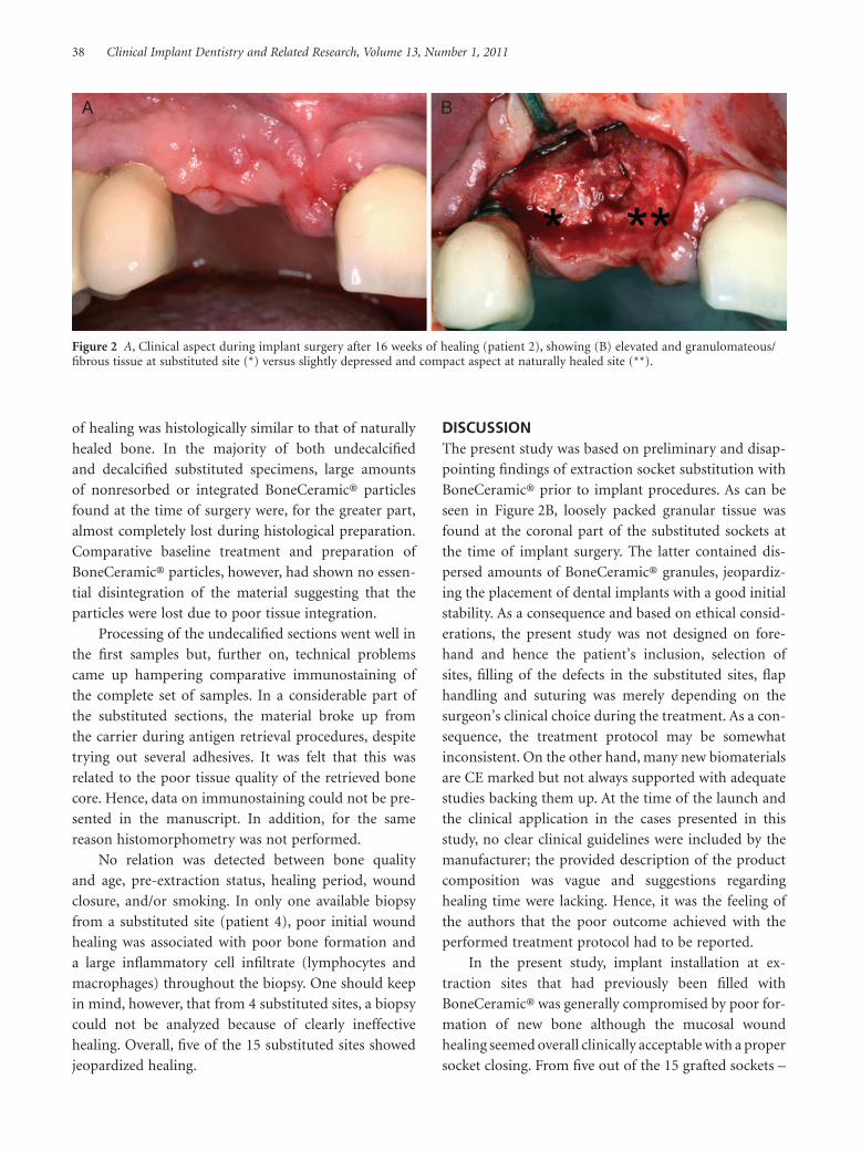
time of implant surgery, there was less resorption of the
alveolar crest at the substituted sites as compared with
the naturally healed sites (Figure 2). Bone at substituted
sites, however, was softer than bone in naturally healed
sites which was irrespective of the individual healing
time. In the majority of cases, large amounts of loose
biomaterial were found at the time of surgery. These
sites were thoroughly debrided prior to implant instal-
lation but sometimes the recipient beds were too large to
get normal diameter implants initially stable. Hence,
wider implants were necessary. Due to differences in
healing, radiographic evidence of graft consolidation,
and patient availability, the time between augmentation
and implantation ranged from 6–74 weeks, with a mean
of 22 weeks. Of the 29 extractions sites, one was not
prepared for implantation and hence not available for
biopsy taking. In two patients (#9 and #10) with four
BoneCeramic® grafts (after 8 and 21 weeks of healing
respectively) sampling of a bone core was impossible
due to a complete lack of mineralization. Implants,
although planned, could not be installed at these sites
and treatment was postponed. In those two patients, as a
consequence, no biopsy was taken from the nongrafted
sites. Hence, in total, 14 control and 10 substituted sites
were harvested. Of the 15 originally substituted sites, five
could not be implanted because of impaired healing and
two of the 10 inserted implants failed within 3 months
after insertion. Thirteen of the 14 naturally healed sites
were implanted; however, three failed of which two in
an immediate loading case. The clinical results and the
36 Clinical Implant Dentistry and Related Research, Volume 13, Number 1, 2011
characteristics of patients, extraction sites, and implants
are summarized in Table 1.
Histological Findings
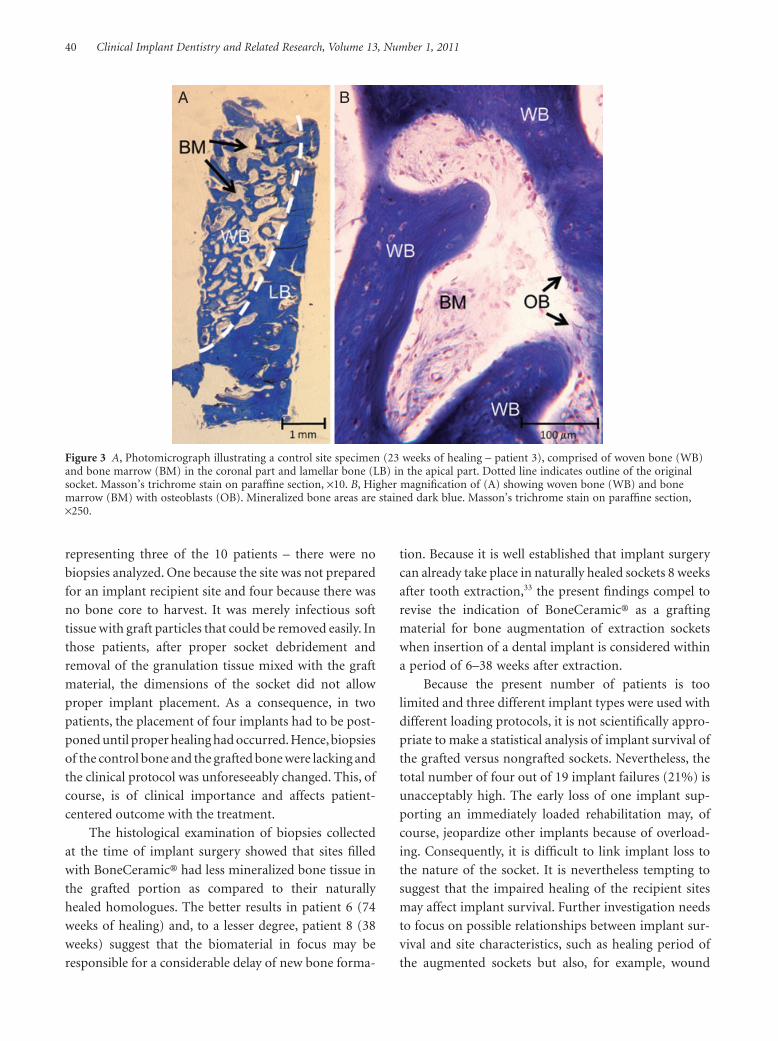
In decalcified sections of biopsies harvested from
untreated control sites, mature bone was present, mainly
comprising lamellar bone outlining the original socket,
and newly formed woven bone at the center of the pre-
vious socket (Figure 3, A and B). Formation of new bone
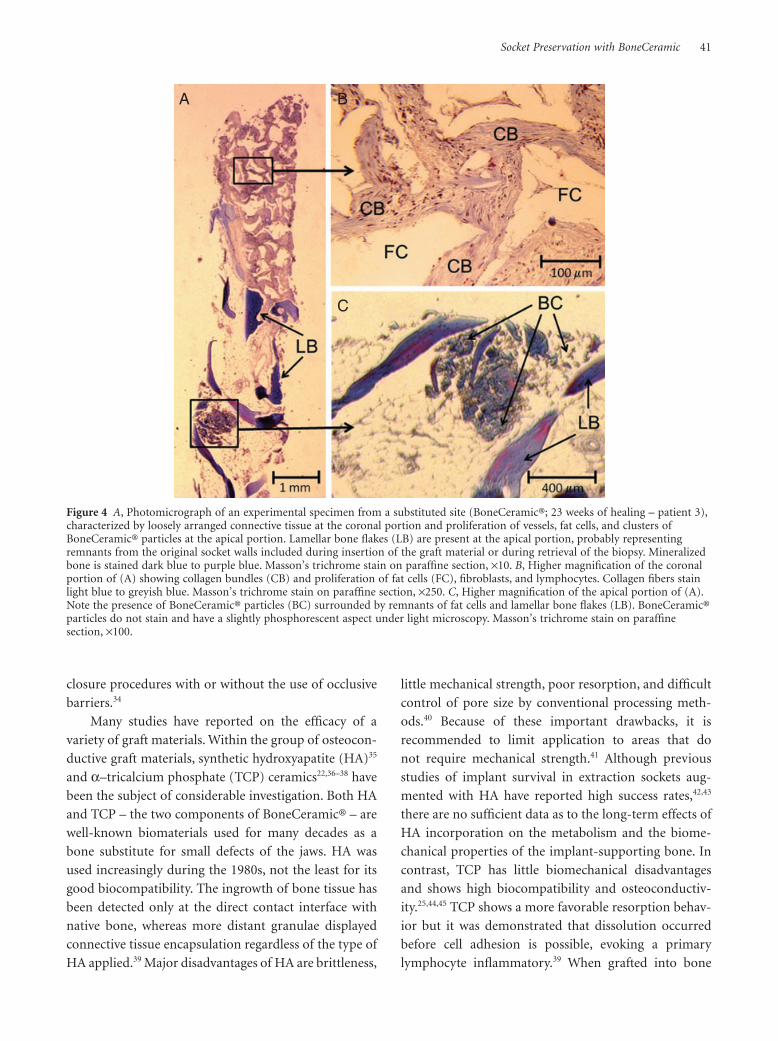
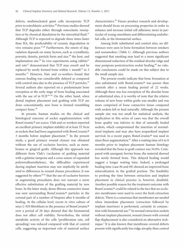
at substituted sites was poorer than controls, mainly
exhibiting loosely arranged connective tissue with sparse
evidence of mineralized bone in the grafted portion of
the cylinders (Figure 4, A–C). In most of the substituted
samples, the coronal portion of the biopsies was pre-
dominantly comprised of irregularly arranged bundles
of collagen fibers, proliferation of blood vessels and
fibroblasts, fat cells and moderate numbers of lympho-
cytes. At the apical portions of some of the samples,
impaction of small clusters of BoneCeramic® particles
could be observed in the fibrotic tissue with weak to no
evidence of new bone formation. The bone tissue api-
cally from the previous socket area was of a lamellar
type. These findings of poor bone formation were most
pronounced as compared with control sites in substi-
tuted sites of patients 2, 4, 5, and 7, and less explicit but
still clearly present in patients 1, 3, and 8. Bone in the
substituted socket of patient 6 harvested after 74 weeks
A
B C D
E F G
�
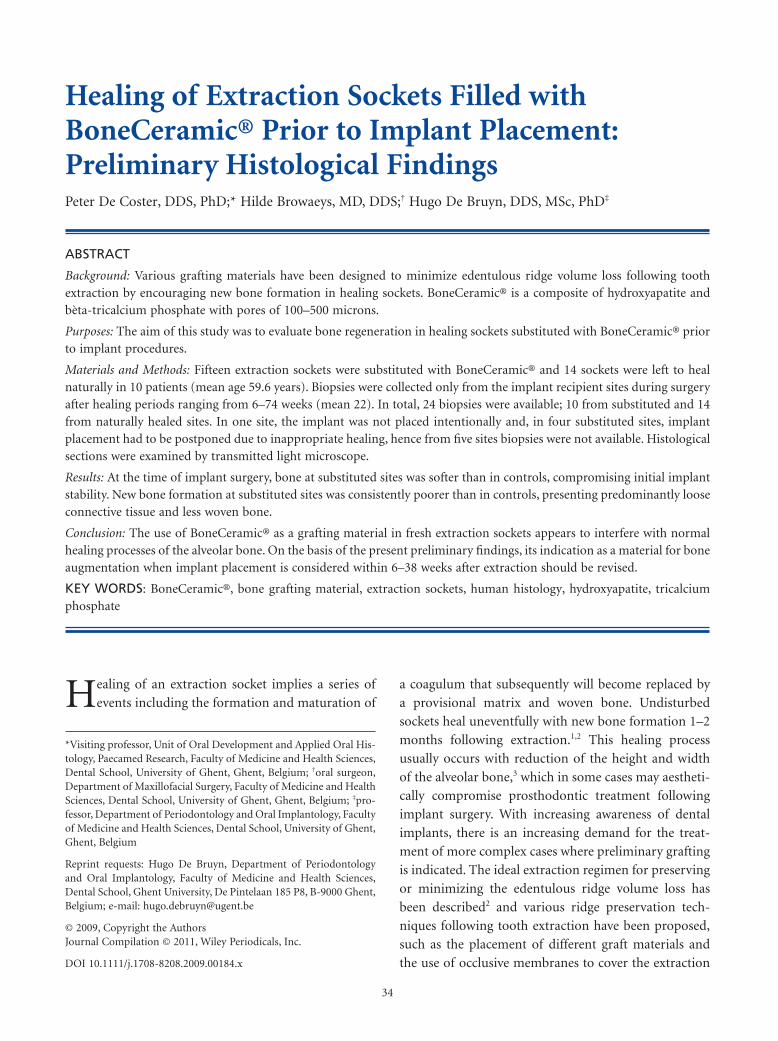
Figure 1 A, Clinical radiographs (patient 1) taken after 15months of implant loading. In total, seven teeth were extractedfor periodontal reasons; three extraction sockets remainedungrafted (22, 13, 14) and four were filled with BoneCeramic®(12, 23, 24, 25). After a healing period of 10 weeks, implantscould be placed with excellent initial stability (13, 14, 15, 21, 22,23) followed by immediate loading with a provisional resinbridge. Implants did not obtain initial stability on 24 and 12,both BoneCeramic®-grafted sites, due to insufficient healing.After 9 months, the final prosthesis was put into loading.Bone-to-implant contact is indicated with arrows and bone lossbelow the first implant thread is eminent on nongrafted sites(14, 13, 22). Furthermore and additionally, a radio-opacity isvisible around the implant in the grafted site (23) and granulaeare present at the grafted but not implanted nor harvested bonesite on position (25), indicating that integration ofBoneCeramic® in the newly-formed bone was incomplete. B,Photomicrograph of a control site biopsy (13) after 10 weeks ofhealing, showing excellent bone formation with woven bone(WB) and bone marrow (BM). Mineralized bone areas arestained dark blue, while bone marrow stains grey to light blue.Masson’s trichrome stain on paraffine section, ¥10. C,Photomicrograph of a substituted site biopsy (12) after same 10weeks of healing, showing predominantly dense connectivetissue (CT) without evidence of bone formation. Most ofBoneCeramic® particles that were still abundantly present as agranular mass at the time of biopsy taking were lost duringhistological processing. Because of ineffective healing, bone atthis site was too soft and no implant could be placed. Masson’strichrome stain on paraffine section, ¥10. D, Photomicrographof a substituted site biopsy (24) displaying a combination ofloosely arranged connective tissue (CT) and few islands ofimmature woven bone (WB). Note residual lamellar bone flakes(LB) at the apical portion of the biopsy. At this position,implant placement also was impossible due to bone softness.Immature woven bone stains blue to purple with central redspots indicating areas of advancing mineralization. Masson’strichrome stain on paraffine section, ¥10. E, Detail of (B)displaying woven bone (WB) with bone marrow (BM); ¥250. F,Detail of (C) presenting dense, blue to greyish stained collagenbundles (CB) and proliferation of vessels, fibroblasts, andlymphocytes. The bright red cells are erythrocytes issuing fromlocal hemorrhage during biopsy taking; ¥250 (G). Detail of (D)showing loose connective tissue abundant with fat cells (FC)and some peripheral islands of mineralized bone (B); ¥250.
Socket Preservation with BoneCeramic 37
of healing was histologically similar to that of naturally
healed bone. In the majority of both undecalcified
and decalcified substituted specimens, large amounts
of nonresorbed or integrated BoneCeramic® particles
found at the time of surgery were, for the greater part,
almost completely lost during histological preparation.
Comparative baseline treatment and preparation of
BoneCeramic® particles, however, had shown no essen-
tial disintegration of the material suggesting that the
particles were lost due to poor tissue integration.
Processing of the undecalified sections went well in
the first samples but, further on, technical problems
came up hampering comparative immunostaining of
the complete set of samples. In a considerable part of
the substituted sections, the material broke up from
the carrier during antigen retrieval procedures, despite
trying out several adhesives. It was felt that this was
related to the poor tissue quality of the retrieved bone
core. Hence, data on immunostaining could not be pre-
sented in the manuscript. In addition, for the same
reason histomorphometry was not performed.
No relation was detected between bone quality
and age, pre-extraction status, healing period, wound
closure, and/or smoking. In only one available biopsy
from a substituted site (patient 4), poor initial wound
healing was associated with poor bone formation and
a large inflammatory cell infiltrate (lymphocytes and
macrophages) throughout the biopsy. One should keep
in mind, however, that from 4 substituted sites, a biopsy
could not be analyzed because of clearly ineffective
healing. Overall, five of the 15 substituted sites showed
jeopardized healing.
DISCUSSIONThe present study was based on preliminary and disap-
pointing findings of extraction socket substitution with
BoneCeramic® prior to implant procedures. As can be
seen in Figure 2B, loosely packed granular tissue was
found at the coronal part of the substituted sockets at
the time of implant surgery. The latter contained dis-
persed amounts of BoneCeramic® granules, jeopardiz-
ing the placement of dental implants with a good initial
stability. As a consequence and based on ethical consid-
erations, the present study was not designed on fore-
hand and hence the patient’s inclusion, selection of
sites, filling of the defects in the substituted sites, flap
handling and suturing was merely depending on the
surgeon’s clinical choice during the treatment. As a con-
sequence, the treatment protocol may be somewhat
inconsistent. On the other hand, many new biomaterials
are CE marked but not always supported with adequate
studies backing them up. At the time of the launch and
the clinical application in the cases presented in this
study, no clear clinical guidelines were included by the
manufacturer; the provided description of the product
composition was vague and suggestions regarding
healing time were lacking. Hence, it was the feeling of
the authors that the poor outcome achieved with the
performed treatment protocol had to be reported.
In the present study, implant installation at ex-
traction sites that had previously been filled with
BoneCeramic® was generally compromised by poor for-
mation of new bone although the mucosal wound
healing seemed overall clinically acceptable with a proper
socket closing. From five out of the 15 grafted sockets –
A B
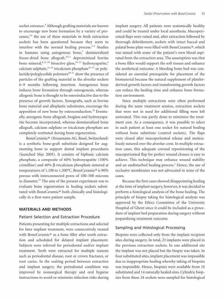
Figure 2 A, Clinical aspect during implant surgery after 16 weeks of healing (patient 2), showing (B) elevated and granulomateous/fibrous tissue at substituted site (*) versus slightly depressed and compact aspect at naturally healed site (**).
38 Clinical Implant Dentistry and Related Research, Volume 13, Number 1, 2011

TAB
LE1
Cas
eD
escr
ipti
on
of
All
Pati
ents
,Tr
eatm
ent
Mo
dal
ity
of
the
Extr
acti
on
Sock
ets,
Imp
lan
tLo
cati
on
,an
dIm
pla
nt
Ch
arac
teri
stic
s.Si
tes
wit
hIn
effe
ctiv
eH
ealin
go
fB
on
ean
dA
bse
nce
of
Bio
psy
are
Ind
icat
edw
ith
#
Cas
eA
ge
(Yea
rs)
Rea
son
for
Extr
acti
on
Smo
ker
Too
thSi
teB
on
eCer
amic
inSo
cket
Hea
ling
inW
eeks
Imp
lan
t/B
iop
syin
Extr
acti
on
Site
Faile
dIm
pla
nt
Imp
lan
tsp
erPa
tien
t
Faile
d/In
stal
led
Imp
lan
tsp
erPa
tien
t
Imp
lan
tLe
ng
th(m
m)
Imp
lan
tW
idth
(mm
)Im
pla
nt
Stab
ility
155
Peri
oN
o22
No
11Ye
s/
Yes
N4
extr
acti
onsi
tes
+2
hea
led
site
s
0/6
astr
a11
.54
Goo
d
155
Peri
oN
o23
Yes
11Ye
s/
Yes
N15
4G
ood
155
Peri
oN
o24
Yes
11N
o/
Yes
not
plac
ed—
——
155
Peri
oN
o12
Yes
11N
o/
No
not
plac
ed—
——
155
Peri
oN
o13
No
11Ye
s/
Yes
N15
4G
ood
155
Peri
oN
o14
No
11Ye
s/
Yes
N10
4G
ood
258
Car
ies
Yes
11N
o16
Yes
/Ye
sN
2ex
trac
tion
site
s0/
2n
obel
134
Goo
d
258
Car
ies
Yes
12Ye
s16
Yes
/Ye
sN
134
Goo
d
357
Frac
ture
No
11Ye
s23
Yes
/Ye
sN
3ex
trac
tion
site
s+
2
hea
led
site
s
1/5
astr
a15
4G
ood
357
Frac
ture
No
23N
o23
Yes
/Ye
sN
154
Goo
d
358
Car
ies
Yes
22N
o23
Yes
/Ye
sN
154
Goo
d
466
Car
ies
Yes
44N
o21
Yes
/Ye
sY
2ex
trac
tion
site
s2/
2n
obel
11.5
4G
ood
466
Car
ies
Yes
45Ye
s21
Yes
/Ye
sY
104
Goo
d
581
Frac
ture
No
12N
o6
Yes
/Ye
sY
5ex
trac
tion
site
s3/
5B
iom
et3i
105
Goo
d
581
Car
ies
No
22Ye
s6
Yes
/Ye
sY
154
Goo
d
581
Car
ies
No
23N
o6
Yes
/Ye
sY
154
Goo
d
581
Car
ies
No
21N
o6
Yes
/Ye
sN
154
Goo
d
581
Car
ies
No
11Ye
s6
Yes
/Ye
sN
154
Goo
d
641
Peri
oN
o41
No
74Ye
s/
Yes
N2
extr
acti
onsi
tes
+2
hea
led
site
s
0/4
nob
el15
4G
ood
641
Peri
oN
o43
Yes
74Ye
s/
Yes
N15
4G
ood
762
Frac
ture
No
12N
o21
Yes
/Ye
sN
2ex
trac
tion
site
s+
2
hea
led
site
s
0/4
Bio
met
3i15
4G
ood
762
Frac
ture
No
22Ye
s21
Yes
/Ye
sN
154.
8Po
or
858
Car
ies
Yes
44Ye
s38
Yes
/Ye
sN
3ex
trac
tion
site
s0
/3N
obel
103.
7G
ood
858
Car
ies
Yes
24N
o38
Yes
/Ye
sN
154
Goo
d
858
Frac
ture
Yes
25N
o38
Yes
/Ye
sN
11.5
4G
ood
958
Car
ies
No
11Ye
s8
No
/N
o#
not
plac
ed0
inse
rted
**
**
958
Car
ies
No
37Ye
s8
No
/N
o#
not
plac
ed*
**
**
1060
Peri
oN
o26
Yes
21N
o/
No
#n
otpl
aced
0in
sert
ed*
**
*
1060
Peri
oN
o37
Yes
21N
o/
No
#n
otpl
aced
**
**
*
n=
10M
ean
age
59.6
Per
io:n
=9
Yes:
n=
7n
=24
Yes:
n=
15M
ean
tim
eYe
s/Ye
s:n
=23
Fail
ed:n
=5
Fail
ed:6
/31
Goo
d:n
=22
Car
ies:
n=
9n
o:n
=16
No:
n=
1422
.7w
eeks
No/
Yes:
n=
1N
otp
lace
d:n
=6
Poo
r:n
=1
Frac
ture
:n=
6N
o/N
o:n
=5
Socket Preservation with BoneCeramic 39
representing three of the 10 patients – there were no
biopsies analyzed. One because the site was not prepared
for an implant recipient site and four because there was
no bone core to harvest. It was merely infectious soft
tissue with graft particles that could be removed easily. In
those patients, after proper socket debridement and
removal of the granulation tissue mixed with the graft
material, the dimensions of the socket did not allow
proper implant placement. As a consequence, in two
patients, the placement of four implants had to be post-
poned until proper healing had occurred.Hence,biopsies
of the control bone and the grafted bone were lacking and
the clinical protocol was unforeseeably changed. This, of
course, is of clinical importance and affects patient-
centered outcome with the treatment.
The histological examination of biopsies collected
at the time of implant surgery showed that sites filled
with BoneCeramic® had less mineralized bone tissue in
the grafted portion as compared to their naturally
healed homologues. The better results in patient 6 (74
weeks of healing) and, to a lesser degree, patient 8 (38
weeks) suggest that the biomaterial in focus may be
responsible for a considerable delay of new bone forma-
tion. Because it is well established that implant surgery
can already take place in naturally healed sockets 8 weeks
after tooth extraction,33 the present findings compel to
revise the indication of BoneCeramic® as a grafting
material for bone augmentation of extraction sockets
when insertion of a dental implant is considered within
a period of 6–38 weeks after extraction.
Because the present number of patients is too
limited and three different implant types were used with
different loading protocols, it is not scientifically appro-
priate to make a statistical analysis of implant survival of
the grafted versus nongrafted sockets. Nevertheless, the
total number of four out of 19 implant failures (21%) is
unacceptably high. The early loss of one implant sup-
porting an immediately loaded rehabilitation may, of
course, jeopardize other implants because of overload-
ing. Consequently, it is difficult to link implant loss to
the nature of the socket. It is nevertheless tempting to
suggest that the impaired healing of the recipient sites
may affect implant survival. Further investigation needs
to focus on possible relationships between implant sur-
vival and site characteristics, such as healing period of
the augmented sockets but also, for example, wound
A B
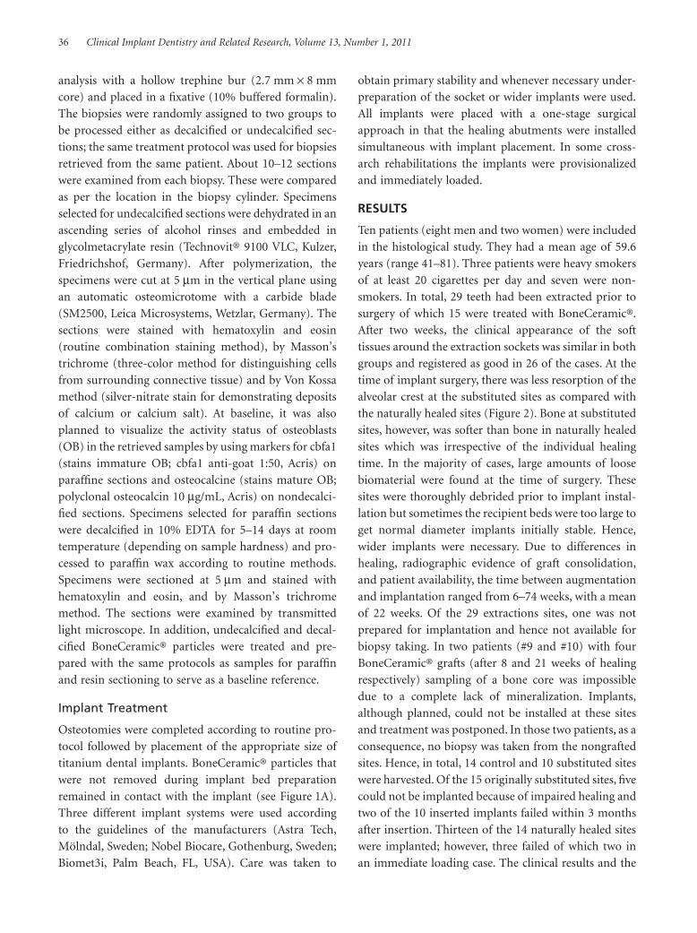
Figure 3 A, Photomicrograph illustrating a control site specimen (23 weeks of healing – patient 3), comprised of woven bone (WB)and bone marrow (BM) in the coronal part and lamellar bone (LB) in the apical part. Dotted line indicates outline of the originalsocket. Masson’s trichrome stain on paraffine section, ¥10. B, Higher magnification of (A) showing woven bone (WB) and bonemarrow (BM) with osteoblasts (OB). Mineralized bone areas are stained dark blue. Masson’s trichrome stain on paraffine section,¥250.
40 Clinical Implant Dentistry and Related Research, Volume 13, Number 1, 2011
closure procedures with or without the use of occlusive
barriers.34
Many studies have reported on the efficacy of a
variety of graft materials. Within the group of osteocon-
ductive graft materials, synthetic hydroxyapatite (HA)35
and a–tricalcium phosphate (TCP) ceramics22,36–38 have
been the subject of considerable investigation. Both HA
and TCP – the two components of BoneCeramic® – are
well-known biomaterials used for many decades as a
bone substitute for small defects of the jaws. HA was
used increasingly during the 1980s, not the least for its
good biocompatibility. The ingrowth of bone tissue has
been detected only at the direct contact interface with
native bone, whereas more distant granulae displayed
connective tissue encapsulation regardless of the type of
HA applied.39 Major disadvantages of HA are brittleness,
little mechanical strength, poor resorption, and difficult
control of pore size by conventional processing meth-
ods.40 Because of these important drawbacks, it is
recommended to limit application to areas that do
not require mechanical strength.41 Although previous
studies of implant survival in extraction sockets aug-
mented with HA have reported high success rates,42,43
there are no sufficient data as to the long-term effects of
HA incorporation on the metabolism and the biome-
chanical properties of the implant-supporting bone. In
contrast, TCP has little biomechanical disadvantages
and shows high biocompatibility and osteoconductiv-
ity.25,44,45 TCP shows a more favorable resorption behav-
ior but it was demonstrated that dissolution occurred
before cell adhesion is possible, evoking a primary
lymphocyte inflammatory.39 When grafted into bone
A B
C
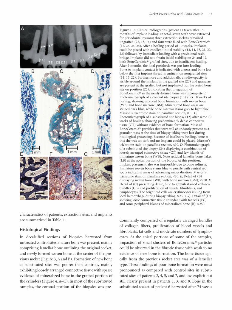
Figure 4 A, Photomicrograph of an experimental specimen from a substituted site (BoneCeramic®; 23 weeks of healing – patient 3),characterized by loosely arranged connective tissue at the coronal portion and proliferation of vessels, fat cells, and clusters ofBoneCeramic® particles at the apical portion. Lamellar bone flakes (LB) are present at the apical portion, probably representingremnants from the original socket walls included during insertion of the graft material or during retrieval of the biopsy. Mineralizedbone is stained dark blue to purple blue. Masson’s trichrome stain on paraffine section, ¥10. B, Higher magnification of the coronalportion of (A) showing collagen bundles (CB) and proliferation of fat cells (FC), fibroblasts, and lymphocytes. Collagen fibers stainlight blue to greyish blue. Masson’s trichrome stain on paraffine section, ¥250. C, Higher magnification of the apical portion of (A).Note the presence of BoneCeramic® particles (BC) surrounded by remnants of fat cells and lamellar bone flakes (LB). BoneCeramic®particles do not stain and have a slightly phosphorescent aspect under light microscopy. Masson’s trichrome stain on paraffinesection, ¥100.
Socket Preservation with BoneCeramic 41
defects, multinucleated giant cells incorporate TCP
prior to osteoblastic activities.46 Previous studies showed
that TCP degrades either through osteoclastic resorp-
tion or by chemical dissolution by the interstitial fluid.37
Although TCP is expected to degrade three times faster
than HA, the predictability of ceramic degradation in
vivo remains poor.47,48 Furthermore, the extent of deg-
radation depends on many factors, such as crystallinity,
porosity, density, particle form and size,49 the host, and
implantation site.50 In vivo experiments using rabbits51
and rats52 demonstrated that TCP may resorb and be
replaced by newly formed bone within 1 month52 to 3
months.51 However, Nair and co-workers found that
osseous healing was considerably delayed as compared
with control sites due to the presence of TCP particles.53
Several authors also reported on a predominant bone
resorption at the early stage of bone healing associated
with the use of a-TCP.54–56 On the other hand, when
dental implant placement and grafting with TCP are
done concomitantly, new bone is formed resembling
compact bone.38
At present, human studies on the clinical and
histological outcome of socket supplementation with
BoneCeramic® are scarce. Vlah and co-workers reported
an excellent primary implant stability of at least 35 Ncm
at sockets that had been augmented with BoneCeramic®
3 months before implant placement.57 In the present
study, a good primary wound closure was obtained
without the use of occlusive barriers, such as mem-
branes or gingival grafts. Although this approach was
different from Vlah’s (occlusion of grafting material
with a gelatine tampone and a cross-suture of expanded
polytetrafluorethylene), the difficulties experienced
during implant insertion may not implicitly be attrib-
uted to differences in wound closure procedures. It was
suggested by others34,58 that the use of occlusive barriers
in augmenting procedures does not warrant a more
effective substitution of the grafting material by new
bone. In the latter study, dense fibrous connective tissue
was seen surrounding BoneCeramic® particles in the
coronal part of a number of biopsies after 8 months of
healing. At the cellular level, recent in vitro culture of
mice L-929 fibroblasts in the presence of BoneCeramic®
over a period of 28 days showed that the biomaterial
does not affect cell viability. Nevertheless, the initial
metabolic activity of the cells (proliferation rate, cell
spreading) was reduced compared with that of control
cells, suggesting an important role of material surface
characteristics.59 Future product research and develop-
ment should focus on processing properties in order to
enhance and increase initial cell adhesion, more in par-
ticular of young osteoblasts and differentiating endothe-
lial cells, at the biomaterial surface.
Among both substituted and control sites, no dif-
ferences were seen in bone formation between smokers
and nonsmokers (Table 1). Although previous authors
suggested that smoking may lead to a more significant
dimensional reduction of the residual alveolar ridge and
may postpone postextraction socket healing,60 no valu-
able conclusions could be made on this subject due to
the small sample size.
The present results indicate that bone formation at
sites substituted with BoneCeramic® was poorer than
controls after a mean healing period of 22 weeks.
Although there was less resorption of the alveolar bone
at substituted sites, it is worthy of note that the relative
volume of new bone within grafts was smaller and was
more comprised of loose connective tissue compared
with sockets left to heal naturally. Although the present
sample size was too small for statistical analysis, the
implication in this series of cases was that the overall
bone quality was inferior to that of naturally healed
sockets, which compromised the insertion of normal-
sized implants and may also have jeopardized implant
survival. In a recent paper, BoneCeramic® was used in
sinus floor augmentation.61 After a waiting time of 6 to 8
months prior to implant placement human histology
revealed that the bone to graft contact was 34.0%. Com-
pared with anorganic bovine bone, the material showed
less newly formed bone. This delayed healing would
suggest a longer waiting time. Indeed, a prolonged
healing time (case #6 and #8) showed an improved bone
mineralization in the grafted portion. The feasibility
to prolong the time between extraction and implant
placement in clinical practice is, however, doubtful.
Another possible reason for the treatment outcome with
BoneCeramic® could be related to the fact that no occlu-
sive membranes were used to cover the fresh extraction
defects. There is a consensus that membranes are needed
when immediate placement (extraction followed by
implant insertion) is performed, certainly in conjunc-
tion with biomaterial use.62 In normal extraction sockets
without implant placement, wound closure with coronal
flap displacement is also considered an alternative tech-
nique.2 It is also known that membrane covered defects
present with significantly less ridge atrophy than control
42 Clinical Implant Dentistry and Related Research, Volume 13, Number 1, 2011
sites. This was, however, also observed in the present
study in the substituted sites without the use of mem-
branes. It is clear from the preliminary findings of our
study that further studies should address the biological
mechanisms as well as material, site and patient charac-
teristics that influence implant integration/survival in
extraction sockets supplemented with BoneCeramic®.
This includes, among others, the long-term influence of
the presence of incorporated HA bodies on both the
metabolic characteristics and physical properties of the
newly formed bone.
CONCLUSIONS
On the basis of the present preliminary findings, it
can be concluded that socket supplementation with
BoneCeramic® yields excellent results with respect to
volume preservation of the alveolar crest but influences
bone healing negatively. That is, new bone formation
is retarded and inefficient compared with naturally
healed sockets after equal healing periods (ranging 6–74
weeks). Further research, based on a proper designed
study with adequate numbers for statistical analysis, is
needed to scrutinize the biological mechanisms and the
long-term clinical benefits of the procedure. The authors
emphasize the need for clear instructions concerning the
minimum healing period at the time of launching of
new grafting materials, which should be based on mul-
tivariate studies implicating different patient and site
characteristics. Unfortunately, until today, the manufac-
turer has not provided evidence based on histological
studies to provide insight in the usage of the biomaterial
discussed in the present study in extraction sockets.
REFERENCES
1. Evian CI, Rosenberg ES, Coslet JG, Com H. The osteogenic
activity of bone removed from healing extraction sockets in
humans. J Periodontol 1982; 53:81–85.
2. Wang HL, Kiyonobu K, Neiva RF. Socket augmentation:
rationale and technique. Implant Dent 2004; 13:286–296.
3. Araújo MG, Lindhe J. Dimensional ridge alterations follow-
ing tooth extraction. An experimental study in the dog. J
Clin Periodontol 2005; 32:212–218.
4. Froum S, Cho SC, Rosenberg E, Rohrer M, Tarnow D. His-
tological comparison of healing sockets implanted with bio-
active glass or demineralized freez-dried bone allograft. J
Periodontol 2002; 73:94–102.
5. Constantino PD, Freidman CD. Synthetic bone graft substi-
tutes. Otolaryngol Clin North Am 1994; 27:1037–1073.
6. Cypher TJ, Grossman JP. Biological principles of bone graft
healing. J Foot Ankle Surg 1996; 35:413–417.
7. Becker W, Becker BE, Caffesse R. A comparison of deminer-
alized freeze-dried bone and autologous bone to induce
bone fromation in human extraction sockets. J Periodontol
1994; 65:1128–1133.
8. Becker W, Urist M, Vincenzi G, DeGeorges D, Niederwanger
M. Clinical and histological observation of sites implanted
with intraoral autologous bone graft or allograft. 15 human
case reports. J Periodontol 1996; 67:1025–1033.
9. Buser D, Hoffmann B, Bernard JP, Lussie A, Mettler D,
Schenk RK. Evaluation of filling materials in membrane-
protected defect. Clin Oral Implants Res 1998; 3:137–150.
10. Becker W, Clokie C, Sennerby L, Urist MR, Becker BE. His-
tologic findings after implantation and evaluation of
different grafting materials and titanium micro screws into
extraction sockets: case reports. J Periodontol 1998; 69:414–
421.
11. Brugnami F, Then PR, Moroi H, Leone CW. Histologic
evaluation of human extraction socket treated with dem-
ineralized bone allograft (DFDBA) and cell occlusive mem-
brane. J Periodontol 1996; 67:821–825.
12. Diès F, Etienne D, Bou Abboud N, Ouhayoun JP. Bone
regeneration in extraction sites after immediate placement
of an e-PTFE membrane with or withou a biomaterial. Clin
Oral Implants Res 1996; 7:277–285.
13. Artzi Z, Tal H, Dayan D. Porous bovine bone mineral in
healing of human extraction socket. Part I. Histometric
evaluation at 9 months. J Periodontol 2000; 71:1015–1023.
14. Carmagnola D, Adriaens PA, Berglundh T. Healing of
human extraction sockets filled with Bio-Oss. Clin Oral
Implants Res 2003; 14:137–143.
15. Iasella JM, Greenwell H, Miller RL, et al. Ridge preservation
with freeze-dried bone allograft and a collagen membrane
compared to extraction alone for implant site development:
a clinical and histologic study in humans. J Periodontol
2003; 74:990–999.
16. Minichetti JC, D’Amore JC, Hong AY, Cleveland DB.
Human histologic analysis of mineralized bone allograft
(Puros®) placement before implant surgery. J Oral Implan-
tol 2004; 30:74–82.
17. Hahn J, Rohrer MD, Tofe AJ. Clinical, radiographic, histo-
logic, and histomorphometric comparison of PepGen P-15
particulate and PepGen P-15 flow in extraction sockets: a
same-mouth case study. Implant Dent 2003; 12:170–174.
18. Norton MR, Odell EW, Thompson ID, Cook RJ. Efficacy of
bovine bone mineral for alveolar augmentation: a human
histologic study. Clin Oral Implants Res 2003; 14:775–783.
19. Stanley HR, Hall MB, Clark AE, King CJ 3rd, Hench LL,
Berte JJ. Using 45S5 bioglass cones as endosseous ridge
maintenance implants to prevent alveolar ridge resorption:
a 5-year evaluation. Int J Oral Maxillofac Implants 1997;
12:95–105.
Socket Preservation with BoneCeramic 43

20. Camargo PM, Lekovic V, Weinlaender M, et al. Influence of
bioactive glass on changes in alvolar dimensions after
exodontia. Oral Surg Oral Med Oral Pathol Oral Radiol
Endod 2000; 90:581–586.
21. Froum S, Cho SC, Elian N, Rosenberg E, Rohrer M, Tarnow
D. Extraction sockets and implantation. Implant Dent 2004;
13:153–164.
22. Shankly PE, Mackie IC, McCord FJ. The use of tricalcium
phosphate to preserve alveolar bone in a patient with ecto-
dermal dysplasia: a case report. Spec Care Dentist 1999;
19:35–39.
23. Guarnieri R, Pecora G, Fini M, et al. Medical grade calcium
sulfate hemihydrate in healing of human extraction sockets:
clinical and histological observations at 3 months. J Period-
ontol 2004; 75:902–908.
24. Guarnieri R, Aldini NN, Pecora G, Fini M, Giardino R.
Medial-grade calcium sulfate hemihydrate (surgiplaster) in
healing of a human extraction socket – histologic observa-
tion at 3 months: a case report. Int J Oral Maxillofac
Implants 2005; 20:636–641.
25. LeGeros RZ. Properties of osetoconductive biomaterials:
calcium phosphates. Clin Orthop Relat Res 2002; 395:81–98.
26. Ormanier Z, Palti A, Shifman A. Survival of immediately
loaded dental implants in deficient alveolar bone sites aug-
mented with beta-tricalcium phosphate. Implant Dent 2006;
15:395–403.
27. Rothamel D, Schwarz F, Herten M, et al. Kiefer-
kammveränderungen nach Versorgung frischer
Extraktionsalveolen mit polylactidvernetzten b-TCP
Wurzelreplikaten – eine histomorphometrische Tierstudie.
Mund Kiefer GesichtsChir 2007; 11:89–97.
28. Lekovic V, Camargo PM, Klokkevold PR, et al. Preservation
of alveolar bone in extraction sockets using bioabsorbable
membranes. J Periodontol 1998; 69:1044–1049.
29. Serino G, Biancu S, Iezzi G, Piattelli A. Ridge preservation
following tooth extraction using a polylactide and polygly-
colide sponge as space filler: a clinical and histological study
in humans. Clin Oral Implants Res 2003; 14:651–658.
30. Nair PN, Schug J. Observations on healing of human tooth
extraction sockets implanted with bioabsorbable polylactic-
polyglycolic acids (PLGA) copolymer root replicas: a clinical,
radiographic, and histologic follow-up report of 8 cases.
Oral Surg Oral Med Oral Pathol Oral Radiol Endod 2004;
97:559–569.
31. Serino G, Rao W, Iezzi G, Piattelli A. Polylactide and polyg-
lycolide sponge used in human extraction sockets: bone for-
mation following 3 months after its application. Clin Oral
Implants Res 2008; 19:26–31.
32. Dietze S, Bayerlein T, Proff P, Hoffmann A, Gedrnge T. The
ultrastructure and processing properties of Struamann Bone
Ceramic® and NanoBone®. Folia Morphol 2006; 65:63–65.
33. Degidi M, Piattelli A, Carinci F. Immediate loaded
dental implants: comparison between fixtures inserted in
postextractive and healed bone sites. J Craniofac Surg 2007;
18:965–971.
34. Tehemar S, Hanes P, Sharawy M. Enhancement of osseoin-
tegration of implants placed into extraction sockets of
healthy and periodontally diseased teeth by using graft mate-
rial, an ePTFE membrane, or a combination. Clin Implant
Dent Relat Res 2003; 5:193–211.
35. Luczyszyn SM, Papalexiou V, Novaes AB Jr, Grisi MF, Souza
SL, Taba M Jr. Acellular dermal matrix and hydroxyapatite.
Implant Dent 2005; 14:176–184.
36. Hempton TJ, Fugazotto PA. Ridge augmentation utilizing
guided tissue regeneration, titanium screws, freeze-dried
bone, and tricalcium phosphate: clinical report. Implant
Dent 1994; 3:35–37.
37. Zerbo IR, Bronckers AL, de Lange G, van Beek GJ, Burger
EH. Localisation of osteogenic and osteoclastic cells in
porous beta-tricalcium-phosphate particles used for human
maxillary sinus floor elevation. Biomaterials 2005; 26:1445–
1451.
38. Nakadate M, Amizuka N, Li M, et al. Histological evaluation
on bone regeneration of dental implant placement sites
grafted with a self-setting alpha-tricalcium phosphate
cement. Microsc Res Tech 2008; 71:93–104.
39. Soost F, Koch S, Stoll C. Validation of bone conversion in
osteoonductive and osteoinductive bone substitutes. Cell
Tissue Bank 2001; 2:77–86.
40. Chu TM, Orton DG, Hollister SJ, et al. Mechanical and in
vivo performance of hydroxyapatite implants with con-
trolled architectures. Biomaterials 2002; 23:1283–1293.
41. Chang BS, Lee CK, Hong KS, et al. Osteoconduction at
porous hydroxyapatite with various pore configurations.
Biomaterials 2000; 21:1291–1298.
42. Remagen W, Prezmecky L. Bone augmentation with
hydroxyapatite: histological findings in 55 cases. Implant
Dent 1995; 4:182–188.
43. Mangano C, Bartolucci EG, Mazzocco C. A new porous
hydroxyapatite for promotion of bone regeneration in max-
illary sinus augmentation: clinical and histologic study in
humans. Int J Oral Maxillofac Implants 2003; 18:23–30.
44. Jarcho M. Calcium phosphate ceramics as hard tissue pros-
thetics. Clin Orthop 1981; 157:260–288.
45. Damien CJ, Parsons JR. Bone graft and bone grafts substi-
tutes: a review of current technology and applications. Appl
Biomater 1991; 2:187–208.
46. Yamada S, Heymann D, Bouler JM, Daculsi G. Osteoclastic
resorption of calcium phosphate ceramics with different
hydroxyapatite/tricalcium phosphate ratios. Biomaterials
1997; 18:1037–1041.
47. Koerten HK, van der Meulen J. Degradation of calcium
phosphate ceramics. J Biomed Mater Res 1999; 44:78–86.
48. Handschel J, Wiesmann HP, Stratmann U, et al. TCP is
hardly resorbed and not osteoconductive in a non-loading
calvarial model. Biomaterials 2002; 23:1689–1695.
44 Clinical Implant Dentistry and Related Research, Volume 13, Number 1, 2011
49. Camiré CL, Gbureck U, Hirsiger W, Bohner M. Correlating
crystallinity and reactivity in an alpha-tricalcium phosphate.
Biomaterials 2005; 26:2787–2794.
50. Theiss F, Apelt D, Brand B, et al. Biocompatibility and
resorption of a brushite calcium phosphate cement. Bioma-
terials 2005; 26:4383–4394.
51. Bhaskar SN, Brady JM, Getter L, Grower MF, Driskell T.
Biodegradable ceramic implants in bone. Electron and light
microscopic analysis. Oral Surg Oral Med Oral Pathol 1971;
32:336–346.
52. Hao H, Amizuka N, Oda K, et al. A histological evaluation
on self-setting a-tricalcium phosphate applied in the rat
bone cavity. Biomaterials 2004; 25:431–442.
53. Nair PN, Luder H-U, Maspero FA, Fischer JH, Schug J. Bio-
compatibility of b-tricalcium phosphate root replicas in
porcine tooth extraction sockets: a correlative histological,
ultrastructural, and X-ray microanalytical pilot study. J Bio-
mater Appl 2006; 20:307–324.
54. Rejda BV, Peelen JG, de Groot K. Tri-calcium phosphate as a
bone substitute. J Bioeng 1977; 1:93–97.
55. Nagase M, Chen RB, Asada Y. Radiographic and microscopic
evaluation of subperiostally implanted blocks of hydrated
and hardened a-TCP in rabbits. J Oral Maxillofac Surg 1989;
47:582–586.
56. Wada T, Hara K, Ozawa H. Ultrastructural and histoche-
mical study of b-tricalcium phosphate resorbing cells in
periodontium of dogs. J Periodont Res 1989; 24:391–401.
57. Vlah M, Bosnjak A, Meniga A. Post-extraction socket aug-
mentation by modified beta-tricalcium phosphate. Acta Sto-
matol Croat 2008; 42. Proceedings of the 2nd Congress of the
Croatian Society of Dental Implantology, October 18–20,
2007. Zagreb, Croatia.
58. Mardas N, Chadha V, Donos N. Socket preservation with
synthetic bone substitute (Straumann Bone Ceramic®) or a
bovine xenograft (Bio-Oss®). IADR 86th General Session
and Exhibition. July 1–5, 2008. Toronto, Ontario, Canada:
Abstract 0800.
59. Kauschke E, Rumpel E, Fanghänel J, Bayerlein T, Gedrange
T, Proff P. The in vitro viability and growth of fibroblasts
cultured in the presence of different bone grafting materials
(NanoBone® and Straumann Bone Ceramic®). Folia
Morphol 2006; 65:37–42.
60. Saldanha JB, Casati MZ, Neto FH, Sallum EA, Nociti FH Jr.
Smoking may affect the alveolar process dimensions and
radiographic bone density in maxillary extraction sites: a
prospective study in humans. J Oral Maxillofac Surg 2006;
64:1359–1365.
61. Cordaro L, Bosshardt DD, Palattella P, Rao W, Serino G,
Chiapasco M. Maxillary sinus grafting with Bio-Oss or
Straumann Bone Ceramic: histomorphometric results from
a randomized controlled multicenter clinical trial. Clin Oral
Implants Res 2008; 19:796–803.
62. Hämmerle CH, Jung RE. Bone augmentation by means of
barrier membranes. Periodontol 2000 2003; 33:36–53.
Socket Preservation with BoneCeramic 45