harmful algal bloom toxins alter c-fos protein expression in the brain of killifish, fundulus...
TRANSCRIPT
Harmful algal bloom toxins alter c-Fos protein expression in thebrain of killifish, Fundulus heteroclitus
J.D. Saliernoa, N.S. Snyderb, A.Z. Murphyc, M. Polid, S. Halle, D. Badenf, and A.S. Kanea,g,*
a Aquatic Pathobiology Center, Department of Epidemiology and Preventive Medicine, University ofMaryland School of Medicine, Baltimore, MD 21201, USA
b Department of Anatomy and Neurobiology, University of Maryland School of Medicine, Baltimore, MD21201, USA
c Department of Biology, Georgia State University, Atlanta, GA 30303, USA
d US Army Medical Research Institute of Infectious Diseases, Integrated Toxicology Division, Fort Detrick,USA
e US Food and Drug Administration, Office of Seafood, Beltsville Research Facility, Laurel, MD 20708, USA
f Center for Marine Science Research, University of North Carolina Wilmington, Wilmington, NC 28409,USA
g Aquatic Pathobiology Center, Virginia-Maryland Regional College of Veterinary Medicine, College Park,MD 20742, USA
AbstractThe immediate early gene c-fos, and its protein product c-Fos, are known to be induced in neuronsof mammals and fish as a result of neuronal stimulation. The purpose of this study was toquantitatively examine CNS alterations in killifish, Fundulus heteroclitus, in relation to harmful algalbloom (HAB) toxin exposure. c-Fos expression was visualized using immunocytochemistry in thebrains of killifish exposed to the excitatory neurotoxins domoic acid (DA) and brevetoxin (PbTx-2),and a paralytic neurotoxin, saxitoxin (STX), released from HABs. In addition, a simulated transportstress experiment was conducted to investigate effects of physical stress on c-Fos induction. Groupsof fish were exposed to the different stress agents, brain sections were processed for c-Fos staining,and expression was quantified by brain region. Fish exposed to DA, STX, and transport stressdisplayed significant alterations in neuronal c-Fos expression when compared to control fish (p ≤0.05). DA, PbTx-2, and transport stress increased c-Fos expression in the optic tecta regions of thebrain, whereas STX significantly decreased expression. This is the first study to quantify c-Fosprotein expression in fish exposed to HAB toxins. General alterations in brain activity, as well asknowledge of specific regions within the brain activated in association with HABs or other stressors,provides valuable insights into the neural control of fish behavior as well as sublethal effects ofspecific stressors in the CNS.
KeywordsFish; c-Fos; Brevetoxin; Saxitoxin; Domoic acid; Fundulus heteroclitus
* Corresponding author. Tel.: +1 301 314 6808. E-mail address: [email protected] (A.S. Kane).
NIH Public AccessAuthor ManuscriptAquat Toxicol. Author manuscript; available in PMC 2009 March 24.
Published in final edited form as:Aquat Toxicol. 2006 July 20; 78(4): 350–357. doi:10.1016/j.aquatox.2006.04.010.
NIH
-PA Author Manuscript
NIH
-PA Author Manuscript
NIH
-PA Author Manuscript

1. IntroductionBehavioral changes in organisms can result from complex alterations at the biochemical andphysiological levels of organization. Knowledge of these underlying processes is crucial tounderstanding the control of behavior and response to stimuli. Organisms can induce cellularchange by adapting to stimuli, and mediating repair and regeneration processes throughalterations in gene expression. Adaptation to environmental conditions is only possible due tothe rapidity and flexibility of neuronal signal transduction, and downstream changes in geneexpression (Rybnikova et al., 2003).
Immediate early genes (IEGs), localized within the nuclei of neurons, are induced as a resultof electrical or second messenger stimulation. Induction requires no synthesis of transcriptionfactors, and IEGs can be induced rapidly with activation within 15–30 min of stimulus reception(Herdegen and Leah, 1998). IEG activation then codes for new proteins such as cytokines,enzymes, HSP70, as well as transcription factors leading to neuronal phenotypic changes(Herrera and Robertson, 1996). These phenotypic changes alter gene expression through thedimerization of c-Fos and AP-1 complex binding (Herdegen and Leah, 1998; Bakin and Curran,1999; Zhang et al., 2002; Hansson et al., 2003). As a result, c-Fos induction can be used as abiomarker of neuronal and regional brain activity when animals are exposed to different typesof stressful stimuli (Martinez et al., 2002; Rybnikova et al., 2003).
Studies have been conducted in rats and birds to investigate the role of the c-fos gene in onphysiological and behavioral responses (Hansson et al., 2003). In rats, it has been demonstratedthat c-Fos expression mediates neuronal excitation and enhances survival (Zhang et al.,2002). c-fos can be induced in rats through glutamate receptor agonists, ion channel flux,dioxins, and the mind altering drugs haloperidol and clozapine (Morgan and Curran, 1986;Sonnenberg et al., 1989; Murphy and Feldon, 2001; Cheng et al., 2002). Further, it appearsthat drug-induced patterns of Fos expression are dependent on the animals' concurrentbehavioral status (Murphy and Feldon, 2001). c-Fos expression is also associated with longterm memory and walking behaviors (rats), and imprinting and courting behaviors (zebrafinches, Taeniopygia guttat) (Sadananda and Bischof, 2002; Espana et al., 2003).
In fish, the mechanisms and outcome of c-Fos protein expression is not well understood. Fishare exposed to similar stressors as mammals and birds, i.e., xenobiotics, neurotoxins, andenvironmental estrogens, and c-Fos expression may serve as an important measure of exposureand response. It has been demonstrated that the c-fos gene is highly conserved betweenvertebrate species (Kindy and Verma, 1990; Schreiberagus et al., 1993; Bosch et al., 1995).Previous studies using rainbow trout have described increased c-Fos expression in thebrainstem following startle responses, indicating that Fos expression can be utilized as aanatomical marker of neuronal activity to investigate higher order behaviors (Bosch et al.,1995, 2001; Matsuoka et al., 1998). However, no studies to date have investigated alterationsin c-Fos expression in fish exposed to ecologically relevant chemical stressors.
Harmful algal blooms (HABs) in the middle Atlantic states, and HAB-associated fish morbidityand mortality, have increased in frequency and severity over the past several decades. Theability to predict and characterize environmental effects of different HAB species are essentialto HAB remediation and control. Domoic acid (DA), brevetoxin (PbTx-2), and saxitoxin (STX)are neurotoxins released by Pseudo-nitzschia, Karenia, and Alexandrium spp., respectively.These species have been demonstrated harmful to fish through behavioral alterations andmortality (Steidinger et al., 1973; White, 1977; Lefebvre et al., 2001; Landsberg, 2002). Thisstudy examined alterations in c-Fos expression in six regions of the brain from killifish exposedto DA, PbTx-2, or STX.
Salierno et al. Page 2
Aquat Toxicol. Author manuscript; available in PMC 2009 March 24.
NIH
-PA Author Manuscript
NIH
-PA Author Manuscript
NIH
-PA Author Manuscript
The goal of this work was to develop and test the utility of c-Fos expression, visualized throughimmunocytochemistry, as a measure of exposure and effect in killifish, Fundulusheteroclitus, exposed to harmful algal bloom (HAB) neurotoxins. The effect of a physicalstressor, simulated transport stress, was also examined. c-Fos expression was evaluated throughthe examination of brains from stressed and control fish using anti c-Fos polyclonal antibodystaining. In addition to general changes in brain activity, knowledge of the specific regionswithin the brain activated during stress provides valuable insight into the neural control of fishbehavior.
2. Methods2.1. Exposures
Fish were exposed for 60 min in 4 L glass beakers containing 3 L of static artificial seawater(6 PSU, pH 8, 25 °C) to DA (IP injection, 0 and 5 mg/kg, Sigma–Aldrich), PbTx-2, (aqueous,0 and 40 ppb, nominal concentration with an emulsifying vehicle (EL-620, 0.001%)), and STX(aqueous, 0, 75, and 150 ppb, nominal concentrations). Nominal toxin exposure concentrationsand duration were based on preliminary LC50 experiments (data not shown) and other studiesinvestigating HAB exposure on fish (Lefebvre et al., 2001, 2004). Actual exposureconcentrations were typically within 10% of nominal concentrations, based on repeatedradioimmunoassay and receptor binding assay results from preliminary experiments withPbTx-2 and STX, respectively (data not shown). Three sets of controls were conducted: a shamIP injection control of phosphate buffered saline (DA), an emulsifying vehicle control inartificial seawater (EL-620, 0.001%, PbTx-2), and an artificial seawater control (STX). Tosimulate transport stress, fish were placed in a 10-L covered transport bucket, and the bucketwas vertically dropped 0.5 cm to the floor at a rate of 15 times per minute for 60 min. This wasreproducibly accomplished with aid of a vertically mounted reciprocating apparatus. Controlfish were placed in the same bucket and affixed to the apparatus, but not dropped. Sample sizeswere four fish per treatment in the DA and PbTx-2 exposures, and six fish per treatment in theSTX and simulated transport stress exposures. Behavioral observations were conducted every20 min during the exposures and any alterations were recorded.
2.2. Tissue preservation and processingAfter exposure, fish were deeply anesthetized with buffered MS-222 and opened on the leftflank to expose the heart. Fish were then perfused by inserting a syringe needle through theventricle and injecting ice cold heparinized PBS. The atrium was then cut to allow bleed-out.The perfusion concluded after the gills blanched, indicating the perfusate had reached the brain.After perfusion, the dorsal aspect of the cranium was dissected away leaving the brain exposed.The brain and cranium of the fish were then preserved in 10% neutral buffered formalincontaining 2.5% acrolein for 3 h. Brains were then removed from the crania and transferred toa solution of 30% sucrose, where they remained until embedding.
Intact brains were then embedded in egg gel molds and post-fixed in 4% paraformaldehydeovernight. Brains were then stored in 30% sucrose, frozen with dry ice, and sectioned on acryomicrotome at a thickness of 25 μm. Section were collected and placed in a cryoprotectantsolution and stored at −20 °C until processed for immunocytochemistry (Watson et al.,1986).
2.3. c-Fos immunocytochemistryEvery third brain section was selected, rinsed in potassium phosphate buffered saline (KPBS),and incubated in sodium borohydride for 20 min to remove any residual acrolein. To enhancesignal resolution and reduce background staining, an antigen retrieval procedure was followedusing a sodium citrate buffer (pH 6.0). Sections were rinsed with KPBS and the citrate buffer
Salierno et al. Page 3
Aquat Toxicol. Author manuscript; available in PMC 2009 March 24.
NIH
-PA Author Manuscript
NIH
-PA Author Manuscript
NIH
-PA Author Manuscript
(5:1 solution of 0.01 sodium citrate and 0.01 M citric acid), and microwaved for 30 s. Sectionswere then immediately rinsed with KPBS, immersed into the polyclonal primary antibody(sheep anti-c-Fos, 1:1000 in KPBS containing 0.4% Triton-X, Chemicon, Temecula, CA),incubated at room temperature for 60 min, and then at 4 °C for 48 h.
After incubation in the primary antibody, sections were rinsed in KPBS, immersed in thesecondary antibody (biotinylated rabbit–anti-sheep, IgG 1:600 in KPBS with 0.4% Triton-X,Vector, Burlingame, CA), and incubated for 60 min at room temperature. Sections were thenrinsed again in KPBS, immersed into avidin–biotin complex (Vector Stain ABC kit, 45 μL ofavidin and 45 μL of biotin per 10 ml of KPBS with 0.4% Triton-X), and incubated at 25 °Cfor 60 min. Sections were then rinsed in KPBS and 0.175 M sodium acetate. For visualizationof c-Fos expression, sections were incubated in nickel-DAB chromogen (0.002 g 3,3diaminobenzidine, Sigma–Aldrich, 0.25 g nickel sulfate, and 8.31 μL of H2O2/10 ml of sodiumacetate) for 20 min and rinsed with the sodium acetate and KPBS. After staining, sections werestored in KPBS at 4 °C until mounting.
Sections of each fish brain were then floated in saline and mounted in order, anterior toposterior, onto gelatin-subbed microscope slides, and allowed to dry overnight. Once dry, slideswere immersed through a series of ethanol concentrations and histoclear. Finally, slides werecover slipped using histomount (National Diagnostics, Atlanta, GA), cleaned, and observedfor differences in c-Fos expression between treatments using standard light microscopy.
Data were recorded from six regions of the killifish brain were selected based on consistent c-Fos expression observed in pilot experiments: the anterior telencephalon (area ventralistelencephali pars ventralis (Vv) and dorsalis (Vd)), the posterior telencephalon (diencephalicventricle (DiV) and anterior parvocellular preoptic nucleus (PPa)), two regions in the optictectum (anterior and posterior periventricular grey zone, L1 and L2), the midbrain tegmentum(ventrolateral nucleus of the torus semicircularis (TSvl), nucleus lateralis valvulae (NLV)),and the rhombencephalon (medial longitudinal fascicle (MLF)) (Fig. 1) (Peter et al.,1975;Wulliman et al., 1996).
2.4. Data collection and analysisImages from each brain section were viewed using a Nikon Eclipse 800 microscope througha 10× objective lens with consistent illumination. Digital images were captured through themicroscope using a Photmetrics Sensys digital camera (Biovision Tecnologies, Exton, PA)connected to a Macintosh G4 computer. Regions of interest from digitized brain section images(Fig. 1) were outlined and imported into NIH-Image for total section area calculation. For c-Fos stain area calculation, the images were consistently processed by “black” level-adjustmentthresholding set to 64% in Adobe Photoshop™ in order to limit the images to contain onlyareas of DAB staining. This level of thresholding was determined to be optimal to consistentlyvisualize areas of staining, based on preliminary experiments. Black areas of stain weresubsequently analyzed in NIH-Image and normalized by the total section area.
The percentage of stain area to total region area was then calculated and the percentages werecompared between treatments. DA, PbTx-2, and transport data were log transformed to meetthe assumptions of the ANOVA procedure prior to analysis. A one-tailed, two-way ANOVAwas used to analyze the effects of concentration (stress verses control) and region of the brain(Fig. 1) on c-Fos expression, with the a priori hypothesis that DA, PbTx-2, and transport stressfish would increase expression (PROC MIXED, SAS, vs. 8.1, Cary, NC). In order to investigatedose response effects of the STX exposure, concentration (0, 75 and 150 ppb) was treated asa continuous variable for the two-tailed ANOVA procedure. For all experiments, a Tukey–Kramer post hoc mean comparison test was used to evaluate differences (α ≤ 0.05) betweencontrol and exposed fish.
Salierno et al. Page 4
Aquat Toxicol. Author manuscript; available in PMC 2009 March 24.
NIH
-PA Author Manuscript
NIH
-PA Author Manuscript
NIH
-PA Author Manuscript
3. ResultsNo mortality occurred in any of the exposure treatments, and the immunocytochemistry methodconsistently revealed reproducible c-Fos expression in the nucleus of killifish neurons. Strong,punctuate nuclear staining was visualized in neurons of the telencephalon (area ventralistelencephali), mesencephalon (optic tectum), and metencephalon (torus semicircularis),combined with cytoplasmic staining in the mylencephalon and rhombencephalon. Althoughnuclear staining of c-Fos in the killifish was found throughout the brain, expression was greatestin the anterior and posterior optic tecta (Fig. 2).
The regional brain data had relatively high variances within all treatments, with control fishexpressing low levels of c-Fos protein in all regions investigated (Figs. 3–6). As a result of thevariability in c-Fos expression, no significant differences were found when comparing wholebrain data from control to exposed fish (p > 0.05). However, statistically significant alterationsin c-Fos expression between treatments were observed within specific regions of exposed fishbrains.
3.1. Simulated transport stressc-Fos expression in fish exposed to simulated transport was significantly increased in theanterior and posterior optic tecta (p = 0.038 and 0.052, respectively; Fig. 3). The posteriortelencephalon (area ventralis telencephali, pars dorsalis, lateralis, and ventralis) and dorsalmidbrain tegmentum also exhibited increased c-Fos expression (p = 0.173 and 0.335,respectively; Fig. 3).
3.2. DA exposurec-Fos expression significantly increased in the anterior optic tectum of DA-exposed fishcompared to controls (p = 0.055, Fig. 4). Increased c-Fos expression was also observed in theanterior telencephalon, and the midbrain tegmentum of DA-exposed fish (p = 0.165 and 0.245,respectively; Fig. 4). Behavioral alterations in DA-exposed killifish included hyperactivity,minimal to moderate corkscrew swimming, and occasional listing.
3.3. PbTx-2 exposurec-Fos expression did not significantly increase in PbTx-2-exposed fish (Fig. 5). However,increased c-Fos labeling in PbTx-2-exposed fish was observed in the anterior telencephalon,posterior optic tectum and midbrain tegmentum (p = 0.231, 0.312, and 0.134, respectively; Fig.5). No behavioral alterations PbTx-2-exposed killifish were observed.
3.4. STX exposureIn contrast to the other stressors, fish exposed to increasing concentrations of STX displayedsignificant decreases in c-Fos expression in the anterior and posterior optic lobes (p = 0.007and 0.001, respectively, Fig. 6). Behavioral alterations in STX-exposed killifish included mildparalysis, a decrease in general activity, occasional floating at the surface and lying on thebottom.
4. DiscussionThis study examined c-Fos protein expression in fish exposed to physical stress and HABneurotoxins in killifish. Stressor-induced alterations in c-Fos expression were found in killifishbrains, primarily in the periventricular gray zone of the optic tecta. In addition, we observedneuronal nuclear staining in the fore- and mid-brain in the killifish in response to stress. Nuclearstaining of c-Fos protein in fish neurons has not been previously reported. Bosch et al.(1995, 2001) described cytoplasmic staining of Fos-like protein without evidence of nuclear
Salierno et al. Page 5
Aquat Toxicol. Author manuscript; available in PMC 2009 March 24.
NIH
-PA Author Manuscript
NIH
-PA Author Manuscript
NIH
-PA Author Manuscript

staining in rainbow trout, Oncorhynchus mykiss. This is the first report we know of describingneuronal nuclear staining of c-Fos protein in a teleost brain using immunocytochemistry, whichis reasonable considering c-Fos is a nuclear transcription protein. However, similar todescriptions by Bosch et al. (1995), cytoplasmic staining was observed in the rhombencephalonand anterior brainstem of killifish.
Alterations in c-Fos expression in this study were observed from killifish exposed to simulatedtransport stress (increased expression), domoic acid (increased expression), and saxitoxin(decreased expression) after sixty minutes. The largest difference between stressed and controlkillifish in this study occurred in the optic tecta, a region where Bosch et al. (1995, 2001) foundno staining in rainbow trout. Increased expression of Fos-like protein in rainbow trout, exposedto vibratory stress, has been previously quantified by in the preoptic nucleus, mesencephalon(medial longitudinal fasiculus), oculomotor nerve, torus semicircularis, and nucleus rubber(Bosch et al., 2001). Bosch et al. also located staining throughout the brain resulting from startleresponse stress, including areas not previously known to participate in startle responsebehavior; significant differences were located only in the rhombencephalon. In addition, Fos-like expression was located in both control and treated brains, with significant increases instartled fish, and controls expressing far less than stressed fish (Bosch et al., 1995, 2001). c-Fos expression was observed in control killifish as well, but in most cases, quantativeexpression was altered compared to exposed fish.
The simulated transport stress experiment in the present study comprised a combination ofstimuli including visual, lateral line, and auditory cues. This overall stress is confirmed by thesignificant increase in c-Fos expression in the anterior and posterior optic tecta, as well asincreases in all other regions investigated. Future work should investigate neuronal activationof c-Fos to simply visual or lateral line stimuli, in an attempt to better locate regional neuronalactivation for each stimulus.
DA-exposed killifish displayed significant increases in c-Fos expression in the anterior opticlobe, and non-significant, but notable, increases in the telencephalon and the midbraintegmentum. In addition, behavioral alterations were observed in DA-exposed fish, includingdisorientation and loss of equilibrium in the water column. DA binds to neuronal NMDA/glutamate channels, AMPA/kinate receptors, and the L-type voltage sensitive calcium channels(VSCC). DA increases intracellular calcium concentrations, reversing the operation of the Na/Ca exchanger, creating alterations in neuronal signaling and survival (Berman et al., 2002). Inthe wild, DA can bioaccumulate in fish, exposing predatory fish, marine mammals, birds, andhumans to DA (Scholin et al., 2000; Lefebvre et al., 2002). Anchovies exposed to DA throughIP injection in the laboratory display alterations in swimming behavior, with an ED50 of 3.2mg/kg (Lefebvre et al., 2001). Our study confirms alterations, at the neuronal level in killifish,resulting from DA exposure in the laboratory. c-Fos labeling may additionally be used toinvestigate the effects of DA on neural activity in fish.
PbTx-2-exposed fish had non-significant increased c-Fos expression compared with controlfish. This finding, although not statistically significant, is consistent with data from DA exposedfish, where alterations in neuronal signaling should be similar. PbTx-2 has a specific bindingaffinity for VSSCs in excitable membranes, holding VSSCs open, increasing the influx ofsodium ions, and increasing Ca2+ influx through the voltage-gated calcium channel (VGCC)(Deshpande et al., 1993; Berman and Murray, 1999). This cascade of events, leading to Ca2+
influx and neuronal depolarization, is similar to the effects of DA exposure, albeit throughVSSCs instead of NMDA receptors.
There are several possible explanations for the less notable c-Fos expression observed inPbTx-2 exposures compared to DA-exposed fish. Different routes of exposure for the two
Salierno et al. Page 6
Aquat Toxicol. Author manuscript; available in PMC 2009 March 24.
NIH
-PA Author Manuscript
NIH
-PA Author Manuscript
NIH
-PA Author Manuscript
neurotoxins (DA were administered by IP injection, while PbTx was aqueous), may, in part,account for some of the differences in outcome. Behavioral alterations observed in the DAexposure were not seen in the PbTx exposed fish, suggesting the dose was not equivalentlyhigh to illicit a behavioral effect. Finally, effects of brevetoxin observed from this experimentare based on relatively low, environmental concentrations, and a small sample size. Higher,but still environmentally relevant brevetoxin concentrations may elicit greater c-Fosexpression. Future studies are required, at higher concentrations with a larger sample size, toinvestigate any significant effect of brevetoxin on c-Fos expression.
In STX-exposed fish, we found a significant depression in optic lobe c-Fos expression, whichis not surprising considering that the mechanism of action is to block Na+ channel conductance.STX mediates toxicity by obstructing the ion pore of the VSSC, thereby stopping flow ofsodium into excitable cells, resulting in nerve dysfunction (Anderson, 1997). Behavioralalterations observed in STX-exposed fish included paralysis and a decrease in general activity,with occasional floating at the surface and lying on the bottom. In zebrafish, Danio rerio, STX-induced significant effects on physiology, growth, and survival of larvae including a rapid lossof sensory function (Lefebvre et al., 2004). Tetrodotoxin (TTX), a neurotoxin with the samemechanism of action as STX, has been demonstrated to inhibit c-Fos expression in the ratcortex, visual systems, and striatum (LaHoste et al., 2000; Hausmann et al., 2001; Lu et al.,2001). STX depression of c-Fos in fish further demonstrates that teleost neurons respondsimilarly to mammalian neurons when exposed to similar stressors. STX could potentiallyaffect neurons that control swimming and other motor functions, which in turn may lead to adecrease in predator avoidance and prey capture behaviors.
Regional alterations in c-Fos expression in the brain of killifish can have higher-level effectson behavior. Schooling and motor behaviors are controlled by the telencephalon, diencephalon,mesencephalon (optic tectum), and medulla (Smith, 1982), regions in which increased c-Fosexpression was observed in killifish. Our study confirms that alterations in behavior observedin DA- and STX-exposed killifish may be related to neuronal stress in the telencephalon andoptic tecta. Future studies should investigate and quantify the relationship between c-Fosexpression and behavior in an attempt to correlate changes in regional brain activity withbehavioral alterations. This is the first study to investigate alterations in c-Fos expressionresulting from sub-lethal HAB toxin exposure in an ecologically relevant fish species. Theseresulting alterations may serve to compromise or enhance the survival of exposed animals(Zhang et al., 2002), including exposed killifish and other estuarine species. Ultimately, thisstudy shows that alterations in c-Fos expression provides a sensitive measure of low-level HABstress exposure in fish.
AcknowledgementsPortions of the project were supported by the U.S. Environmental Protection Agency, Science to Achieve Results(STAR) program (#R82-8224). We thank Gloria Hoffman for her support and insights, and Mohamed Ali and TraceyDavid for their assistance with the data collection and processing. All experiments within this manuscript compliedwith the current regulations set by the Institutional Animal Care and Use Committee (IACUC) of the University ofMaryland (Protocol #R-00-36B).
ReferencesAnderson DM. Bloom dynamics of toxic Alexandrium species in the northeastern US. Limnol Oceanogr
1997;42:1009–1022.Bakin AV, Curran T. Role of DNA 5-methylcytosine transferase in cell transformation by fos. Science
1999;283:387–390. [PubMed: 9888853]
Salierno et al. Page 7
Aquat Toxicol. Author manuscript; available in PMC 2009 March 24.
NIH
-PA Author Manuscript
NIH
-PA Author Manuscript
NIH
-PA Author Manuscript

Berman FW, Lepage KT, Murray TF. Domoic acid neurotoxicity in cultured cerebellar granule neuronsis controlled preferentially by the NMDA receptor Ca2+ influx pathway. Brain Res 2002;924:20–29.[PubMed: 11743991]
Berman FW, Murray TF. Brevetoxins cause acute excitotoxicity in primary cultures of rat cerebellargranule neurons. J Pharmacol Exp Ther 1999;290:439–444. [PubMed: 10381810]
Bosch TJ, Maslam S, Roberts BL. Fos-like immunohistochemical identification of neurons active duringthe startle response of the rainbow trout. J Comp Neurol 2001;439:306–314. [PubMed: 11596056]
Bosch TJ, Maslam S, Roberts BL. A polyclonal antibody against mammalian Fos can be used as acytoplasmic neuronal-activity marker in a teleost fish. J Neurosci Methods 1995;58:173–179.[PubMed: 7475225]
Cheng SB, Kuchiiwa S, Nagatomo I, Akasaki Y, Uchida M, Tominaga M, Hashiguchi W, Kuchiiwa T,Nakagawa S. 2,3,7,8-Tetrachlorodibenzo-p-dioxin treatment induces c-Fos expression in the forebrainof the Long-Evans rat. Brain Res 2002;931:176–180. [PubMed: 11897103]
Deshpande SS, Adler M, Sheridan RE. Differential actions of brevetoxin on phrenic-nerve and diaphragmmuscle in the rat. Toxicon 1993;31:459–470. [PubMed: 8503134]
Espana RA, Valentino RJ, Berridge CW. Fos immunoreactivity in hypocretin-synthesizing andhypocretin-1 receptor-expressing neurons: effects of diurnal and nocturnal spontaneous waking, stressand hypocretin-1 administration. Neuroscience 2003;121:201–217. [PubMed: 12946712]
Hansson AC, Sommer W, Rimondini R, Andbjer B, Stromberg L, Fuxe K. c-fos reduces corticosterone-mediated effects on neurotrophic factor expression in the rat hippocampal CA1 region. J Neurosci2003;23:6013–6022. [PubMed: 12853419]
Hausmann A, Marksteiner J, Hinterhuber H, Humpel C. Magnetic stimulation induces neuronal c-fos viatetrodotoxin-sensitive sodium channels in organotypic cortex brain slices of the rat. Neurosci Lett2001;310:105–108. [PubMed: 11585578]
Herdegen T, Leah JD. Inducible and constitutive transcription factors in the mammalian nervous system:control of gene expression by Jun, Fos and Krox, and CREB/ATF proteins. Brain Res Rev1998;28:370–490. [PubMed: 9858769]
Herrera DG, Robertson HA. Activation of c-fos in the brain. Prog Neurobiol 1996;50:83–107. [PubMed:8971979]
Kindy MS, Verma IM. Developmental expression of the Xenopus-Laevis Fos protooncogene. CellGrowth Differ 1990;1:27–37. [PubMed: 2127691]
LaHoste GJ, Henry BL, Marshall JF. Dopamine D-l receptors synergize with D-2, but not D-3 or D-4,receptors in the striatum without the involvement of action potentials. J Neurosci 2000;20:6666–6671. [PubMed: 10964971]
Landsberg JH. The effects of harmful algal blooms on aquatic organisms. Rev Fish Sci 2002;10:113–390.
Lefebvre KA, Bargu S, Kieckhefer T, Silver MW. From sanddabs to blue whales: the pervasiveness ofdomoic acid. Toxicon 2002;40:971–977. [PubMed: 12076651]
Lefebvre KA, Dovel SL, Silver MW. Tissue distribution and neurotoxic effects of domoic acid in aprominent vector species, the northern anchovy Engraulis mordax. Mar Biol 2001;138:693–700.
Lefebvre KA, Trainer VL, Scholz NL. Morphological abnormalities and sensorimotor deficits in larvalfish exposed to dissolved saxitoxin. Aquat Toxicol 2004;66:159–170. [PubMed: 15036871]
Lu B, Lund RD, Coffey PJ. Basal increase in c-Fos-like expression in superior colliculus of royal collegeof surgeons dystrophic rats can be abolished by intraocular injection of tetrodotoxin. Neuroscience2001;107:109–115. [PubMed: 11744251]
Martinez M, Calvo-Torrent A, Herbert J. Mapping brain response to social stress in rodents with c-fosexpression: a review. Stress 2002;5:3–13. [PubMed: 12171762]
Matsuoka L, Fuyuki K, Shoji T, Kurihara K. Identification of c-fos related genes and their induction byneural activation in rainbow trout brain. Bba-Gene Struct Expr 1998;1395:220–227.
Morgan JI, Curran T. Role of ion flux in the control of C-Fos expression. Nature 1986;322:552–555.[PubMed: 2426600]
Murphy CA, Feldon J. Interactions between environmental stimulation and antipsychotic drug effects onforebrain C-FOS activation. Neuroscience 2001;104:717–730. [PubMed: 11440804]
Salierno et al. Page 8
Aquat Toxicol. Author manuscript; available in PMC 2009 March 24.
NIH
-PA Author Manuscript
NIH
-PA Author Manuscript
NIH
-PA Author Manuscript
Peter RE, Macey MJ, Gill VE. Stereotaxic atlas and technique for forebrain nuclei of killifish, Fundulusheteroclitus. J Comp Neurol 1975;159:103–127. [PubMed: 1088949]
Rybnikova EA, Pelto-Huikko M, Shalyapina VG. Expression of early genes in the rat brain afteradministration of corticoliberin into the neostriatum. Neurosci Behav Physiol 2003;33:81–84.[PubMed: 12617307]
Sadananda M, Bischof HJ. Enhanced fos expression in the zebra finch (Taeniopygia guttata) brainfollowing first courtship. J Comp Neurol 2002;448:150–164. [PubMed: 12012427]
Scholin CA, Gulland F, Doucette GJ, Benson S, Busman M, Chavez FP, Cordaro J, DeLong R, DeVogelaere A, Harvey J, Haulena M, Lefebvre K, Lipscomb T, Loscutoff S, Lowenstine LJ, MarinR, Miller PE, McLellan WA, Moeller PDR, Powell CL, Rowles T, Silvagni P, Silver M, Spraker T,Trainer V, Van Dolah FM. Mortality of sea lions along the central California coast linked to a toxicdiatom bloom. Nature 2000;403:80–84. [PubMed: 10638756]
Schreiberagus N, Horner J, Torres R, Chiu FC, Depinho RA. Zebra fish myc family and max genes—differential expression and oncogenic activity throughout vertebrate evolution. Mol Cell Biol1993;13:2765–2775. [PubMed: 8474440]
Smith, JR. Fish Neurotoxicology. In: Weber, LJ., editor. Fish Neurotoxicology. Raven Press; New York:1982. p. 107-151.
Sonnenberg JL, Mitchelmore C, Macgregorleon PF, Hempstead J, Morgan JI, Curran T. Glutamatereceptor agonists increase the expression of Fos, Fra, and Ap-1 DNA-binding activity in themammalian brain. J Neurosci Res 1989;24:72–80. [PubMed: 2553994]
Steidinger, KA.; Burklew, M.; Ingle, RM. The Effects of Gymnodinium Breve Toxin on EstuarineAnimals. Academic Press; New York: 1973.
Watson RE, Wiegand SJ, Clough RW, Hoffman GE. Use of cryoprotectant to maintain long-term peptideimmunoreactivity and tissue morphology. Peptides 1986;7:155–159. [PubMed: 3520509]
White AW. Dinoflagellate toxins as a probable cause of an Atlantic herring (Clupea harengusharengus) kill and pteropods as apparent vector. J Fisheries Res Board, Can 1977;34:2421–2424.
Wulliman, MF.; Rupp, B.; Reichert, H. Neuroanatomy of the Zebrafish Brain: A Topological Atlas.Birkhauser Verlag; Basel: 1996.
Zhang JH, Zhang DS, McQuade JS, Behbehani M, Tsien JZ, Xu M. c-fos regulates neuronal excitabilityand survival. Nat Genet 2002;30:416–420. [PubMed: 11925568]
Salierno et al. Page 9
Aquat Toxicol. Author manuscript; available in PMC 2009 March 24.
NIH
-PA Author Manuscript
NIH
-PA Author Manuscript
NIH
-PA Author Manuscript
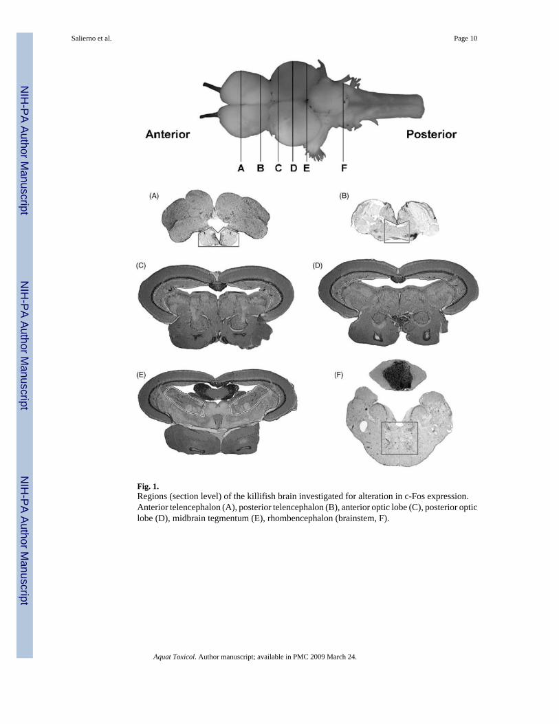
Fig. 1.Regions (section level) of the killifish brain investigated for alteration in c-Fos expression.Anterior telencephalon (A), posterior telencephalon (B), anterior optic lobe (C), posterior opticlobe (D), midbrain tegmentum (E), rhombencephalon (brainstem, F).
Salierno et al. Page 10
Aquat Toxicol. Author manuscript; available in PMC 2009 March 24.
NIH
-PA Author Manuscript
NIH
-PA Author Manuscript
NIH
-PA Author Manuscript
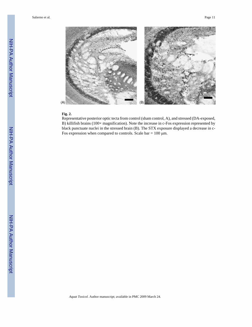
Fig. 2.Representative posterior optic tecta from control (sham control, A), and stressed (DA-exposed,B) killifish brains (100× magnification). Note the increase in c-Fos expression represented byblack punctuate nuclei in the stressed brain (B). The STX exposure displayed a decrease in c-Fos expression when compared to controls. Scale bar = 100 μm.
Salierno et al. Page 11
Aquat Toxicol. Author manuscript; available in PMC 2009 March 24.
NIH
-PA Author Manuscript
NIH
-PA Author Manuscript
NIH
-PA Author Manuscript

Fig. 3.c-Fos expression in control (light gray) and transport stressed (dark gray) killifish brains (N =6, data are means ± S.E.). Ant. Telen., anterior telencephalon; Post. Telen., posteriortelencephalon; Ant. Optic, anterior optic lobe; Post. Optic, posterior optic lobe; Midbrain,midbrain tegmentum; and Brainstem, rhombencephalon. The y-axis is the percentage of c-Fosexpression per area (mm2). Significant increases in c-Fos expression occurred in the Ant. andPost. Optic regions (*p = 0.05).
Salierno et al. Page 12
Aquat Toxicol. Author manuscript; available in PMC 2009 March 24.
NIH
-PA Author Manuscript
NIH
-PA Author Manuscript
NIH
-PA Author Manuscript
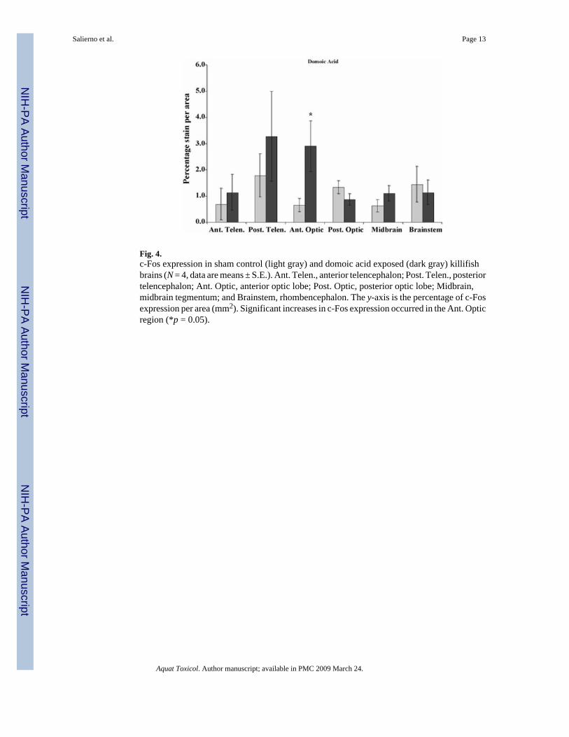
Fig. 4.c-Fos expression in sham control (light gray) and domoic acid exposed (dark gray) killifishbrains (N = 4, data are means ± S.E.). Ant. Telen., anterior telencephalon; Post. Telen., posteriortelencephalon; Ant. Optic, anterior optic lobe; Post. Optic, posterior optic lobe; Midbrain,midbrain tegmentum; and Brainstem, rhombencephalon. The y-axis is the percentage of c-Fosexpression per area (mm2). Significant increases in c-Fos expression occurred in the Ant. Opticregion (*p = 0.05).
Salierno et al. Page 13
Aquat Toxicol. Author manuscript; available in PMC 2009 March 24.
NIH
-PA Author Manuscript
NIH
-PA Author Manuscript
NIH
-PA Author Manuscript
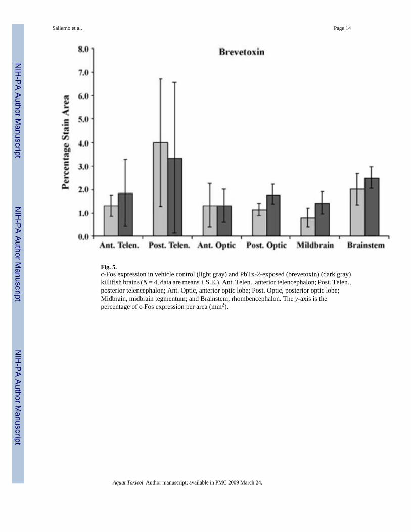
Fig. 5.c-Fos expression in vehicle control (light gray) and PbTx-2-exposed (brevetoxin) (dark gray)killifish brains (N = 4, data are means ± S.E.). Ant. Telen., anterior telencephalon; Post. Telen.,posterior telencephalon; Ant. Optic, anterior optic lobe; Post. Optic, posterior optic lobe;Midbrain, midbrain tegmentum; and Brainstem, rhombencephalon. The y-axis is thepercentage of c-Fos expression per area (mm2).
Salierno et al. Page 14
Aquat Toxicol. Author manuscript; available in PMC 2009 March 24.
NIH
-PA Author Manuscript
NIH
-PA Author Manuscript
NIH
-PA Author Manuscript
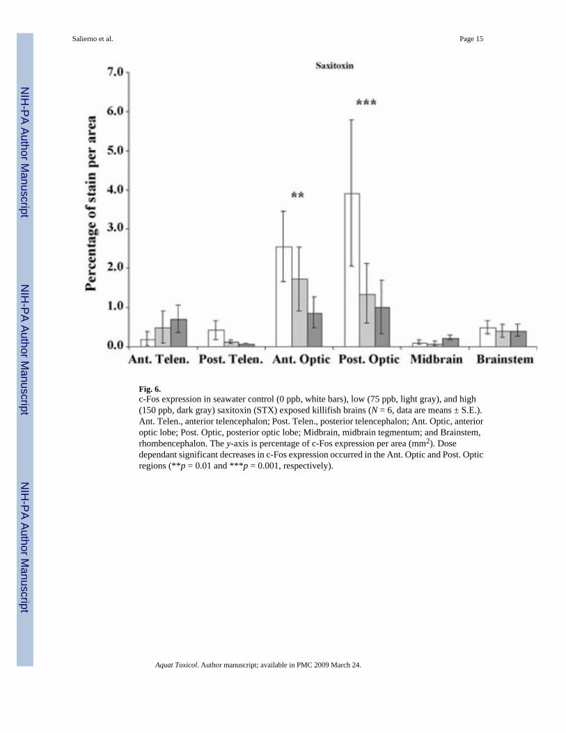
Fig. 6.c-Fos expression in seawater control (0 ppb, white bars), low (75 ppb, light gray), and high(150 ppb, dark gray) saxitoxin (STX) exposed killifish brains (N = 6, data are means ± S.E.).Ant. Telen., anterior telencephalon; Post. Telen., posterior telencephalon; Ant. Optic, anterioroptic lobe; Post. Optic, posterior optic lobe; Midbrain, midbrain tegmentum; and Brainstem,rhombencephalon. The y-axis is percentage of c-Fos expression per area (mm2). Dosedependant significant decreases in c-Fos expression occurred in the Ant. Optic and Post. Opticregions (**p = 0.01 and ***p = 0.001, respectively).
Salierno et al. Page 15
Aquat Toxicol. Author manuscript; available in PMC 2009 March 24.
NIH
-PA Author Manuscript
NIH
-PA Author Manuscript
NIH
-PA Author Manuscript