haploinsufficiency of the autism-associated shank3 gene leads to deficits in synaptic function,...
TRANSCRIPT
RESEARCH Open Access
Haploinsufficiency of the autism-associatedShank3 gene leads to deficits in synapticfunction, social interaction, and socialcommunicationOzlem Bozdagi1,2†, Takeshi Sakurai1,2†, Danae Papapetrou3, Xiaobin Wang4, Dara L Dickstein3, Nagahide Takahashi2,Yuji Kajiwara2, Mu Yang6, Adam M Katz6, Maria Luisa Scattoni6,7, Mark J Harris6, Roheeni Saxena6, Jill L Silverman6,Jacqueline N Crawley6, Qiang Zhou4,8, Patrick R Hof3, Joseph D Buxbaum1,2,3,5*
Abstract
Background: SHANK3 is a protein in the core of the postsynaptic density (PSD) and has a critical role in recruitingmany key functional elements to the PSD and to the synapse, including components of a-amino-3-hydroxyl-5-methyl-4-isoxazole-propionic acid (AMPA), metabotropic glutamate (mGlu) and N-methyl-D-aspartic acid (NMDA)glutamate receptors, as well as cytoskeletal elements. Loss of a functional copy of the SHANK3 gene leads to theneurobehavioral manifestations of 22q13 deletion syndrome and/or to autism spectrum disorders. The goal of thisstudy was to examine the effects of haploinsufficiency of full-length Shank3 in mice, focusing on synapticdevelopment, transmission and plasticity, as well as on social behaviors, as a model for understanding SHANK3haploinsufficiency in humans.
Methods: We used mice with a targeted disruption of Shank3 in which exons coding for the ankyrin repeatdomain were deleted and expression of full-length Shank3 was disrupted. We studied synaptic transmission andplasticity by multiple methods, including patch-clamp whole cell recording, two-photon time-lapse imaging andextracellular recordings of field excitatory postsynaptic potentials. We also studied the density of GluR1-immunoreactive puncta in the CA1 stratum radiatum and carried out assessments of social behaviors.
Results: In Shank3 heterozygous mice, there was reduced amplitude of miniature excitatory postsynaptic currentsfrom hippocampal CA1 pyramidal neurons and the input-output (I/O) relationship at Schaffer collateral-CA1synapses in acute hippocampal slices was significantly depressed; both of these findings indicate a reduction inbasal neurotransmission. Studies with specific inhibitors demonstrated that the decrease in basal transmissionreflected reduced AMPA receptor-mediated transmission. This was further supported by the observation of reducednumbers of GluR1-immunoreactive puncta in the stratum radiatum. Long-term potentiation (LTP), induced eitherwith θ-burst pairing (TBP) or high-frequency stimulation, was impaired in Shank3 heterozygous mice, with nosignificant change in long-term depression (LTD). In concordance with the LTP results, persistent expansion ofspines was observed in control mice after TBP-induced LTP; however, only transient spine expansion was observedin Shank3 heterozygous mice. Male Shank3 heterozygotes displayed less social sniffing and emitted fewerultrasonic vocalizations during interactions with estrus female mice, as compared to wild-type littermate controls.
Conclusions: We documented specific deficits in synaptic function and plasticity, along with reduced reciprocalsocial interactions in Shank3 heterozygous mice. Our results are consistent with altered synaptic development and
* Correspondence: [email protected]† Contributed equally1Seaver Autism Center for Research and Treatment, Mount Sinai School ofMedicine, New York, NY 10029, USAFull list of author information is available at the end of the article
Bozdagi et al. Molecular Autism 2010, 1:15http://www.molecularautism.com/content/1/1/15
© 2010 Bozdagi et al; licensee BioMed Central Ltd. This is an Open Access article distributed under the terms of the Creative CommonsAttribution License (http://creativecommons.org/licenses/by/2.0), which permits unrestricted use, distribution, and reproduction inany medium, provided the original work is properly cited.
function in Shank3 haploinsufficiency, highlighting the importance of Shank3 in synaptic function and supporting alink between deficits in synapse function and neurodevelopmental disorders. The reduced glutamatergictransmission that we observed in the Shank3 heterozygous mice represents an interesting therapeutic target inShank3-haploinsufficiency syndromes.
BackgroundThe Shank protein familyShank proteins, which include Shanks 1, 2 and 3, werefirst identified in a yeast two-hybrid assay using the gua-nylate kinase-associated protein (GKAP, also calledSAPAP1, DLGAP1 and DAP-1) as bait [1]. GKAP is aPSD-95-binding protein that forms an important compo-nent of the postsynaptic density (PSD), where protein-protein interactions between scaffolding proteins andreceptors are a key mechanism in assembling a functionalsynapse [2]. Shanks are highly enriched in the PSD andcontain five domains for protein-protein interactions,including an ankyrin repeat domain, an Src homology 3(SH3) domain, a PSD-95/discs large/zonula occludens-1(PDZ) domain, several proline-rich regions and aC-terminal sterile a-motif (SAM) domain [3] (Figure 1).The PDZ domain mediates the interaction of Shankswith the COOH terminus of several different proteins,including GKAP [1,4], G protein-coupled receptors [5,6]and the COOH terminus of group I metabotropic gluta-mate receptors (mGluRs) [7]. GKAP mediates the bind-ing of Shanks to N-methyl-D-aspartic acid (NMDA) anda-amino-3-hydroxyl-5-methyl-4-isoxazole-propionic acid(AMPA) receptors [8]. In addition, the SAM domaininteracts with GKAP [7], while proline-rich regions ofthe Shank proteins bind the mGluR-binding proteinHomer [7], actin-binding protein Abp-1 [9,10] and cor-tactin (cortical actin-binding protein) [11], which pro-motes polymerization of the actin cytoskeleton, animportant modulator of long-term synaptic plasticity[12]. Shanks interact directly with AMPA receptorsthrough the SH3 domain [13]. Interaction of the ankyrinrepeats of Shanks with a-fodrin may mediate calmodu-lin-mediated processes after synaptic stimulation throughinteractions with actin and calmodulin [14]. Sharpin canalso interact with the Shanks [15] and may be involved inactivity-dependent modification in dendritic spines.
Shank proteins in synaptic biologyThere are ~300 individual Shank molecules in a singlepostsynaptic site, representing ~5% of the total proteinmolecules and total protein mass in the site [16]. Itwould therefore not be surprising that alteration inShank expression could profoundly affect synaptic mor-phology and function. Such a crucial role for Shanklevels in synaptic function is supported by the observa-tion that overexpression of Shank1 led to increased
spine size in neurons in culture [17]. Additional studiesshowed that knockout of Shank1 leads to a decrease inspine number and spine and PSD size, decreased levelsof GKAP and Homer, and reduced basal synaptic trans-mission [18]. Furthermore, inhibition of Shank3 expres-sion has been shown to reduce numbers of spines inhippocampal neurons in culture and, conversely, whenShank3 was introduced into aspiny cerebellar neuronsin vitro, the neurons developed spines with functionalglutamatergic synapses expressing NMDA, AMPA andmGlu receptors [19].
Haploinsufficiency of SHANK3 in neurodevelopmentalsyndromesChromosome 22q13 deletion syndrome (22q13DS, alsocalled Phelan-McDermid syndrome) was first describedin the early 1990s (reviewed in [20]). Affected indivi-duals show global developmental delays with absent orseverely delayed expressive speech, and they may haveautism spectrum disorders (ASDs). Careful analysis ofthe extent of the deletion in dozens of independentcases indicated the presence of a small “critical region”encompassing SHANK3 [21,22], providing the first lineof evidence that a dysfunction in SHANK3 may beresponsible for the neurobehavioral aspects of 22q13DS.The second line of evidence was the demonstration of arecurrent breakpoint in SHANK3 in some cases with22q13DS [23,24], which led these authors to concludethat disruption of this single gene might be sufficient tocause 22q13DS. Third, there are translocations inSHANK3 that lead to 22q13DS [25]. Finally, severalrecent studies demonstrated that even de novo pointmutations in SHANK3 can produce the entirety of neu-rodevelopmental symptoms of 22q13DS, including glo-bal developmental delay, absent or severely delayedexpressive speech, and ASDs [26-28]. Neurobehavioralphenotypes associated with mutation or deletion ofSHANK3 are here referred to as SHANK3-haploinsuffi-ciency syndromes and, as noted above, can be associatedwith ASDs. Interestingly, overexpression of SHANK3may also result in an ASD as evidenced by reports ofAsperger syndrome in an individual with three copies ofthe SHANK3 locus [27]. Recently, de novo missense andnonsense mutations in SHANK3 have been described inatypical schizophrenia (with mild to moderate intellec-tual disability, early onset and dysmorphic features [29]).Mutations in the highly related gene SHANK2 have also
Bozdagi et al. Molecular Autism 2010, 1:15http://www.molecularautism.com/content/1/1/15
Page 2 of 15
recently been associated with ASDs and/or intellectualdisability [29-31].In the current study, we characterized mice with a tar-
geted disruption of Shank3 as a model for SHANK3-haploinsufficiency syndromes. We focused on synaptic
biology and synaptic function as the most proximal tar-get for altered Shank3 expression. We also examinedsocial interaction and social communication in Shank3heterozygotes and their wild-type littermates. Theresults are consistent with an important role for
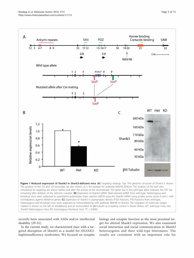
Figure 1 Reduced expression of Shank3 in Shank3-deficient mice. (A) Targeting strategy. Top: The genomic structure of Shank3 is shown.The position of the 22t and 32t transcripts are also shown, as is the epitope for antibody N69/46. Bottom: The location of the loxP sitesintroduced for targeting are shown before and after the activity of the recombinase. The green bar in the wild-type allele indicates the FRT siteremaining after deletion of the selection cassette. (B) Expression of Shank3 mRNA. Brain-derived mRNA from wild-type, heterozygous andknockout mice were subjected to quantitative polymerase chain reaction (qPCR) assay for Shank3 mRNA using probes across exons 6 and 7 withnormalization against reference genes. (C) Expression of Shank3 in postsynaptic density (PSD) fractions. PSD fractions from wild-type,heterozygous and knockout mice were subjected to immunoblotting with antibody N69/46 to Shank3. The migration of molecular weightmarkers is shown on the left (in kilodaltons) and an immunoblot for bIII-tubulin as a loading control is shown below. WT, wild-type mice; Het,Shank3 heterozygous mice; KO, homozygous knockout mice. *P < 0.0003.
Bozdagi et al. Molecular Autism 2010, 1:15http://www.molecularautism.com/content/1/1/15
Page 3 of 15
SHANK3 in synaptic function and plasticity and impli-cate specific pathways as potential therapeutic targetsfor SHANK3-haploinsufficiency syndromes.
MethodsGeneration of mice with a disruption of the Shank3 geneAnimal procedures were approved by the Mount SinaiSchool of Medicine and the National Institute of MentalHealth Animal Care and Use committees. We made useof gene targeting in Bruce4 C57BL/6 embryonic stem(ES) cells [32] to generate a mouse line that has loxPsites inserted before exon 4 and after exon 9 (encodingthe ankyrin repeats), with the selection cassette (flankedby FRT sites) excised by FLP recombinase. This floxedstrategy was chosen to allow us the option of doing con-ditional (region-specific) knockouts if needed. C57BL/6was used as the chosen embryonic stem cell linebecause of the more robust social and cognitive abilitiesin this line as compared to many of the 129-derived ESlines. For all studies reported here, the floxed allele wasfirst excised by crossing with a CMV-Cre transgenic line(again on a C57BL/6 background) that has ubiquitousCre expression and a line maintained with a deletion ofexons 4 through 9. This Shank3-deficient line was car-ried forward by crossing heterozygotes with the C57BL/6 strain to maintain a pure C57 background suitable forelectrophysiology and behavioral analyses.
Quantitative polymerase chain reactionRNA was extracted from brain cortex using the RNeasyMini Kit (Qiagen, Valencia, CA, USA) according to themanufacturer’s instructions. cDNA was synthesized withthe High Capacity cDNA Reverse Transcription Kit(Applied Biosystems, Carlsbad, CA, USA). The universalprobe library (UPL) system (Roche, Indianapolis, IN,USA) was used to perform quantitative polymerase chainreaction (qPCR). Primers located in exons 6 and 7 ofShank3 (NM_021423) were designed using ProbeFinderSoftware (Roche). Three reference genes (Actb, Gapd andRpl13a) were used for normalization, and relative expres-sion levels were calculated using qBase software [33],now available from Biogazelle (Ghent, Belgium). Primersequences and UPL probe numbers were Shank3, for-ward tggttggcaagagatccat, reverse ttggccccatagaacaaaag,#1; Actb, forward ggatgcagaaggagattactgc, reverse ccacc-gatccacacagagta, #63; Gapd, Fw gccaaaagggtcatcatctc,reverse cacacccatcacaaacatgg, #29; Rpl 13a, forwardtccctgctgctctcaagg, reverse gccccaggtaagcaaactt, #41.Unpaired t-tests were used for group comparisons.
ImmunoblottingPSD fractions were prepared as follows. Hemibrains ofwild-type, heterozygous and homozygous Shank3 micewere homogenized in 2-[4-(2-hydroxyethyl)piperazin-1-
yl]ethanesulfonic acid (HEPES)-A containing 4 mMHEPES, pH 7.4, 0.32 M sucrose, Protease InhibitorCocktail and PhoSTOP Phosphatase Inhibitor Cocktail(both from Roche). Nuclear fractions were precipitatedby centrifuging twice at 700 g for 15 min, and theresulting supernatants were further centrifuged at21,000 g for 15 min. The precipitates were resuspendedin HEPES-B containing 4 mM HEPES, pH 7.4, ProteaseInhibitor Cocktail and PhoSTOP Phosphatase InhibitorCocktail, homogenized and rotated at 4°C for 1 hour.The lysates were centrifuged at 32,000 g for 20 min andwashed twice with HEPES-C containing 50 mM HEPES,pH 7.4, 0.5% Triton X-100, Protease Inhibitor Cocktailand PhoSTOP Phosphatase Inhibitor Cocktail. Finally,postsynaptic density fractions were resuspended inHEPES-C containing 1.8% sodium dodecyl sulfate (SDS)and 2.5 M urea. Fifty-two μg of each PSD fraction wereloaded to 4-12% SDS-polyacrylamide gel electrophoresis(PAGE gel (Invitrogen, Carlsbad, CA, USA), transferredto polyvinylidene fluoride membrane and immuno-blotted with either the N69/46 anti-Shank3 antibodydirected against an epitope downstream of the PDZdomain (UC Davis/NIH NeuroMab Facility, Davis, CA)or the anti-ProSAP2 anti-Shank3 antibody directedagainst the last 100 amino acids of Shank3 (Millipore,Billerica, MA, USA). For bIII tubulin, the membranewas stripped and immunoblotted with an anti-bIII tubu-lin antibody (Abcam, Cambridge, MA, USA).
Hippocampal slice electrophysiologyWhole cell recording, two-photon time-lapse imagingand analysisMethods of recording, imaging and analysis were carriedout according to our previously published protocols[34,35]. All experiments were conducted on CA1 pyrami-dal cells at 32°C in acute slices taken from Shank3 hetero-zygous mice and wild-type littermates. Spines werevisualized using calcein contained in the patch pipette,making use of a two-photon laser scanning system modi-fied from Olympus Fluoview FV 300 driven by a Chame-leon two-photon laser (Coherent, Santa Clara, CA, USA).Baseline synaptic responses were evoked using a glass pip-ette positioned ~20 μm away from the imaged spines andrecorded at the soma. Long-term potentiation (LTP) wasinduced with a θ-burst pairing (TBP) protocol in whichtwo trains of θ-burst stimuli (each train, separated by 20 s,consisted of five bursts at 5 Hz, and each burst containedfive pulses at 100 Hz) were paired with brief, small postsy-naptic depolarization. Volume analysis of individual spineswas performed as detailed previously [34]. Briefly, the inte-grated fluorescence intensity inside a spine head was mea-sured for individual spines at different time points andnormalized to the fluorescence intensity of the dendritesfrom the same image stack to correct for potential changes
Bozdagi et al. Molecular Autism 2010, 1:15http://www.molecularautism.com/content/1/1/15
Page 4 of 15
in excitation [36]. Spine volume in the θ-burst stimulation(TBS) experiments was also calculated using the Rayburstalgorithm in NeuronStudio software (available from theComputational Neurobiology and Imaging Center, MountSinai School of Medicine, New York, NY, USA) followingdeconvolution of the data [37-39], and we obtained similarresults using either approach.Extracellular recordingsHippocampal slices (350 μm thick) were prepared from4- to 6-week-old heterozygous mice and their wild-typelittermate controls. Slices were perfused with Ringer’ssolution containing (in mM): NaCl, 125.0; KCl, 2.5;MgSO4, 1.3; NaH2PO4, 1.0; NaHCO3, 26.2; CaCl2, 2.5; andglucose, 11.0. The Ringer’s solution was bubbled with 95%O2 and 5% CO2 at 32°C during extracellular recordings(electrode solution: 3 M NaCl). Slices were maintained for1 hour prior to establishment of a baseline of field excita-tory postsynaptic potentials (fEPSPs) recorded from stra-tum radiatum in area CA1, evoked by stimulation of theSchaffer collateral-commissural afferents (100-μs pulsesevery 30 s) with bipolar tungsten electrodes placed intoarea CA3 [40]. Test stimulus intensity was adjusted toobtain fEPSPs with amplitudes that were one-half of themaximal response. The EPSP initial slope (mV/ms) wasdetermined from the average waveform of four consecu-tive responses. Input-output (I/O) curves were generatedby plotting the fEPSP slope versus fiber volley amplitudein low-Mg2+ (0.1 mM) solution. AMPA receptor-mediatedand NMDA receptor-mediated I/O relationships weremeasured in the presence of 2-amino-5-phosphonopenta-noic acid (APV; 50 μM) and 6-cyano-7-nitroquinoxaline-2,3-dione (CNQX; 100 μM), respectively (Sigma, St. Louis,MO, USA).Paired-pulse responses were measured with an inter-
stimulus interval (ISI) of 50 ms and are expressed as theratio of the average responses to the second stimulationpulse (FP2) to the first stimulation pulse (FP1). LTP wasinduced by either a high-frequency stimulus (four trainsof 100-Hz, 1-s stimulations separated by 5 min) or TBS(15 bursts of four pulses at 100 Hz separated by 200ms). To induce long-term depression (LTD), Schaffercollaterals were stimulated by low-frequency stimulation(LFS; 900 pulses at 1 Hz, 15 min) or by a paired-pulselow-frequency stimulation (PP-LFS; 1 Hz for 20 min,50-ms interstimulus interval [41]) to induce mGlureceptor-dependent LTD. Data were expressed as means± SD, and statistical analyses were performed using ana-lysis of variance (ANOVA) or Student’s t-test, with sig-nificance set at an a level of 0.05.
Measurement of GluR1-immunoreactive punctaImmunohistochemistryThree-month-old animals were anesthetized with 250 μlof 15% chloral hydrate and perfused transcardially with
1% paraformaldehyde for 1 min followed by 4% parafor-maldehyde for a total of 13 min. The brains were thenremoved, hemisected, cut in 50-μm-thick sections usinga Leica VT1000S Vibratome (Vibratome, Bannockburn,IL, USA) and subsequently stored in phosphate-bufferedsaline (PBS) until use. Sections were incubated at 37°Cfor 5 min, followed by incubation in acidified pepsin(1 ml in a 0.2 N HCl solution) for 6.5 min. The tissuewas then washed at room temperature in PBS-B (3 × 20min) and incubated in a 0.3% Triton X-100, 0.5% bovineserum albumin (BSA), 5% normal goat serum for 1 h onan orbital shaker. The blocking step was followed byovernight incubation in the primary antibody (rabbitpolyclonal antiglutamate receptor 1 AB1504; Millipore,Billerica, MA, USA), which was made in blocking solu-tion at the appropriate dilution (1 μg/ml). The tissuesections were then washed in PBS-B (5 × 5 min) andincubated in secondary antibody (goat antirabbit AlexaFluor 488, Invitrogen) in a 2% BSA and 0.3% TritonPBS-B solution at the appropriate dilution (1:400) for1 h at room temperature on an orbital shaker. Finally,the tissue was washed in PBS-B (3 × 5 min), stainedwith 4”,6"-diamino-2-phenylindole-2HCl (DAPI) andmounted on charged Aqua ColorFrost slides using Vec-tashield mounting medium (Vector Laboratories, Burlin-game, CA, USA).Tissue samplingTo quantify puncta, we used a systematic random sam-pling approach whereby a 1:6 series of sections werestained, the stratum radiatum of the CA1 was contouredusing SteroInvestigator (MBF Bioscience, Williston, VT,USA) and the sampling sites were determined using anoptical fractionator with the size of the grid set at 18 μm2
(the dimension of the confocal image stacks to be latersampled with a ×100 lens objective), at a digital zoom of5 on a Zeiss LSM510 META confocal microscope (Zeiss,Oberkochen, Germany). The trace of the contoured areawith an optical fractionator sampling grid placed on itwas used as a guide to obtain confocal image stacks ofthe above-mentioned dimensions that were 100 μm apartfrom each other (i.e., the size of the probe).Confocal imaging and puncta quantificationThe fluorescent puncta were visualized under a ×100 oilimmersion objective (1.4 numerical aperture) in a seriesof Z-stacks using an Argon/2 laser (488 nm wavelength)at 50% output (tube current of 6.4 A and maximumpower of 30 mW), with a collection band pass spectrumof 505-550 nm (with the following laser and microscopesettings: image frame size of 512 × 512, 1 Airy unit,refractive index correction of 0.9144 and Z stack intervalof 0.1 μm (x,y pixel size = 0.05 μm)) and their intensity,number and size quantified. These settings were opti-mized during pilot studies and held constant throughoutthe study.
Bozdagi et al. Molecular Autism 2010, 1:15http://www.molecularautism.com/content/1/1/15
Page 5 of 15
The resultant stacks were then deconvolved usingAutoDeblur 1.4.1 (Media Cybernetics, Bethesda, MD,USA), using an adaptive point-spread function (PSF)deconvolution method with a theoretical PSF, and thenanalyzed with custom Vamp2D software [42] that reli-ably calculates the size of individual puncta on the basisof three-dimensional estimates and circumvents theproblem of object superimposition found with moretraditional methods that collapse the stacks into two-dimensional projections. The relative density of punctawas then calculated per cubic micrometer, and the dif-ferences between groups were assessed using the non-parametric Mann-Whitney U test.
Behavioral analysesShank3 wild-type and heterozygote breeding pairs wereimported from Mount Sinai School of Medicine to theNational Institute of Mental Health. Mice were main-tained by breeding C57BL/6 wild-type mice with Shank3heterozygotes and housed in a conventional tempera-ture- and humidity-controlled vivarium. Littermateswere housed by sex in mixed genotype groups of two tofour per cage on a 12:12-h circadian cycle with lights onat 0600. Behavioral experiments were conductedbetween 1000 and 1600 in dedicated testing rooms.Developmental milestones were tested across postnatal
days 2-14, including measures of body weight, bodylength, tail length, pinnea detachment, eye opening, inci-sor eruption, fur development, righting reflex, negativegeotaxis, cliff avoidance, grasping reflex, auditory startle,bar holding, level screen and vertical screen as pre-viously described [43,44]. In addition, the mice wereevaluated in a standard, automated three-chamberedsocial approach task as previously described [44].Male-female social interactions were evaluated in a
5-min test session as previously described [43,45], withthe exception that subject males were group-housed andindividually tested in clean cages with clean litter. Eachof the 12 wild-type and 14 heterozygous male subjectmice, ages 2.5-4 months, was paired with a differentunfamiliar estrus C57BL/6J female. A digital closed-circuit television camera (Panasonic, Secaucus, NJ, USA)was positioned horizontally 30 cm from the cage. Anultrasonic microphone (Avisoft UltraSoundGate conden-ser microphone capsule CM15; Avisoft Bioacoustics,Berlin, Germany) was mounted 20 cm above the cage.Sampling frequency for the microphone was 250 kHz,and the resolution was 16 bits. The entire apparatus wascontained in a sound-attenuating environmental cham-ber (ENV-018V; Med Associates, St. Albans, VT, USA)illuminated by a single 25-Watt red light. Videos fromthe male subjects were subsequently scored by an inves-tigator uninformed of the subject’s genotype on mea-sures of nose-to-nose sniffing, nose-to-anogenital
sniffing and sniffing of other body regions, using NoldusObserver software (Noldus Information Technology,Leesburg, VA, USA) as previously described. Ultrasonicvocalizations were played back and spectrograms weredisplayed using Avisoft software [43,45]. Ultrasonicvocalizations were identified manually by two highlytrained investigators blinded to genotype information,and summary statistics were calculated using the Avisoftpackage. Interrater reliability was 95%. Data were ana-lyzed using an unpaired Student’s t-test.Olfactory habituation/dishabituation testing was con-
ducted in male and female Shank3 wild-type and hetero-zygous mice ages 2.5-4 months using methodspreviously described [44,46,47]. Nonsocial and socialodors were presented on a series of cotton swabsinserted into the home cage sequentially, each for2 min, in the following order: water, water, water (dis-tilled water); almond, almond, almond (1:100 dilutionalmond extract); banana, banana, banana (1:100 dilutionartificial banana flavoring); social 1, social 1, social1 (swiped from the bottom of a cage housing unfamiliarsex-matched B6 mice); and social 2, social 2, social2 (swiped from the bottom of a second cage housing adifferent group of unfamiliar sex-matched 129/SvImJmice). One-way repeated measures ANOVA was per-formed within each genotype for each set of habituationevents and each dishabituation event, followed by aTukey post hoc test.
ResultsGeneration and characterization of aShank3-deficient mouseWe made use of gene targeting in Bruce4 C57BL/6embryonic stem (ES) cells [32] to generate a mouse linethat has loxP sites inserted before exon 4 and after exon9 (Figure 1A). For all studies reported here, the floxedallele was excised and a line was maintained with a dele-tion of exons 4 through 9. This line, which completelydeletes the ankyrin repeat domains of Shank3, producedwild-type (+/+), heterozygous (+/-) and knockout (-/-)animals with Mendelian frequencies from heterozygote-heterozygote crosses.qPCR showed 50% reduction of full-length Shank3
mRNA in the heterozygotes and complete loss in knock-outs (Figure 1B). Moreover, there was no expression offull-length Shank3 protein in PSD fractions fromShank3-knockout mice and reduced expression in theheterozygotes, using antibodies which cross-react eitherwith an epitope downstream of the PDZ domain (anti-body N69/46; see Figure 1A) (Figure 1C) or with theCOOH terminal (data not shown), consistent withhaploinsufficiency.Heterozygous and homozygous animals were viable
and showed no obvious alterations in gross brain
Bozdagi et al. Molecular Autism 2010, 1:15http://www.molecularautism.com/content/1/1/15
Page 6 of 15
structure or hippocampal cytoarchitecture, nor werethere any obvious seizures. There was evidence forsubtle motor abnormalities in homozygotes, which isbeing further characterized in ongoing studies. Bothgenotypes were normal on measures of general health,developmental milestones and exploratory activity, aswell as on social approach as measured in an automatedthree-chambered social approach task.Because SHANK3 haploinsufficiency is responsible for
the neurobehavioral phenotype in individuals with22q13DS or SHANK3 mutations, we focused the studiesreported here on Shank3 heterozygous mice generatedby crossing wild-type mice with heterozygotes to bemost relevant to the clinical syndromes and to be mostuseful in ultimately assessing potential therapeutic inter-ventions in preclinical studies. A comprehensive investi-gation of the knockout mice obtained through analternate breeding strategy (heterozygote-heterozygotematings) is now in progress in independent experiments.
Basal glutamatergic synaptic transmission is reduced inShank3 heterozygous miceTo examine the role of Shank3 in regulating synapticglutamate receptor function, we studied glutamatergicsynaptic transmission in hippocampal slices. We exam-ined the properties of basic transmission at Schaffer col-lateral-CA1 synapses in hippocampal slices from 3- to4-week-old Shank3 heterozygous mice and their litter-mates using extracellular recordings. Plotting field exci-tatory postsynaptic potential (fEPSP) slope versusstimulus intensity demonstrated a reduction in the I/Ocurves in the heterozygotes (data not shown), promptingus to then further examine synaptic transmission in thepresence of inhibitors of specific subtypes of glutamatereceptors. In the presence of the NMDA receptorantagonist APV, a decrease in AMPA receptor-mediatedfield potentials in the heterozygous mice was seen,reflected as a 50% decrease in the average slope of I/Ofunction compared with wild-type mice (n = 4 mice pergenotype, two to three slices per mouse; P = 0.001;Figure 2A). In contrast, when the I/O relationship wasanalyzed in the presence of the competitive AMPA/kai-nate receptor antagonist CNQX to measure synapticNMDA receptor function, there was no differencebetween genotypes (n = 4 mice per genotype, two tothree slices per mouse; P = 0.1; Figure 2B). These resultsindicate that there is a specific reduction in AMPAreceptor-mediated basal transmission in the Shank3 het-erozygous mice.We asked whether the impairment of evoked synaptic
transmission in heterozygous mice is caused by altera-tions in presynaptic and/or postsynaptic parameters. Weperformed whole cell patch-clamp recordings in3-month-old littermates and monitored spontaneous
miniature postsynaptic currents in the presence of tetro-dotoxin (TTX). The amplitude of miniature excitatorypostsynaptic currents (mEPSCs) from hippocampal CA1pyramidal neurons of heterozygous mice were signifi-cantly smaller than those in control mice (n = 7 forwild-type and n = 8 for heterozygous mice, P < 0.01;Figures 2C and 2D), which was evident by the signifi-cant shift of the cumulative probability to the left(Figure 2E), again indicating a reduction in basal trans-mission. However, in heterozygous mice, the frequencyof miniature excitatory postsynaptic currents was signifi-cantly higher (n = 7 for wild-type and n = 8 for hetero-zygous mice, P < 0.03; Figures 2C and 2D) and paired-pulse ratio was decreased (n = 6 for wild-type and n = 7for heterozygous mice, P < 0.05; Figure 2F), whichrevealed an additional, presynaptic alteration in the het-erozygotes as well.
Long-term potentiation is impaired in Shank3heterozygous miceWe next examined long-term potentiation (LTP) withextracellular fEPSP recordings at Schaffer collateral/CA1synapses. In the first set of experiments, LTP was inducedby tetanic stimulation of the Schaffer collaterals (fourtrains of 100 Hz separated by 5 min). While initial expres-sion of LTP was identical across the two genotypes, themaintenance of LTP was clearly impaired in the heterozy-gous mice (average percentage of baseline 120 min aftertetanus: 165.1 ± 8.8% in wild-type and 117.1 ± 9.5% in het-erozygous mice, P = 0.004; Figure 3A). In an additional setof experiments, we further tested TBS LTP (10 bursts offour pulses at 100 Hz separated by 200 ms), which alsoshowed a significant decrease in the potentiation at 60min after TBS in heterozygous mice (156.3 ± 9.2% of base-line in wild-type and 126.0 ± 8.9% in heterozygous mice,measured 60 min after TBS, P = 0.007; Figure 3B).In contrast to the altered synaptic plasticity observed
with LTP, long-term depression (LTD) induced byeither low-frequency stimulation (LFS) (82.6 ± 1.35% ofbaseline in wild-type and 79.9 ± 2.5% in heterozygousmice, measured 60 min after LFS, P > 0.1; Figure 3G) orpaired-pulse LFS (PP-LFS) stimulation (81.6 ± 6% ofbaseline in wild-type and 82.7 ± 1.9% in heterozygousmice, P > 0.1; Figure 3H) was not significantly changedin heterozygotes.Previous studies have shown that LTP is accompanied
by spine enlargement [35,48]. Therefore, it was of inter-est to determine whether the deficits in LTP in theShank3 heterozygous mice were associated with alteredspine remodeling. We first established that spines fromwild-type mice were capable of structural modificationby simultaneously monitoring spine size and synapticresponses in CA1 neurons before and after TBP [34,35].We found that TBP produced a rapid and persistent
Bozdagi et al. Molecular Autism 2010, 1:15http://www.molecularautism.com/content/1/1/15
Page 7 of 15
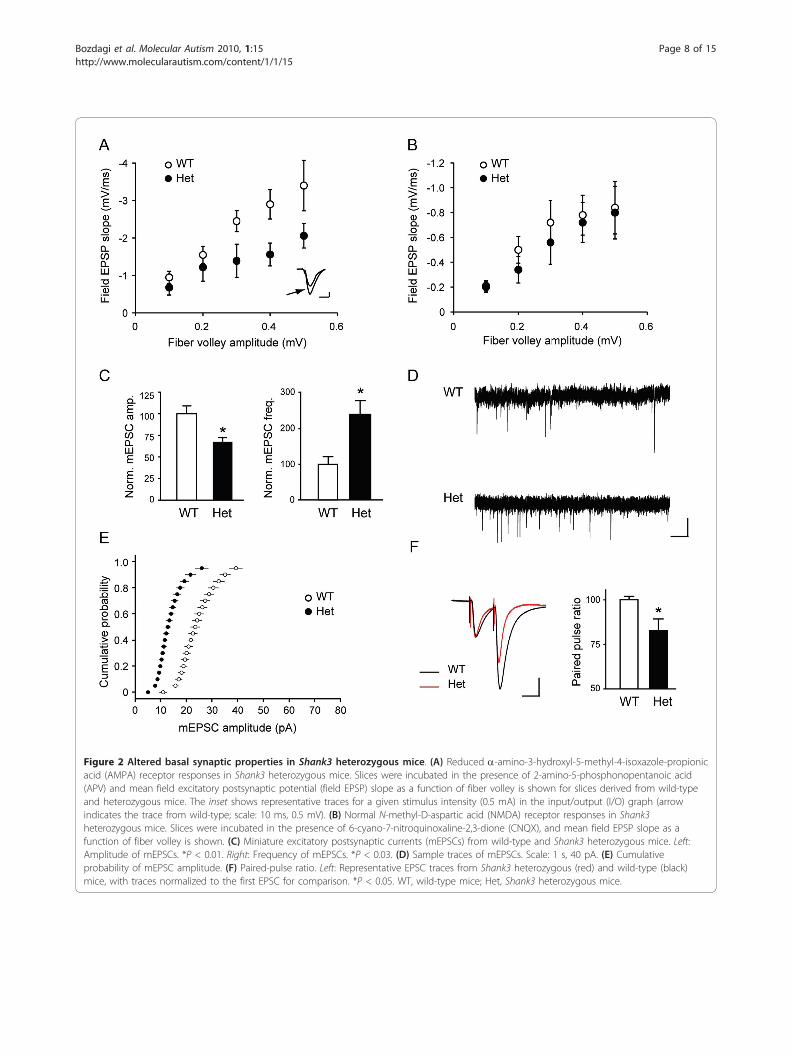
Figure 2 Altered basal synaptic properties in Shank3 heterozygous mice. (A) Reduced a-amino-3-hydroxyl-5-methyl-4-isoxazole-propionicacid (AMPA) receptor responses in Shank3 heterozygous mice. Slices were incubated in the presence of 2-amino-5-phosphonopentanoic acid(APV) and mean field excitatory postsynaptic potential (field EPSP) slope as a function of fiber volley is shown for slices derived from wild-typeand heterozygous mice. The inset shows representative traces for a given stimulus intensity (0.5 mA) in the input/output (I/O) graph (arrowindicates the trace from wild-type; scale: 10 ms, 0.5 mV). (B) Normal N-methyl-D-aspartic acid (NMDA) receptor responses in Shank3heterozygous mice. Slices were incubated in the presence of 6-cyano-7-nitroquinoxaline-2,3-dione (CNQX), and mean field EPSP slope as afunction of fiber volley is shown. (C) Miniature excitatory postsynaptic currents (mEPSCs) from wild-type and Shank3 heterozygous mice. Left:Amplitude of mEPSCs. *P < 0.01. Right: Frequency of mEPSCs. *P < 0.03. (D) Sample traces of mEPSCs. Scale: 1 s, 40 pA. (E) Cumulativeprobability of mEPSC amplitude. (F) Paired-pulse ratio. Left: Representative EPSC traces from Shank3 heterozygous (red) and wild-type (black)mice, with traces normalized to the first EPSC for comparison. *P < 0.05. WT, wild-type mice; Het, Shank3 heterozygous mice.
Bozdagi et al. Molecular Autism 2010, 1:15http://www.molecularautism.com/content/1/1/15
Page 8 of 15

Figure 3 Reduced long-term potentiation in Shank3 heterozygous mice. (A) Long-term potentiation (LTP) following high-frequencystimulation. Field recordings of LTP induced with high-frequency stimulation (HFS; 4 times 100 Hz, separated by 5 min) as a function of time inslices from wild-type and Shank3 heterozygous mice. (B) LTP following θ-burst stimulation (TBS). (C) Long-term depression (LTD) following low-frequency stimulation (LFS), an NMDA receptor-dependent form of LTD. (D) LTD following paired-pulse low-frequency stimulation (PP-LFS), aprotein synthesis-dependent form of LTD. (E) LTP recorded with whole cell patch-clamp method. LTP was induced with θ-burst pairing (TBP). (E)Normalized EPSP slope is shown as a function of time. (G) Representative EPSP traces before and after (arrow) LTP induction (scale bar: 5 mV, 10ms). (F) Changes in normalized spine volume following LTP. (H) Representative images showing TBP-induced spine expansion in Shank3heterozygotes. Images were acquired before and 5 min and 45 min after TBP. Transiently increased and stable spines are indicated byarrowheads and arrow, respectively (scale bar: 1 μm). WT, wild-type mice; Het, Shank3 heterozygous mice.
Bozdagi et al. Molecular Autism 2010, 1:15http://www.molecularautism.com/content/1/1/15
Page 9 of 15
increase in spine volume concurrent with an immediateincrease in EPSP slope in whole cell recordings, whichgradually reached a plateau by ~30 min (Figures 3C-F).However, in recordings from heterozygous mice, wefound that the stabilization in synaptic potentiation andspine expansion were impaired in CA1 neurons (Figures3C-F). In the heterozygous mice, both EPSP slope andspine volume increased immediately to values compar-able to those of control spines, but synaptic potentiationand spine expansion failed to be sustained.
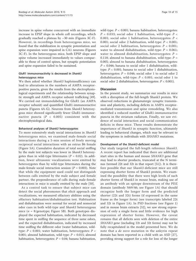
GluR1 immunoreactivity is decreased in Shank3heterozygous miceWe then asked whether Shank3 haploinsufficiency canlead to alterations in the numbers of AMPA receptor-positive puncta, given the results from the electrophysio-logical experiments and the relationship between synap-tic strength and AMPA receptor subunit trafficking [49].We carried out immunolabeling for GluR1 (an AMPAreceptor subunit) and quantified GluR1-immunoreactivepuncta (Figures 4A-G). Neurons from Shank3 heterozy-gous mice showed significantly fewer GluR1-immunor-eactive puncta (P < 0.005) consistent with theelectrophysiological data.
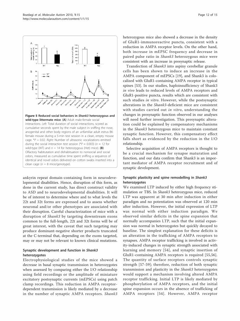
Behavioral analyses of Shank3 heterozygotesTo more extensively study social interactions in Shank3heterozygous mice, we examined male-female socialinteractions during a 5-min session of freely movingreciprocal social interactions with an estrus B6 female(Figure 5A). Cumulative duration of total social sniffingby the male test subjects was lower in Shank3 heterozy-gotes than in wild-type littermates (P = 0.02). In addi-tion, fewer ultrasonic vocalizations were emitted byheterozygotes than by wild-type littermates during themale-female social interaction session (P = 0.003). Notethat while the equipment used could not distinguishbetween calls emitted by the male subject and femalepartner, the preponderance of calls during male-femaleinteractions in mice is usually emitted by the male [50].As a control task to ensure that subject mice can
detect the social pheromones that elicit approach andvocalizations, we measured olfactory abilities using theolfactory habituation/dishabituation test. Habituationand dishabituation were normal for social and nonsocialodor cues in both wild-type and Shank3 heterozygousmice (n = 8/genotype; Figure 5B). Both genotypes dis-played the expected habituation, indicated by decreasedtime spent in sniffing the sequence of three same odors,and the expected dishabituation, indicated by increasedtime sniffing the different odor (water habituation, wild-type: P < 0.001; water habituation, heterozygotes: P <0.001; almond habituation, wild-type: P = 0.015; almondhabituation, heterozygotes: P = 0.04; banana habituation,
wild-type: P < 0.001; banana habituation, heterozygotes:P = 0.033; social odor 1 habituation, wild-type: P <0.001; social odor 1 habituation, heterozygotes: P <0.001; social odor 2 habituation, wild-type: P < 0.001;social odor 2 habituation, heterozygotes: P < 0.001;water to almond dishabituation, wild-type: P = 0.061;water to almond dishabituation, heterozygotes: P =0.118; almond to banana dishabituation, wild-type: P =0.005; almond to banana dishabituation, heterozygotes:P = 0.046; banana to social odor 1 dishabituation, wild-type: P < 0.001; banana to social odor 1 dishabituation,heterozygotes: P = 0.046; social odor 1 to social odor 2dishabituation, wild-type: P < 0.001; social odor 1 tosocial odor 2 dishabituation, heterozygotes: P < 0.001).
DiscussionIn the present study, we summarize our results in micewith a disruption of the full-length Shank3 protein. Weobserved reductions in glutamatergic synaptic transmis-sion and plasticity, including deficits in AMPA receptor-mediated transmission and spine remodeling. We alsoidentified a reduced number of GluR1-immunoreactivepuncta in the striatum radiatum. Finally, we saw evi-dence of social interaction and social communicationdeficits in these mice. These results demonstrate theimportance of Shank3 in synaptic function, ultimatelyleading to behavioral changes, which may be relevant tothe symptoms in individuals with SHANK3 mutations.
Development of the Shank3-deficient modelOur study targeted the full-length reference Shank3.A recent report has indicated that there are additionalstart sites in the human and mouse SHANK3 gene thatmay lead to shorter products, truncated at the N termi-nus (termed 22t and 32t in that report [51]). It is there-fore possible that our Shank3-deficient mice are stillexpressing shorter forms of Shank3 protein. We exam-ined the possibility that there were high levels of suchshorter forms of Shank3 in mouse brain, making use ofan antibody with an epitope downstream of the PDZdomain (antibody N69/46; see Figure 1A) that shouldrecognize both the longer form and the predictedshorter (22t and 32t) forms (if expressed in the sameframe as the longer form) (see transcripts labeled 22tand 32t in Figure 1A). In PSD fractions (see Figure 1)and in mouse brain extracts [52], we saw strong expres-sion of only a single form and little evidence for highexpression of shorter forms. However, the caveatremains that all deficits seen with deletion of the entireSHANK3 gene (including 22t and 32t forms) may not befully recapitulated in the model presented here. We donote that a de novo mutation in the ankyrin repeatdomain has been reported in a child with an ASD [27],providing strong support for a role for loss of the longer
Bozdagi et al. Molecular Autism 2010, 1:15http://www.molecularautism.com/content/1/1/15
Page 10 of 15
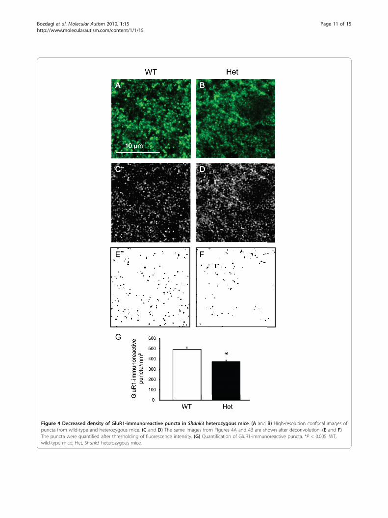
Figure 4 Decreased density of GluR1-immunoreactive puncta in Shank3 heterozygous mice. (A and B) High-resolution confocal images ofpuncta from wild-type and heterozygous mice. (C and D) The same images from Figures 4A and 4B are shown after deconvolution. (E and F)The puncta were quantified after thresholding of fluorescence intensity. (G) Quantification of GluR1-immunoreactive puncta. *P < 0.005. WT,wild-type mice; Het, Shank3 heterozygous mice.
Bozdagi et al. Molecular Autism 2010, 1:15http://www.molecularautism.com/content/1/1/15
Page 11 of 15
ankyrin repeat domain-containing form in neurodeve-lopmental disabilities. Hence, disruption of this form, asdone in the current study, has direct construct validityto ASD and to neurodevelopmental disabilities. It willbe of interest to determine where and to what levels the22t and 32t forms are expressed and to assess whetherneuronal and/or other phenotypes are associated withtheir disruption. Careful characterization of mice with adisruption of Shank3 by targeting downstream exonscommon to the full-length, 22t and 32t forms will be ofgreat interest, with the caveat that such targeting mayproduce dominant-negative shorter products truncatedat the C-terminal that, depending on the exons targeted,may or may not be relevant to known clinical mutations.
Synaptic development and function in Shank3heterozygotesElectrophysiological studies of the mice showed adecrease in basal synaptic transmission in heterozygoteswhen assessed by comparing either the I/O relationshipusing field recordings or the amplitude of miniatureexcitatory postsynaptic currents (mEPSCs) using patchclamp recordings. This reduction in AMPA receptor-dependent transmission is likely mediated by a decreasein the number of synaptic AMPA receptors. Shank3
heterozygous mice also showed a decrease in the densityof GluR1-immunoreactive puncta, consistent with areduction in AMPA receptor levels. On the other hand,both increase in mEPSC frequency and decrease inpaired pulse ratio in Shank3 heterozygous mice wereconsistent with an increase in presynaptic release.Transfection of Shank3 into aspiny cerebellar granule
cells has been shown to induce an increase in theAMPA component of mEPSCs [19], and Shank3 is colo-calized with GluR1-containing AMPA receptor in typicalspines [53]. In our studies, haploinsufficiency of Shank3in vivo leads to reduced levels of AMPA receptors andGluR1-positive puncta, results which are consistent withsuch studies in vitro. However, while the postsynapticalterations in the Shank3-deficient mice are consistentwith studies carried out in vitro, understanding thechanges in presynaptic function observed in our analyseswill need further investigation. This presynaptic altera-tion could be explained by compensatory mechanismsin the Shank3 heterozygous mice to maintain constantsynaptic function. However, this compensatory effectfalls short as evidenced by the reduction in the I/Orelationship.Selective acquisition of AMPA receptors is thought to
be a crucial mechanism for synapse maturation andfunction, and our data confirm that Shank3 is an impor-tant mediator of AMPA receptor recruitment and ofsynaptic development.
Synaptic plasticity and spine remodelling in Shank3heterozygotesWe examined LTP induced by either high frequency sti-mulation or TBS. In Shank3 heterozygous mice, reducedLTP was apparent at 30 min after induction in eitherparadigm and no potentiation was observed at 120 minafter induction. However, the initial expression of LTPwas normal with either induction paradigm. Weobserved similar deficits in the spine expansion thatnormally accompanies LTP, such that the initial expan-sion was normal in heterozygotes but quickly decayed tobaseline. The simplest explanation for these deficits isan alteration in the trafficking of AMPA receptors tosynapses. AMPA receptor trafficking is involved in activ-ity-induced changes in synaptic strength associated withlearning and memory [54], and synaptic insertion ofGluR1-containing AMPA receptors is required [55,56].The quantity of surface receptors controls synapticstrength [57-59]; therefore, reduction of both synaptictransmission and plasticity in the Shank3 heterozygoteswould support a mechanism involving altered AMPAreceptor trafficking. Initial LTP is likely mediated byphosphorylation of AMPA receptors, and the initialspine expansion occurs in the absence of trafficking ofAMPA receptors [54]. However, AMPA receptor
Figure 5 Reduced social behaviors in Shank3 heterozygous andwild-type littermate mice. (A) Adult male-female socialinteractions. Left: Total duration of social interactions, scored ascumulative seconds spent by the male subject in sniffing the nose,anogenital and other body regions of an unfamiliar adult estrus B6female mouse during a 5-min test session in a clean, empty mousecage. *P = 0.02. Right: Number of ultrasonic vocalizations emittedduring the social interaction test session (*P = 0.003) (n = 12 forwild-type (WT) and n = 14 for heterozygous (Het) mice). (B)Olfactory habitutation and dishabituation to nonsocial and socialodors, measured as cumulative time spent sniffing a sequence ofidentical and novel odors delivered on cotton swabs inserted into aclean cage (n = 8 mice/genotype).
Bozdagi et al. Molecular Autism 2010, 1:15http://www.molecularautism.com/content/1/1/15
Page 12 of 15
trafficking to the synapse is critical to sustain LTP andspine expansion. The transient nature of synaptic poten-tiation and spine enlargement in heterozygous mice isconsistent with normal phosphorylation of existingsynaptic AMPA receptors with a deficit in subsequentconsolidation, the process in which both structural andfunctional forms of plasticity are progressively stabilizedover 30 min [34].
Social behavior in Shank3 heterozygotesMale Shank3 heterozygous mice displayed reducedresponses to female social cues compared to their wild-type littermates on parameters of social sniffing andultrasonic vocalizations in a male-female reciprocalsocial interaction context. Given that Shank3 has a cen-tral role in synaptic function, Shank3-related changes incellular and network components that underlie socialcognition could underlie the reduced social interactionsin Shank3 heterozygous mice. It was interesting thatsocial deficits were not observed in a three-chambersocial approach task, implying some specificity of thesocial alterations. It will be of interest to better under-stand the frequency of ASD and the characteristics ofsocial deficits in SHANK3-haploinsufficiency syndromes,as it is already evident that this is not a universal findingin these syndromes when considering 22q13DS orschizophrenia.The social deficits observed in the mice serve as
important reminders that systems outside the hippo-campus that are involved in social behaviors will needto be examined in these mice and that additional beha-viors known to be mediated by the hippocampus will beimportant for further investigations. Cellular and elec-trophysiological analyses in additional brain regions andlarge-scale behavioral studies are now underway for theShank3-deficient mice in the authors’ laboratories.
ConclusionsHaploinsufficiency of full-length Shank3 resulted in adecrease in synaptic transmission, altered functional andstructural plasticity of synapses and reduced social beha-viors. These findings are consistent with a model ofdelayed synaptic development in Shank3 haploinsuffi-ciency, together with a reduction in AMPA receptortrafficking. The results highlight the importance ofShank3 in synaptic development and function and sup-port a link between deficits in synapse function andneurodevelopmental disorders. To date, pharmacologicaltreatments for ASDs and other developmental disorders(including SHANK3-haploinsufficiency syndromes) areprimarily ameliorative, focusing on managing associatedsystems such as anxiety, aggression, repetitive behaviors,attention deficits and epilepsy, among others (see, forexample, [60]). Pharmacological treatments addressing
core diagnostic symptoms, including, in ASDs, altera-tions in reciprocal social interactions and communica-tion, do not yet exist. Recently, the field has begun tosee the evaluation of therapies targeted to etiology (thatis, “personalized medicine”) using models of neurodeve-lopmental disorders including fragile X, tuberous sclero-sis, and Rett syndromes (see, for example, [61-64]). Theuse of model systems such as the Shank3-deficient micereported here could lead to similar advances in the caseof SHANK3-haploinsufficiency syndromes. One interest-ing outcome of the current study is that compoundsthat enhance glutamatergic transmission, includingthose that specifically enhance AMPA transmission(AMPAkines) could possibly represent therapeuticapproaches in these conditions. Further analysis of thisand additional models may identify targets for noveltherapeutics for individuals with developmental delaysarising from 22q13 deletion syndrome or SHANK3mutations.
AcknowledgementsThis work was supported by the Seaver Foundation, the Simons Foundation,the National Institute of Mental Health Intramural Research Program, and bya gift from Paulina Rychenkova, PhD, and William Gibson. OB and TS areSeaver Junior Faculty Fellows. We thank Lisa Krug and Shekhar Patil for helpat early stages of the project.
Author details1Seaver Autism Center for Research and Treatment, Mount Sinai School ofMedicine, New York, NY 10029, USA. 2Department of Psychiatry, Mount SinaiSchool of Medicine, New York, NY 10029, USA. 3Department ofNeuroscience, Mount Sinai School of Medicine, New York, NY 10029, USA.4Department of Neurology, Mount Sinai School of Medicine, New York, NY10029, USA. 5Department of Genetics and Genomic Sciences, Mount SinaiSchool of Medicine, New York, NY 10029, USA. 6Laboratory of BehavioralNeuroscience, National Institute of Mental Health, Bethesda, MD 20892-3730,USA. 7Istituto Superiore di Sanità, Rome, Italy. 8Genentech, South SanFrancisco, CA 94080, USA.
Authors’ contributionsTS and JDB generated and biochemically characterized the mice. JDB, PRH,JNC, TS, QZ and OB designed the experiments. OB, XW and QZ performedthe electrophysiology experiments and analysis. Immunohistochemistry aswell as confocal microscopy and analysis were conducted by DP, DLD andPRH. Behavioral experiments and analysis were conducted by MY, AMK, MLS,MJH, RS, JLS and JNC. qPCR was conducted by NT. Immunoblotting wasconducted by YK. The manuscript was written by OB and JDB, and allauthors reviewed the manuscript before submission.
Competing interestsOB, TS and JDB have submitted a patent on this work.
Received: 14 October 2010 Accepted: 17 December 2010Published: 17 December 2010
References1. Naisbitt S, Kim E, Tu JC, Xiao B, Sala C, Valtschanoff J, Weinberg RJ,
Worley PF, Sheng M: Shank, a novel family of postsynaptic densityproteins that binds to the NMDA receptor/PSD-95/GKAP complex andcortactin. Neuron 1999, 23:569-582.
2. Sheng M: Excitatory synapses. Glutamate receptors put in their place.Nature 1997, 386:221-223.
3. Sheng M, Kim E: The Shank family of scaffold proteins. J Cell Sci 2000,113(Pt 11):1851-1856.
Bozdagi et al. Molecular Autism 2010, 1:15http://www.molecularautism.com/content/1/1/15
Page 13 of 15
4. Yao I, Iida J, Nishimura W, Hata Y: Synaptic localization of SAPAP1, asynaptic membrane-associated protein. Genes Cells 2003, 8:121-129.
5. Zitzer H, Richter D, Kreienkamp HJ: Agonist-dependent interaction of therat somatostatin receptor subtype 2 with cortactin-binding protein 1. JBiol Chem 1999, 274:18153-18156.
6. Kreienkamp HJ, Soltau M, Richter D, Bockers T: Interaction of G-protein-coupled receptors with synaptic scaffolding proteins. Biochem Soc Trans2002, 30:464-468.
7. Tu JC, Xiao B, Naisbitt S, Yuan JP, Petralia RS, Brakeman P, Doan A,Aakalu VK, Lanahan AA, Sheng M, Worley PF: Coupling of mGluR/Homerand PSD-95 complexes by the Shank family of postsynaptic densityproteins. Neuron 1999, 23:583-592.
8. Kim JH, Kim JH, Yang E, Park JH, Yu YS, Kim KW: Shank 2 expressioncoincides with neuronal differentiation in the developing retina. Exp MolMed 2009, 41:236-242.
9. Qualmann B, Boeckers TM, Jeromin M, Gundelfinger ED, Kessels MM:Linkage of the actin cytoskeleton to the postsynaptic density via directinteractions of Abp1 with the ProSAP/Shank family. J Neurosci 2004,24:2481-2495.
10. Haeckel A, Ahuja R, Gundelfinger ED, Qualmann B, Kessels MM: The actin-binding protein Abp1 controls dendritic spine morphology and isimportant for spine head and synapse formation. J Neurosci 2008,28:10031-10044.
11. Du Y, Weed SA, Xiong WC, Marshall TD, Parsons JT: Identification of anovel cortactin SH3 domain-binding protein and its localization togrowth cones of cultured neurons. Mol Cell Biol 1998, 18:5838-5851.
12. Cingolani LA, Goda Y: Actin in action: the interplay between the actincytoskeleton and synaptic efficacy. Nat Rev Neurosci 2008, 9:344-356.
13. Sheng M, Kim MJ: Postsynaptic signaling and plasticity mechanisms.Science 2002, 298:776-780.
14. Vanderklish PW, Krushel LA, Holst BH, Gally JA, Crossin KL, Edelman GM:Marking synaptic activity in dendritic spines with a calpain substrateexhibiting fluorescence resonance energy transfer. Proc Natl Acad Sci USA2000, 97:2253-2258.
15. Lim S, Naisbitt S, Yoon J, Hwang JI, Suh PG, Sheng M, Kim E:Characterization of the Shank family of synaptic proteins. Multiplegenes, alternative splicing, and differential expression in brain anddevelopment. J Biol Chem 1999, 274:29510-29518.
16. Sugiyama Y, Kawabata I, Sobue K, Okabe S: Determination of absoluteprotein numbers in single synapses by a GFP-based calibrationtechnique. Nat Methods 2005, 2:677-684.
17. Sala C, Piech V, Wilson NR, Passafaro M, Liu G, Sheng M: Regulation ofdendritic spine morphology and synaptic function by Shank and Homer.Neuron 2001, 31:115-130.
18. Hung AY, Futai K, Sala C, Valtschanoff JG, Ryu J, Woodworth MA, Kidd FL,Sung CC, Miyakawa T, Bear MF, et al: Smaller dendritic spines, weakersynaptic transmission, but enhanced spatial learning in mice lackingShank1. J Neurosci 2008, 28:1697-1708.
19. Roussignol G, Ango F, Romorini S, Tu JC, Sala C, Worley PF, Bockaert J,Fagni L: Shank expression is sufficient to induce functional dendriticspine synapses in aspiny neurons. J Neurosci 2005, 25:3560-3570.
20. Phelan MC, Rogers RC, Saul RA, Stapleton GA, Sweet K, McDermid H,Shaw SR, Claytor J, Willis J, Kelly DP: 22q13 deletion syndrome. Am J MedGenet 2001, 101:91-99.
21. Luciani JJ, de Mas P, Depetris D, Mignon-Ravix C, Bottani A, Prieur M,Jonveaux P, Philippe A, Bourrouillou G, de Martinville B, et al: Telomeric22q13 deletions resulting from rings, simple deletions, andtranslocations: cytogenetic, molecular, and clinical analyses of 32 newobservations. J Med Genet 2003, 40:690-696.
22. Wilson HL, Wong AC, Shaw SR, Tse WY, Stapleton GA, Phelan MC, Hu S,Marshall J, McDermid HE: Molecular characterisation of the 22q13deletion syndrome supports the role of haploinsufficiency of SHANK3/PROSAP2 in the major neurological symptoms. J Med Genet 2003,40:575-584.
23. Bonaglia MC, Giorda R, Borgatti R, Felisari G, Gagliardi C, Selicorni A,Zuffardi O: Disruption of the ProSAP2 gene in a t(12;22)(q24.1;q13.3) isassociated with the 22q13.3 deletion syndrome. Am J Hum Genet 2001,69:261-268.
24. Bonaglia MC, Giorda R, Mani E, Aceti G, Anderlid BM, Baroncini A,Pramparo T, Zuffardi O: Identification of a recurrent breakpoint within the
SHANK3 gene in the 22q13.3 deletion syndrome. J Med Genet 2006,43:822-828.
25. Phelan MC: Deletion 22q13.3 syndrome. Orphanet Journal of Rare Diseases2008, 3:14.
26. Durand CM, Betancur C, Boeckers TM, Bockmann J, Chaste P, Fauchereau F,Nygren G, Rastam M, Gillberg IC, Anckarsater H, et al: Mutations in thegene encoding the synaptic scaffolding protein SHANK3 are associatedwith autism spectrum disorders. Nat Genet 2007, 39:25-27.
27. Moessner R, Marshall CR, Sutcliffe JS, Skaug J, Pinto D, Vincent J,Zwaigenbaum L, Fernandez B, Roberts W, Szatmari P, Scherer SW:Contribution of SHANK3 mutations to autism spectrum disorder. Am JHum Genet 2007, 81:1289-1297.
28. Gauthier J, Spiegelman D, Piton A, Lafreniere RG, Laurent S, St-Onge J,Lapointe L, Hamdan FF, Cossette P, Mottron L, et al: Novel de novoSHANK3 mutation in autistic patients. Am J Med Genet B NeuropsychiatrGenet 2009, 150B:421-424.
29. Gauthier J, Champagne N, Lafreniere RG, Xiong L, Spiegelman D, Brustein E,Lapointe M, Peng H, Cote M, Noreau A, et al: De novo mutations in thegene encoding the synaptic scaffolding protein SHANK3 in patientsascertained for schizophrenia. Proc Natl Acad Sci USA 2010, 107:7863-7868.
30. Berkel S, Marshall CR, Weiss B, Howe J, Roeth R, Moog U, Endris V,Roberts W, Szatmari P, Pinto D, et al: Mutations in the SHANK2 synapticscaffolding gene in autism spectrum disorder and mental retardation.Nat Genet 2010, 42:489-491.
31. Pinto D, Pagnamenta AT, Klei L, Anney R, Merico D, Regan R, Conroy J,Magalhaes TR, Correia C, Abrahams BS, et al: Functional impact of globalrare copy number variation in autism spectrum disorders. Nature 2010,466:368-372.
32. Hughes ED, Qu YY, Genik SJ, Lyons RH, Pacheco CD, Lieberman AP,Samuelson LC, Nasonkin IO, Camper SA, Van Keuren ML, Saunders TL:Genetic variation in C57BL/6 ES cell lines and genetic instability in theBruce4 C57BL/6 ES cell line. Mamm Genome 2007, 18:549-558.
33. Hellemans J, Mortier G, De Paepe A, Speleman F, Vandesompele J: qBaserelative quantification framework and software for management andautomated analysis of real-time quantitative PCR data. Genome Biol 2007,8:R19.
34. Yang Y, Wang XB, Frerking M, Zhou Q: Spine expansion and stabilizationassociated with long-term potentiation. J Neurosci 2008, 28:5740-5751.
35. Bozdagi O, Wang XB, Nikitczuk JS, Anderson TR, Bloss EB, Radice GL,Zhou Q, Benson DL, Huntley GW: Persistence of coordinated long-termpotentiation and dendritic spine enlargement at mature hippocampalCA1 synapses requires N-cadherin. J Neurosci 2010, 30:9984-9989.
36. Holtmaat AJ, Trachtenberg JT, Wilbrecht L, Shepherd GM, Zhang X,Knott GW, Svoboda K: Transient and persistent dendritic spines in theneocortex in vivo. Neuron 2005, 45:279-291.
37. Wearne SL, Rodriguez A, Ehlenberger DB, Rocher AB, Henderson SC, Hof PR:New techniques for imaging, digitization and analysis of three-dimensional neural morphology on multiple scales. Neuroscience 2005,136:661-680.
38. Rodriguez A, Ehlenberger DB, Dickstein DL, Hof PR, Wearne SL: Automatedthree-dimensional detection and shape classification of dendritic spinesfrom fluorescence microscopy images. PLoS One 2008, 3:e1997.
39. Rodriguez A, Ehlenberger DB, Hof PR, Wearne SL: Rayburst sampling, analgorithm for automated three-dimensional shape analysis from laserscanning microscopy images. Nat Protoc 2006, 1:2152-2161.
40. Bozdagi O, Rich E, Tronel S, Sadahiro M, Patterson K, Shapiro ML,Alberini CM, Huntley GW, Salton SR: The neurotrophin-inducible gene Vgfregulates hippocampal function and behavior through a brain-derivedneurotrophic factor-dependent mechanism. J Neurosci 2008,28:9857-9869.
41. Huber KM, Kayser MS, Bear MF: Role for rapid dendritic protein synthesisin hippocampal mGluR-dependent long-term depression. Science 2000,288:1254-1257.
42. Dumutriu D, Hara Y, Berger S, Wadsworth S, Morrison J: Volume assistedmeasurement of puncta in 2 dimensions (VAMP2D): A new tool foraccurate size quantification in fluorescent imaging. Society for NeurosienceAnnual Meeting Chicago, IL: Society for Neuroscience Abstracts; 2009.
43. Scattoni ML, Gandhy SU, Ricceri L, Crawley JN: Unusual repertoire ofvocalizations in the BTBR T+tf/J mouse model of autism. PLoS One 2008,3:e3067.
Bozdagi et al. Molecular Autism 2010, 1:15http://www.molecularautism.com/content/1/1/15
Page 14 of 15
44. Silverman JL, Turner SM, Barkan CL, Tolu SS, Saxena R, Hung AY, Sheng M,Crawley JN: Sociability and motor functions in Shank1 mutant mice. BrainRes 2010.
45. Scattoni ML, Ricceri L, Crawley JN: Unusual repertoire of vocalizations inadult BTBR T+tf/J mice during three types of social encounters. GenesBrain Behav 2010.
46. Yang M, Crawley JN: Simple behavioral assessment of mouse olfaction.Curr Protoc Neurosci 2009, Chapter 8(Unit 8):24.
47. Silverman JL, Yang M, Lord C, Crawley JN: Behavioural phenotyping assaysfor mouse models of autism. Nat Rev Neurosci 2010, 11:490-502.
48. Matsuzaki M, Honkura N, Ellis-Davies GC, Kasai H: Structural basis of long-term potentiation in single dendritic spines. Nature 2004, 429:761-766.
49. Kopec CD, Real E, Kessels HW, Malinow R: GluR1 links structural andfunctional plasticity at excitatory synapses. J Neurosci 2007,27:13706-13718.
50. Nyby J: Ultrasonic vocalizations during sex behavior of male house mice(Mus musculus): a description. Behav Neural Biol 1983, 39:128-134.
51. Maunakea AK, Nagarajan RP, Bilenky M, Ballinger TJ, D’Souza C, Fouse SD,Johnson BE, Hong C, Nielsen C, Zhao Y, et al: Conserved role of intragenicDNA methylation in regulating alternative promoters. Nature 2010,466:253-257.
52. Kolevzon A, Cai G, Soorya L, Takahashi N, Grodberg D, Kajiwara Y,Willner JP, Tryfon A, Buxbaum JD: Analysis of a purported SHANK3mutation in a boy with autism: Clinical impact of rare variant research inneurodevelopmental disabilities. Brain Res 2010.
53. Uchino S, Wada H, Honda S, Nakamura Y, Ondo Y, Uchiyama T, Tsutsumi M,Suzuki E, Hirasawa T, Kohsaka S: Direct interaction of post-synapticdensity-95/Dlg/ZO-1 domain-containing synaptic molecule Shank3 withGluR1 alpha-amino-3-hydroxy-5-methyl-4-isoxazole propionic acidreceptor. J Neurochem 2006, 97:1203-1214.
54. Malinow R, Malenka RC: AMPA receptor trafficking and synaptic plasticity.Annu Rev Neurosci 2002, 25:103-126.
55. Shi S, Hayashi Y, Esteban JA, Malinow R: Subunit-specific rules governingAMPA receptor trafficking to synapses in hippocampal pyramidalneurons. Cell 2001, 105:331-343.
56. Park M, Penick EC, Edwards JG, Kauer JA, Ehlers MD: Recycling endosomessupply AMPA receptors for LTP. Science 2004, 305:1972-1975.
57. Ehrlich I, Malinow R: Postsynaptic density 95 controls AMPA receptorincorporation during long-term potentiation and experience-drivensynaptic plasticity. J Neurosci 2004, 24:916-927.
58. El-Husseini AE, Schnell E, Chetkovich DM, Nicoll RA, Bredt DS: PSD-95involvement in maturation of excitatory synapses. Science 2000,290:1364-1368.
59. Stein V, House DR, Bredt DS, Nicoll RA: Postsynaptic density-95 mimicsand occludes hippocampal long-term potentiation and enhances long-term depression. J Neurosci 2003, 23:5503-5506.
60. King BH, Bostic JQ: An update on pharmacologic treatments for autismspectrum disorders. Child Adolesc Psychiatr Clin N Am 2006, 15:161-175.
61. Bear MF, Huber KM, Warren ST: The mGluR theory of fragile X mentalretardation. Trends Neurosci 2004, 27:370-377.
62. Ehninger D, Silva AJ: Genetics and neuropsychiatric disorders: treatmentduring adulthood. Nat Med 2009, 15:849-850.
63. Tropea D, Giacometti E, Wilson NR, Beard C, McCurry C, Fu DD, Flannery R,Jaenisch R, Sur M: Partial reversal of Rett Syndrome-like symptoms inMeCP2 mutant mice. Proc Natl Acad Sci USA 2009, 106:2029-2034.
64. Ehninger D, de Vries PJ, Silva AJ: From mTOR to cognition: molecular andcellular mechanisms of cognitive impairments in tuberous sclerosis. JIntellect Disabil Res 2009, 53:838-851.
doi:10.1186/2040-2392-1-15Cite this article as: Bozdagi et al.: Haploinsufficiency of the autism-associated Shank3 gene leads to deficits in synaptic function, socialinteraction, and social communication. Molecular Autism 2010 1:15.
Submit your next manuscript to BioMed Centraland take full advantage of:
• Convenient online submission
• Thorough peer review
• No space constraints or color figure charges
• Immediate publication on acceptance
• Inclusion in PubMed, CAS, Scopus and Google Scholar
• Research which is freely available for redistribution
Submit your manuscript at www.biomedcentral.com/submit
Bozdagi et al. Molecular Autism 2010, 1:15http://www.molecularautism.com/content/1/1/15
Page 15 of 15