guidelines - austrian board of orthodontists
TRANSCRIPT
Guidelines
Contents page
Preamble………………………………………………………………………. 3
1 Application……………………………………………………………… 4
2 Board of examiners……………………………………………........... 4
3 Place and time of the ABO examination……………………………. 4
4 Requirements for submitting an application………………............ 5
5 Fees……………………………………………………………………... 5
6 Guidelines for the choice of cases…………………………………… 6
7 Course of the examination……………………………………………. 9
7.1 Part 1 – Case presentations………………………………………….. 9
7.2 Part 2 – The oral examination…………………………………………10
8 Guidelines for case presentations (Part 1)……………………………11
8.1 General guidelines………………………………………………………11
8.2 Presentation of written material/X-rays……………………………….12
8.3 Presentation of dental casts……………………………………………13
9 Guidelines for the oral examination (Part 2)………………………….14
10 Appendix………………………………………………………………….15
10.1 Orthodontic analysis of cephalometric radiographs…………………
of the European Board of Orthodontists………………………………16
10.2 Template for the trimming of dental casts…………………………….18
10.3 Template for the labelling of dental casts……………………………..19
10.4 Assessment scale of the ABO examination board…………………...20
1.0 ABO GUIDELINES 2019.docx 2
Preamble
The ABO examination board is not a legal personality. The examination to attain the ABO membership is organised by the Association of Austrian Orthodontists / Verband Österreichischer Kieferorthopäden (VÖK).
Unless stated otherwise in these guidelines, decisions about all matters concerning the ABO are subject to the jurisdiction of the expanded executive board of the VÖK.
All decisions taken in the expanded executive board require approval by a two-thirds majority.
The ABO is meant to be a set of rules which not only offers orthodontists, but all committed colleagues in the orthodontic domain, the chance to have their knowledge and skills tested by a neutral expert committee.
Membership in the ABO is a voluntary proof of the ability to carry out orthodontic treatment with very good results in accordance with the state of science.
Membership in the ABO by itself does not entitle to practise orthodontics. However, itis evidence of its holder’s very high clinical standard by passing the ABO examination.
ABO Members are obliged to exercise their great clinical abilities in accordance with medical ethics to the benefit of their patients and the repute of the profession.
Gross ethical negligence may lead to annulation of Membership with the VÖK. This measure must be based on a unanimous decision of the expanded executive board of the VÖK.
1.0 ABO GUIDELINES 2019.docx 3
1 Application
Applications to acquire the ABO degree must be submitted before 31 July of a calendar year at the office of the VÖK and the examination fee must have been paid by this time.
The executive board will decide by simple majority whether the candidate is granted permission to take the ABO examination. The office of the VÖK will inform the candidate of the result of this evaluation and of the place and time of the examinationat least 3 months before the date of the examination. No later than 2 months before the date of the examination, the candidate will receive the candidate number assigned to them from a notary public.
Members of the “European Board of Orthodontists” may be awarded the certificate ofthe Austrian Board of Orthodontists by the decision of the executive board of the VÖK. The holders must submit their application in writing along with the certificate of the European Board of Orthodontists.
2 Board of examiners
The executive board will select the members of the board of examiners from an examiners’ pool by simple majority. The examiners’ pool will be determined by the extended executive board.
The board of examiners consists of three members who are either holders of the certificate of the European Board of Orthodontists, of the Austrian Board of Orthodontists, or an equivalent board, or habilitated university teachers of orthodontics. One member should be a habilitated university teacher and one member should be a practising orthodontist.
The board of examiners elects its chairperson and establishes the procedure for the assessment of cases.
3 Place and time of the ABO examination
The examination to acquire the ABO degree is held once a year. The candidates will be informed of the place and time of the examination in a good time, i.e. at least 3 months before the set date. If the number of received applications is too small, the examination may be deferred by a maximum of one year.
1.0 ABO GUIDELINES 2019.docx 4
4 Requirements for the submission of applications
The candidate must produce evidence of at least three years of mainly or exclusively orthodontic work (in their own office and/or the orthodontic department of a university clinic and/or a comparable specialised institution). Documentary proof of this work / practice must be submitted.
The candidate must confirm with their signature that the cases presented by them have been treated by them alone.
At the same time, the candidate irrevocably consents to one person who is committed to professional secrecy reviewing their patient files to determine whether a case submitted to the ABO has been taken from the circle of orthodontic patients treated by the candidate, should any arguable doubt arise.
The candidate must sign an agreement wherein he accepts the decisions of the board of examiners as final.
5 Fees
The current fee (as of 2019/2020) is € 750.- for members of the VÖK and € 1.400.- for all other candidates. The amount of the fee is determined every other year by simple majority by the expanded executive board of the VÖK.
The full fee must be paid at the time of registration for the ABO examination. If the application is withdrawn at least 2 months before the examination date, one half of the fee will be retained as cancellation charge; if the application is withdrawn less than 2 months before the examination date, there will be no reimbursement of the fee.
If Part 1 (case presentations) and Part 2 (oral examination) are repeated, the full fee must be paid again.
No additional fee will be charged if Part 2 (oral examination) is repeated.
1.0 ABO GUIDELINES 2019.docx 5
6 Guidelines for the choice of cases
A total of eight cases must be selected from the three main groups as described below and the following must be kept in mind:
At least one case must be presented from each Angle Class.
At least three cases must be extraction cases.
At least one case must have extractions in all four quadrants.
Extraction cases include cases with already missing teeth (agenesis, extractions that date further back, etc.) on the condition that orthodontic space closure is carried out.
At least one case must have a marked skeletal discrepancy on the sagittal or vertical plane, disregarding the fact whether the correction is achieved with the help of orthognatic surgery or solely by orthodontic treatment.
Only one case may involve orthognatic surgery.
Group 1 – Early treatment (decidous teeth or early mixed dentition)
At least one case and a maximum of three cases from different Angle Classes have to be chosen.
If more than one case is presented from group 1, the second and, if applicable, the third case must have been started in primary or early mixed dentition stage and finished in the permanent dentition. If the treatment is divided into several stages, an additional documentation before the beginning of the last stage is required. If a cephalometric morphological analysis at the beginning of the treatment can be assumed not to produce any additional cues, it may be omitted in group 1 cases if a written justification is provided.
Group 2 –Treatment of children/adolescents (late mixed dentition to end of growth)
At least two cases from different Angle Classes have to be chosen.
The end-of-growth stadium is defined as the age of 17 for female patients and the age of 18 for male patients. Alternatively, valid documentation of the respective skeletal maturity may be provided.
Group 3 –Treatment of adults
At least two cases from different Angle Classes have to be chosen.
1.0 ABO GUIDELINES 2019.docx 6
Additional information / Notes on the definitions of Angle Classes
Source: Edward H. Angle, Classification of MalocclusionDental Cosmos 1899, Vol. 49, pp. 248 – 264
”In diagnosing cases of malocclusion we must consider, first, the mesio-distal relations of the dental arches; second, the individual positions of the teeth.“
”The key to occlusion is the relative position of the first molars. In normal occlusion the mesio-buccal cusp of the upper first molar is received in the sulcus between the mesial and distal buccal cusps of the lower.“
Therefore, the categorisation of a malocclusion according to Angle is based on two criteria:
a) Relative sagittal position of upper and lower jaw (skeletal relation)
b) Relative position of 6-year-molars (and canines) (dental relation)
N. B.:
Mesial movement or mesial tipping of molars because of loss of supporting decidous teeth, tooth extraction, agenesis, etc., obscure the actual, original occlusal relation. This must be considered when the Angle Class is determined.
Recommendation:
In order to avoid mistakes in the choice of cases and to avoid unnecessary discussions with the examination board, cases with clearly definable malocclusion and an according jaw base relation should be chosen.
1.0 ABO GUIDELINES 2019.docx 7
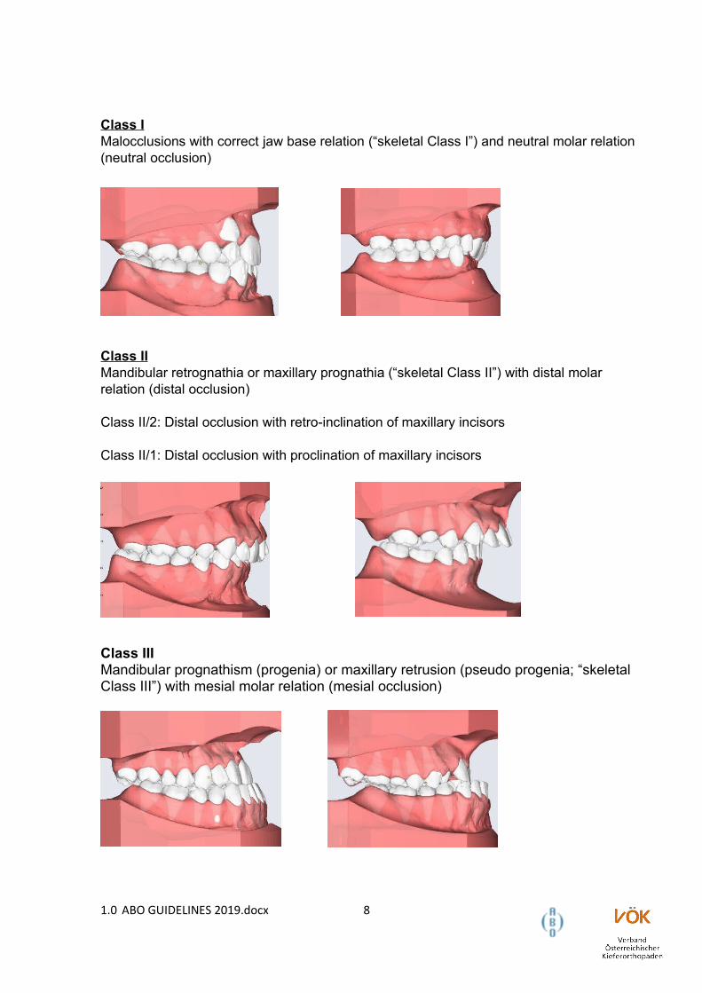
Class IMalocclusions with correct jaw base relation (“skeletal Class I”) and neutral molar relation(neutral occlusion)
Class IIMandibular retrognathia or maxillary prognathia (“skeletal Class II”) with distal molar relation (distal occlusion)
Class II/2: Distal occlusion with retro-inclination of maxillary incisors
Class II/1: Distal occlusion with proclination of maxillary incisors
Class IIIMandibular prognathism (progenia) or maxillary retrusion (pseudo progenia; “skeletal Class III”) with mesial molar relation (mesial occlusion)
1.0 ABO GUIDELINES 2019.docx 8
7 Course of the examination
The qualification will be evaluated in two parts.
7.1 Part 1 – Case presentations
Part 1 is based on the presentation and evaluation of the documents presented concerning eight completed cases (see section 8), which comprise a suitable spectrum of malocclusions (see section 6). The board of examiners will evaluate the cases according to their complexity and the results of treatment. To a lesser extent, the presentation of documentation (written information, dental casts, X-rays, etc.) are part of the evaluation.
All documents required for submission (see section 8) must be placed on the table marked with the candidate’s number at least two hours before the beginning of the examination. This must be done either by the candidate himself or by a person appointed by the candidate.
In addition, the written presentation must be made available to the examining board electronically (on CD or USB-stick).
All eight cases must be presented in a standardised form (see section 8). If any of the eight cases is inadequately documented (e.g. missing written information, error incase selection, insufficient quality of dental casts), no further cases will be evaluated.Consequently, all cases will be rated inadequate and will be returned. In such an event, the candidate may re-submit the cases with the corresponding documents to the board of examiners at the next ABO examination (without a waiting period).
Exceptions from the compulsory documentation: If a cephalometric radiograph analysis at the beginning of the treatment can be assumed not to produce any additional cues, a cephalometric analysis at the beginning of the treatment may be omitted in group 1 cases if justification is provided.
If it was not possible to produce a lateral skull radiograph at the end of the treatment due to protection against radiation (e. g. because of pregnancy), a cephalometric-analysis may be omitted in groups 1, 2, or 3 if justification is provided.
The outcome of part 1 may be “permitted to appear at the oral examination” or “not permitted to appear at the oral examination” and will be communicated to the candidate before the oral examination (part 2).
In the event of a negative evaluation, the candidate will be informed about the reasonfor not being permitted to take the oral examination. If a candidate falls ill after havingreceived permission to take the oral examination, they may register for the next ABO Part 2 examination without having to present their cases again.
1.0 ABO GUIDELINES 2019.docx 9
7.2 Part 2 – The oral examination
The oral examination is conducted in German or in English.
Its purpose is to determine the knowledge, comprehension and abilities of the candidate with regard to performing orthodontic treatment.
The candidate will be given the initial documents of two cases unknown to them with regard to a diagnostic analysis and a treatment plan. The presented materials consistof dental casts, photographs and X-rays. The candidate has to perform tracing of the lateral skull radiograph and analysis in accordance with the guidelines of the European Board of Orthodontics. The candidate has also the possibility to perform additional tracings of the cephalometric radiographs if they wish to do so.
The candidate will be given one hour in total to draft the diagnosis, the treatment goaland the treatment plan for both cases. After that, the candidate will be asked questions about the cases for about 30 minutes (see section 9).
The overall result of part 1 and part 2 will be “passed” or “failed”.
In the event of failure, the candidate will be allowed to retake the examination twice, each time after a period of at least one year.
The candidate will be notified of the result of the oral examination and the overall result immediately after part 2 of the examination.
Case presentations of successful ABO candidates may be presented by the VÖK at the congress at Kitzbühel to colleagues who would like to study them.
1.0 ABO GUIDELINES 2019.docx 10
8 Guidelines for case presentations (Part 1)
To ensure an accurate and thorough evaluation of all materials presented, the cases must be presented in a standardised fashion (see section 10.4 Assessment scale of the examination board).
8.1 General guidelines
The candidate is obliged to use the ABO forms. The summaries must be limited to the space provided for them. The forms must be filled in with a typewriter or computer. The font size may be altered; line spacing, however, should not be changed.
All material (dental casts, cephalometric tracings, X-rays, etc.) must be labelled as follows:
1. Candidate number (will be issued by the notary)
2. Case number
3. Date of the documents
4. Age of the patient
5. Phase of treatment
Phase I: Beginning of treatment – black
Phase II: Completion of treatment – red
Phase III: Retention documents – green (voluntary)
The tracings have to be done in the prescribed colours, black/red/green, on a transparency with a 0.5 mm fine liner. Please note: Do not use tracing sheets!
All documents (each sheet separately) have to be placed in sheet protectors and filedin a ring binder.
Digital X-rays have to be submitted in printed form in a resolution of at least 150 dpi in calibrated original size and evaluable quality.
1.0 ABO GUIDELINES 2019.docx 11
8.2 Presentation of written material / X-rays
General presentation of the case
Page 1 Title page (candidate number, case number)
Page 2 Summary
Documents before the beginning of treatment (Phase I)
Page 3 Description of the case/diagnosis (description of malocclusion and functional status; excess space/lack of space measurements in mm; classification of lateral dentition according to Angle categories I, II and III with the corresponding measure of premolar width; do not forget the wisdom teeth)
Page 4 Photographs of the face before case start (for cases begun after 1st July 2010)
Page 5 Intraoral photographs before case start (for cases begun after 1st July 2010)
Page 6 Lateral cephalometric radiograph before case start
Page 7 Tracing of the lateral cephalometric radiograph before case start and analysis in accordance with the guidelines of the European Board of Orthodontics (see Appendix 10.1), on a transparency, in black ink.
Page 8 Evaluation of the tracing of the cephalometric radiograph before case start
Page 9 Miniature X-rays or panoramic radiograph before case start
Page 10 Treatment goals/treatment plan/rationale behind
Combined cases treated with orthodontics and orthognatic surgery should include information about the planned operation and its consequences.
Page 11 Summary of the course of treatment including any difficulties encountered
Digital X-rays have to be printed on transparencies with a resolution of at least 150 dpi in calibrated original size and evaluable quality.
Documents during treatment
Photographs taken during treatment can be included; this is optional and may result in merit points in the assessment – see page 20.
1.0 ABO GUIDELINES 2019.docx 12
Documents at the end of treatment (Phase II)
Page 12 Photographs of the face at the end of treatment
Page 13 Intraoral photographs at the end of treatment
Page 14 Lateral skull radiograph at the end of treatment
Page 15 Tracing of the lateral skull radiograph at the end of treatment and analysis in accordance with the guidelines of the European Board of Orthodontics (see Appendix 10.1), on a transparency, in red ink
Page 16 Evaluation of the tracing of the lateral skull radiograph after completion of treatment
Page 17 Miniature X-ray or panoramic radiograph at the end of treatment
Page 18 Description of the outcome of treatment and retention
Digital X-rays have to be printed on transparencies with a resolution of at least 150 dpi in calibrated original size and evaluable quality.
Documents after the phase of retention (Phase III)
Retention documents can be included; this is optional and may result in merit points in the assessment, see p. 17)
8.3 Presentation of the dental casts
The candidate will present plaster casts made immediately before the beginning oftreatment and after completion of treatment (after removal of the brackets; seesections 10.2 and 10.3).
Dental casts must show all anatomical details of teeth and periodontium (apical basisincluding vestibulum oris, palate including raphe palatina mediana, frenula etc.).
Digital models have to be printed with the highest possible detail accuracy.
The treatment must end in correct occlusion.
1.0 ABO GUIDELINES 2019.docx 13
9 Guidelines for the oral examination (Part 2)
The candidate has to evaluate the initial documents of two cases unknown to him (see section 7.2) and then present to the board of examiners the diagnosis, goal of treatment and treatment plan according to the following structure:
Diagnosis
Anamnesis (general findings, concerns of the patients)
Analysis of the panoramic radiograph
Evaluation of the dental substance (caries, devital teeth), agenesis or excess teeth, wisdom teeth, displaced/impacted teeth, etc.
Analysis of the dental casts
Description of malocclusion (description of the Angle Class and the corresponding measure of the premolar width; sagittal/transversal/vertical deviations; excess space/lack of space – measurements in mm; loss of supporting decidous teeth, single tooth deviations, etc.)
Analysis of photographs
Extraoral: evaluation of soft tissue, labial closure, muscular tension, facial asymmetry, etc.
Intraoral: oral hygiene, assessment of the periodontium
Analysis of the cephalometric radiograph (EBO analysis; see section 10.1)
Skeletal diagnosis (description of the tendency of growth, assessment of the relation between the jaws and the skull base and the relation between the maxilla and the mandible)
Dental diagnosis (assessment of the position and inclination of incisors)
functional findings (if applicable)
Treatment plan
Goals of treatment / alternatives
Suggested therapy / treatment plan and the reason for it / possible treatment alternatives
1.0 ABO GUIDELINES 2019.docx 14
10 Appendix
10.1 Analysis of the cephalometric radiograph according to the European Board of
Orthodontics (2 pages) – see also https://www.eoseurope.org/ebo/instructions
10.2 Template for the trimming of dental casts; base trays from Dr. Hinz Dental-
Vertriebsgesellschaft in Herne, Germany, may be used
(https://www.dr-hinz-dental.de)
10.3 Template for the labelling the dental casts
10.4 Assessment scale of the ABO examination board
1.0 ABO GUIDELINES 2019.docx 15
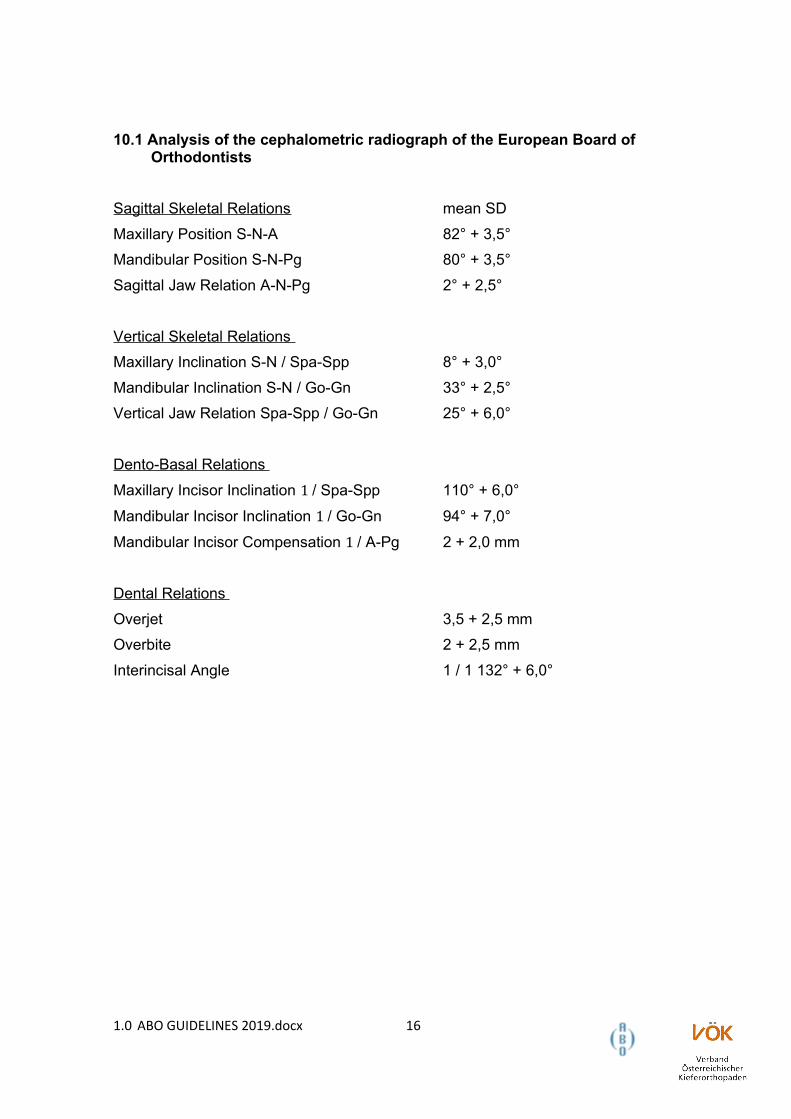
10.1 Analysis of the cephalometric radiograph of the European Board of Orthodontists
Sagittal Skeletal Relations mean SD
Maxillary Position S-N-A 82° + 3,5°
Mandibular Position S-N-Pg 80° + 3,5°
Sagittal Jaw Relation A-N-Pg 2° + 2,5°
Vertical Skeletal Relations
Maxillary Inclination S-N / Spa-Spp 8° + 3,0°
Mandibular Inclination S-N / Go-Gn 33° + 2,5°
Vertical Jaw Relation Spa-Spp / Go-Gn 25° + 6,0°
Dento-Basal Relations
Maxillary Incisor Inclination 1 / Spa-Spp 110° + 6,0°
Mandibular Incisor Inclination 1 / Go-Gn 94° + 7,0°
Mandibular Incisor Compensation 1 / A-Pg 2 + 2,0 mm
Dental Relations
Overjet 3,5 + 2,5 mm
Overbite 2 + 2,5 mm
Interincisal Angle 1 / 1 132° + 6,0°
1.0 ABO GUIDELINES 2019.docx 16
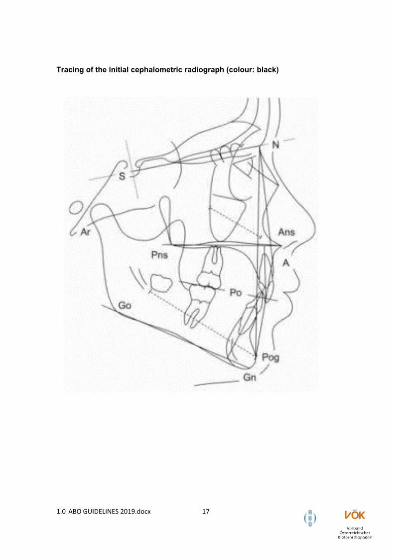
Tracing of the initial cephalometric radiograph (colour: black)
1.0 ABO GUIDELINES 2019.docx 17
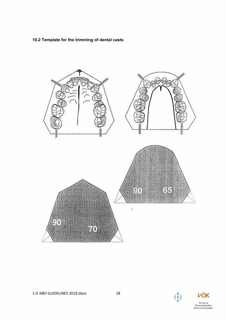
10.2 Template for the trimming of dental casts
1.0 ABO GUIDELINES 2019.docx 18
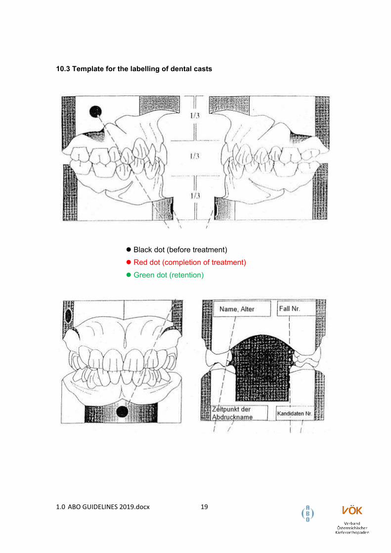
10.3 Template for the labelling of dental casts
Black dot (before treatment)
Red dot (completion of treatment)
Green dot (retention)
1.0 ABO GUIDELINES 2019.docx 19
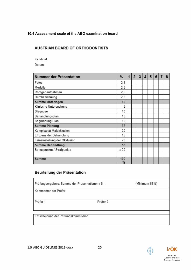
10.4 Assessment scale of the ABO examination board
1.0 ABO GUIDELINES 2019.docx 20