gonzalez-barriga et al. mtna 2013 article + suppl info
TRANSCRIPT

Citation: Molecular Therapy–Nucleic Acids (2013) 2, e81; doi:10.1038/mtna.2013.9© 2013 American Society of Gene & Cell Therapy All rights reserved 2158-3188/11
www.nature.com/mtna
The first two authors contributed equally to this work.1Department of Cell Biology, Nijmegen Centre for Molecular Life Sciences, Radboud University Nijmegen Medical Centre, Nijmegen, The Netherlands; 2Prosensa Therapeutics B.V., Leiden, The Netherlands; 3Current address: Department of Human Genetics, Radboud University Nijmegen Medical Centre, Nijmegen, The Netherlands. Correspondence: Derick G Wansink, Radboud University Nijmegen Medical Centre, NCMLS, Department of Cell Biology (code 283), P.O. Box 9101, 6500 HB Nijmegen, The Netherlands. E-mail: [email protected]: antisense oligonucleotides; myotonic dystrophy; RNA processing; RNA silencing; triplet repeat expansionReceived 8 October 2012; accepted 7 February 2013; advance online publication 19 March 2013. doi:10.1038/mtna.2013.9
Design and Analysis of Effects of Triplet Repeat Oligonucleotides in Cell Models for Myotonic DystrophyAnchel González-Barriga1,2, Susan AM Mulders1,2, Jeroen van de Giessen2, Jeroen D Hooijer1,3, Suzanne Bijl2, Ingeborg DG van Kessel1, Josee van Beers1, Judith CT van Deutekom2, Jack AM Fransen1, Bé Wieringa1 and Derick G Wansink1
Myotonic dystrophy type 1 (DM1) is caused by DM protein kinase (DMPK) transcripts containing an expanded (CUG)n repeat. Antisense oligonucleotide (AON)-mediated suppression of these mutant RNAs is considered a promising therapeutic strategy for this severe disorder. Earlier, we identified a 2′-O-methyl (2′-OMe) phosphorothioate (PT)–modified (CAG)7 oligo (PS58), which selectively silences mutant DMPK transcripts through recognition of the abnormally long (CUG)n tract. We present here a comprehensive collection of triplet repeat AONs and found that oligo length and nucleotide chemistry are important determinants for activity. For significant reduction of expanded DMPK mRNAs, a minimal length of five triplets was required. 2′-O,4′-C-ethylene-bridged nucleic acid (ENA)–modified AONs appeared not effective, probably due to lack of nuclear internalization. Selectivity for products from the expanded DMPK allele in patient myoblasts, an important requirement to minimize unwanted side effects, appeared also dependent on AON chemistry. In particular, RNase-H–dependent (CAG)n AONs did not show (CUG)n length specificity. We provide evidence that degradation of long DMPK transcripts induced by PS58-type AONs is an RNase-H independent process, does not involve oligo-intrinsic RNase activity nor does it interfere with splicing of DMPK transcripts. Our collection of triplet repeat AONs forms an important resource for further development of a safe therapy for DM1 and other unstable microsatellite diseases.Molecular Therapy–Nucleic Acids (2013) 2, e81; doi:10.1038/mtna.2013.9; published online 19 March 2013Subject Category: Nucleic Acid Chemistry; Antisense Oligonucleotides
Introduction
Myotonic dystrophy type 1 (DM1) is an autosomal dominant, trinucleotide repeat expansion disease and the most common form of adult onset muscular dystrophy.1–3 The multisystemic symptoms are highly variable and involve different tissues and organs, including skeletal muscle, heart, brain, eyes, reproductive systems, and the gastrointestinal tract. No cure to slow or stop the disease process is currently available.
The genetic basis of DM1 is an unstable (CTG)n expan-sion in the 3′-untranslated region of the DM protein kinase (DMPK) gene. Length of the (CTG)n segment is correlated with age of onset and severity of disease.1 In the healthy population, the CTG repeat is polymorphic in length, with a triplet number that ranges from 5 to 37 (mostly below 15),4 whereas disease alleles may contain between 50 and >2,000 triplets.1
The DM1 pathogenic mechanism involves an RNA gain-of-function. Repeat-containing transcripts from the expanded allele accumulate in cell nuclei and alter the function of RNA-binding proteins, whose association with disease has been most intensively studied for muscleblind (MBNL) and CUG-binding protein 1 (CUGBP1).2 In adult DM1 tissue, the result-ing imbalance in availability of these antagonistic splicing regulators causes misregulation of events in which embry-onic patterns of splicing occur. For example, myotonia and insulin resistance, two key features of DM1, may be caused by altered splicing of muscle chloride channel and insulin
receptor transcripts, respectively.2 Also effects on microRNA processing and function have been reported.5
Results obtained with DM1 mouse models that inducibly express (CUG)n RNA show that features of RNA toxicity are reversible,6 strongly suggesting that reducing the level of expanded DMPK transcripts will be beneficial to patients. In the past decade, different antisense strategies have been developed with the intention to eliminate expanded DMPK transcripts via either antisense RNA expression, ribozyme activity, oligo-mediated RNase-H activity or RNA interference (RNAi) technology.7 Reduction of DMPK transcript levels was achieved by several of these methods, but selectivity between toxic and normal-sized transcripts was generally low.
To be functional and safe for therapeutic use in DM1 patients, an antisense approach should ideally silence tran-scripts from the expanded allele only and leave normal-sized DMPK transcripts or unrelated transcripts containing (CUG)n segments intact. Since length of the (CUG)n repeat is the only consistent polymorphic difference between mutant and normal DMPK (pre)mRNAs, the repeat tract itself forms an attractive therapeutic target. Besides, despite the fact that normal and mutant DMPK transcripts are both made in the nucleus, (CUG)n length selectivity may be enhanced by directing active compounds to this compartment where mutant transcripts accumulate after synthesis.
Earlier, we published on the identification and character-ization of PS58, an antisense oligonucleotide (AON) com-plementary to the (CUG)n repeat.8 PS58 is a (CAG)7 fully

Molecular Therapy–Nucleic Acids
Triplet Repeat Oligos for Myotonic Dystrophy González-Barriga et al.
2
2′-O-methyl (2′-OMe) phosphorothioate (PT)-modified AON, which silences expanded (CUG)n transcripts in DM1 cell and animal models. Other in vivo AON studies directed at the expansion mutation made use of a (CAG)n morpholino to interfere with the (CUG)n repeat–MBNL interaction9 or LNA (locked nucleic acid)/MOE (2′-O-methoxyethyl)-PT–type (CAG)n gapmers to recruit RNase-H activity for deg-radation of (CUG)n-containing transcripts.10 François et al. engineered a hU7-(CAG)15-small nuclear RNA which prefer-entially reduced pathogenic DMPK mRNA in DM1 cells, via a yet unknown mechanism.11
Here, we report on specific properties that (CUG)n-di-rected AONs require for effective knockdown of expanded DMPK RNA. Triplet repeat AONs with different chemistries and lengths were tested in an unbiased screen for silenc-ing capacity in a transgenic DM1 cell culture model express-ing human DMPK (CUG)500 transcripts. AON length and nucleotide chemistry appeared to be major parameters for therapeutic efficacy. A selection of effective AONs was tested in DM1 patient myoblasts. Findings of this study are important for transition towards in vivo studies with AONs and ultimately for development of a specific AON-mediated therapy for DM1. As a first step in support of this transition, we report on effects on in vitro complement activation by some of these AONs.
ResultsAONs complementary to DMPK’s (CUG)n repeatA series of triplet repeat AONs that differed in length, chemis-try, and nucleotide composition was designed and analyzed for their ability to silence expanded DMPK transcripts in cul-tured cells (see AON description in Figure 1a). AONs were tested via transfection in the DM500 myoblast–myotube cell model.8 The human DMPK (hDMPK) transgene in this model is under near-normal myogenic transcriptional control and produces hDMPK transcripts bearing a (CUG)500 repeat at levels comparable to that of endogenous mouse Dmpk (mDmpk) transcripts. It should be noted that mDmpk tran-scripts contain a CCG(CUG)2(CAG)2CUG sequence instead of the generally believed to be uninterrupted (CUG)n seg-ment found in hDMPK transcripts. Monitoring AON-silencing effects was done by northern blotting to follow steady-state levels of full-length transgenic hDMPK transcripts and possible stable breakdown products. Analysis of endog-enous mDmpk transcripts was used as a control. Reverse transcription-quantitative PCR (RT-qPCR) was included for quantitative assessment of expression of defined transcript segments.
DNA-based (CAG)n AONsDNA-type oligonucleotides are known to allow specific RNase-H–mediated cleavage of RNA in a DNA–RNA het-eroduplex.12 For these AONs, several chemical modifications have been developed that improve their metabolic stabil-ity, while maintaining hybridization affinity to target RNA and ability to recruit RNase H.13 We tested three DNA-type (CAG)7 AONs in DM500 myotubes: PS56, a pure DNA AON; PS142, a PT-modified DNA AON; and PS260, a PT-modi-fied chimeric AON, so called gapmer, comprised of 2′-OMe wings on the 5′ and 3′ sides of a DNA gap. DNA AON PS56
did not show silencing activity (Figure 1a), probably because it was quickly degraded inside cells (Supplementary Figure S1). In contrast, PS142 and PS260–both stabilized against nuclease breakdown–were able to silence expanded hDMPK mRNA for ∼50 and 80%, respectively (Figure 1a,b). Endogenous mDmpk mRNA was left intact. A concentration–response analysis yielded an IC50 (half maximal inhibitory concentration) of 0.3 ± 0.4 μmol/l for DNA/PT oligo PS142, which is about 1,000-fold lower than that of 2′-OMe/PT lead compound PS58 (Figure 1c, Supplementary Figure S2 and ref. 8).
Oligo length and base variations of 2′-OMe AONsTo investigate length as a potential parameter in the per-formance of PS58-type oligos, we tested a series of fully 2′-OMe–modified (CAG)n AONs consisting of 4–13 triplets in the DM500 myotube culture system. All, except PS251 (CAG)4, effectively and selectively silenced (CUG)500 transcripts (>80% efficiency) (Figure 1a,b). A (CUG)7 2′-OMe sense oligo or 2′-OMe oligos directed against tar-get sequences in the hDMPK transcript outside the repeat did not have a silencing effect (data not shown).8 The lack of activity of the (CAG)4 AON is best explained by assuming that a minimum oligo length is necessary for effective hybrid-ization to the (CUG)n target needed for AON activity.
We wondered whether the nature of the nucleotide residue at the 5′ or 3′ end of the AON might be crucial for silenc-ing and tested a repeat-shifted version, 2′-OMe–modified (AGC)7 (PS259). PS259 treatment resulted in a significant reduction of 85% of (CUG)500 transcripts without silencing mDmpk mRNA (Figure 1a).
Finally, a 2′-OMe–modified (CIG)7 AON was tested. The inosine residues should enable this AON, PS261, to hybrid-ize to (CUG)n, (CAG)n, (CCG)n repeats and any combination of these triplets. Similar to PS58, PS261 induced 85% reduc-tion of expanded mRNA (Figure 1a). Notably, endogenous mDmpk mRNA was also significantly reduced, albeit with only ~35% efficiency. This effect may be best explained by binding of (CIG)7 to the CCG(CUG)2(CAG)2CUG sequence in mDmpk mRNA. Besides, three low-affinity–binding sites were identified that could have a cooperative, additive effect (11–13 nucleotides match; not present in hDMPK transcripts).
Silencing of expanded hDMPK transcripts by triplet repeat 2′-OMe AONs in DM500 myotubes was independently con-firmed by RT-qPCR analysis (Figure 1d). The lower silenc-ing efficiency measured by RT-qPCR (60–90 versus 85–95% detected by northern blotting) is attributed to technical differ-ences in sensitivity intrinsic to the methods.
Morpholino phosphorodiamidate (CAG)n AONsMorpholino phosphorodiamidate oligos are uncharged AONs widely used as blocking oligos for selective inhibi-tion of gene expression.14 A (CAG)8 morpholino (PS304) was tested for its ability to silence expanded hDMPK RNA in DM500 myotubes. The morpholino was coupled to an octaguanidine dendrimer (so called Vivo-Porter) to facili-tate cellular uptake. PS304 treatment caused a 70% reduc-tion of (CUG)500 transcripts, leaving mDmpk mRNA intact (Figure 1a).

www.moleculartherapy.org/mtna
Triplet Repeat Oligos for Myotonic Dystrophy González-Barriga et al.
3
Sequence and chemistrya
(5′ -> 3′)
mDmpk
hDMPK
mDmpk
Gapdh
hDMPK
PS56
0 25 50
mRNA level (%)
75
***
***
***
*********
******
**
**
**
* **
* ****
**
** ** **
***
***
***
*** **
*
***
***
**
**
100
Oligo name
(CAG)7
PS142 (CAG)7
PS260 (CAG)7
PS251 (CAG)4
PS147 (CAG)5
PS253 (CAG)6
PS58 (CAG)7
PS255 (CAG)8
PS256 (CAG)9
PS146 (CAG)10
PS258 (CAG)13
PS259 (AGC)7
PS261 (CIG)7
PS304c (CAG)8
PS139b (CAG)3
PS138b (CAG)4PS137 (CAG)5
PS136 (CAG)7
PS135
0−11 −10 −9
Log (AON)
PS58 PS142
−8 −7 −6
25
50
mR
NA
leve
l (%
) 75
100
0
Mock
Mock
Mock
PS142
Mock
PS147 (
CAG)5
PS253 (
CAG)6
PS58 (C
AG)7
PS255 (
CAG)8
PS256 (
CAG)9
PS146 (
CAG)10
PS258 (
CAG)13
PS261 (
CIG)7
PS136
PS58
25
50
mR
NA
leve
l (%
)
75
100
e15 5′
e15 3′
EN
AP
MO
2′ -OM
e
(CAG)10
DN
A
a
b c d
Figure 1 Collection of triplet repeat AONs tested for their ability to silence expanded hDMPK mRNA in DM500 myotubes. (a) Summary of silencing efficiency. Endogenous mDmpk RNA was included as a negative control, since it lacks a pure (CUG)n repeat. AONs are grouped according to their main chemical modification: aDNA (capital); RNA (capital, underlined); PT, phosphorothioate linkage (*); 2′-OMe, 2′-O-methyl sugar modification (capital, underlined, bold); ENA, 2′-O,4′-C-ethylene-bridged nucleic acid (capital, italics); PMO, morpholino phosphorodiamidate (lower case); bCy3-labeled; cCoupled to octaguanidine dendrimer. The means of at least three independent experiments for each AON are shown. Dashed line indicates 100% levels (mock samples). (b) Representative northern blots with RNA isolated from AON-treated DM500 myotubes probed with a hDMPK and a Gapdh probe. Results of treatment with three AONs (200 nmol/l) or mock treatment is shown. (c) Concentration–response curves of PS58 and PS142. DM500 myotubes were treated with a concentration series of 0.01–500 nmol/l AON (see also Supplementary Figure S2). (d) RT-qPCR analysis of hDMPK RNA levels in DM500 myotubes after treatment with a selection of 2′-OMe AONs (200 nmol/l, n ≥ 3). PCR amplicons were located in exon 15, either 5′ or 3′ to the (CUG)n tract. *P < 0.05; **P < 0.01; ***P < 0.001. AON, antisense oligonucleotide; Gapdh, glyceraldehyde 3-phosphate dehydrogenase; hDMPK, human DM protein kinase; mDmpk, mouse Dmpk; RT-qPCR, reverse transcription-quantitative PCR.

Molecular Therapy–Nucleic Acids
Triplet Repeat Oligos for Myotonic Dystrophy González-Barriga et al.
4
2′-O,4′-C-ethylene-bridged nucleic acid-modified (CAG)n AONsThe bicyclic modification in 2′-O,4′-C-ethylene-bridged nucleic acid (ENA) confers strong nuclease resistance and high-binding affinity to complementary single-stranded RNA (average ΔTm (Δmelting temperature)/modification = 5.5 ver-sus 1.3 °C for 2′-OMe)15 (Supplementary Figure S1). We tested two fully ENA-modified AONs consisting of three and four CAG triplets, and three ENA/2′-OMe gapmers of 5, 7, and 10 CAGs (PS139-135, Figure 1a). Only treatment with PS136 resulted in a small, yet significant reduction (30%) of hDMPK RNA expression.
The discrepancy in efficacy between ENA- and 2′-OMe–modified AONs may be caused by differences in subcellular targeting. To verify this possibility, we followed cellular uptake and routing of two fluorescently labeled AONs with these modifications. For these analyses, we switched to the use of DM500 myoblasts, which display a rather flat morphology, rendering them more suitable for live cell imaging of AON localization than myotubes. Forty minutes after transfection, fluorescent PS58 oligo was present in the cell nucleus and sig-nal intensity further increased up to 60 minutes (Supplemen-tary Figure S3). This behavior was independent of the type of fluorophore used and fluorophore coupling did not affect PS58 silencing activity (data not shown). In contrast, labeled PS138, as a representative of the ENA AON collection, was concentrated in vesicle-like structures and rarely present in the nucleus itself. This pattern remained unchanged up to 48 hours after transfection (data not shown). We conclude that lower silencing efficiency of ENA-modified (CAG)n AONs observed in this study is due to their inability to reach muscle cell nuclei, where most expanded (CUG)n transcripts reside.
Triplet repeat siRNAsKrol et al.16 reported that (CXG)n triplet repeat hairpins in mRNAs are substrates for Dicer and produce short repeat RNAs that in turn may act as short-interfering RNAs (siRNAs) against complementary repeats. We wanted to investigate whether in our cell model expanded hDMPK mRNAs could be silenced by introducing synthetic (CAG)n/(CUG)n siRNAs. Two different RNA duplexes were transfected in DM500 myotubes, PI-01 ((CAG)7/(GCU)7) and PI-02 ((GCA)7/(CUG)7) (Sup-plementary Table S1). An unrelated siRNA and PS58 were included as controls. RT-qPCR analysis demonstrated that none of the repeat siRNAs silenced hDMPK mRNA expres-sion (Figure 2). From our AON stability assay, we conclude that it is unlikely that the inability to reduce expanded hDMPK transcripts is due to rapid intracellular degradation of the chemically unprotected siRNAs (Supplementary Figure S1).
Mouse transcripts containing a small (CUG)n segmentTranscripts that contain a (CUG)n segment, other than encoded by the hDMPK transgene, may also serve as targets for (CAG)n AONs in DM500 cells. Genes carrying a (CTG)n segment are rare, but at least nine transcripts in the mouse transcriptome contain a repeat of more than six triplets.8 We investigated whether 2′-OMe/PT AONs of variable length and sequence ((CAG)5, (CAG)7, (CAG)10, and (CIG)7) acted on a subset of these transcripts expressed in DM500 myotubes.
We analyzed transcript levels by RT-qPCR based on ampli-cons next to the repeat tracts (Figure 3; primer locations are depicted in more detail in Supplementary Figure S4). Ptbp1 (CUG)6, Txlnb (CUG)9, and Mapkap1 (CUG)26–the longest (CUG)n tract in the mouse transcriptome–showed no signifi-cant changes in expression after AON treatment (Figure 3). Note that due to the location of the (CUG)9 tract in the very 5′ end of the Txlnb gene, no suitable 5′ amplicon could be designed to measure Txlnb transcripts.
We also wanted to analyze RNA expression via amplifica-tion across the (CUG)n triplet repeat. Semi-quantitative RT-PCR for Ptbp1 and Txlnb transcripts confirmed the RT-qPCR findings and showed no significant change in expression (Supplementary Figure S5a,b). Remarkably, quantifica-tion across the Mapkap1 (CUG)26 repeat did record sig-nificant silencing after AON treatment, especially by PS146 (CAG)10 (Supplementary Figure S5c). This effect was not seen, however, when segments flanking the (CUG)26 tract were amplified in the same assay. Also northern blotting, visualizing two types of Mapkap1 transcripts as a result of alternative polyadenylation17 (Supplementary Figure S4), revealed only a minor reduction in RNA expression, whereas western blotting revealed no significant changes in Mapkap1 protein expression (Supplementary Figure S5d,e). We therefore decided that PCR-based quantifica-tion should not rely on amplification across a medium-sized (CUG)n tract.
Given these observations, we wanted to exclude the possi-bility that traces of AONs that might still be present in purified RNA samples would interfere with RT-PCR analysis. Using a hybridization–ligation method, we determined that free AON concentration in our RNA preparations was <0.2 nmol/l. To rule out interference of (CAG)n AONs with RT or PCR ampli-fication, untreated DM500 RNA samples were spiked with 0.1–10 nmol/l PS147, PS58 or PS146 and correspondingly used in RT-PCR analysis. Presence of AONs during RT-PCR
0
Moc
k
PS58 (C
AG)7
Contro
l siR
NAPI-0
1PI-0
2
25
50
mR
NA
leve
l (%
)
75**
100
Figure 2 RT-qPCR analysis of hDMPK (CUG)500 RNA levels in DM500 myotubes after treatment with siRNAs (50 nmol/l) targeting the (CUG)n repeat (n = 6). PS58 (50 nmol/l) was used as a control. Sequences of PI-01 ((CAG)7/(GCU)7 duplex), PI-02 ((GCA)7/(CUG)7 duplex), and control siRNA are listed in Supplementary Table S1. **P < 0.01. hDMPK, human DM protein kinase; RT-qPCR, reverse transcription-quantitative PCR; siRNA, short-interfering RNA.

www.moleculartherapy.org/mtna
Triplet Repeat Oligos for Myotonic Dystrophy González-Barriga et al.
5
amplification did not have an effect on Mapkap1 (CUG)26 levels nor on quantification of hDMPK (CUG)500 RNA (Sup-plementary Figure S6). In sum, we conclude that transcripts containing small (CUG)n tracts were largely unaffected by the (CAG)n and (CIG)n repeat oligos tested.
Mouse transcripts carrying a small (CAG)n segmentTo examine the possibility that oligo-mediated hDMPK RNA breakdown would generate short (CUG)n RNAs that in turn might act as siRNAs on complementary (CAG)n transcripts (compare, ref. 16), we measured levels of mouse transcripts with a small (CAG)n segment: Sec24b (CAG)15, Hcn1 (CAG)7+(CAG)20, and Nr3c1 (CAG)17. After treatment with PS147, PS58, PS146, and PS261; no significant change in expression of any of these transcripts was detectable by
RT-qPCR analysis (Figure 4) ruling out an indirect effect of (CAG)n AONs on transcripts bearing a (CAG)n segment in the transcriptome.
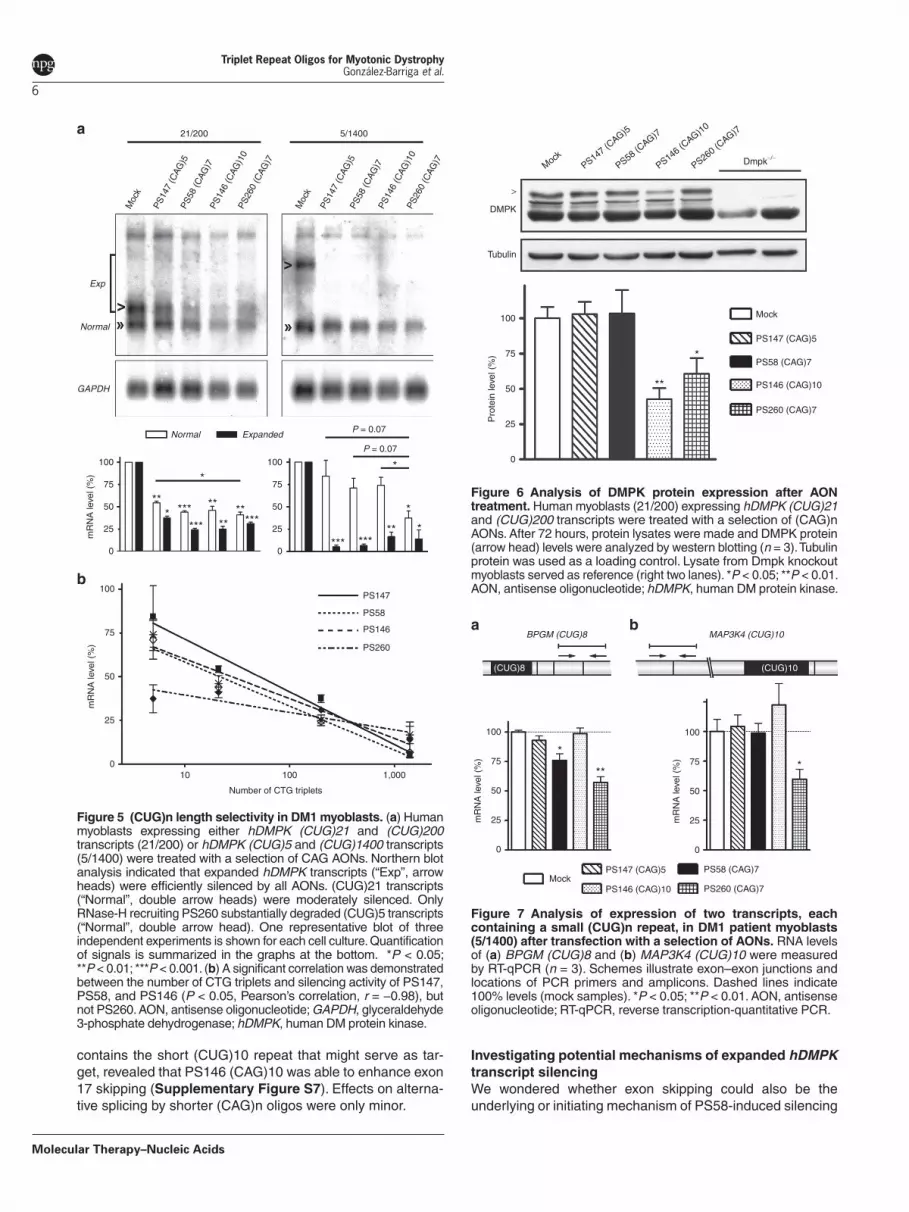
(CUG)n length selectivity in DM1 patient myoblastsA safe, antisense strategy for DM1 should differentiate between expanded and normal-sized hDMPK transcripts. To test this requirement, we used two DM1 myoblast cul-tures: one expressing hDMPK transcripts with 21 (normal) and 200 (mutant) triplets (abbreviated 21/200) and another with 5 and 1,400 CUG triplets (abbreviated 5/1,400).18 Activi-ties of three 2′-OMe AONs, PS147, PS58, and PS146 were examined in these cultures, with use of northern blotting to discriminate between normal and mutant hDMPK RNA. The three pure 2′-OMe/PT AONs behaved equally (Figure 5a): levels of (CUG)1400 and (CUG)200 transcripts were strongly decreased (65–95%). (CUG)21 transcripts appeared less susceptible to breakdown (45–55%) and as expected, (CUG)5 hDMPK mRNA was left intact. Silencing efficacy sig-nificantly correlated with the number of CUG triplets in the transcripts (Figure 5b).
Western blot analysis showed that silencing of hDMPK transcripts by PS146 (CAG)10 resulted in around 50% loss of DMPK protein after 72 hours in 21/200 myoblasts (Figure 6). No effect of PS147 (CAG)5 or PS58 (CAG)7 treatment was observed at this time point.
Human transcripts containing a small (CUG)n segmentAlso in the human transcriptome (CUG)n tracts are relatively rare.8,19 Effects of a subset of 2′-OMe/PT AONs on tran-scripts bearing a (CUG)n repeat expressed in 5/1,400 myo-blasts were examined for two transcripts: BPGM (CUG)8 and MAP3K4 (CUG)10 by RT-qPCR. No reduction of BPGM tran-scripts was detected after treatment with PS147 and PS146, whereas only a small effect was seen with PS58 (25% reduc-tion) (Figure 7a). Similar effects for BPGM (CUG)8 were detected in 21/200 myoblast cultures (data not shown).
Total MAP3K4 mRNA levels were not affected by 2′-OMe AON treatment (Figure 7b). A conspicuous observation was made with regard to an effect on MAP3K4 pre-mRNA pro-cessing. Analysis of the fate of alternative exon 17,20 which
05′
Mock PS147 (CAG)5
(CUG)6 (CUG)9 (CUG)26
PS58 (CAG)7 PS146 (CAG)10 PS261 (CIG)7
5′3′
5′ 5′3′Ptbp1 (CUG)6 Txlnb (CUG)9 Mapkap1 (CUG)26
3′ 3′
3′ 3′
25
50
mR
NA
leve
l (%
)
75
100
0
25
50
mR
NA
leve
l (%
)
75
100
0
25
50
mR
NA
leve
l (%
)
75
100
a b c
Figure 3 Expression of three mouse transcripts, carrying a small (CUG)n repeat, after transfecting DM500 myotubes with a selection of AONs. (a) Ptbp1 (CUG)6, (b) Txlnb (CUG)9, and (c) Mapkap1 (CUG)26. RNA levels were analyzed by RT-qPCR amplifying segments flanking the triplet repeat (n = 3). Schemes illustrate exon–exon junctions, locations of PCR primers, and amplicons. Dashed lines indicate 100% levels (mock samples). AON, antisense oligonucletide; RT-qPCR, reverse transcription-quantitative PCR.
05′
MockPS147 (CAG)5
PS146 (CAG)10
PS58 (CAG)7
PS261 (CIG)7
(CAG)15 (CAG)7+20 (CAG)17
3′
5′ 5′ 5′3′ 3′
5′ 5′ 3′
25
50
mR
NA
leve
l (%
)
75
100
Sec24b(CAG)15
Hcn1(CAG)7/(CAG)20
Nr3c1(CAG)17
a b c
Figure 4 Analysis of expression levels of three mouse transcripts, containing a (CAG)n repeat, in DM500 myotubes after transfection with a selection of AONs. (a) Sec24b (CAG)15, (b) Hcn1 carrying a (CAG)7 and a (CAG)20 repeat located close together (see Supplementary Figure S4), and (c) Nr3c1 (CAG)17. Expression was analyzed by RT-qPCR amplifying segments flanking the triplet repeat (n = 3). Schemes illustrate exon–exon junctions, locations of PCR primers, and amplicons. Dashed lines indicate 100% levels (mock samples). AON, antisense oligonucletide; RT-qPCR, reverse transcription-quantitative PCR.
Molecular Therapy–Nucleic Acids
Triplet Repeat Oligos for Myotonic Dystrophy González-Barriga et al.
6
contains the short (CUG)10 repeat that might serve as tar-get, revealed that PS146 (CAG)10 was able to enhance exon 17 skipping (Supplementary Figure S7). Effects on alterna-tive splicing by shorter (CAG)n oligos were only minor.
Investigating potential mechanisms of expanded hDMPK transcript silencingWe wondered whether exon skipping could also be the underlying or initiating mechanism of PS58-induced silencing
0
MockPS147 (C
AG)5
PS58 (CAG)7
PS146 (CAG)10
PS260 (CAG)7
Dmpk−/−
DMPK
>
Mock
PS147 (CAG)5
PS58 (CAG)7
PS146 (CAG)10
PS260 (CAG)7
Tubulin
25
50
Pro
tein
leve
l (%
) 75
100
*
**
Figure 6 Analysis of DMPK protein expression after AON treatment. Human myoblasts (21/200) expressing hDMPK (CUG)21 and (CUG)200 transcripts were treated with a selection of (CAG)n AONs. After 72 hours, protein lysates were made and DMPK protein (arrow head) levels were analyzed by western blotting (n = 3). Tubulin protein was used as a loading control. Lysate from Dmpk knockout myoblasts served as reference (right two lanes). *P < 0.05; **P < 0.01. AON, antisense oligonucleotide; hDMPK, human DM protein kinase.
0
(CUG)8 (CUG)10
MockPS147 (CAG)5 PS58 (CAG)7
PS146 (CAG)10 PS260 (CAG)7
BPGM (CUG)8 MAP3K4 (CUG)10
25
50
mR
NA
leve
l (%
) 75
100
0
25
50
****
mR
NA
leve
l (%
) 75
100
a b
Figure 7 Analysis of expression of two transcripts, each containing a small (CUG)n repeat, in DM1 patient myoblasts (5/1400) after transfection with a selection of AONs. RNA levels of (a) BPGM (CUG)8 and (b) MAP3K4 (CUG)10 were measured by RT-qPCR (n = 3). Schemes illustrate exon–exon junctions and locations of PCR primers and amplicons. Dashed lines indicate 100% levels (mock samples). *P < 0.05; **P < 0.01. AON, antisense oligonucleotide; RT-qPCR, reverse transcription-quantitative PCR.
0
Moc
k
Moc
k
PS14
7 (C
AG)5
PS14
7 (C
AG)5
PS58
(CAG
)7
PS58
(CAG
)7
PS14
6 (C
AG)1
0
PS14
6 (C
AG)1
0
PS26
0 (C
AG)7
PS26
0 (C
AG)7
21/200 5/1400
25
50
mR
NA
leve
l (%
)
75
100
Normal Expanded
Normal
GAPDH
Exp
0
25
50
75
100
P = 0.07
P = 0.07
*
*
*********
******
***
*
****
*****
010 100
Number of CTG triplets
1,000
25
50
mR
NA
leve
l (%
)
75
100PS147
PS58
PS146
PS260
b
a
Figure 5 (CUG)n length selectivity in DM1 myoblasts. (a) Human myoblasts expressing either hDMPK (CUG)21 and (CUG)200 transcripts (21/200) or hDMPK (CUG)5 and (CUG)1400 transcripts (5/1400) were treated with a selection of CAG AONs. Northern blot analysis indicated that expanded hDMPK transcripts (“Exp”, arrow heads) were efficiently silenced by all AONs. (CUG)21 transcripts (“Normal”, double arrow heads) were moderately silenced. Only RNase-H recruiting PS260 substantially degraded (CUG)5 transcripts (“Normal”, double arrow head). One representative blot of three independent experiments is shown for each cell culture. Quantification of signals is summarized in the graphs at the bottom. *P < 0.05; **P < 0.01; ***P < 0.001. (b) A significant correlation was demonstrated between the number of CTG triplets and silencing activity of PS147, PS58, and PS146 (P < 0.05, Pearson’s correlation, r = −0.98), but not PS260. AON, antisense oligonucleotide; GAPDH, glyceraldehyde 3-phosphate dehydrogenase; hDMPK, human DM protein kinase.

www.moleculartherapy.org/mtna
Triplet Repeat Oligos for Myotonic Dystrophy González-Barriga et al.
7
of expanded hDMPK transcripts. hDMPK pre-mRNAs are subject to extensive alternative splicing and events involving final exon 15, in which the (CUG)n tract is located, have been reported.21,22 For analysis of fate of different hDMPK RNA segments in presence of AONs, we used a transcript scan-ning RT-qPCR approach for amplicons along the expanded hDMPK transcript in DM500 myotubes. Amplicons were designed in the 5′ end of the gene (exon 1–exon 2, exon 4– exon 5) and on either side of the (CUG)n tract in exon 15 (Figure 8). No significant differences were found in signal strength for any of these hDMPK segments (70–85% reduc-tion for all segments across the transcript, compared with mock-treated samples). This finding indicates that PS58-induced skipping of the (CUG)n tract during hDMPK pre-mRNA splicing is unlikely to be mechanistically involved.
Test tube incubation of radioactively labeled, in vitro- transcribed (CUG)90 RNA with a concentration series of PS58 (CAG)7 caused a mobility shift illustrating direct oligo–RNA association (Supplementary Figure S8). Similar shifts were seen after incubation of different (CAG)n and (CUG)n AONs with (CUG)90 or (CAG)90 RNAs (Supplementary Fig-ure S8). To exclude that triplet repeat 2′-OMe AONs had an intrinsic RNase activity, similar to ribozymes or DNAzymes,23 (CUG)n RNA was incubated with a series of different AONs in the presence of various concentrations of Mg2+, Mn2+ or Zn2+ (Supplementary Figure S8; data not shown). No (CUG)n breakdown products could be detected, indicating that under the conditions tested, none of these AONs displayed RNase-like activity when complexed to (CUG)n RNA.
Using the same (CUG)90 RNA template, we could con-firm that PS58, a fully modified 2′-OMe AON, was unable to induce RNase-H–mediated degradation of expanded (CUG)n transcripts in vitro (Supplementary Figure S8). As posi-tive controls, we used DNA-type AON PS142 and gapmer PS260, both of which were able to recruit RNase-H to cleave
(CUG)90 RNA. Activity of gapmer PS260 was also examined in human myoblasts, but with an apparent lack of specificity (Figures 5 and 6). More than 60% of both expanded and normal-sized hDMPK transcripts were degraded. Similar lack of specificity was observed for BPGM (CUG)8 and MAP3K4 (CUG)10 RNAs, for which a 40% reduction in steady-state copy number was observed (Figure 7). Since the MAP3K4 exon 17+ variant and the exon 17− variant (which lacks the AON target site) were equally reduced by PS260, we con-clude that RNase-H–dependent degradation must have occurred in the nucleus, before exon 17 splicing (Supple-mentary Figure S7).
Complement activation by triplet repeat AONsAll triplet repeat oligos were well tolerated by cells in culture and essentially no toxic effects on growth or behavior were observed (data not shown). A well-known toxic side effect of AONs in vivo, especially PT-modified versions, is activation of the complement pathway.24 As a prelude to later preclinical studies in animals and humans, we tested a selection of our PT-modified AONs with different chemistries and sequences for their ability to activate complement in plasma in vitro: full 2′-OMe (PS58, PS146, PS147, PS261), chimeric 2′-OMe/DNA (PS260), and pure DNA (PS142). A first generation oli-gonucleotide known to activate the complement pathway was included as a positive control.25 Formation of complement split products C3a (marker for the classical and alternative path-way) and Bb (marker for the alternative complement pathway) was measured in plasma from cynomolgus monkeys and humans after addition of AONs (Supplementary Figure S9). In particular, PS261 (CIG)7, and to a lesser extent PS146, PS147, and PS260, caused a significant dose-dependent increase in the concentration of Bb in cynomolgus plasma. Only PS261 and PS260 activated C3a formation in human plasma. Other AONs showed no significant or minor effects.
Discussion
This study demonstrates that a range of triplet repeat AONs is capable of silencing expanded hDMPK transcripts. The long (CUG)n target that characterizes toxic hDMPK transcripts is a special RNA sequence in the sense that it can form a stem-loop structure that folds almost like an antiparallel double-stranded helix, despite the periodic U•U mismatches.26 We conclude that the thermodynamically stable hairpin structure, known to sequester members of the MBNL protein family, does not hamper accessibility of antisense repeat AONs, when in the cellular context. The (CUG)n repeat, whose length differs between normal-short and mutant-expanded hDMPK tran-scripts, is therefore an attractive therapeutic target.
Chemical modifications of the sugar phosphate backbone of AONs serve to confer resistance against nucleases to extend cellular half-life.13 The silencing activity of modified AONs PS142 and PS260 versus the inability of pure DNA AON PS56 to induce breakdown clearly illustrates the rela-tionship between stability and efficacy. Oligo chemistry may also determine nucleocytoplasmic distribution of AONs. As target transcripts in DM1 are concentrated in the nucleus, an AON must be capable to enter this compartment to
0
Mock
e1–e2
******
*** ***
e4–e5 e15 5′ e15 3′
Scrambled-PS58 PS58
hDMPK (CUG)500
25
50
mR
NA
leve
l (%
) 75
100
1 2 3 4 5 6 7 8 9 10 11 12 14 15 (CUG)500
Figure 8 Human DMPK (CUG)500 RNA expression in DM500 myoblasts after transfection with PS58 or scrambled-PS58. Expression of four different transcript segments (little black bars) was analyzed by RT-qPCR (n = 3): two in the 5′ end of the transcript and two flanking the triplet repeat in the 3′ end of the transcript. Scheme illustrates exon composition (numbered boxes) of hDMPK transcript, arrowhead indicates reported cryptic splicing site 3′ to the triplet repeat.22 Dashed lines indicate 100% levels (mock samples). No significant differences in response to the oligos were observed between the four segments. ***P < 0.001. hDMPK, human DM protein kinase; RT-qPCR, reverse transcription-quantitative PCR.

Molecular Therapy–Nucleic Acids
Triplet Repeat Oligos for Myotonic Dystrophy González-Barriga et al.
8
become useful. Microscopic evidence is provided that ENA-modified AONs remain in vesicular structures and only inefficiently reach the nucleus, rendering them a poor choice for repeat-directed antisense DM1 strategies. We have no simple explanation for this finding, as similarly modified 2′-OMe RNA/ENA chimeras have shown activity in the nucleus during exon skipping for Duchenne muscular dystrophy.27 Effects of nucleotide sequence on subcellular oligo localization cannot be excluded at this point. Also the type of cell model and transfection method used may have an effect.
Not all types of AONs were equally efficient and specific, but as we compared only triplet repeat oligos here, some conspicuous observations are worth mentioning. First, as an explanation for the failure of (CUG)n/(CAG)n triplet repeat siRNAs to induce an RNAi response, we assume that a com-bination of chemistry and oligo sequence must be respon-sible. The poor performance of the two duplexes tested can be the result of unfavorable subcellular distribution of siR-NAs, although the RNAi machinery is known to be active in the nucleus.28 Alternatively, the (CUG)n RNA hairpin may be insufficiently accessible to certain components of the RNAi pathway in our myogenic cell model. Very recently, a com-parable triplet repeat siRNA was successfully tested in vivo in skeletal muscle in a mouse model for DM1. (CUG)220 expanded RNA and also Txlnb (CUG)9 transcripts were reduced, but not Mapkap1 (CUG)25 RNA.29
Second, oligo PS261 (CIG)7 was initially considered a promising new lead compound, because of its predicted ability to bind to (CUG)n as well as (CAG)n transcripts, with poten-tial for treatment of DM1 and polyQ diseases like Hunting-ton’s disease and several spinocerebellar ataxias. From the complement activation assay we learned, however, that this AON activated the complement cascade, rendering it a less favorable candidate for further development. Virtually none of the other AONs showed major adverse effects in vitro, with the best performing oligo being PS58. Screening for toxicity profiles in animals in vivo will be a logical next step.
Third, RNase-H recruiting AON PS260 appeared to have prominent silencing capacity, but also suffered of lack of target specificity. Further characteristics of these DNA-type AONs will be discussed below in the context of mechanistic considerations.
The main focus of our study had to do with AONs based on 2′-OMe/PT chemistry. For these AONs, we demonstrated that oligo length may provide a means to introduce selectivity between expanded transcripts from a mutated hDMPK allele and RNA containing a normal, short (CUG)n tract. The short-est variant in our series of 4–13 CAG triplets was unable to knockdown expanded transcripts in DM500 myotubes. The hybridization strength of the four CAG triplet AON may have been insufficient for a stable interaction with a (CUG)n seg-ment in the living cell, even though the theoretical T
m for this interaction is around 39 °C.
AONs longer than four triplets efficiently silenced transgenic (CUG)500 transcripts. Significant (CUG)n repeat length-dependent activity in patient cells was displayed by (CAG)5, (CAG)7, and (CAG)10 2′-OMe/PT AONs. Between them they showed no significant difference in activity towards normal-sized hDMPK transcripts: (CUG)5 transcripts, expressed
by ~40% of the Caucasian population,4 were left intact and (CUG)21 transcripts were ~50% reduced by all three AONs. In contrast, differential effects were observed at the DMPK protein level, which might indicate a correlation with transla-tion efficiency of remaining hDMPK transcripts. It should be noted that we cannot predict at the moment whether partial reduction of normal-sized transcripts will have any adverse consequences. We know that Dmpk knockout mice have rela-tively mild phenotypes,30 hence a low level of remaining Dmpk protein might be sufficient to protect functional and structural integrity of cells in which it is expressed. Another still unre-solved issue is the minimum efficacy needed for a beneficial effect. Partial suppression of toxic (CUG)n RNA may already be of clinical relevance in DM1 patients.
Expression of genes with a small (CTG)n repeat in myo-genic cells was essentially unchanged upon treatment with 2′-OMe/PT AONs, except for MAP3K4. PS146 (CAG)10 enhanced skipping of MAP3K4 exon 17 in which the (CUG)10 repeat is located. This (CUG)10 repeat is unusual as it is located in an alternative exon, where it might function as an exon-splicing enhancer.31 To what extent exon 17+ and exon 17− transcript variants contribute to protein production is unknown. More importantly, it is unclear whether a shift in MAP3K4 isoform ratio would have a biological effect. Nothing is known about functional differentiation generated by inclu-sion of the 50 amino acid stretch encoded by in-frame exon 17 (including 10 alanines encoded by the repeat tract). The minor shift in MAP3K4 variant ratio induced by smaller (CAG)n AONs is essentially within the normal variation observed in mock samples. Note that since splicing is a nuclear process, we can conclude that PS146 (CAG)10 must have been active inside the nucleus.
It is difficult to reconcile all observations regarding Map-kap1 (CUG)26 transcripts. First, as also seen with MAP3K4 transcripts, oligo length is an important parameter for AON activity and side effects seem to intensify with increased oligo length. Second, we provide ample evidence that our oligos did not interfere with RT-PCR quantification. Third, we pro-pose that special care should be taken with RNA quantifi-cation based on amplicons covering a triplet repeat of >10 units. These amplicons will never meet requirements for RT-qPCR analysis, but they may also be useless in semi-quantitative RT-PCR. For that reason, we generally used segments outside the repeat region for PCR quantification. Similar warnings regarding RT-PCR and use of AONs have been issued by others.32 Given the location of the (CTG)26 repeat in between two alternative polyadenylation sites of the Mapkap1 gene,17 we cannot exclude length-dependent AON effects on splicing, transcription termination or polyadenyla-tion. Clearly, more work is needed to be able to unequivocally interpret these data.
What do we know about the molecular mechanism under-lying silencing activity of hDMPK transcripts by triplet repeat AONs examined here? Although the goal of this study was not to identify distinct pathways or describe novel interfer-ence principles, our findings provide important new insight in potential activities of triplet repeat AONs.
In a series of simple biochemical test tube experiments, we first investigated whether triplet repeat 2′-OMe/PT AONs might carry intrinsic endoribonuclease activity, similar to

www.moleculartherapy.org/mtna
Triplet Repeat Oligos for Myotonic Dystrophy González-Barriga et al.
9
ribozymes or DNAzymes.23 In none of the experiments RNase activity was detected. Although we also tested several cell-free or protein-free conditions (data not shown), it cannot be excluded that the right cellular reaction conditions were not fully reproduced.
We provide evidence that PS260, a (CAG)7 gapmer, effi-ciently reached various (CUG)n RNA targets, irrespective of repeat length, and recruited endogenous RNase-H for a first endonucleolytic cleavage. We expect that unprotected 5′ and 3′ RNA segments will be degraded after initial cleav-age by conventional exonucleases as part of the quality con-trol system in the cell.33 Surprisingly, PS260 showed only little selectivity between expanded and normal-sized hDMPK transcripts in patient cells, suggesting that either RNase-H activity took place in both the nucleus and the cytosol, or that degradation already occurred swiftly, during or shortly after RNA synthesis in the nucleus. Our observations contrast with a report that an LNA-based repeat gapmer did cleave hDMPK (CUG)40, but not (CUG)12 transcripts;10 and also with a study in which a (CAG)6 LNA gapmer did not reduce CASK (CUG)16 RNA.34 The reason for these discrepancies should probably be found in oligo chemistry, concentration or model system used.
Nucleotide analogs like morpholinos or modifications like 2′-OMe do not support RNase-H–dependent degradation. These AONs must therefore use distinct pathways, explain-ing why PS58, a 2′-OMe/PT (CAG)7 AON, and PS260, dis-played differential (CUG)n length-dependent silencing.
The RNAi pathway is unlikely to be involved in activity of fully 2′-OMe/PT AONs or (CAG)n morpholinos. Imperfect hybrids of 2′-OMe/PT or morpholino (CAG)n AONs, or perfect hybrids that these AONs may form with (CUG)n transcripts, will not be recognized by the RNAi machinery.35–37 Further-more, the size of the shortest effective AON (15 nucleotides) and tolerated 5′ modifications like a fluorescent group (as in FAM-PS58) or a peptide (data not shown) render involvement of the RNAi pathway highly unlikely.38,39 Also, we did not find evidence for AON-induced processing of expanded hDMPK transcripts into (CUG)n siRNAs that could trigger degrada-tion of (CAG)n transcripts.
Fully 2′-OMe/PT-modified AONs, like PS58, and morpholi-nos are usually designed and employed to serve as block-ing oligos to modulate splicing or prevent translation.13,40 As discussed above, PS146, a 30 nucleotide long 2′-OMe/PT AON, exhibits splicing modulating activity. We could not detect a similar activity of (CAG)n AONs towards hDMPK (CUG)500 transcripts. The most logical explanation for this observation is the location of the (CUG)n repeat in the final exon of the hDMPK gene. The only reported splice accep-tor, located around 35 nucleotides 3′ of the (CUG)n tract, is only rarely used22,41 and probably too weak to be amenable to manipulation.
A (AGC)8A morpholino reportedly blocked Mbnl1 protein binding to transgenic (CUG)250 transcripts in a mouse model for DM1-related RNA toxicity.9 In that study, cytoplasmic lev-els of expanded transcripts raised upon treatment, but the total amount of (CUG)250 transcripts was ~50% reduced. A comparable reduction was reported for expanded Huntingtin (CAG)n RNA using a (GCT)6G LNA-type blocking oligonu-cleotide.42 The molecular mechanism underlying the activity
of AGC morpholino is unknown, but it was suggested that the displacement of expanded transcripts to the cytoplasm may have induced hDMPK mRNA decay. We propose that a simi-lar mechanism may be at play for our (CAG)n morpholino and the 2′-OMe/PT AONs examined here. Alternatively, this class of AONs may activate a yet to be identified RNase or RNA decay program in nucleus or cytoplasm. Our current working hypotheses are that the interaction between (CAG)n AONs and the (CUG)n repeat attracts proteins involved in RNA degradation or that AON binding displaces RNA-protecting proteins resulting in naked RNA, which is extremely sensi-tive to RNases. Further detailed studies into these interesting possibilities are warranted.
In sum, we provide further evidence that the expanded (CUG)n segment in hDMPK mRNA, being the product from the only known mutation in DM1, forms an attractive target for AON therapy. For development of future treatment modalities, chemical structure and oligo length in combination with the rather special sequence content needed—i.e., the comple-mentary (CAG)n repeat character—have now been identi-fied as crucial determinants. By extrapolation of findings, our results also open up interesting possibilities for therapy of other triplet repeat diseases.
Materials and methods
Cell culture. Immortalized DM500 myoblasts were derived from DM300-328 mice43 and cultured and differentiated to myotubes as described.8 Human DM1 myoblast lines were derived and cultured as described.18
Oligonucleotides. AONs and siRNAs were complementary to the (CUG)n repeat. Their sequence and chemistry are listed in Figure 1a and Supplementary Table S1, except for control oligos scrambled-PS58: 5′-CAGAGGACCACC AGACCAAGG-3′; and PS57: 5′-CUGCUGCUGCUGCUGCU GCUG-3′ (both fully 2′-OMe/PT-modified). All AONs and siR-NAs were provided by Prosensa (Leiden, The Netherlands), except morpholino PS304, which was purchased coupled to an octaguanidine dendrimer from Gene Tools (Philomath, OR) and control siRNA, purchased from Thermo Scientific Dharmacon (Lafayette, CO).
Oligotransfection. AONs were transfected using polyethyle-neimine (ExGen 500; Fermentas, Glen Burnie, MD) according to the manufacturer’s instructions. Typically, 5 μl polyethyl-eneimine/μg AON was added at a final AON concentration of 200 nmol/l in differentiation medium to myotubes on day 5 of myogenesis or in Opti-MEM (Invitrogen, Carlsbad, CA) to myo-blasts. For concentration–response experiments, oligo con-centrations between 0.01 and 500 nmol/l were applied. Four hours later, fresh medium was supplemented to a maximum volume of 2 ml medium/well. Medium was changed 20 hours later. RNA was isolated 48 hours after start of transfection.
PS304 was tested following the protocol above with the exception that no transfection reagent was used.
siRNAs were administered twice (on subsequent days: day 4 and 5 of myogenesis) using a Lipofectamine 2000-based transfection protocol (Invitrogen) according to the manufac-turer’s instructions, at a final concentration of 50 nmol/l. Four hours following addition of the transfection mix and 24 hours

Molecular Therapy–Nucleic Acids
Triplet Repeat Oligos for Myotonic Dystrophy González-Barriga et al.
10
after start of the second transfection, medium was changed. RNA was isolated 48 hours after the second transfection.
Oligo stability. AONs (7.5 pmol) were radioactively labeled by T4 polynucleotide kinase in the presence of [γ-32P]-ATP and then purified on G50 columns followed by ethanol precipitation.
DM500 myoblast whole cell extract was prepared by scraping ~1 × 10⁶ cells per well in phosphate-buffered saline (PBS) containing 1 mmol/l EDTA. Cells were collected via centrifugation (1,000 rpm, 5 minutes), packed cell volume was estimated and cells were resuspended in 1× packed cell volume of 10 mmol/l Tris-HCl; pH 8.0, 1.5 mmol/l MgCl2, 10 mmol/l KCl, 1 mmol/l DTT and 1× Protease Inhibitory Cock-tail (Roche, Almere, The Netherlands). Cell suspensions were placed on ice for 15 minutes and homogenized using a pestle. Whole cell lysate was collected after centrifugation (14,000g, 10 minutes).
AONs (~2 pmol) were incubated with 5 μl whole cell lysate at 37 °C for 0, 3 or 24 hours, reaction was stopped by adding 140 mmol/l sodium acetate; pH 5.0, phenol/CHCI3 and water phase was collected (14,000g, 10 minutes, 4 °C). Oligo was precipitated by the addition of three volumes of ethanol. The oligo precipitate was washed with 70% (vol/vol) ethanol, air dried and dissolved in sample buffer (75% deionized forma-mide, 25% glycerol). Samples were loaded on a 12% (wt/vol) acrylamide/bisacrylamide (37.5:1) denaturing urea (8 mol/l) PAGE gel in 1× MOPS buffer. After running, gels were dried at 70 °C for 2 hours and exposed to X-ray film (X-Omat AR; Kodak, Rochester, NY).
RNA isolation. RNA was isolated using the Aurum Total RNA Mini Kit (Bio-Rad, Hercules, CA), according to the manufac-turer’s protocol.
Northern blotting. Northern blotting was done as described.8 Random-primed 32P-labeled human DMPK (2.6 kb cDNA), rat Gapdh (1.1 kb cDNA), and mouse Mapkap1 (1.1 kb cDNA) probes were used. Signals were quantified by phospho-im-ager analysis (GS-505 or Molecular Imager FX; Bio-Rad) and analyzed with Quantity One (Bio-Rad) or ImageJ software. Gapdh levels were used for normalization. RNA levels of con-trol samples were set at 100%.
RT-PCR. Primer sets for PCR were designed using Primer-BLAST44 in the National Center for Biotechnology Information database and validated in silico using OligoAnalyzer 3.145 to prevent formation of hairpins and dimers during amplifica-tion. Resulting products were visualized on agarose gels and sequenced to verify identity and triplet repeat length. Primer sequences are listed in Supplementary Table S2.
For RT, typically 0.5 μg RNA was subjected to cDNA syn-thesis using the SuperScript first-strand synthesis system with random hexamer primers in a total volume of 20 μl. For RT-PCR experiments in presence of AONs, AONs were added to the RNA to reach the indicated final concentration and incubated for 15 minutes on ice before starting the RT reaction.
For semi-quantitative RT-PCR, 1 μl of cDNA preparation was used in a PCR according to standard procedures. In RT− control experiments, reverse transcriptase was omitted. The
signal for β-actin was used for normalization. Cycle number was 18 for β-actin and 22-31 for other primer sets. PCR prod-ucts were analyzed on 1.5–2.5% agarose gels stained by ethidium bromide. Quantification of signals was done using Labworks 4.0 software (UVP BioImaging Systems, Cam-bridge, UK). Values for control samples were set at 100%.
For RT-qPCR, all procedures were performed following the Minimum Information for Publication of Quantitative Real-Time PCR Experiments (MIQE) guidelines.46 Amplification efficiency of primer sets was determined using standard curves (serial dilutions; tenfold to 5,000-fold range). Primers sets displaying <90% efficiency in product amplification were ruled out and redesigned. cDNA preparations (see RT reaction above) were diluted 200–500-fold to avoid PCR inhibition by RT components; 3 μl of this dilution was mixed in a final volume of 10 μl contain-ing 5 μl 2X Sybr Green mix (Roche Applied Science, Almere, The Netherlands) and 400 nmol/l of each primer using the CAS-1200 automated pipetting system (Corbett Life Science/Qiagen, Venlo, The Netherlands). No template control and no reverse transcriptase control (RT−) were included in each qPCR run to detect possible contaminations. Samples were analyzed using the Rotor-Gene 6000 (Corbett Life Science) or CFX96 Real-time System (Bio-Rad). A melting curve was obtained for each sample to confirm single product amplification. Relative mRNA levels were calculated using the ΔΔCt method.47 Both Actb and Gapdh were used for normalization. We validated that these reference genes were not affected by the treatment and had an optimal M stability value48 under all experimental condi-tions. Values of control samples were set at 100%.
AON hybridization–ligation assay. PS58 concentrations were determined by a hybridization–ligation assay as published by Yu et al.49 with a few modifications: Template DNA probe (5′-GAATAGACGCTGCTGCTGCTGCTGCTGCTG-biotin-3′) and ligation DNA probe (5′-P-CGTCTATTC-digoxigenin (DIG)-3′) were used. Samples were incubated with the tem-plate probe (50 nmol/l) at 37 °C for 1 hour and hybridized samples were transferred to streptavidin-coated plates. Sub-sequently, plates were washed four times and the DIG-labeled ligation probe (2 nmol/l) was added and incubated for 30 min-utes at ambient temperature. DIG label was detected using an anti-DIG-POD (1:7,500; Roche Diagnostics, Almere, The Netherlands), visualized with a 3,3′,5,5′-tetramethylbenzidine substrate (Sigma Aldrich, Zwijndrecht, The Netherlands). The reaction was stopped using maleic acid (345 mmol/l; Sigma Aldrich). Absorption was measured at 450 nm using a BioTek Synergy HT plate reader (Beun de Ronde, Abcoude, The Netherlands). All samples and calibration curves were diluted (fit to criteria of the assay) in PBS. Absorption was read against a calibration curve of PS58 in PBS. A sample containing only PBS was used as correction for background.
Live cell imaging. DM500 myoblasts were cultured on gelatin-coated glass-bottom Willco dishes (Willco Wells, Amsterdam, The Netherlands). Cells were analyzed directly after addi-tion of the transfection mix containing FAM-PS58 or Cy3-PS138 in RPMI medium without phenol red and including 10 mmol/l HEPES and 10% fetal calf serum (Invitrogen). A Zeiss LSM510 meta confocal laser-scanning microscope was used, equipped with a temperature controlled CO
2 incubator

www.moleculartherapy.org/mtna
Triplet Repeat Oligos for Myotonic Dystrophy González-Barriga et al.
11
(type S) and sample stage. For recording of oligo uptake, we used a PlanApochromatic 63×, 1.4 NA oil immersion DIC lens (Carl Zeiss, Jena, Germany).
Western blotting. DM1 patient 21/200 myoblasts or DM500 mouse myotubes were transfected with PS147, PS58, PS146, PS260 or PS261 as indicated (200 nmol/l) or mock transfected and 72 hours later lysed in lysis buffer (TBS, 1% Nonidet P-40, 1 mmol/l PMSF, and Protease Inhibitory Mixture (Roche)) on ice. Cleared lysates were separated on an 8% SDS/PAGE gel and transferred to PVDF (Amersham Pharmacia Biotech, Piscataway, NJ). Blots were incubated with DMPK-specific antibody B7921 or anti-SIN1/Mapkap1 antibody (ab71152; Abcam, Cambridge, UK) and β-tubulin monoclonal antibody E7 (Developmental Studies Hybridoma Bank, University of Iowa, Iowa City, IA), followed by electrochemiluminescence detection. Quantification of signals was done using ImageJ software. Values for mock-treated samples were set at 100%.
Experiments using in vitro-transcribed triplet repeat RNA. (CUG)n and (CAG)n RNA was generated by in vitro tran-scription of a hDMPK (CTG)90 DNA template carrying flank-ing T7 and SP6 promoters. The hDMPK (CTG)90 template was generated by PCR using a hDMPK cDNA construct bearing a (CTG)90 repeat,50 T7-hDMPK forward primer: 5′-GAATTTAATACGACTCACTATAGGGAGAACGGGGCTC-GAAGGGT-3′ and SP6-hDMPK reverse primer: 5′-ATTTAG-GTGACACTATAGAAGGGCGTCATGCACAAGAAA-3′ and sequenced to verify triplet repeat number. (CUG)90 and (CAG)90 RNAs were synthesized in presence of [α-32P]GTP, using the MEGAscript in vitro transcription kit (Applied Bio-systems/Ambion, Austin, TX) according to the manufacturer’s protocol.
To confirm AON binding to expanded triplet repeat RNA, (CUG)90 RNA was incubated with a concentration series (1, 10, and 100 μmol/l) of PS58 in 50 mmol/l Tris-HCl; pH 7.5 and 25 mmol/l MgCl2 for 2 hours at 37 °C. Sample buffer was added to a final concentration of 50% deionized formamide and 30% glycerol and samples were run at room tempera-ture on a denaturing polyacrylamide gel (5% acryl/bisacryl, 8 mol/l urea) in 1× MOPS. Gels were dried and exposed to X-ray film (X-Omat AR; Kodak).
To reveal binding specificity, (CUG)90 and (CAG)90 RNAs were incubated with 100 μmol/l AONs in the presence of 50 mmol/l Tris-HCl; pH 7.5, 50 mmol/l NaCl, 25 mmol/l MgCl2, and 1 mmol/l EDTA for 2 hours at 37 °C. Samples were next analyzed by electrophoresis as described above.
To examine RNase-H–dependent activity, (CUG)n RNA was dissolved in 50 mmol/l Tris-HCl; pH 7.5, 50 mmol/l KCl, 5 mmol/l MgCl2, and 1 mmol/l DTT and incubated with 60–100 μmol/l AON in the presence of 20 U/ml RNase-H for 1 hour at 37 °C. Samples were analyzed by electrophoresis as described above with the exception that the 1× MOPS run-ning buffer was heated to 70 °C before start of electrophore-sis to induce dissociation between AONs and (CUG)n RNA.
To examine intrinsic oligo catalytic activity, (CUG)n RNA was dissolved in 100 mmol/l Tris-HCl; pH 7.5, 100 mmol/l NaCl, and 1 mmol/l EDTA and incubated with 100 μmol/l AON for 2 hours at 37 °C in the presence of 3.5 or 25 mmol/l MgCl2. Samples were analyzed by electrophoresis as described above.
Complement activation assay. The potency of AONs to acti-vate the complement pathway was demonstrated using Li+ heparin plasma from cynomolgus monkeys (kindly provided by CIT, France) and Li+ heparin plasma from healthy human donors (Sanquin, Amsterdam, The Netherlands). AONs were added to plasma in various concentrations (25, 50, 150, and 300 μg/ml in a dilution of ~1:20 (volume AON/volume plasma)) and the samples were incubated at 37 °C for 30 minutes. The reaction was terminated by putting the samples on ice and adding ice-cold diluents according to the manufac-turer’s protocol (Quidel, San Diego, CA). Bb and C3a concen-trations were determined by ELISA (Quidel). First generation oligonucleotide PS455 (ISIS 5132) was included as a positive control, as this oligo has been shown to activate the comple-ment pathway.25
Statistical analysis. Northern blot and PCR signals were ana-lyzed using an unpaired Student’s t-test or a one-way analysis of variance. All values in graphs are presented as the mean ± SEM. Pearson’s correlation was applied to evaluate whether a correlation exists between silencing efficacy and number of CTG triplets in hDMPK alleles in human myoblasts. Differences between groups were considered significant when P < 0.05: *P < 0.05; **P < 0.01; ***P < 0.001. Statistical analyses and sig-moidal concentration curve fitting were performed with Graph-Pad Prism 4 software (GraphPad Software, La Jolla, CA).
Supplementary materialFigure S1. AON stability in cell extract.Figure S2. Concentration–response curve of PS142 includ-ing raw data.Figure S3. Differential uptake and localization of AONs in DM500 myoblasts.Figure S4. Schematic representation of triplet repeat tran-scripts and segments thereof, analyzed by RT-(q)PCR.Figure S5. Expression of mouse transcripts, carrying a small (CUG)n repeat, after transfecting DM500 myotubes with a selection of AONs.Figure S6. Control experiments to exclude perturbing effects of AONs during RT-(q)PCR quantification.Figure S7. (CAG)n oligos can modulate alternative splic-ing of the (CUG)n-containing exon in the MAP3K4 primary transcript.Figure S8. Test tube experiments using in vitro-synthesized RNA bearing a (CUG)90 or (CAG)90 tract.Figure S9. Complement activation assay.Table S1. siRNAs (RNA duplexes) used in this study.Table S2. PCR primers used in this study (5′ -> 3′).
Acknowledgments. This work was supported by grants from SenterNovem (a Dutch agency in the Ministry of Economic Affairs), the Prinses Beatrix Spierfonds (in particular, the Stichting Spieren voor Spieren (War07-34)), the Association Française contre les Myopathies, and the Muscular Dystrophy Association–USA. We thank members of the Department of Cell Biology for discussions and continued support and Sjef de Kimpe and Gerard Platenburg (Prosensa Therapeutics, Leiden, The Netherlands) for help during the initial period of this project. We are grateful to Geneviève Gourdon (Hôpital Necker-Enfants Malades, Paris, France) for kindly providing mice from the DM300-328 line to establish the immorto DM500

Molecular Therapy–Nucleic Acids
Triplet Repeat Oligos for Myotonic Dystrophy González-Barriga et al.
12
cell model and to Denis Furling (Institut de Myologie, Paris, France) for providing human DM1 myoblast lines. We thank Makoto Koizumi (Sankyo Co., Ltd., Tokyo, Japan) for providing ethylene-bridged nucleic acid nucleosides as building blocks for the synthesis of ethylene-bridged nucleic acid-modified oligonucleotides. Data described in this paper are the subject of a patent application (inventors D.G.W. et al.). A.G.-B., S.A.M.M., J.van de G., S.B., and J.C.T.van D. report being employed by or having an equity interest in Prosensa Therapeutics B.V. The other authors declared no conflict of interest.
1. Groenen, P and Wieringa, B (1998). Expanding complexity in myotonic dystrophy. Bioessays 20: 901–912.
2. Wheeler, TM and Thornton, CA (2007). Myotonic dystrophy: RNA-mediated muscle disease. Curr Opin Neurol 20: 572–576.
3. Harper, PS (2001). Myotonic Dystrophy. WB Saunders: London, UK.4. Zerylnick, C, Torroni, A, Sherman, SL and Warren, ST (1995). Normal variation at the
myotonic dystrophy locus in global human populations. Am J Hum Genet 56: 123–130.5. Rau, F, Freyermuth, F, Fugier, C, Villemin, JP, Fischer, MC, Jost, B et al. (2011).
Misregulation of miR-1 processing is associated with heart defects in myotonic dystrophy. Nat Struct Mol Biol 18: 840–845.
6. Mahadevan, MS, Yadava, RS, Yu, Q, Balijepalli, S, Frenzel-McCardell, CD, Bourne, TD et al. (2006). Reversible model of RNA toxicity and cardiac conduction defects in myotonic dystrophy. Nat Genet 38: 1066–1070.
7. Mulders, SA, van Engelen, BG, Wieringa, B and Wansink, DG (2010). Molecular therapy in myotonic dystrophy: focus on RNA gain-of-function. Hum Mol Genet 19(R1): R90–R97.
8. Mulders, SA, van den Broek, WJ, Wheeler, TM, Croes, HJ, van Kuik-Romeijn, P, de Kimpe, SJ et al. (2009). Triplet-repeat oligonucleotide-mediated reversal of RNA toxicity in myotonic dystrophy. Proc Natl Acad Sci USA 106: 13915–13920.
9. Wheeler, TM, Sobczak, K, Lueck, JD, Osborne, RJ, Lin, X, Dirksen, RT et al. (2009). Reversal of RNA dominance by displacement of protein sequestered on triplet repeat RNA. Science 325: 336–339.
10. Lee, JE, Bennett, CF and Cooper, TA (2012). RNase H-mediated degradation of toxic RNA in myotonic dystrophy type 1. Proc Natl Acad Sci USA 109: 4221–4226.
11. François, V, Klein, AF, Beley, C, Jollet, A, Lemercier, C, Garcia, L et al. (2011). Selective silencing of mutated mRNAs in DM1 by using modified hU7-snRNAs. Nat Struct Mol Biol 18: 85–87.
12. Tashiro, F and Ueno, Y (1978). Ribonuclease H from rat liver. I. Partial purification and characterization of nuclear ribonuclease H1. J Biochem 84: 385–393.
13. Kurreck, J (2003). Antisense technologies. Improvement through novel chemical modifications. Eur J Biochem 270: 1628–1644.
14. Eisen, JS and Smith, JC (2008). Controlling morpholino experiments: don’t stop making antisense. Development 135: 1735–1743.
15. Morita, K, Hasegawa, C, Kaneko, M, Tsutsumi, S, Sone, J, Ishikawa, T et al. (2002). 2’-O,4’-C-ethylene-bridged nucleic acids (ENA): highly nuclease-resistant and thermodynamically stable oligonucleotides for antisense drug. Bioorg Med Chem Lett 12: 73–76.
16. Krol, J, Fiszer, A, Mykowska, A, Sobczak, K, de Mezer, M and Krzyzosiak, WJ (2007). Ribonuclease dicer cleaves triplet repeat hairpins into shorter repeats that silence specific targets. Mol Cell 25: 575–586.
17. Schroder, W, Cloonan, N, Bushell, G and Sculley, T (2004). Alternative polyadenylation and splicing of mRNAs transcribed from the human Sin1 gene. Gene 339: 17–23.
18. Furling, D, Lemieux, D, Taneja, K and Puymirat, J (2001). Decreased levels of myotonic dystrophy protein kinase (DMPK) and delayed differentiation in human myotonic dystrophy myoblasts. Neuromuscul Disord 11: 728–735.
19. Kozlowski, P, de Mezer, M and Krzyzosiak, WJ (2010). Trinucleotide repeats in human genome and exome. Nucleic Acids Res 38: 4027–4039.
20. Gerwins, P, Blank, JL and Johnson, GL (1997). Cloning of a novel mitogen-activated protein kinase kinase kinase, MEKK4, that selectively regulates the c-Jun amino terminal kinase pathway. J Biol Chem 272: 8288–8295.
21. Groenen, PJ, Wansink, DG, Coerwinkel, M, van den Broek, W, Jansen, G and Wieringa, B (2000). Constitutive and regulated modes of splicing produce six major myotonic dystrophy protein kinase (DMPK) isoforms with distinct properties. Hum Mol Genet 9: 605–616.
22. Tiscornia, G and Mahadevan, MS (2000). Myotonic dystrophy: the role of the CUG triplet repeats in splicing of a novel DMPK exon and altered cytoplasmic DMPK mRNA isoform ratios. Mol Cell 5: 959–967.
23. Bhindi, R, Fahmy, RG, Lowe, HC, Chesterman, CN, Dass, CR, Cairns, MJ et al. (2007). Brothers in arms: DNA enzymes, short interfering RNA, and the emerging wave of small-molecule nucleic acid-based gene-silencing strategies. Am J Pathol 171: 1079–1088.
24. Henry, SP, Giclas, PC, Leeds, J, Pangburn, M, Auletta, C, Levin, AA et al. (1997). Activation of the alternative pathway of complement by a phosphorothioate oligonucleotide: potential mechanism of action. J Pharmacol Exp Ther 281: 810–816.
25. Rudin, CM, Holmlund, J, Fleming, GF, Mani, S, Stadler, WM, Schumm, P et al. (2001). Phase I Trial of ISIS 5132, an antisense oligonucleotide inhibitor of c-raf-1, administered by 24-hour weekly infusion to patients with advanced cancer. Clin Cancer Res 7: 1214–1220.
26. Mooers, BH, Logue, JS and Berglund, JA (2005). The structural basis of myotonic dystrophy from the crystal structure of CUG repeats. Proc Natl Acad Sci USA 102: 16626–16631.
27. Koizumi, M (2006). ENA oligonucleotides as therapeutics. Curr Opin Mol Ther 8: 144–149.28. Robb, GB, Brown, KM, Khurana, J and Rana, TM (2005). Specific and potent RNAi in the
nucleus of human cells. Nat Struct Mol Biol 12: 133–137.29. Sobczak, K, Wheeler, TM, Wang, W and Thornton, CA (2013). RNA interference targeting
CUG repeats in a mouse model of myotonic dystrophy. Mol Ther 21: 380–387.30. Wansink, DG and Wieringa, B (2003). Transgenic mouse models for myotonic dystrophy
type 1 (DM1). Cytogenet Genome Res 100: 230–242.31. Smith, PJ, Zhang, C, Wang, J, Chew, SL, Zhang, MQ and Krainer, AR (2006). An
increased specificity score matrix for the prediction of SF2/ASF-specific exonic splicing enhancers. Hum Mol Genet 15: 2490–2508.
32. Fiszer, A, Olejniczak, M, Switonski, PM, Wroblewska, JP, Wisniewska-Kruk, J, Mykowska, A et al. (2012). An evaluation of oligonucleotide-based therapeutic strategies for polyQ diseases. BMC Mol Biol 13: 6.
33. Doma, MK and Parker, R (2007). RNA quality control in eukaryotes. Cell 131: 660–668.34. Nakamori, M, Gourdon, G and Thornton, CA (2011). Stabilization of expanded
(CTG)•(CAG) repeats by antisense oligonucleotides. Mol Ther 19: 2222–2227.35. Chiu, YL and Rana, TM (2003). siRNA function in RNAi: a chemical modification analysis.
RNA 9: 1034–1048.36. Amarzguioui, M, Holen, T, Babaie, E and Prydz, H (2003). Tolerance for mutations and
chemical modifications in a siRNA. Nucleic Acids Res 31: 589–595.37. Braasch, DA, Jensen, S, Liu, Y, Kaur, K, Arar, K, White, MA et al. (2003). RNA
interference in mammalian cells by chemically-modified RNA. Biochemistry 42: 7967–7975.
38. Sun, X, Rogoff, HA and Li, CJ (2008). Asymmetric RNA duplexes mediate RNA interference in mammalian cells. Nat Biotechnol 26: 1379–1382.
39. Chu, CY and Rana, TM (2008). Potent RNAi by short RNA triggers. RNA 14: 1714–1719.40. Summerton, JE (2007). Morpholino, siRNA, and S-DNA compared: impact of structure and
mechanism of action on off-target effects and sequence specificity. Curr Top Med Chem 7: 651–660.
41. Wansink, DG, van Herpen, RE, Coerwinkel-Driessen, MM, Groenen, PJ, Hemmings, BA and Wieringa, B (2003). Alternative splicing controls myotonic dystrophy protein kinase structure, enzymatic activity, and subcellular localization. Mol Cell Biol 23: 5489–5501.
42. Hu, J, Matsui, M, Gagnon, KT, Schwartz, JC, Gabillet, S, Arar, K et al. (2009). Allele-specific silencing of mutant huntingtin and ataxin-3 genes by targeting expanded CAG repeats in mRNAs. Nat Biotechnol 27: 478–484.
43. Seznec, H, Lia-Baldini, AS, Duros, C, Fouquet, C, Lacroix, C, Hofmann-Radvanyi, H et al. (2000). Transgenic mice carrying large human genomic sequences with expanded CTG repeat mimic closely the DM CTG repeat intergenerational and somatic instability. Hum Mol Genet 9: 1185–1194.
44. Ye, J, Coulouris, G, Zaretskaya, I, Cutcutache, I, Rozen, S and Madden, TL (2012). Primer-BLAST: a tool to design target-specific primers for polymerase chain reaction. BMC Bioinformatics 13: 134.
45. Owczarzy, R, Tataurov, AV, Wu, Y, Manthey, JA, McQuisten, KA, Almabrazi, HG et al. (2008). IDT SciTools: a suite for analysis and design of nucleic acid oligomers. Nucleic Acids Res 36(Web Server issue): W163–W169.
46. Bustin, SA, Benes, V, Garson, JA, Hellemans, J, Huggett, J, Kubista, M et al. (2009). The MIQE guidelines: minimum information for publication of quantitative real-time PCR experiments. Clin Chem 55: 611–622.
47. Livak, KJ and Schmittgen, TD (2001). Analysis of relative gene expression data using real-time quantitative PCR and the 2(-Delta Delta C(T)) Method. Methods 25: 402–408.
48. Vandesompele, J, De Preter, K, Pattyn, F, Poppe, B, Van Roy, N, De Paepe, A et al. (2002). Accurate normalization of real-time quantitative RT-PCR data by geometric averaging of multiple internal control genes. Genome Biol 3: RESEARCH0034.
49. Yu, RZ, Baker, B, Chappell, A, Geary, RS, Cheung, E and Levin, AA (2002). Development of an ultrasensitive noncompetitive hybridization-ligation enzyme-linked immunosorbent assay for the determination of phosphorothioate oligodeoxynucleotide in plasma. Anal Biochem 304: 19–25.
50. van den Broek, WJ, Nelen, MR, Wansink, DG, Coerwinkel, MM, te Riele, H, Groenen, PJ et al. (2002). Somatic expansion behaviour of the (CTG)n repeat in myotonic dystrophy knock-in mice is differentially affected by Msh3 and Msh6 mismatch-repair proteins. Hum Mol Genet 11: 191–198.
Supplementary Information accompanies this paper on the Molecular Therapy–Nucleic Acids website (http://www.nature.com/mtna)
Molecular Therapy–Nucleic Acids is an open-access journal published by Nature Publishing Group. This work
is licensed under a Creative Commons Attribution-NonCommercial-NoDerivative Works 3.0 License. To view a copy of this license, visit http://creativecommons.org/licenses/by-nc-nd/3.0/

0 3 24 0 3 24 0 3 24 0 3 24
PS56 PS58 PS136 PI-02
DNA 2’-OMe/PT ENA/2’-OMe dsRNA/DNA
Supplementary Figure S1.
AON stability in cell extract. AONs were end-labeled with [ -32P] ATP,
incubated in DM500 whole cell extract for 0, 3 or 24 hours and then
analyzed by polyacrylamide gel electrophoresis. PS56 was almost
completely degraded after 24 hours. Chemically modified AONs
PS58 and PS136, and siRNA PI-02 were still present after 24 hours
incubation. Numerals at the bottom indicate quantification by
phosphoimager analysis.
FIGURE S1
100 62 5 100 71 64 100 331 173 100 118 46

0
50
100
-11 -10 -9 -8 -7 -6
mR
NA
le
vel (%
)
Log [PS142 (CAG)7]
Supplementary Figure S2.
Concentration-response curve of PS142
including raw data. DM500 myotubes were
treated with a concentration series of 0.01-500
nM of this AON. RNA was isolated 48 hours
post-transfection and hDMPK transcript levels
were analyzed by Northern blotting. Data
points are the mean of at least three
measurements.
FIGURE S2

Supplementary Figure S3.
Differential uptake and localization of AONs in DM500 myoblasts. DM500 myoblasts were
transfected with (a-f) FAM-PS58 or (g-l) Cy3-PS138. Uptake was visualized by live cell imaging.
Time points indicate minutes after addition of the oligo-transfection reagent complex to the culture
medium. Phase contrast (e, k) and dashed lines help to recognize cell boundaries (see also
merge). Essentially no nuclear localization was detected for ENA-modified PS138, whereas
2'-OMe/PT-modified PS58 showed prominent nuclear localization. Intense signals in panels a-f
represent FAM-PS58-polyethyleneimine complexes in the medium. Representative images of two
independent experiments are shown. The type of fluorophore used did not affect AON behaviour
(data not shown). Bars: 20 m.
FIGURE S3
FA
M-P
S5
8C
y3
-PS
13
8
30 min 40 min 50 min 60 min
a b c d
g h i j
phase contrast merge
e f
k l

Txlnb
e1e1
628 45
e25’ 3’
5220
e2 e3
319 216
39
e24
95
(CAG)15
3494
Sec24b
e1
181 51
e25’ 3’
91
e1
54 1035
42
e8
78
(CAG)17
5018
Nr3c1
Hcn1
BPGM
24
e15’ 3’(CUG)8
1072
25 88
891120
e2 e3 e4 e4
Supplementary Figure S4.
Schematic representation of triplet repeat transcripts and segments thereof, analyzed by RT-(q)PCR.
Boxes indicate exons. (CUG)n and (CAG)n tracts are highlighted in red and blue, respectively. PCR
primers are represented by arrows. Transcript lengths are based on NCBI Reference Sequences and
indicate nucleotides. pA indicates alternative polyadenylation site in Mapkap1.
Ptbp1
120 18 127
e9
69 3127 50
e8e7e1 e10 e11 e145’ 3’(CUG)6
998 1659
FIGURE S4
MAP3K4
Mapkap1 e10e1
1026 78
e115’ 3’(CUG)26
2952
e11 e11
1438 98 380
86pA
30
e1 e18 e275’ 3’(CUG)10
131 160 1601
e17e16e4e3
47 103
16241952
321
e1 e7
2100 517 21
34 104
60
(CUG)75’ 3’
5202
e8 e8 e8(CUG)20
496
e1 e2
111 27 124
25 59
4195
(CUG)9 e105’ 3’
69

PS14
7 (C
AG)5
100 96 91 73 73
Supplementary Figure S5.
Expression of mouse transcripts, carrying a small (CUG)n repeat, after transfecting DM500 myotubes
with a selection of AONs: (a) Ptbp1 (CUG)6, (b) Txlnb (CUG)9 and (c) Mapkap1 (CUG)26.
Semi-quantitative RT-PCR was used to be able to amplify a segment across the repeat. Schemes on top
illustrate exon-exon junctions, locations of PCR primers and amplicons. Dashed lines indicate 100%
levels (mock samples). (d) Northern blot containing RNA isolated from DM500 myotubes treated with
repeat AONs or mock-treated. The blot was incubated with a Mapkap1 probe and a Gapdh probe for
normalization. Two Mapkap1 transcripts were detected which result from alternative polyadenylation
(Schroder et al. (2004) Gene, 339, 17-23). The (CUG)26 tract is only present in the long form
(Supplemental Figure S4). Only a minor reduction in the level of both transcripts was observed upon AON
treatment (indicated at the bottom). (e) Mapkap1 protein levels were measured by Western blotting 72 h
after mock or AON treatment (n=3). No significant changes in expression were observed.
FIGURE S5
PS58
(CAG)7
PS14
6 (C
AG)1
0
PS26
1 (C
IG)7
Moc
k
100 90 77 70 60
long form (%)
short form (%)
28S
>
>
Gapdh
Mapkap1
(± 3.8 kb)
Mapkap1
(± 2.6 kb)
18S
mR
NA
level (%
)
100
75
50
25
0
a Ptbp1 (CUG)6 Txlnb (CUG)9
Mock PS147 (CAG)5 PS58 (CAG)7 PS146 (CAG)10 PS261 (CIG)7
b
mR
NA
level (%
)
75
50
25
*
***
100
0
***
5’ 3’across repeat
c Mapkap1 (CUG)26
100
75
50
25
0
mR
NA
level (%
)
5’ 3’
(CUG)26(CUG)6 (CUG)9
across repeat across repeat across repeat
d
across repeatacross repeat
100
75
50
25
0
Pro
tein
level (%
) 125
Mock PS147 (CAG)5 PS58 (CAG)7 PS146 (CAG)10 PS261 (CIG)7
Tubulin
Mapkap1
e

Mapkap1
Actb
0.1 0.1 0.1 0 0111 101010 RT(-)[AON] (nM)
PS147
(CAG)5
Relative Mapkap1 level (%) 87 115 96 109 97 107 104 112 98 104 0 113
PS58
(CAG)7
PS146
(CAG)10
Semi-quantitative RT-PCRa
b
mR
NA
le
ve
l (%
) 100
75
50
25
00 0.1 1 10 0.1 1 10 0.1 1 10
[AON] (nM)
PS147
(CAG)5
hDMPK e15 (5’)
mR
NA
le
ve
l (%
) 100
75
50
25
00 0.1 1 10 0.1 1 10 0.1 1 10
[AON] (nM)
hDMPK e15 (3’)
PS58
(CAG)7
PS146
(CAG)10
PS147
(CAG)5
PS58
(CAG)7
PS146
(CAG)10
Supplementary Figure S6.
Control experiments to exclude perturbing effects of AONs during RT-PCR quantification. (a)
Purified RNA from DM500 myotubes was incubated with PS147, PS58 or PS146 (0 - 10 nM)
and subsequently used in a semi-quantitative RT-PCR assay based on an amplicon containing
the (CUG)26 tract to measure Mapkap1 (CUG)26 levels. Actb RNA was used for normalization
(see numerals at the bottom). None of the three AONs influenced RT-PCR efficiency, not even
at high concentration. (b) The same RNA samples were used in a RT-qPCR assay to measure
hDMPK (CUG)500 RNA levels. Amplicons were located in exon 15, 5’ and 3’ of the (CUG)500
tract. Again, no effect of (CAG)n AON pre-incubation on RT-PCR efficiency was observed.
Dashed lines indicate 100% level (mock samples).
FIGURE S6
RT-qPCR

FIGURE S7
a
ex17-
fraction (
%)
100
80
60
40
0
Supplementary Figure S7.
(CAG)n oligos can modulate alternative splicing of the (CUG)n-containing exon in the MAP3K4
primary transcript. (a) DM1 patient 5/1400 myoblasts were transfected with a selection of AONs
followed by semi-quantitative RT-PCR amplifying a segment containing MAP3K4 exon 17, including
the (CUG)10 repeat. The ex17- variant increased upon PS146 (CAG)10 treatment, while the level of
the ex17+ variant decreased. Gapmer PS260 (CAG)7 reduced expression of both MAP3K4 variants.
Quantification of results obtained with all four AONs are shown in b (ex17- fraction) and c (total
MAP3K4 RNA levels). Dashed line indicates 100% level (mock sample).
c
16 17 18
16 18
ACTB
Moc
k
PS26
0
PS14
6
Moc
k
MAP3K4 (CUG)10 b
(CUG)10e16 e17 e18
20
Moc
k
PS14
7
PS58
PS14
6
PS26
0
Moc
k
PS14
7
PS58
PS14
6
PS26
0
mR
NA
level (%
)
100
75
50
0
25
**** *
***
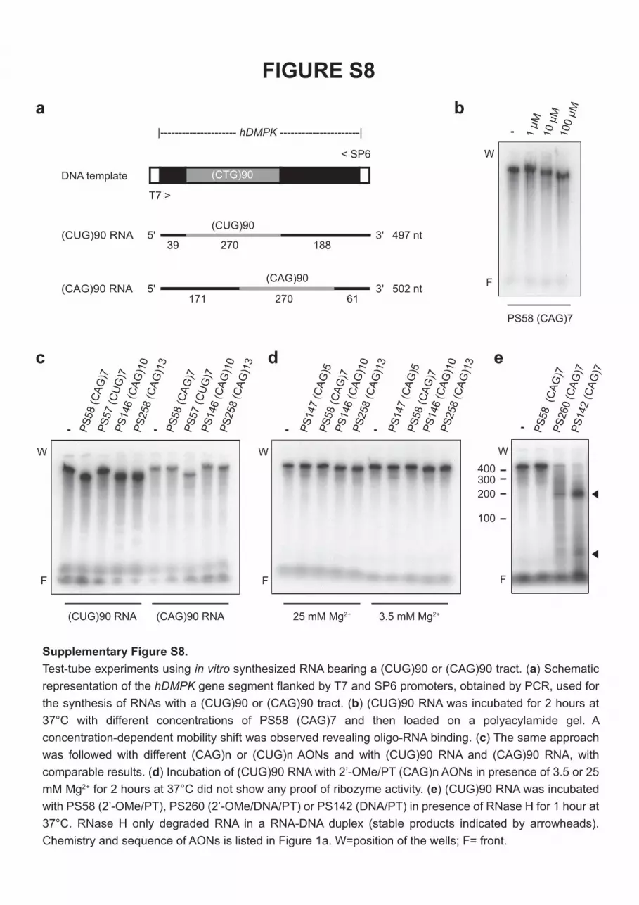
T7 >
< SP6
(CTG)90
497 nt5' 3'
5' 3' 502 nt
(CUG)90
(CAG)90
DNA template
(CUG)90 RNA 39 270 188
|--------------------- hDMPK ----------------------|
171 270 61(CAG)90 RNA
PS
58 (
CA
G)7
PS
258 (
CA
G)1
3
PS
146 (
CA
G)1
0
PS
57 (
CU
G)7
PS
58 (
CA
G)7
PS
258 (
CA
G)1
3
PS
146 (
CA
G)1
0
PS
57 (
CU
G)7
(CUG)90 RNA (CAG)90 RNA
PS
147 (
CA
G)5
P
S58 (
CA
G)7
PS
258 (
CA
G)1
3
PS
146 (
CA
G)1
0
PS
147 (
CA
G)5
P
S58 (
CA
G)7
PS
258 (
CA
G)1
3
PS
146 (
CA
G)1
0
25 mM Mg2+ 3.5 mM Mg2+
a
d ec
b
Supplementary Figure S8.
Test-tube experiments using in vitro synthesized RNA bearing a (CUG)90 or (CAG)90 tract. (a) Schematic
representation of the hDMPK gene segment flanked by T7 and SP6 promoters, obtained by PCR, used for
the synthesis of RNAs with a (CUG)90 or (CAG)90 tract. (b) (CUG)90 RNA was incubated for 2 hours at
37°C with different concentrations of PS58 (CAG)7 and then loaded on a polyacylamide gel. A
concentration-dependent mobility shift was observed revealing oligo-RNA binding. (c) The same approach
was followed with different (CAG)n or (CUG)n AONs and with (CUG)90 RNA and (CAG)90 RNA, with
comparable results. (d) Incubation of (CUG)90 RNA with 2’-OMe/PT (CAG)n AONs in presence of 3.5 or 25
mM Mg2+ for 2 hours at 37°C did not show any proof of ribozyme activity. (e) (CUG)90 RNA was incubated
with PS58 (2’-OMe/PT), PS260 (2’-OMe/DNA/PT) or PS142 (DNA/PT) in presence of RNase H for 1 hour at
37°C. RNase H only degraded RNA in a RNA-DNA duplex (stable products indicated by arrowheads).
Chemistry and sequence of AONs is listed in Figure 1a. W=position of the wells; F= front.
FIGURE S8
- - - -
F
W
F
W
100
200
300400
PS
58 (C
AG
)7
PS
142 (
CA
G)7
PS
260 (
CA
G)7
-
F
W
1 µ
M10 µ
M100 µ
M
F
-
W
PS58 (CAG)7

100 100200 200300 3000 00
15
5
10
0
10
20
30
40
50
60 Positive
control
PS147
PS58
PS146
PS260
PS142
PS261
AON [ g/ml] AON [ g/ml]
Bb
[
g/m
l]
C3
a [
g/m
l]
a
100 100200 200300 3000 0
00
2
4
6
AON [ g/ml] AON [ g/ml]
Bb
[
g/m
l]
C3
a [
g/m
l]
8
10
1
2
3
4
b
Supplementary Figure S9.
Complement activation assay. Complement activation by a selection of AONs was examined in
plasma from (a) cynomolgus monkeys or (b) humans. Formation of split products Bb (left
panels) and C3a (right panels) was determined by ELISA. See Figure 1a for AON sequence
and chemistry. ISIS 5132 was used as a positive control (Rudin et al. (2001) Clin. Cancer Res.,
7, 1214-1220).
FIGURE S9
Cynomolgus monkey plasma
Human plasma

TABLE S1
SiRNAs (RNA duplexes) used in this study.
PI-01:
5’-CAGCAGCAGCAGCAGCAGCAG-3’ + 5'-GCUGCUGCUGCUGCUGCUGCU -3'
PI-02:
5’-GCAGCAGCAGCAGCAGCAGCA-3’ + 5’-CUGCUGCUGCUGCUGCUGCUG-3’
Control:
5’-CAAGAACGAUUUGCUGACATT-3’ + 5’-UGUCAGCAAAUCGUUCUUGTT-3'
Capital: DNA. Capital, underlined: RNA.

TABLE S2
PCR primers used in this study (5' -> 3'):
qPCR:
mActb Fw GCTCTGGCTCCTAGCACCAT
mActb Rv GCCACCGATCCACACAGAGT
mGapdh Fw GTCGGTGTGAACGGATTTG
mGapdh Rv GAACATGTAGACCATGTAGTTG
mPtbp1 (5’) Fw GCATAATGTCAGCCTCTCCGTAT
mPtbp1 (5’) Rv CTCCATGGACATTAGGGACAGAG
mPtbp1 (3’) Fw TGTCCTTTTGGTCAGCAATCTG
mPtbp1 (3’) Rv ACATCACCGTAGACGCCGA
mTxlnb (3’) Fw TTCCTGCTTAGCTCCCTTCCT
mTxlnb (3’) Rv GAGTTGGGTGCTCCACTGAGAG
mSec24b (5’) Fw GGCACCGCCGCAGCAGAA
mSec24b (5’) Rv TGATGAGGCAGCCCATACCCAGAT
mSec24b (3’) Fw GATCCTTGGCTACAGCGAACAGC
mSec24b (3’) Rv AACTGGGTGACTTGGAGCAGGT
mHcn1 (5’) Fw GAGACAGTTGCTATTGACCGACTCG
mHcn1 (5’) Rv GTCATGCTTCACGATCTGCTTCAGG
mNr3c1 (5’) Fw AGTCCCCAGCAGTTTGCTTGGC
mNr3c1 (5’) Rv CGCAGAAACCTTGACTGTAGCTCCA
mNr3c1 (3’) Fw AACTTCGCAGGCCGCTCAGTGT
mNr3c1 (3’) Rv CAGGCAGAGTTTGGGAGGTGGTC
mMapkap1 (5’) Fw GCACCTGTACTTCGAGTCCGA
mMapkap1 (5’) Rv CTCGCTCGTGATTCCAGGAT
mMapkap1 (3’) Fw AGGGAGAAGGAGAGCAAGCC
mMapkap1 (3’) Rv GCATCTGGTGTAGACAGAGGTGTG
hDMPK e1-e2 Fw ACTGGCCCAGGACAAGTACG
hDMPK e1-e2 Rv CCTCCTTAAGCCTCACCACG
hDMPK e4-e5 Fw GACCGGCGGTGGATCACG
hDMPK e4-e5 Rv GGTCCCCGCCCACGTAATAC
hDMPK e15(5’) Fw AGAACTGTCTTCGACTCCGGG
hDMPK e15(5’) Rv TCGGAGCGGTTGTGAACTG
hDMPK e15(3’) Fw TGCCTGCTTACTCGGGAAATT
hDMPK e15(3’) Rv GAGCAGCGCAAGTGAGGAG
hACTB Fw CGGGCCGTCTTCCCCTCCAT
hACTB Rv TGGGCCTCGTCGCCCACATA
hGAPDH Fw CCCGCTTCGCTCTCTGCTCC
hGAPDH Rv CCTTCCCCATGGTGTCTGAGCG
hMAP3K4 e16-e17 Fw TCGGAACAGCCCCCGTCCTATGA
hMAP3K4 e16-e17 Rv CCGCGCGTCGGAAGGCAT
hMAP3K4 e17-e18 Fw CCCTCTGGTGGTGACTCTGTGCT
hMAP3K4 e17-e18 Rv GGGCTTGAGTGACGACTCAGGGAC
hMAP3K4 e3-e4 Fw TGCAAAGGGCACGTATAGCATTGG
hMAP3K4 e3-e4 Rv GCTCCTCCCACGACACAGCA
hBPGM (3’) Fw AGGGCACTCCTAAAACACCTGGAAG
hBPGM (3’) Rv CCAACAGCACGCAGGTTTTCATCC
Primers for mGapdh were described earlier (Fu et al. (2007) J. Biomed. Mater. Res. A, 83, 770-778).
Semi-quantitative PCR:
Primers for Ptbp1, Txlnb and Mapkap1 were described earlier (Mulders et al. (2009) Proc. Natl. Acad.
Sci. USA, 106, 13915-13920).