glycosphingolipid synthesis requires fapp2 transfer of glucosylceramide
TRANSCRIPT
ARTICLES
Glycosphingolipid synthesis requiresFAPP2 transfer of glucosylceramideGiovanni D’Angelo1, Elena Polishchuk1, Giuseppe Di Tullio1, Michele Santoro1, Antonella Di Campli1, Anna Godi1{,Gun West2, Jacek Bielawski3, Chia-Chen Chuang4, Aarnoud C. van der Spoel4, Frances M. Platt4, Yusuf A. Hannun3,Roman Polishchuk1, Peter Mattjus2 & Maria Antonietta De Matteis1
The molecular machinery responsible for the generation of transport carriers moving from the Golgi complex to the plasmamembrane relies on a tight interplay between proteins and lipids. Among the lipid-binding proteins of this machinery, wepreviously identified the four-phosphate adaptor protein FAPP2, the pleckstrin homology domain of which bindsphosphatidylinositol 4-phosphate and the small GTPase ARF1. FAPP2 also possesses a glycolipid-transfer-protein homologydomain. Here we show that human FAPP2 is a glucosylceramide-transfer protein that has a pivotal role in the synthesisof complex glycosphingolipids, key structural and signalling components of the plasma membrane. The requirement forFAPP2 makes the whole glycosphingolipid synthetic pathway sensitive to regulation by phosphatidylinositol 4-phosphateand ARF1. Thus, by coupling the synthesis of glycosphingolipids with their export to the cell surface, FAPP2 emerges ascrucial in determining the lipid identity and composition of the plasma membrane.
The Golgi complex (GC) is responsible for processing, trafficking andsorting of proteins and lipids synthesized in the endoplasmic reticu-lum (ER). The trans-Golgi network (TGN) is the major sorting com-partment of the GC, where cargoes are packaged into distinct carriersfor transport to their final destinations. Our present understandingof the formation of carriers moving from the TGN to the plasmamembrane (PM) is limited1,2. Several proteins involved in their bio-genesis act through a tight interplay with lipids1–3; among these weidentified FAPP1 and FAPP2 (ref. 4). The FAPPs are recruited to theTGN through their pleckstrin homology (PH) domain, which bindsphosphatidylinositol 4-phosphate (PtdIns4P), and the small GTPaseARF1. The FAPPs are involved specifically in transport from the TGNto the PM4,5 and control cilium formation in polarized cells6.
The mechanisms of action of the FAPPs remain to be defined.FAPP2 has sequence similarity to glycolipid-transfer protein(GLTP)4,7, a protein able to mediate the intermembrane transfer ofglycosphingolipids (GSLs) in vitro8. GSLs are important componentsof the PM, with key roles in cell signalling, adhesion, proliferationand differentiation9.
GSL synthesis starts in the ER from ceramide. Ceramide is glyco-sylated to glucosylceramide (GlcCer) on the cytosolic leaflet of cis-Golgi membranes by GlcCer synthase (GCS)10. GlcCer has then to betransported to late-Golgi compartments and translocated to theluminal leaflet, because all further glycosylation steps leading to com-plex GSLs take place in the lumen of these compartments11,12. Thus,its presence on the cytosolic face of the GC gives GlcCer the correcttopology to be ‘intercepted’ by cytosolic proteins that can bind toand/or transfer it. The transfer of GlcCer by cytosolic GLTPs wouldpermit the non-vesicular (that is, non-membrane-bound) transportof GlcCer, as opposed to or in addition to its vesicular transport (bymeans of membrane flow).
Here we show that FAPP2 is required for the synthesis of complexGSLs because it mediates the non-vesicular transport of GlcCer to
distal Golgi compartments. This puts the GSL biosynthetic pathwayunder the control of PtdIns4P and the small GTPase ARF1, the twoknown regulators of FAPP2.
FAPP2 is a GlcCer-transfer protein
Despite the limited sequence similarity of the GLTP-homologydomain of FAPP2 to GLTP itself, the residues required for the glyco-lipid transfer activity of GLTP13 are all conserved in FAPP2 (Sup-plementary Fig. 1). Furthermore, homology modelling of FAPP2 onthe structure of GLTP13 produces reliable and significant overlap(Fig. 1a). These considerations prompted us to investigate whetherFAPP2 is indeed a glycolipid-transfer protein.
Figure 1b shows that FAPP2 can transfer dodecanoyl-dipyrrometheneboron difluoride-GlcCer (C12-BODIPY-GlcCer) between 1-palmitoyl-2-oleoyl-sn-glycero-3-phosphocholine (POPC) vesicles in vitro10,whereas a mutated FAPP2 designed on an inactive mutant of GLTP13
(FAPP2-W407A) cannot (Fig. 1a–c).To define the lipid specificity of FAPP2, its ability to transfer
sphingomyelin (SM), phosphatidylcholine and ceramide was exam-ined. As shown in Fig. 1b, c, FAPP2 did not transfer SM, phospha-tidylcholine or ceramide. Because the FAPP PH domain bindsPtdIns4P and ARF1 (ref. 4), we assessed whether these two PH-domain ligands affect the GlcCer transfer activity of FAPP2. Theaddition of PtdIns4P and ARF1 to acceptor (but not to donor) vesi-cles increased the rate of GlcCer transfer by FAPP2 (Fig. 1d).
The FAPP2 lipid-transfer activity for longer and unmodifiedacyl-chain glycolipids was assessed with the use of [14C]stearoyl-GlcCer. As shown in Fig. 1e, the addition of FAPP2 and GLTPproduced substantial transfer of [14C]stearoyl-GlcCer to acceptorvesicles.
These data demonstrate that FAPP2 acts in vitro as a GLTP, trans-ferring both BODIPY-labelled and unmodified long-acyl-chainGlcCer.
1Department of Cell Biology and Oncology, Consorzio Mario Negri Sud, Via Nazionale 8/A, 66030 Santa Maria Imbaro, Chieti, Italy. 2Department of Biochemistry and Pharmacy, AboAkademi University, Artillerigatan 6 A III, BioCity FI-20520 Turku, Finland. 3Department of Biochemistry and Molecular Biology, Medical University of South Carolina, 173 AshleyAvenue, Charleston, South Carolina 29425, USA. 4Department of Pharmacology, University of Oxford, Mansfield Road, Oxford OX1 3QT, UK. {Present address: Section of Cell andMolecular Biology, Institute of Cancer Research Chester Beatty Laboratories, 237 Fulham Road, London SW3 6JB, UK.
Vol 449 | 6 September 2007 | doi:10.1038/nature06097
62Nature ©2007 Publishing Group
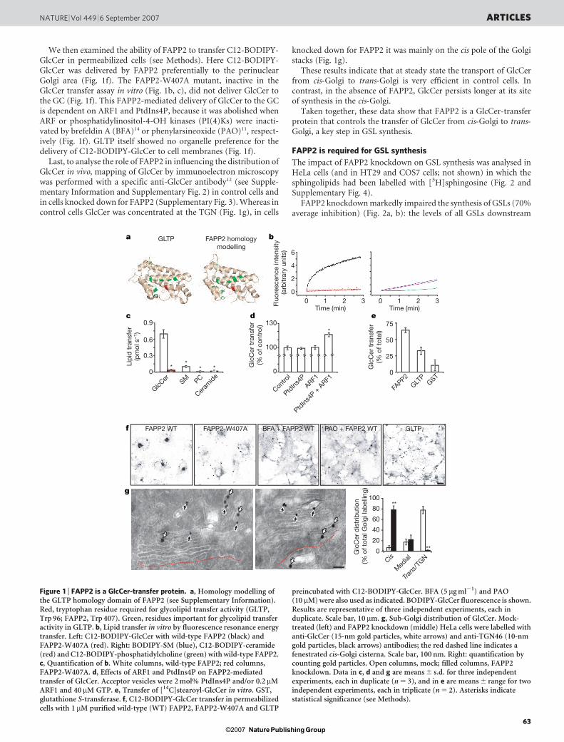
We then examined the ability of FAPP2 to transfer C12-BODIPY-GlcCer in permeabilized cells (see Methods). Here C12-BODIPY-GlcCer was delivered by FAPP2 preferentially to the perinuclearGolgi area (Fig. 1f). The FAPP2-W407A mutant, inactive in theGlcCer transfer assay in vitro (Fig. 1b, c), did not deliver GlcCer tothe GC (Fig. 1f). This FAPP2-mediated delivery of GlcCer to the GCis dependent on ARF1 and PtdIns4P, because it was abolished whenARF or phosphatidylinositol-4-OH kinases (PI(4)Ks) were inacti-vated by brefeldin A (BFA)14 or phenylarsineoxide (PAO)11, respect-ively (Fig. 1f). GLTP itself showed no organelle preference for thedelivery of C12-BODIPY-GlcCer to cell membranes (Fig. 1f).
Last, to analyse the role of FAPP2 in influencing the distribution ofGlcCer in vivo, mapping of GlcCer by immunoelectron microscopywas performed with a specific anti-GlcCer antibody12 (see Supple-mentary Information and Supplementary Fig. 2) in control cells andin cells knocked down for FAPP2 (Supplementary Fig. 3). Whereas incontrol cells GlcCer was concentrated at the TGN (Fig. 1g), in cells
knocked down for FAPP2 it was mainly on the cis pole of the Golgistacks (Fig. 1g).
These results indicate that at steady state the transport of GlcCerfrom cis-Golgi to trans-Golgi is very efficient in control cells. Incontrast, in the absence of FAPP2, GlcCer persists longer at its siteof synthesis in the cis-Golgi.
Taken together, these data show that FAPP2 is a GlcCer-transferprotein that controls the transfer of GlcCer from cis-Golgi to trans-Golgi, a key step in GSL synthesis.
FAPP2 is required for GSL synthesis
The impact of FAPP2 knockdown on GSL synthesis was analysed inHeLa cells (and in HT29 and COS7 cells; not shown) in which thesphingolipids had been labelled with [3H]sphingosine (Fig. 2 andSupplementary Fig. 4).
FAPP2 knockdown markedly impaired the synthesis of GSLs (70%average inhibition) (Fig. 2a, b): the levels of all GSLs downstream
f FAPP2 WT FAPP2-W407A GLTP
GLTP FAPP2 homologymodelling
g
BFA + FAPP2 WT PAO + FAPP2 WT
c
Lip
id t
rans
fer
(pm
ol s
–1)
0
GlcCer
0.3
0.6
0.9
SM PC
Ceram
ide*
** *
d
Glc
Cer
tra
nsfe
r(%
of c
ontr
ol)
0
100
130*
Glc
Cer
dis
trib
utio
n (%
of t
otal
Gol
gi la
bel
ling)
0
20
40
60
80
100**
**
6
4
2
0
Fluo
resc
ence
inte
nsity
(a
rbitr
ary
units
)
ba
e
0G
lcC
er t
rans
fer
(% o
f tot
al)
50
75
25
0 1 2 3Time (min)
0 1 2 3Time (min)
FAPP2
GLTP
GST
Contro
l
PtdIns
4PARF1
PtdIns
4P +
ARF1
Trans
/TGN
Med
ialCis
Figure 1 | FAPP2 is a GlcCer-transfer protein. a, Homology modelling ofthe GLTP homology domain of FAPP2 (see Supplementary Information).Red, tryptophan residue required for glycolipid transfer activity (GLTP,Trp 96; FAPP2, Trp 407). Green, residues important for glycolipid transferactivity in GLTP. b, Lipid transfer in vitro by fluorescence resonance energytransfer. Left: C12-BODIPY-GlcCer with wild-type FAPP2 (black) andFAPP2-W407A (red). Right: BODIPY-SM (blue), C12-BODIPY-ceramide(red) and C12-BODIPY-phosphatidylcholine (green) with wild-type FAPP2.c, Quantification of b. White columns, wild-type FAPP2; red columns,FAPP2-W407A. d, Effects of ARF1 and PtdIns4P on FAPP2-mediatedtransfer of GlcCer. Acceptor vesicles were 2 mol% PtdIns4P and/or 0.2mMARF1 and 40 mM GTP. e, Transfer of [14C]stearoyl-GlcCer in vitro. GST,glutathione S-transferase. f, C12-BODIPY-GlcCer transfer in permeabilizedcells with 1 mM purified wild-type (WT) FAPP2, FAPP2-W407A and GLTP
preincubated with C12-BODIPY-GlcCer. BFA (5 mg ml21) and PAO(10 mM) were also used as indicated. BODIPY-GlcCer fluorescence is shown.Results are representative of three independent experiments, each induplicate. Scale bar, 10 mm. g, Sub-Golgi distribution of GlcCer. Mock-treated (left) and FAPP2 knockdown (middle) HeLa cells were labelled withanti-GlcCer (15-nm gold particles, white arrows) and anti-TGN46 (10-nmgold particles, black arrows) antibodies; the red dashed line indicates afenestrated cis-Golgi cisterna. Scale bar, 100 nm. Right: quantification bycounting gold particles. Open columns, mock; filled columns, FAPP2knockdown. Data in c, d and g are means 6 s.d. for three independentexperiments, each in duplicate (n 5 3), and in e are means 6 range for twoindependent experiments, each in triplicate (n 5 2). Asterisks indicatestatistical significance (see Methods).
NATURE | Vol 449 | 6 September 2007 ARTICLES
63Nature ©2007 Publishing Group
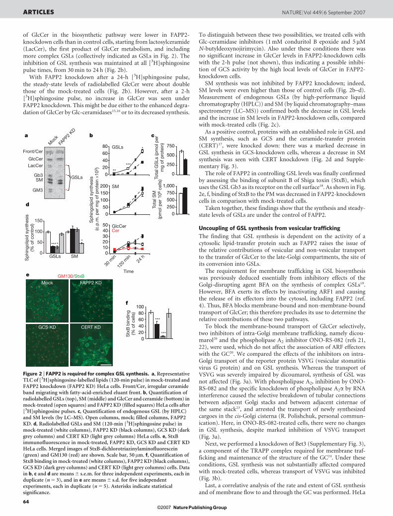
of GlcCer in the biosynthetic pathway were lower in FAPP2-knockdown cells than in control cells, starting from lactosylceramide(LacCer), the first product of GlcCer metabolism, and includingmore complex GSLs (collectively indicated as GSLs in Fig. 2). Theinhibition of GSL synthesis was maintained at all [3H]sphingosinepulse times, from 30 min to 24 h (Fig. 2b).
With FAPP2 knockdown after a 24-h [3H]sphingosine pulse,the steady-state levels of radiolabelled GlcCer were about doublethose of the mock-treated cells (Fig. 2b). However, after a 2-h[3H]sphingosine pulse, no increase in GlcCer was seen underFAPP2 knockdown. This might be due either to the enhanced degra-dation of GlcCer by Glc-ceramidases15,16 or to its decreased synthesis.
To distinguish between these two possibilities, we treated cells withGlc-ceramidase inhibitors (1 mM conduritol B epoxide and 5mMN-butyldeoxynojirimycin). Also under these conditions there wasno significant increase in GlcCer levels in FAPP2-knockdown cellswith the 2-h pulse (not shown), thus indicating a possible inhibi-tion of GCS activity by the high local levels of GlcCer in FAPP2-knockdown cells.
SM synthesis was not inhibited by FAPP2 knockdown; indeed,SM levels were even higher than those of control cells (Fig. 2b–d).Measurement of endogenous GSLs (by high-performance liquidchromatography (HPLC)) and SM (by liquid chromatography–massspectrometry (LC–MS)) confirmed both the decrease in GSL levelsand the increase in SM levels in FAPP2-knockdown cells, comparedwith mock-treated cells (Fig. 2c).
As a positive control, proteins with an established role in GSL andSM synthesis, such as GCS and the ceramide-transfer protein(CERT)17, were knocked down: there was a marked decrease inGSL synthesis in GCS-knockdown cells, whereas a decrease in SMsynthesis was seen with CERT knockdown (Fig. 2d and Supple-mentary Fig. 3).
The role of FAPP2 in controlling GSL levels was finally confirmedby assessing the binding of subunit B of Shiga toxin (StxB), whichuses the GSL Gb3 as its receptor on the cell surface18. As shown in Fig.2e, f, binding of StxB to the PM was decreased in FAPP2-knockdowncells in comparison with mock-treated cells.
Taken together, these findings show that the synthesis and steady-state levels of GSLs are under the control of FAPP2.
Uncoupling of GSL synthesis from vesicular trafficking
The finding that GSL synthesis is dependent on the activity of acytosolic lipid-transfer protein such as FAPP2 raises the issue ofthe relative contributions of vesicular and non-vesicular transportto the transfer of GlcCer to the late-Golgi compartments, the site ofits conversion into GSLs.
The requirement for membrane trafficking in GSL biosynthesiswas previously deduced essentially from inhibitory effects of theGolgi-disrupting agent BFA on the synthesis of complex GSLs19.However, BFA exerts its effects by inactivating ARF1 and causingthe release of its effectors into the cytosol, including FAPP2 (ref.4). Thus, BFA blocks membrane-bound and non-membrane-boundtransport of GlcCer; this therefore precludes its use to determine therelative contributions of these two pathways.
To block the membrane-bound transport of GlcCer selectively,two inhibitors of intra-Golgi membrane trafficking, namely dicou-marol20 and the phospholipase A2 inhibitor ONO-RS-082 (refs 21,22), were used, which do not affect the association of ARF effectorswith the GC20. We compared the effects of the inhibitors on intra-Golgi transport of the reporter protein VSVG (vesicular stomatitisvirus G protein) and on GSL synthesis. Whereas the transport ofVSVG was severely impaired by dicoumarol, synthesis of GSL wasnot affected (Fig. 3a). With phospholipase A2, inhibition by ONO-RS-082 and the specific knockdown of phospholipase A2a by RNAinterference caused the selective breakdown of tubular connectionsbetween adjacent Golgi stacks and between adjacent cisternae ofthe same stack23, and arrested the transport of newly synthesizedcargoes in the cis-Golgi cisterna (R. Polishchuk, personal commun-ication). Here, in ONO-RS-082-treated cells, there were no changesin GSL synthesis, despite marked inhibition of VSVG transport(Fig. 3a).
Next, we performed a knockdown of Bet3 (Supplementary Fig. 3),a component of the TRAPP complex required for membrane traf-ficking and maintenance of the structure of the GC24. Under theseconditions, GSL synthesis was not substantially affected comparedwith mock-treated cells, whereas transport of VSVG was inhibited(Fig. 3b).
Last, a correlative analysis of the rate and extent of GSL synthesisand of membrane flow to and through the GC was performed. HeLa
***
*
a
Front/Cer
GlcCerLacCer
Gb3SM
GM3
d
0
50
100
150
Sp
hing
olip
id s
ynth
esis
(% o
f con
trol
)
GSLs
Time
SM
e
f
Mock FAPP2 KD
GCS KD CERT KD
GM130/StxB
0204060
100
Stx
B b
ind
ing
(% o
f cel
ls) 80
b
*
*
* GSLs0
20
40
60
80
**
GSLs
*** **
***
***
Tota
l GS
Ls (p
mol
per
mg
of p
rote
in)
c
0
250
500
750
Sp
hing
olip
id s
ynth
esis
(c
.p.m
. per
mg
of p
rote
in ×
103 )
01020304050 GlcCer
Cer
0
50
100
150
200 SM
Tota
l SM
(pm
ol p
er 1
06 c
ells
)
0
250
500
750
1,000
FAPP2
KD
24 h
120
min
30 m
in
Moc
k
Figure 2 | FAPP2 is required for complex GSL synthesis. a, RepresentativeTLC of [3H]sphingosine-labelled lipids (120-min pulse) in mock-treated andFAPP2 knockdown (FAPP2 KD) HeLa cells. Front/Cer, irregular ceramideband migrating with fatty-acid-enriched eluant front. b, Quantification ofradiolabelled GSLs (top), SM (middle) and GlcCer and ceramide (bottom) inmock-treated (open squares) and FAPP2 KD (filled squares) HeLa cells after[3H]sphingosine pulses. c, Quantification of endogenous GSL (by HPLC)and SM levels (by LC–MS). Open columns, mock; filled columns, FAPP2KD. d, Radiolabelled GSLs and SM (120-min [3H]sphingosine pulse) inmock-treated (white columns), FAPP2 KD (black columns), GCS KD (darkgrey columns) and CERT KD (light grey columns) HeLa cells. e, StxBimmunofluorescence in mock-treated, FAPP2 KD, GCS KD and CERT KDHeLa cells. Merged images of StxB-dichlorotriazinylaminofluorescein(green) and GM130 (red) are shown. Scale bar, 50 mm. f, Quantification ofStxB binding in mock-treated (white columns), FAPP2 KD (black columns),GCS KD (dark grey columns) and CERT KD (light grey columns) cells. Datain b, c and d are means 6 s.e.m. for three independent experiments, each induplicate (n 5 3), and in e are means 6 s.d. for five independentexperiments, each in duplicate (n 5 5). Asterisks indicate statisticalsignificance.
ARTICLES NATURE | Vol 449 | 6 September 2007
64Nature ©2007 Publishing Group
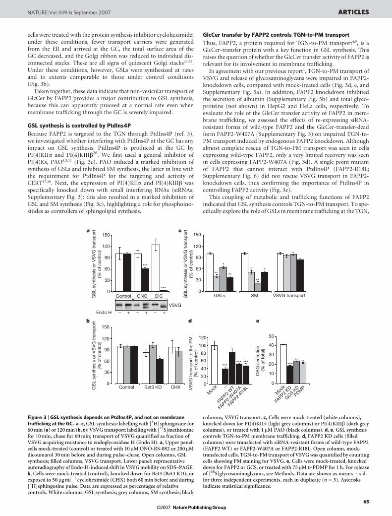
cells were treated with the protein synthesis inhibitor cycloheximide;under these conditions, fewer transport carriers were generatedfrom the ER and arrived at the GC, the total surface area of theGC decreased, and the Golgi ribbon was reduced to individual dis-connected stacks. These are all signs of quiescent Golgi stacks23,25.Under these conditions, however, GSLs were synthesized at ratesand to extents comparable to those under control conditions(Fig. 3b).
Taken together, these data indicate that non-vesicular transport ofGlcCer by FAPP2 provides a major contribution to GSL synthesis,because this can apparently proceed at a normal rate even whenmembrane trafficking through the GC is severely impaired.
GSL synthesis is controlled by PtdIns4P
Because FAPP2 is targeted to the TGN through PtdIns4P (ref. 3),we investigated whether interfering with PtdIns4P at the GC has anyimpact on GSL synthesis. PtdIns4P is produced at the GC byPI(4)KIIa and PI(4)KIIIb26. We first used a general inhibitor ofPI(4)Ks, PAO4,5,11 (Fig. 3c). PAO induced a marked inhibition ofsynthesis of GSLs and inhibited SM synthesis, the latter in line withthe requirement for PtdIns4P for the targeting and activity ofCERT17,26. Next, the expression of PI(4)KIIa and PI(4)KIIIb wasspecifically knocked down with small interfering RNAs (siRNAs;Supplementary Fig. 3): this also resulted in a marked inhibition ofGSL and SM synthesis (Fig. 3c), highlighting a role for phosphoino-sitides as controllers of sphingolipid synthesis.
GlcCer transfer by FAPP2 controls TGN-to-PM transport
Thus, FAPP2, a protein required for TGN-to-PM transport4,5, is aGlcCer-transfer protein with a key function in GSL synthesis. Thisraises the question of whether the GlcCer transfer activity of FAPP2 isrelevant for its involvement in membrane trafficking.
In agreement with our previous report4, TGN-to-PM transport ofVSVG and release of glycosaminoglycans were impaired in FAPP2-knockdown cells, compared with mock-treated cells (Fig. 3d, e, andSupplementary Fig. 5a). In addition, FAPP2 knockdown inhibitedthe secretion of albumin (Supplementary Fig. 5b) and total glyco-proteins (not shown) in HepG2 and HeLa cells, respectively. Toevaluate the role of the GlcCer transfer activity of FAPP2 in mem-brane trafficking, we assessed the effects of re-expressing siRNA-resistant forms of wild-type FAPP2 and the GlcCer-transfer-deadform FAPP2-W407A (Supplementary Fig. 3) on impaired TGN-to-PM transport induced by endogenous FAPP2 knockdown. Althoughalmost complete rescue of TGN-to-PM transport was seen in cellsexpressing wild-type FAPP2, only a very limited recovery was seenin cells expressing FAPP2-W407A (Fig. 3d). A single point mutantof FAPP2 that cannot interact with PtdIns4P (FAPP2-R18L;Supplementary Fig. 6) did not rescue VSVG transport in FAPP2-knockdown cells, thus confirming the importance of PtdIns4P incontrolling FAPP2 activity (Fig. 3e).
This coupling of metabolic and trafficking functions of FAPP2indicated that GSL synthesis controls TGN-to-PM transport. To spe-cifically explore the role of GSLs in membrane trafficking at the TGN,
a
Control ONO DICGS
L sy
nthe
sis
or V
SV
G t
rans
por
t(%
of c
ontr
ol)
GS
L sy
nthe
sis
or V
SV
G t
rans
por
t(%
of c
ontr
ol)
GS
L sy
nthe
sis
or V
SV
G t
rans
por
t(%
of c
ontr
ol)
0
30
60
90
120
150
VSVG
Endo H – + – + – +
***
***
Control CHXBet3 KD0
30
60
90
120
150b
*
c
0
30
60
90
120
150
GSLs
**
*
**
SM
* *
**
VSVG transport
d e
***
0
40
60
80
120
100
20
VS
VG
tra
nsp
ort
to t
he P
M(%
of c
ontr
ol)
GA
G s
ecre
tion
(% o
f tot
al)
****** **
0
10
20
30
40
50
*****
Moc
kM
ock
FAPP2
WT
FAPP2
KD
FAPP2-
W40
7A
GCS KD
PDMP
FAPP2-
R18L–
Figure 3 | GSL synthesis depends on PtdIns4P, and not on membranetrafficking at the GC. a–c, GSL synthesis: labelling with [3H]sphingosine for60 min (a) or 120 min (b, c); VSVG transport: labelling with [35S]methioninefor 10 min, chase for 60 min; transport of VSVG quantified as fraction ofVSVG acquiring resistance to endoglycosidase H (Endo H). a, Upper panel:cells mock-treated (control) or treated with 10mM ONO-RS-082 or 200mMdicoumarol 30 min before and during pulse–chase. Open columns, GSLsynthesis; filled columns, VSVG transport. Lower panel: representativeautoradiography of Endo-H-induced shift in VSVG mobility on SDS–PAGE.b, Cells were mock-treated (control), knocked down for Bet3 (Bet3 KD), orexposed to 50mg ml21 cycloheximide (CHX) both 60 min before and during[3H]sphingosine pulse. Data are expressed as percentages of relativecontrols. White columns, GSL synthesis; grey columns, SM synthesis; black
columns, VSVG transport. c, Cells were mock-treated (white columns),knocked down for PI(4)KIIa (light grey columns) or PI(4)KIIIb (dark greycolumns), or treated with 1mM PAO (black columns). d, e, GSL synthesiscontrols TGN-to-PM membrane trafficking. d, FAPP2 KD cells (filledcolumns) were transfected with siRNA-resistant forms of wild-type FAPP2(FAPP2 WT) or FAPP2-W407A or FAPP2-R18L. Open column, mock-transfected cells. TGN-to-PM transport of VSVG was quantified by countingcells showing PM staining for VSVG. e, Cells were mock-treated, knockeddown for FAPP2 or GCS, or treated with 75mM D-PDMP for 1 h. For releaseof [35S]glycosaminoglycans, see Methods. Data are shown as means 6 s.d.for three independent experiments, each in duplicate (n 5 3). Asterisksindicate statistical significance.
NATURE | Vol 449 | 6 September 2007 ARTICLES
65Nature ©2007 Publishing Group
GSL synthesis was inhibited by GCS knockdown (SupplementaryFig. 3). The TGN-to-PM transport of glycosaminoglycans in GCS-knockdown cells was compared with control cells: it was impairedto a similar extent to that seen in FAPP2-knockdown cells and incells treated with the GCS inhibitor phenyl-2-decanoyl-amino-3-morpholino-1-propanol-hydrochloride27 (PDMP; Fig. 3e).
In agreement with our previous reports4, impairment of membranetrafficking by FAPP2 knockdown was through specific inhibition ofTGN-to-PM transport, and did not involve other Golgi functions4
(Supplementary Fig. 5c, d). In addition, the ultrastructural organiza-tion of Golgi stacks was preserved in FAPP2-knockdown cells, withregular, although shorter, cisternae. However, the TGN was less deve-loped and trans cisternae appeared swollen in FAPP2-knockdowncells (as well as in GCS-knockdown cells; Supplementary Fig. 7 andSupplementary Movies 1–3).
Taken together, our findings demonstrate that the GlcCertransfer activity of FAPP2 and GCS control TGN-to-PM transport,providing further evidence linking GSL metabolism to membranetrafficking27–32.
Discussion
We show here that FAPP2, a protein binding PtdIns4P and ARF1 thatcontrols TGN-to-PM trafficking, is an essential component of the
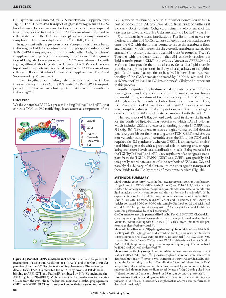
GSL synthetic machinery, because it mediates non-vesicular trans-port of the common GSL precursor GlcCer from its site of synthesis atthe early Golgi to distal Golgi compartments, where most of theenzymes involved in complex GSLs assembly are located33 (Fig. 4).
Our findings have many implications. The first is that newly syn-thesized proteins and GlcCer can use different transport pathways tocross the GC, with the former bound to move via membrane flow,and the latter, which is present in the cytosolic membrane leaflet, alsoamenable for cytosolic transport via lipid-transfer proteins (Fig. 4a).Together with the demonstration that SM synthesis requires thelipid-transfer protein CERT17 (previously known as GPBPD26 (ref.34)), our data provide the most direct evidence that lipid-transferproteins occupy key positions in the synthetic pathway of the sphin-golipids. An issue that remains to be solved is how cis-to-trans vec-toriality of the GlcCer transfer operated by FAPP2 is achieved. Theenrichment of PtdIns4P in TGN membranes4 is likely to be importantin this process.
Another important implication is that our data reveal a previouslyunrecognized and key component of the molecular machineryresponsible for generation of the lipid identity of the PM. Indeed,although connected by intense bidirectional membrane trafficking,the PM–endosome–TGN and the early-Golgi–ER membrane systemshave completely distinct lipid compositions, with the former highlyenriched in GSLs, SM and cholesterol compared with the latter9.
The precursors of GSLs, SM and cholesterol itself, are the ligandsfor the family of lipid-binding proteins to which FAPP2 belongs,which includes CERT and oxysterol-binding protein 1 (OSBP1; ref.35) (Fig. 5b). These members share a highly conserved PH domainthat is responsible for their targeting to the TGN. CERT mediates thenon-vesicular transport of ceramide from the ER to the TGN and isrequired for SM synthesis17, whereas OSBP1 is an oxysterol–choles-terol-binding protein with a proposed role in sensing and/or regu-lating cholesterol levels and distribution in cells. Being recruited tothe TGN by PtdIns4P and ARF1, key regulators of anterograde trans-port from the TGN36, FAPP2, CERT and OSBP1 can spatially andtemporally coordinate and couple the synthesis of GSLs and SM, andpossibly the delivery of cholesterol, to the anterograde transport ofthese lipids to the PM by means of membrane carriers (Fig. 5b).
METHODS SUMMARYLipid transfer assays in vitro. In the fluorescence resonance energy transfer assay,
10mg of proteins, C12-BODIPY-lipids (1 mol%) and DiI-C18 (1,19-dioctadecyl-
3,3,39,39-tetramethylindodicarbocyanine, perchlorate) were used to monitor the
lipid-transfer activity in continuous real time, as described previously10. In the
experiments using ARF1 and PtdIns4P, donor vesicles contained 2 mol% PtdIns,
3 mol% DiI-C18, 0.5 mol% BODIPY-GlcCer and 94.5 mol% POPC. Acceptor
vesicles contained POPC or POPC with 2 mol% PtdIns4P or 0.2mM ARF1 and
40mM GTP. The lipid transfer assay with [14C]stearoyl-GlcCer and 1 mM pro-
teins was performed as described previously37.
GlcCer transfer assay in permeabilized cells. The C12-BODIPY-GlcCer deliv-
ery assay in streptolysin-O-permeabilized cells was performed as described in
Methods. Protein loading with C-12-BODIPY-GlcCer from lipid films was per-
formed as described previously37.
Metabolic labelling with [3H]sphingosine and sphingolipid analysis. Metabolic
labelling with [3H]sphingosine, GSL extraction and high-performance thin-layer
chromatography (HPTLC) were performed as described38. HPTLC plates were
analysed by using a Raytest TLC Analyser V2.15, and then imaged with a Fujifilm
BAS 1800-II phosphor imaging system. Endogenous sphingolipids were analysed
by HPLC and LC–MS, as described39,40.
Membrane trafficking assays. Transport of the temperature-sensitive mutant of
VSVG (ts045-VSVG) and [35S]glycosaminoglycan secretion were assessed as
described previously41,42. ts045-VSVG transport to the PM was evaluated by ana-
lysing the PM staining of at least 200 cells after 30 min of release from a 20 uCtemperature block. Albumin secretion was assessed by immunoprecipitating
radiolabelled albumin from medium or cell lysates of HepG2 cells pulsed with
[35S]methionine for 5 min and chased for 20 min, as described previously43.
Immunolocalization of endogenous GlcCer. Ultrathin cell cryosectioning was
performed at 4 uC, as described44. Morphometric analysis was performed as
described previously45.
a b
ARF1GTP
PI(4)KIIIβ
PHARF1GTP
PH
FAPP2
GCS
PH
PH
PH
PH
OSBP1
FAPP2
PH
PH
CERT
ER
GSLs
PtdIns4P
GlcCer
CeramideCholesterolSM
FAPP2
Figure 4 | Model of FAPP2 mechanism of action. Schematic diagram of themechanism of action and regulation of FAPP2 (a) and other lipid-transferproteins (b) at the GC. See the text and Supplementary Discussion fordetails. Inset: FAPP2 is recruited to the TGN by means of PH-domainbinding to ARF1-GTP and PtdIns4P (produced by PI(4)Ks, including theARF1-regulated PI(4)KIIIb). Violet arrow, GlcCer translocator transferringGlcCer from the cytosolic to the luminal membrane leaflet; grey square inCERT and OSBP1, FFAT motif responsible for their targeting to the ER.
ARTICLES NATURE | Vol 449 | 6 September 2007
66Nature ©2007 Publishing Group
Full Methods and any associated references are available in the online version ofthe paper at www.nature.com/nature.
Received 20 June; accepted 20 July 2007.Published online 8 August; corrected 6 September 2007.
1. Bard, F. & Malhotra, V. The formation of TGN-to-plasma-membrane transportcarriers. Annu. Rev. Cell Dev. Biol. 22, 439–455 (2006).
2. Luini, A., Ragnini-Wilson, A., Polishchuk, R. S. & De Matteis, M. A. Largepleiomorphic traffic intermediates in the secretory pathway. Curr. Opin. Cell Biol.17, 353–361 (2005).
3. De Matteis, M. A. & Godi, A. Protein–lipid interactions in membrane trafficking atthe Golgi complex. Biochim. Biophys. Acta 1666, 264–274 (2004).
4. Godi, A. et al. FAPPs control Golgi-to-cell-surface membrane traffic by binding toARF and PtdIns(4)P. Nature Cell Biol. 6, 393–404 (2004).
5. Vieira, O. V., Verkade, P., Manninen, A. & Simons, K. FAPP2 is involved in thetransport of apical cargo in polarized MDCK cells. J. Cell Biol. 170, 521–526(2005).
6. Vieira, O. V. et al. FAPP2, cilium formation, and compartmentalization of theapical membrane in polarized Madin–Darby canine kidney (MDCK) cells. Proc.Natl Acad. Sci. USA 103, 18556–18561 (2006).
7. Levine, T. P. & Munro, S. Targeting of Golgi-specific pleckstrin homology domainsinvolves both PtdIns 4-kinase-dependent and -independent components. Curr.Biol. 12, 695–704 (2002).
8. Brown, R. E. & Mattjus, P. Glycolipid transfer proteins. Biochim. Biophys. Acta 1771,746–760 (2007).
9. Holthuis, J. C., Pomorski, T., Raggers, R. J., Sprong, H. & Van Meer, G. Theorganizing potential of sphingolipids in intracellular membrane transport. Physiol.Rev. 81, 1689–1723 (2001).
10. Nylund, M. et al. Molecular features of phospholipids that affect glycolipidtransfer protein-mediated galactosylceramide transfer between vesicles. Biochim.Biophys. Acta 1758, 807–812 (2006).
11. Wiedemann, C., Schafer, T. & Burger, M. M. Chromaffin granule-associatedphosphatidylinositol 4-kinase activity is required for stimulated secretion. EMBOJ. 15, 2094–2101 (1996).
12. Brade, L., Vielhaber, G., Heinz, E. & Brade, H. In vitro characterization of anti-glucosylceramide rabbit antisera. Glycobiology 10, 629–636 (2000).
13. Malinina, L., Malakhova, M. L., Teplov, A., Brown, R. E. & Patel, D. J. Structuralbasis for glycosphingolipid transfer specificity. Nature 430, 1048–1053 (2004).
14. Klausner, R. D., Donaldson, J. G. & Lippincott-Schwartz, J. Brefeldin A: insights intothe control of membrane traffic and organelle structure. J. Cell Biol. 116, 1071–1080(1992).
15. Boot, R. G. et al. Identification of the non-lysosomal glucosylceramidase asb-glucosidase 2. J. Biol. Chem. 282, 1305–1312 (2007).
16. Yildiz, Y. et al. Mutation of b-glucosidase 2 causes glycolipid storage disease andimpaired male fertility. J. Clin. Invest. 116, 2985–2994 (2006).
17. Hanada, K. et al. Molecular machinery for non-vesicular trafficking of ceramide.Nature 426, 803–809 (2003).
18. Mallard, F. et al. Direct pathway from early/recycling endosomes to the Golgiapparatus revealed through the study of Shiga toxin B-fragment transport. J. CellBiol. 143, 973–990 (1998).
19. Young, W. W. Jr, Lutz, M. S., Mills, S. E. & Lechler-Osborn, S. Use of brefeldin A todefine sites of glycosphingolipid synthesis: GA2/GM2/GD2 synthase is trans tothe brefeldin A block. Proc. Natl Acad. Sci. USA 87, 6838–6842 (1990).
20. Mironov, A. A. et al. Dicumarol, an inhibitor of ADP-ribosylation of CtBP3/BARS,fragments golgi non-compact tubular zones and inhibits intra-golgi transport. Eur.J. Cell Biol. 83, 263–279 (2004).
21. Brown, W. J., Chambers, K. & Doody, A. Phospholipase A2 (PLA2) enzymes inmembrane trafficking: mediators of membrane shape and function. Traffic 4,214–221 (2003).
22. Drecktrah, D. & Brown, W. J. Phospholipase A2 antagonists inhibit nocodazole-induced Golgi ministack formation: evidence of an ER intermediate andconstitutive cycling. Mol. Biol. Cell 10, 4021–4032 (1999).
23. Trucco, A. et al. Secretory traffic triggers the formation of tubular continuitiesacross Golgi sub-compartments. Nature Cell Biol. 6, 1071–1081 (2004).
24. Yu, S. et al. mBet3p is required for homotypic COPII vesicle tethering inmammalian cells. J. Cell Biol. 174, 359–368 (2006).
25. Marra, P. et al. The biogenesis of the Golgi ribbon: the roles of membrane inputfrom the ER and of GM130. Mol. Biol. Cell 18, 1595–1608 (2007).
26. Toth, B. et al. Phosphatidylinositol 4-kinase IIIb regulates the transport ofceramide between the endoplasmic reticulum and Golgi. J. Biol. Chem. 281,36369–36377 (2006).
27. Rosenwald, A. G., Machamer, C. E. & Pagano, R. E. Effects of a sphingolipidsynthesis inhibitor on membrane transport through the secretory pathway.Biochemistry 31, 3581–3590 (1992).
28. Sprong, H. et al. Glycosphingolipids are required for sorting melanosomal proteinsin the Golgi complex. J. Cell Biol. 155, 369–380 (2001).
29. Schwarz, A. & Futerman, A. H. Distinct roles for ceramide and glucosylceramideat different stages of neuronal growth. J. Neurosci. 17, 2929–2938 (1997).
30. Boldin, S. A. & Futerman, A. H. Up-regulation of glucosylceramide synthesis uponstimulation of axonal growth by basic fibroblast growth factor. Evidence for
post-translational modification of glucosylceramide synthase. J. Biol. Chem. 275,9905–9909 (2000).
31. Chang, M. C., Wisco, D., Ewers, H., Norden, C. & Winckler, B. Inhibition ofsphingolipid synthesis affects kinetics but not fidelity of L1/NgCAM transportalong direct but not transcytotic axonal pathways. Mol. Cell. Neurosci. 31, 525–538(2006).
32. Tamboli, I. Y. et al. Inhibition of glycosphingolipid biosynthesis reduces secretionof the b-amyloid precursor protein and amyloid b-peptide. J. Biol. Chem. 280,28110–28117 (2005).
33. Lannert, H., Gorgas, K., Meissner, I., Wieland, F. T. & Jeckel, D. Functionalorganization of the Golgi apparatus in glycosphingolipid biosynthesis.Lactosylceramide and subsequent glycosphingolipids are formed in the lumen ofthe late Golgi. J. Biol. Chem. 273, 2939–2946 (1998).
34. Raya, A. et al. Goodpasture antigen-binding protein, the kinase thatphosphorylates the Goodpasture antigen, is an alternatively spliced variantimplicated in autoimmune pathogenesis. J. Biol. Chem. 275, 40392–40399(2000).
35. De Matteis, M. A., Di Campli, A. & D’Angelo, G. Lipid-transfer proteins inmembrane trafficking at the Golgi complex. Biochim. Biophys. Acta 1771, 761–768(2007).
36. De Matteis, M. A. & Godi, A. PI-loting membrane traffic. Nature Cell Biol. 6,487–492 (2004).
37. Malakhova, M. L. et al. Point mutational analysis of the liganding site in humanglycolipid transfer protein. Functionality of the complex. J. Biol. Chem. 280,26312–26320 (2005).
38. Sala, G., Dupre, T., Seta, N., Codogno, P. & Ghidoni, R. Increased biosynthesis ofglycosphingolipids in congenital disorder of glycosylation Ia (CDG-Ia) fibroblasts.Pediatr. Res. 52, 645–651 (2002).
39. Wing, D. R. et al. High-performance liquid chromatography analysis of gangliosidecarbohydrates at the picomole level after ceramide glycanase digestion andfluorescent labeling with 2-aminobenzamide. Anal. Biochem. 298, 207–217(2001).
40. Bielawski, J., Szulc, Z. M., Hannun, Y. A. & Bielawska, A. Simultaneous quantitativeanalysis of bioactive sphingolipids by high-performance liquidchromatography–tandem mass spectrometry. Methods 39, 82–91 (2006).
41. Godi, A. et al. ADP ribosylation factor regulates spectrin binding to the Golgicomplex. Proc. Natl Acad. Sci. USA 95, 8607–8612 (1998).
42. Marra, P. et al. The GM130 and GRASP65 Golgi proteins cycle through and definea subdomain of the intermediate compartment. Nature Cell Biol. 3, 1101–1113(2001).
43. Lodish, H. F. & Kong, N. Glucose removal from N-linked oligosaccharides isrequired for efficient maturation of certain secretory glycoproteins from the roughendoplasmic reticulum to the Golgi complex. J. Cell Biol. 98, 1720–1729 (1984).
44. Watt, S. A., Kular, G., Fleming, I. N., Downes, C. P. & Lucocq, J. M. Subcellularlocalization of phosphatidylinositol 4,5-bisphosphate using the pleckstrinhomology domain of phospholipase Cd1. Biochem. J. 363, 657–666 (2002).
45. Polishchuk, E. V., Di Pentima, A., Luini, A. & Polishchuk, R. S. Mechanism ofconstitutive export from the golgi: bulk flow via the formation, protrusion, and enbloc cleavage of large trans-golgi network tubular domains. Mol. Biol. Cell 14,4470–4485 (2003).
Supplementary Information is linked to the online version of the paper atwww.nature.com/nature.
Acknowledgements We thank A. Luini, R. Ghidoni, P. Viani, L. Riboni fordiscussions; A. Luini, D. Corda and M. Gimona for critical reading of themanuscript; T. Scanu for sharing the data on Bet3 knockdown; C. Iurisci fortechnical assistance; A. Spaar for the bioinformatic analysis; G. Perinetti for thestatistic analysis of the data; J. P. Slotte for the fluorescence facility; J. Saus foranti-CERT antibody; C. P. Berrie for editorial assistance; and E. Fontana for theartwork. This work was supported by the Telethon Electron Microscopy CoreFacility, by a fellowship from FIRC to G.D.A., and by Telethon, AIRC, the Academyof Finland, Sigrid Juselius, Magnus Ehrnrooth, K. Albin Johansson Foundations,Medicinska Understodsforeningen Liv och Halsa, ISB Graduate School and AboAkademi University.
Author Contributions M.A.D.M. supervised the entire project and wrote themanuscript with G.D.A. and with comments from all coauthors; G.D.A designedand conducted the experiments of sphingolipid labelling; G.D.A. designed theexperiments of membrane trafficking, which were conducted by A.D.C. and G.D.T.;G.D.A. designed the cloning of FAPP2, which was conducted by G.D.T. and M.S.;G.D.T. and M.S. prepared all the constructs, recombinant proteins and anti-FAPP1,FAPP2, PI(4)KIIIb, PI(4)KIIa, BET3 and GM130 antibodies; E.P. and R.P. designedand conducted the experiments for electron microscopy; A.G. performed thetrafficking experiment in FAPP1-knockdown cells; P.M. designed the experimentsof intervesicular lipid transfer, which were conducted by G.W.; A.S. and C.C.C.conducted the HPLC measurements of GSLs under the supervision of F.M.P.; J.B.conducted the sphingolipid analysis by LC–MS under the supervision of Y.A.H.
Author Information Reprints and permissions information is available atwww.nature.com/reprints. The authors declare no competing financial interests.Correspondence and requests for materials should be addressed to M.A.D.M([email protected]).
NATURE | Vol 449 | 6 September 2007 ARTICLES
67Nature ©2007 Publishing Group

METHODSMaterials. All chemical reagents were of analytical grade or higher and pur-
chased from Sigma unless otherwise specified. Cell culture media were from
Invitrogen. Polyclonal antibodies against human FAPP1, FAPP2, PI(4)KIIIb,
PI(4)KIIa, Bet3 and GM130 were raised in rabbits, with glutathione
S-transferase fusion proteins as immunogens. All were affinity purified on their
corresponding immunogens. The rabbit polyclonal antibody against GlcCer was
from Glycobiotech GmbH. Rabbit polyclonal antibodies against GCS were from
Exalpha Biologicals. The anti-ts045-VSVG clone P5D4, Cy3-conjugated and
anti-Flag M5 monoclonal antibodies, and the anti-rabbit and anti-mouse IgGCy3-conjugated antibodies were from Sigma. Sheep polyclonal antibodies
against TGN46 were from AbD Serotech. The mouse monoclonal antibody
against CERT was a gift from J. Saus. The Alexa 488 goat anti-mouse and
anti-rabbit IgG (H1L) antibodies were from Molecular Probes. All unlabelled
purified lipids were from Avanti Polar Lipids. BODIPY-labelled lipids were from
Invitrogen Molecular Probes. [14C]Stearoyl-GlcCer was from American
Radiolabelled Chemicals, and [3H]sphingosine was from PerkinElmer.
Binding assay for dichlorotriazinylaminofluorescein-conjugated StxB. HeLa
cells that were mock-treated or siRNA-treated for the knockdown of FAPP2,
GCS or CERT were incubated on ice for 25 min with 0.5 mg ml21 dichlorotria-
zinylaminofluorescein-conjugated StxB. The cells were washed three times with
ice-cold PBS to remove the unbound toxin and were subsequently fixed in 4%
paraformaldehyde for 10 min (ref. 18). The samples were then processed for
immunofluorescence and analysed with an LSM 510 confocal microscope
(Zeiss).
Electron microscopy, immunoelectron microscopy, electron tomography andmorphometric analysis. Detection of green fluorescent protein-conjugated
FAPP2 by immunoelectron microscopy was performed in transfected HeLa cellsas described previously45. To distinguish membranes of endosomal origin from
the TGN in the trans-Golgi area, cells were incubated with 25mg ml21 transferrin
conjugated with horseradish peroxidase, as described46. The surface areas of
round and tubular profiles at the TGN and of corresponding Golgi stacks were
measured and compared in 30 control and 30 FAPP2-knockdown cells. Electron
tomography of chemically fixed samples of HeLa cells was performed on sections
250 nm thick, as described previously23.
RNA interference. The siRNAs for human FAPP2 (AF380162), CERT
(NM_031361), GCS (NM_003358), Bet3 (NM_014408), PI(4)KIIIb (NM_
002651), PI(4)KIIa (NM_018425.2) and FAPP1 (NM_019091) comprised mix-
tures of four siRNA duplexes (Supplementary Fig. 3) and were obtained from
Dharmacon. HeLa and COS7 cells were plated at 30% confluence in 12-well
plates and transfected with 120–150 pmol of siRNAs with Oligofectamine
(Invitrogen), in accordance with the manufacturer’s protocol. At 72 h after the
initial treatment with siRNA, the cells were processed directly for the different
experiments.
To test the effects of FAPP2-siRNA-resistant forms, a single siRNA duplex was
used to knock down endogenous FAPP2. The duplex sequence was 59-
GAAUUGAUGUGGGAACUUU-39. The 3 3 Flag-FAPP2-519 and the single-
point mutant W407A of 3 3 Flag-FAPP2-519 were mutagenized to produce
mismatches in the duplex region without inducing any amino-acid change in
the protein sequence. The DNA sequence in the duplex region after mutations
was (mutated bases in bold): 59-GAAATCGATGTCGAACCTT-39. At 48 h after
the initial siRNA treatment, the cells were transfected with plasmids expressing
3 3 Flag-FAPP2 or the siRNA-resistant versions of FAPP2, and the levels of the
transfected and endogenous FAPP2 were assessed by western blotting with spe-
cific antibodies (Supplementary Fig. 3b).
Statistical analysis. Two-tailed Student t-tests were applied to the data. Asterisk,
P # 0.05; two asterisks, P # 0.01; three asterisks, P # 0.005.
46. Millar, C. A. et al. Adipsin and the glucose transporter GLUT4 traffic to the cellsurface via independent pathways in adipocytes. Traffic 1, 141–151 (2000).
doi:10.1038/nature06097
Nature ©2007 Publishing Group