glycoproteomics based on tandem mass spectrometry of glycopeptides
TRANSCRIPT
Journal of Chromatography B, 849 (2007) 115–128
Review
Glycoproteomics based on tandem mass spectrometry of glycopeptides�
Manfred Wuhrer ∗, M. Isabel Catalina, Andre M. Deelder, Cornelis H. HokkeBiomolecular Mass Spectrometry Unit, Department of Parasitology, Leiden University Medical Center, P.O. Box 9600, 2300 RC Leiden, The Netherlands
Received 13 June 2006; accepted 8 September 2006Available online 17 October 2006
Abstract
Next to the identification of proteins and the determination of their expression levels, the analysis of post-translational modifications (PTM)is becoming an increasingly important aspect in proteomics. Here, we review mass spectrometric (MS) techniques for the study of proteinglycosylation at the glycopeptide level. Enrichment and separation techniques for glycoproteins and glycopeptides from complex (glyco-)proteinmixtures and digests are summarized. Various tandem MS (MS/MS) techniques for the analysis of glycopeptides are described and comparedwith respect to the information they provide on peptide sequence, glycan attachment site and glycan structure. Approaches using electrosprayionization and matrix-assisted laser desorption/ionization (MALDI) of glycopeptides are presented and the following fragmentation techniques inglycopeptide analysis are compared: collision-induced fragmentation on different types of instruments, metastable fragmentation after MALDIionization, infrared multi-photon dissociation, electron-capture dissociation and electron-transfer dissociation. This review discusses the potential
and limitations of tandem mass spectrometry of glycopeptides as a tool in structural glycoproteomics.© 2006 Elsevier B.V. All rights reserved.Keywords: Collision-induced dissociation; Electron-capture dissociation; Electron-transfer-dissociation; Glycopeptide; Glycosylation
Contents
1. Introduction . . . . . . . . . . . . . . . . . . . . . . . . . . . . . . . . . . . . . . . . . . . . . . . . . . . . . . . . . . . . . . . . . . . . . . . . . . . . . . . . . . . . . . . . . . . . . . . . . . . . . . . . . . . . 1162. Sample preparation and chromatography . . . . . . . . . . . . . . . . . . . . . . . . . . . . . . . . . . . . . . . . . . . . . . . . . . . . . . . . . . . . . . . . . . . . . . . . . . . . . . . . . . 116
2.1. Glycopeptide enrichment . . . . . . . . . . . . . . . . . . . . . . . . . . . . . . . . . . . . . . . . . . . . . . . . . . . . . . . . . . . . . . . . . . . . . . . . . . . . . . . . . . . . . . . . . . 1162.2. Glycopeptide separation . . . . . . . . . . . . . . . . . . . . . . . . . . . . . . . . . . . . . . . . . . . . . . . . . . . . . . . . . . . . . . . . . . . . . . . . . . . . . . . . . . . . . . . . . . . 1172.3. Diagnostic fragment ions of glycopeptides . . . . . . . . . . . . . . . . . . . . . . . . . . . . . . . . . . . . . . . . . . . . . . . . . . . . . . . . . . . . . . . . . . . . . . . . . . . 117
3. Electrospray ionization–tandem mass spectrometry of glycopeptides . . . . . . . . . . . . . . . . . . . . . . . . . . . . . . . . . . . . . . . . . . . . . . . . . . . . . . . . . 1173.1. Collision-induced dissociation. . . . . . . . . . . . . . . . . . . . . . . . . . . . . . . . . . . . . . . . . . . . . . . . . . . . . . . . . . . . . . . . . . . . . . . . . . . . . . . . . . . . . . 117
3.1.1. CID of N-glycopeptides . . . . . . . . . . . . . . . . . . . . . . . . . . . . . . . . . . . . . . . . . . . . . . . . . . . . . . . . . . . . . . . . . . . . . . . . . . . . . . . . . . . 1173.1.2. CID of O-glycopeptides . . . . . . . . . . . . . . . . . . . . . . . . . . . . . . . . . . . . . . . . . . . . . . . . . . . . . . . . . . . . . . . . . . . . . . . . . . . . . . . . . . . 120
3.2. Sustained off-resonance irradiation collision-induced dissociation (SORI-CID) and infraredmulti photon dissociation (IRMPD) . . . . . . . . . . . . . . . . . . . . . . . . . . . . . . . . . . . . . . . . . . . . . . . . . . . . . . . . . . . . . . . . . . . . . . . . . . . . . . . . 121
3.3. Electron-capture dissociation (ECD) . . . . . . . . . . . . . . . . . . . . . . . . . . . . . . . . . . . . . . . . . . . . . . . . . . . . . . . . . . . . . . . . . . . . . . . . . . . . . . . . 1223.4. Electron-transfer dissociation (ETD) . . . . . . . . . . . . . . . . . . . . . . . . . . . . . . . . . . . . . . . . . . . . . . . . . . . . . . . . . . . . . . . . . . . . . . . . . . . . . . . . 123
4. MALDI–MS/MS of glycopeptides . . . . . . . . . . . . . . . . . . . . . . . . . . . . . . . . . . . . . . . . . . . . . . . . . . . . . . . . . . . . . . . . . . . . . . . . . . . . . . . . . . . . . . . . 1245. Discussion and perspectives . . . . . . . . . . . . . . . . . . . . . . . . . . . . . . . . . . . . . . . . . . . . . . . . . . . . . . . . . . . . . . . . . . . . . . . . . . . . . . . . . . . . . . . . . . . . . . 125
5.1. Which is the preferable mass spectrometric technique for glycopeptide analysis? . . . . . . . . . . . . . . . . . . . . . . . . . . . . . . . . . . . . . . . . 1255.2. Structural characterization of glycans by MS/MS of glycopeptides . . . . . . . . . . . . . . . . . . . . . . . . . . . . . . . . . . . . . . . . . . . . . . . . . . . . . 1265.3. Towards a higher throughput in glycopeptide-based glycoproteomics . . . . . . . . . . . . . . . . . . . . . . . . . . . . . . . . . . . . . . . . . . . . . . . . . . . 126Acknowledgements . . . . . . . . . . . . . . . . . . . . . . . . . . . . . . . . . . . . . . . . . . . . . . . . . . . . . . . . . . . . . . . . . . . . . . . . . . . . . . . . . . . . . . . . . . . . . . . . . . . . . 126References . . . . . . . . . . . . . . . . . . . . . . . . . . . . . . . . . . . . . . . . . . . . . . . . . . . . . . . . . . . . . . . . . . . . . . . . . . . . . . . . . . . . . . . . . . . . . . . . . . . . . . . . . . . . . 126
� This paper is part of a special volume entitled “Analytical Tools for Proteomics”, guest edited by Erich Heftmann.∗ Corresponding author. Tel.: +31 71 526 5077; fax: +31 71 526 6907.
E-mail address: [email protected] (M. Wuhrer).
1570-0232/$ – see front matter © 2006 Elsevier B.V. All rights reserved.doi:10.1016/j.jchromb.2006.09.041
1 matog
1
ladtbp
os[srpmy
2
2
tf(gglgettcp
othNttiuabNseeMttDsr
tbr
cmfabCdttrbOgapglIbaimsm
flssaa
irptfiIpct
mivcrit
16 M. Wuhrer et al. / J. Chro
. Introduction
In proteomics, the incorporation of methods to target and ana-yze protein glycosylation has initially been a largely neglectedrea. This has radically changed in the last few years, with theevelopment of more sophisticated MS techniques, as well ashrough the realization that many research questions addressedy proteomics approaches cannot be answered without takingrotein glycosylation into account.
A number of reviews have been published in the last few yearsn analytical methods including chromatography, electrophore-is and MS for the characterization of glycans and glycoproteins1–4] and on general MS-based proteome and peptide analy-is methods [5–7]. The current review specifically summarizesecent developments and insights into the direct targeting ofrotein glycosylation at the level of glycopeptides, focusing onethods for sample preparation of glycopeptides and their anal-
sis with different MS ionization and fragmentation techniques.
. Sample preparation and chromatography
.1. Glycopeptide enrichment
Conventional MS-based proteomics strategies often includehe analysis of tryptic peptides and glycopeptides createdrom a diversity of samples ranging from individual purifiedglyco)proteins or sets of co-migrating proteins in an SDS-PAGEel, to the complete proteome of an organism. In these mixtures,lycopeptides are usually hard to analyze since most glycosy-ation sites carry a multitude of glycans (microheterogeneity)iving rise to different glycoforms of glycosylated peptides,ach present at a relatively low concentration in the total pep-ide pool. Therefore, several approaches have been developedo target general or specific glycoproteomes and to enrich gly-oproteins and glycopeptides from complex samples such aslasma, urine or cells.
The methods that may be chosen to study glycoproteinsr glycopeptides in MS-based analyzes vary according tohe specific research question. In many cases, these methodsave been developed with the aim of selectively obtaining the-glycoproteome of a particular sample by MS analysis of
he corresponding (deglycosylated) tryptic peptides to iden-ify the underlying proteins [8–24]. In these types of stud-es, N-glycosylation sites in (enriched) tryptic glycopeptidessually are identified by conventional LC–MS/MS or matrix-ssisted laser desorption/ionization (MALDI)–MS/MS analysisased on the conversion of Asn to Asp upon treatment with-glycanase (PNGase) [9,10,15,20,21,23–25], or on the locali-
ation of the remaining GlcNAc-Asn tag upon treatment withndo-N-acetylglucosaminidases [19]. Large or medium scalenrichment techniques applied in combination with advancedS/MS methods for the direct analysis of intact glycopeptides
o obtain sequence information of both the glycan and the pep-
ide moiety have so far been less commonly used [26–31].etailed knowledge of protein glycosylation at the proteome-cale is, however, becoming an important aspect of post-genomicesearch. The development and application of intact glycopep-
mgbt
r. B 849 (2007) 115–128
ide analysis techniques to increase this knowledge will stronglyenefit from advanced MS/MS technologies, which are summa-ized and discussed in this review (see Sections 3 and 4).
For the isolation of glycoproteins or glycopeptides by affinityhromatography various lectins can be used [8,32]. Lectin chro-atography using concanavalin A (Con A) has been reported
or the enrichment of N-glycoproteins from diverse sources suchs human urine or serum, Caenorhabditis elegans plasma mem-rane proteins and glycoproteins in the murine dermis [9–11,26].on A binds with preference to oligomannosidic, hybrid andi-antennary N-glycans, either unconjugated or attached to pro-eins or peptides. Con A has a relatively broad specificity, butriantennary and tetraantennary complex-type glycans are notetained on Con A, and binding is sometimes also hinderedy fucosylation of GlcNAc in the antennae [32]. Moreover,-glycopeptides, or glycoproteins that contain exclusively O-lycosylation sites will not be bound at all by this lectin. Tofford a more complete coverage of the glycoproteome of humanlasma or serum, lectin columns that combine Con A with wheaterm agglutinin (WGA), which binds to many complex-type sia-ylated and non-sialylated glycans, have been used [10,13,27].n the study of Yang and Hancock [12], jacalin was used in com-ination with Con A and WGA in a multi-lectin column in anttempt to include O-glycoproteins in the enriched fractions. Its not clear from this study [12] whether jacalin, which binds to
ucin-type O-glycopeptides that contain GalNAc-Ser/Thr (notubstituted at C6 [33]), contributes significantly to the enrich-ent of plasma or serum glycoproteins.Multi-dimensional lectin chromatography is an effective tool
or comparative analysis of sub-glycoproteomes [14]. Sialy-ation and branching of tryptic N-glycopeptides from humanerum has recently been reported in an ICAT-type study usingerial lectin affinity chromatography with Sambucus nigragglutinin (SNA) which binds to �2-6-sialylated glycopeptides,nd Con A [15].
Regarding a more focused affinity approach, elegant stud-es specifically targeting the lysosomal glycoproteome wereecently published [17,34]. Using immobilised mannose-6-hosphate (Man6-P) receptor, Man6-P containing glycopro-eins, normally targeted to the lysosome based on this modi-cation, were purified from human brain [19] and plasma [17].n a similarly focused glycoproteomic study, mouse liver glyco-roteins that bind to galectin-1 have been identified [35]. In thisase, a conventional peptide sequencer instead of MS was usedo identify the captured glycopeptides.
In addition to the specific lectin-affinity glycopeptide enrich-ent methods, approaches based on general physical and chem-
cal properties of glycopeptides and glycoproteins are mostaluable. Among them, the simple fact that most tryptic gly-opeptides in a complex peptide/glycopeptide mixture have aelatively high mass is very useful. Glycopeptides can be signif-cantly enriched by size exclusion chromatography [18]. Usinghe hydrophilicity of the glycan moiety, another enrichment
ethod is based on hydrophilic interaction with carbohydrateel matrices such as cellulose or Sepharose [29]. Extractiony hydrophilic interaction has been performed on glycopep-ides from individual glycoproteins of human serum or plasma
matog
[hmftcciRitctbafd[stapPrcct[
agbemgmabo
2
2acoccc
bt[(pt
so
2
eettttdp(sd(m(vaat[ObeiiM[cw
3o
3
amfcbuEp
3
m
M. Wuhrer et al. / J. Chro
28,30] and to total serum glycoproteins [29]. Alternatively,ydrophilic interaction-liquid chromatography (HILIC; or nor-al phase-chromatography) of (glyco-)peptides may be per-
ormed. As glycopeptide retention is mainly determined byhe hydrophilic character and size of the glycan moiety, gly-opeptides are singled out in the late-eluting fractions. HILICan be on-line coupled to nano-electrospray (ESI-)MS, allow-ng direct analysis of the enriched glycopeptides [19,28,36,37].ecently, beads functionalized with di-boronic acid have been
ntroduced for the enrichment of glycoproteins and glycopep-ides from serum and other body fluids [38]. Since boronic acidan form diesters with all glycans and glycoconjugates that con-ain cis-diol groups (as in Man, Glc, Gal) [39], this matrix maye particularly useful for the unbiased enrichment of both N-nd O-glycopeptides. A similarly generally applicable matrixor the enrichment of glycopeptides which is largely indepen-ent of the structure of the glycan moiety is graphitized carbon40]. Graphite microcolumns have successfully been applied toelectively purify glycopeptides from gel-separated glycopro-eins [31]. Finally, a good matrix for trapping glycopeptidesnd glycoproteins by covalent bonding after oxidation witheriodate appears to be hydrazide functionalized beads [20].eptide moieties of the covalently captured glycopeptides areeleased by treatment with PNGase F to allow peptide and gly-osylation site identification. Information on the glycan partan, however, not be obtained. The method has been appliedo analyze the glycoproteome of plasma [21] and platelets22].
Non-biased matrices that do not rely on the presence orbsence of particular structural elements in glycoproteins orlycopeptides, such as HILIC material, Sepharose, carbon ororonic acid are the matrices of choice for the purpose of overallnrichment. On the other hand, the use of lectin-chromatographyethods may be advantageous for targeting particular sub-
lycoproteomes. The use of specific carbohydrate bindingolecules such as the mannose-6-phosphate receptor, galectins,
nti-carbohydate monoclonal antibodies and other carbohydrateinding proteins will help to identify sub-glycoproteomes basedn particular glycosylation-defined properties.
.2. Glycopeptide separation
All glycopeptide enrichment techniques described in Section.1 are compatible with MS in an off-line mode, for examplefter clean-up using C18 tips or cartridges. For on-line MS appli-ations, some techniques may not be suitable because of the usef MS-incompatible elution media such as concentrated sac-haride solutions in the case of lectin-chromatography. Liquidhromatography systems compatible with on-line MS of gly-opeptides have recently been reviewed [36].
In addition, capillary electrophoresis with on-line MS hasecome an established separation and detection technique forhe analysis of peptides and other biomolecules (reviewed in
41]) and some reports on the use of capillary electrophoresisCE)–ESI–MS/MS for the analysis of glycopeptides from a com-lex matrix such as urine [42,43] or antithrombin as a simpleest substance [44] have appeared. The separation power of CEawmt
r. B 849 (2007) 115–128 117
uggests that this technique will be very useful for the analysisf glycopeptides in complex mixtures.
.3. Diagnostic fragment ions of glycopeptides
MS analysis of glycopeptides may be performed afterxtensive purification (see also above). In practice, how-ver, glycopeptides are often analyzed from complex pep-ide/glycopeptide mixtures. When such samples are subjectedo LC–ESI–MS/MS analysis with collision-induced fragmenta-ion (see Section 3.1), data evaluation methods which highlighthe relevant glycopeptide MS data within the complex overallata set are required. Strategies applied to achieve this com-rise the generation of diagnostic fragment ions in the MS-modewithout precursor selection) and/or MS/MS-mode (with precur-or selection). Glycopeptide-marker ions in collision-inducedissociation are normally low-molecular-weight oxonium ionsB-type fragmentation according to Domon and Costello [45]) of/z 204 (N-acetylhexosamine; abbreviated as HexNAc), m/z 366
hexose1HexNAc1), m/z 292 (N-acetylneuraminic acid; abbre-iated as NANA), and m/z 657 (hexose1HexNAc1NANA1),mong others. In particular, the marker ion at m/z 204 [46–49]s well as ions at m/z 186 and m/z 168 arising from the elimina-ion of 1 or 2 water molecules from the HexNAc oxonium ion50] have been shown to be indicative for both N-glycans and-glycans. Scanning for these diagnostic fragment ions haveeen classically performed on triple-quadrupole mass spectrom-ters in the precursor-ion scanning mode [46–48]. Glycopeptidedentification can also be achieved by selected ion monitor-ng (SIM) with ion trap (IT–)MS [51,52], by magnetic sector
S instrumentation [49] and quadrupole-TOF mass analyzers53,54]. In addition, mass spectra datasets can be screened foronstant neutral losses of terminal monosaccharides, which like-ise highlights glycopeptides [37,55].
. Electrospray ionization–tandem mass spectrometryf glycopeptides
.1. Collision-induced dissociation
Early experiments by Carr and co-workers [46,47] with ESInd collision-induced dissociation (CID) on a triple-quadrupoleass spectrometer have already established several of the key
eatures of CID of glycopeptides. Similarly, Burlingame ando-workers [56–59] have shown similarities in fragmentationehaviour and information content with liquid secondary ion MSsing high-energy CID. On the basis of this pioneering work,SI with CID of glycopeptides has become a key tool in glyco-roteomics.
.1.1. CID of N-glycopeptidesThe fragmentation of electrospray-ionized N-glycopeptides
ay be performed by CID using a variety of instruments
nd experimental setups. A common approach is IT–MS/MS,hich allows repetitive ion isolation/fragmentation cycles. Inost cases, IT–MS/MS of N-glycopeptides is dominated byhe B-type and Y-type fragmentation of glycosidic linkages,
118 M. Wuhrer et al. / J. Chromatogr. B 849 (2007) 115–128
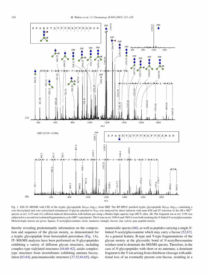
Fig. 1. ESI–IT–MS/MS with CID of the tryptic glycopeptide Ser295-Arg313 from HRP. The RP-HPLC-purified tryptic glycopeptide Ser295-Arg313 containing ac was a 3+
s ing as -ions aM ; trian
ttaIectl
mlAg
ore-fucosylated and core-xylosylated trimannosyl N-glycan attached to N298
pecies at m/z 1119 and (A) collision-induced dissociation with helium gas usubjected to a second ion isolation/fragmentation cycle (MS3 experiment). The b
onoisotopic masses are given. Square, N-acetylglucosamine; circle, mannose
hereby revealing predominantly information on the composi-ion and sequence of the glycan moiety, as demonstrated for
tryptic glycopeptide from horseradish peroxidase (Fig. 1A).T–MS/MS analyzes have been performed on N-glycopeptides
xhibiting a variety of different glycan structures, includingomplex-type sialylated structures [44,60–62], asialo complex-ype structures from invertebrates exhibiting antenna fucosy-ation [63,64], paucimannosidic structures [37,52,64,65], oligo-rcft
nalyzed by direct infusion with nano-ESI and IT selection of the [M + 3H]Bruker high capacity trap (HCT) ultra. (B) The fragment ion at m/z 1194 wast m/z 1894.6 and 1965.6 were both retaining the N-linked N-acetylglucosamine.
gle, fucose; star, xylose; pep, peptide moiety.
annosidic species [66], as well as peptides carrying a single N-inked N-acetylglucosamine which may carry a fucose [52,67].s a general feature, B-type and Y-type fragmentations of thelycan moiety at the glycosidic bond of N-acetylhexosamine
esidues tend to dominate the MS/MS spectra. Therefore, in thease of N-glycopeptides with short or no antennae, a dominantragment is the Y-ion arising from chitobiose cleavage with addi-ional loss of an eventually present core-fucose, resulting in a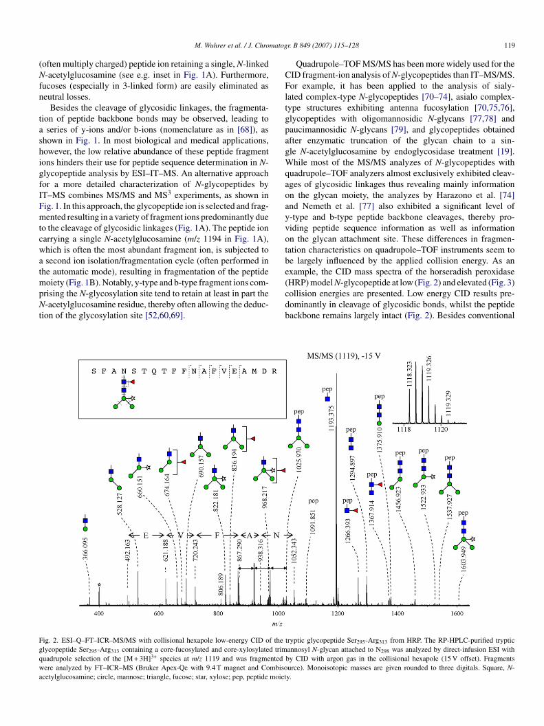
matog
(Nfn
tashigfIFmtcwatmpNt
CFltgpagWqaoayvotbe(
Fgqwa
M. Wuhrer et al. / J. Chro
often multiply charged) peptide ion retaining a single, N-linked-acetylglucosamine (see e.g. inset in Fig. 1A). Furthermore,
ucoses (especially in 3-linked form) are easily eliminated aseutral losses.
Besides the cleavage of glycosidic linkages, the fragmenta-ion of peptide backbone bonds may be observed, leading to
series of y-ions and/or b-ions (nomenclature as in [68]), ashown in Fig. 1. In most biological and medical applications,owever, the low relative abundance of these peptide fragmentons hinders their use for peptide sequence determination in N-lycopeptide analysis by ESI–IT–MS. An alternative approachor a more detailed characterization of N-glycopeptides byT–MS combines MS/MS and MS3 experiments, as shown inig. 1. In this approach, the glycopeptide ion is selected and frag-ented resulting in a variety of fragment ions predominantly due
o the cleavage of glycosidic linkages (Fig. 1A). The peptide ionarrying a single N-acetylglucosamine (m/z 1194 in Fig. 1A),hich is often the most abundant fragment ion, is subjected tosecond ion isolation/fragmentation cycle (often performed in
he automatic mode), resulting in fragmentation of the peptideoiety (Fig. 1B). Notably, y-type and b-type fragment ions com-
rising the N-glycosylation site tend to retain at least in part the-acetylglucosamine residue, thereby often allowing the deduc-
ion of the glycosylation site [52,60,69].
cdb
ig. 2. ESI–Q–FT–ICR–MS/MS with collisional hexapole low-energy CID of thelycopeptide Ser295-Arg313 containing a core-fucosylated and core-xylosylated trimuadrupole selection of the [M + 3H]3+ species at m/z 1119 and was fragmentedere analyzed by FT–ICR–MS (Bruker Apex-Qe with 9.4 T magnet and Combiso
cetylglucosamine; circle, mannose; triangle, fucose; star, xylose; pep, peptide moiet
r. B 849 (2007) 115–128 119
Quadrupole–TOF MS/MS has been more widely used for theID fragment-ion analysis of N-glycopeptides than IT–MS/MS.or example, it has been applied to the analysis of sialy-
ated complex-type N-glycopeptides [70–74], asialo complex-ype structures exhibiting antenna fucosylation [70,75,76],lycopeptides with oligomannosidic N-glycans [77,78] andaucimannosidic N-glycans [79], and glycopeptides obtainedfter enzymatic truncation of the glycan chain to a sin-le N-acetylglucosamine by endoglycosidase treatment [19].hile most of the MS/MS analyzes of N-glycopeptides with
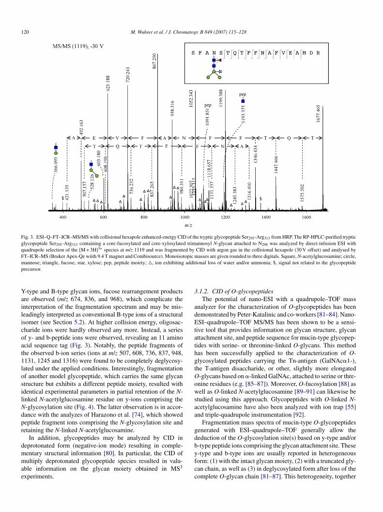
uadrupole–TOF analyzers almost exclusively exhibited cleav-ges of glycosidic linkages thus revealing mainly informationn the glycan moiety, the analyzes by Harazono et al. [74]nd Nemeth et al. [77] also exhibited a significant level of-type and b-type peptide backbone cleavages, thereby pro-iding peptide sequence information as well as informationn the glycan attachment site. These differences in fragmen-ation characteristics on quadrupole–TOF instruments seem toe largely influenced by the applied collision energy. As anxample, the CID mass spectra of the horseradish peroxidaseHRP) model N-glycopeptide at low (Fig. 2) and elevated (Fig. 3)
ollision energies are presented. Low energy CID results pre-ominantly in cleavage of glycosidic bonds, whilst the peptideackbone remains largely intact (Fig. 2). Besides conventionaltryptic glycopeptide Ser295-Arg313 from HRP. The RP-HPLC-purified trypticannosyl N-glycan attached to N298 was analyzed by direct-infusion ESI withby CID with argon gas in the collisional hexapole (15 V offset). Fragmentsurce). Monoisotopic masses are given rounded to three digitals. Square, N-y.
120 M. Wuhrer et al. / J. Chromatogr. B 849 (2007) 115–128
Fig. 3. ESI–Q–FT–ICR–MS/MS with collisional hexapole enhanced-energy CID of the tryptic glycopeptide Ser295-Arg313 from HRP. The RP-HPLC-purified trypticglycopeptide Ser295-Arg313 containing a core-fucosylated and core-xylosylated trimannosyl N-glycan attached to N298 was analyzed by direct-infusion ESI withquadrupole selection of the [M + 3H]3+ species at m/z 1119 and was fragmented by CID with argon gas in the collisional hexapole (30 V offset) and analysed byFT–ICR–MS (Bruker Apex-Qe wiith 9.4 T magnet and Combisource). Monoisotopic masses are given rounded to three digitals. Square, N-acetylglucosamine; circle,m additp
Yailicoat1losilNdpr
dmmae
3
adEtathgtOowsaa
gdb
annose; triangle, fucose; star, xylose; pep, peptide moiety; �, ion exhibitingrecursor.
-type and B-type glycan ions, fucose rearrangement productsre observed (m/z 674, 836, and 968), which complicate thenterpretation of the fragmentation spectrum and may be mis-eadingly interpreted as conventional B-type ions of a structuralsomer (see Section 5.2). At higher collision energy, oligosac-haride ions were hardly observed any more. Instead, a seriesf y- and b-peptide ions were observed, revealing an 11 aminocid sequence tag (Fig. 3). Notably, the peptide fragments ofhe observed b-ion series (ions at m/z 507, 608, 736, 837, 948,131, 1245 and 1316) were found to be completely deglycosy-ated under the applied conditions. Interestingly, fragmentationf another model glycopeptide, which carries the same glycantructure but exhibits a different peptide moiety, resulted withdentical experimental parameters in partial retention of the N-inked N-acetylglucosamine residue on y-ions comprising the-glycosylation site (Fig. 4). The latter observation is in accor-ance with the analyzes of Harazono et al. [74], which showedeptide fragment ions comprising the N-glycosylation site andetaining the N-linked N-acetylglucosamine.
In addition, glycopeptides may be analyzed by CID ineprotonated form (negative-ion mode) resulting in comple-
entary structural information [80]. In particular, the CID ofultiply deprotonated glycopeptide species resulted in valu-ble information on the glycan moiety obtained in MS3
xperiments.
yfcc
ional loss of water and/or ammonia; $, signal not related to the glycopeptide
.1.2. CID of O-glycopeptidesThe potential of nano-ESI with a quadrupole–TOF mass
nalyzer for the characterization of O-glycopeptides has beenemonstrated by Peter-Katalinic and co-workers [81–84]. Nano-SI–quadrupole–TOF MS/MS has been shown to be a sensi-
ive tool that provides information on glycan structure, glycanttachment site, and peptide sequence for mucin-type glycopep-ides with serine- or threonine-linked O-glycans. This methodas been successfully applied to the characterization of O-lycosylated peptides carrying the Tn-antigen (GalNAc�1-),he T-antigen disaccharide, or other, slightly more elongated-glycans based on �-linked GalNAc, attached to serine or thre-nine residues (e.g. [85–87]). Moreover, O-fucosylation [88] asell as O-linked N-acetylglucosamine [89–91] can likewise be
tudied using this approach. Glycopeptides with O-linked N-cetylglucosamine have also been analyzed with ion trap [55]nd triple-quadrupole instrumentation [92].
Fragmentation mass spectra of mucin-type O-glycopeptidesenerated with ESI–quadrupole–TOF generally allow theeduction of the O-glycosylation site(s) based on y-type and/or-type peptide ions comprising the glycan attachment site. These
-type and b-type ions are usually reported in heterogeneousorm: (1) with the intact glycan moiety, (2) with a truncated gly-an chain, as well as (3) in deglycosylated form after loss of theomplete O-glycan chain [81–87]. This heterogeneity, together
M. Wuhrer et al. / J. Chromatogr. B 849 (2007) 115–128 121
Fig. 4. ESI–Q–FT–ICR–MS/MS with collisional hexapole enhanced-energy CID of the tryptic glycopeptide Gly272-Arg294 from HRP. The RP-HPLC-purifiedtryptic glycopeptide Gly272-Arg294 containing a core-fucosylated and core-xylosylated trimannosyl N-glycan attached to N285 was analyzed by direct-infusion ESIwith quadrupole selection of the [M + 3H]3+ species at m/z 1225 and was fragmented by CID with argon gas in the collisional hexapole (30 V offset) and analysedb oisotoc bitingt
wf
bivTc
3dd
tfiplehBso
to
huNis[FhtgsMfmc
i
y FT–ICR–MS (Bruker Apex-Qe with 9.4 T magnet and Combisource). Monircle, mannose; triangle, fucose; star, xylose; pep, peptide moiety, �, ion exhihe N-glycosylation site which lacks the complete glycan chain.
ith the superimposition of various charge stages, makes theragment ion spectra rather complex.
Another type of glycosylation, which has been analyzedy nano-ESI–quadrupole–TOF is C-mannosylation. Mannoses found linked to the C2-atom of the tryptophan indole ring inarious proteins, e.g. the human complement system [93,94].he C-linked mannose appeared to be very stable in CID, inontrast to O-glycans and N-glycans.
.2. Sustained off-resonance irradiation collision-inducedissociation (SORI-CID) and infrared multi photonissociation (IRMPD)
Two so-called “slow-heating” techniques available in Fourierransform ion cyclotron resonance (FT-ICR) MS, which are use-ul for the analysis of glycopeptides, are sustained off-resonancerradiation collision-induced dissociation [95] and infrared multihoton dissociation [96,97]. In SORI-CID, precursor ions col-ide with an inert gas leaked into the ICR cell while they arexcited slightly off-resonance. In IRMPD, precursor ions are
eated by infrared irradiation until they begin to dissociate.oth fragmentation mechanisms result in similar fragmentationpectra of peptides or proteins, i.e., b-type and y-type ions arebserved as well as water and ammonia neutral losses. The pep-
hTao
pic masses are given rounded to three digitals. Square, N-acetylglucosamine;additional loss of water and/or ammonia; diamond, fragment ion comprising
ide fragmentation spectra are thus similar to the CID spectra inther types of instruments (see above).
Glycan sequencing of glycopeptides by SORI-CID MS/MSas been shown for O-glycosylated sialylated peptides fromrine of patients suffering from Schindler’s disease (hereditary-acetylhexosaminidase deficiency). It has to be mentioned that
n these experiments the peptide moiety was restricted to either aingle Ser/Thr amino acid or the di-amino acid peptide Thr-Pro98,99], and therefore, glycan attachment was readily obtained.or the structural characterization of N-glycopeptides, IRMPDas been shown to preferably cleave glycosidic linkages ratherhan peptide linkages, thus offering structural information on thelycan moiety. This preference has been shown for paucimanno-idic [100,101] as well as for complex type glycopeptides [101].
ore recently, Adamson and Hakansson [102] have shown thator high-mannose type glycopeptides, peptide backbone frag-entation can effectively compete with glycosidic backbone
leavages.We have performed SORI-CID MS/MS of an electrospray-
onized glycopeptide after selection in the quadrupole and with
elium as collision gas in the cell of the FT-ICR MS (Fig. 5).he obtained MS/MS spectrum showed predominantly cleav-ges of the glycosidic linkages with mainly neutral losses ofne or several terminal monosaccharide units. In addition, a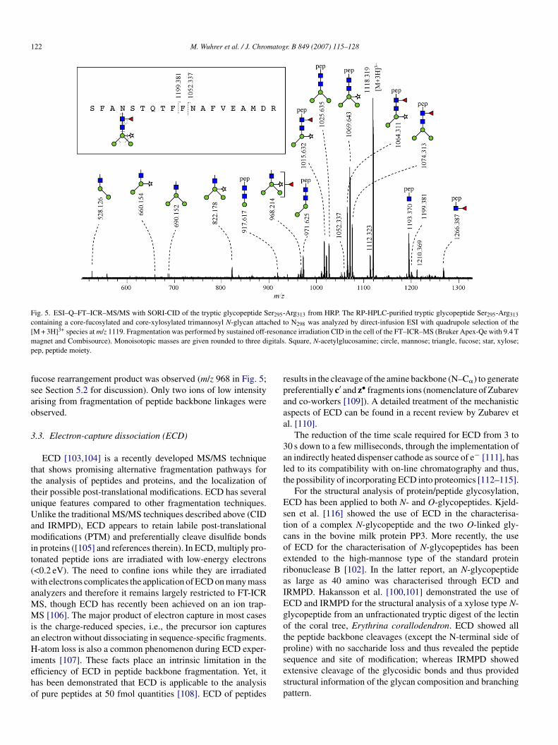
122 M. Wuhrer et al. / J. Chromatogr. B 849 (2007) 115–128
Fig. 5. ESI–Q–FT–ICR–MS/MS with SORI-CID of the tryptic glycopeptide Ser295-Arg313 from HRP. The RP-HPLC-purified tryptic glycopeptide Ser295-Arg313
containing a core-fucosylated and core-xylosylated trimannosyl N-glycan attached to N298 was analyzed by direct-infusion ESI with quadrupole selection of the[ 3+ resonam gitalsp
fsao
3
tttuUamit(waMMiaHieho
rpaaa
3alt
EstcoeraIEgotp
M + 3H] species at m/z 1119. Fragmentation was performed by sustained off-agnet and Combisource). Monoisotopic masses are given rounded to three di
ep, peptide moiety.
ucose rearrangement product was observed (m/z 968 in Fig. 5;ee Section 5.2 for discussion). Only two ions of low intensityrising from fragmentation of peptide backbone linkages werebserved.
.3. Electron-capture dissociation (ECD)
ECD [103,104] is a recently developed MS/MS techniquehat shows promising alternative fragmentation pathways forhe analysis of peptides and proteins, and the localization ofheir possible post-translational modifications. ECD has severalnique features compared to other fragmentation techniques.nlike the traditional MS/MS techniques described above (CID
nd IRMPD), ECD appears to retain labile post-translationalodifications (PTM) and preferentially cleave disulfide bonds
n proteins ([105] and references therein). In ECD, multiply pro-onated peptide ions are irradiated with low-energy electrons<0.2 eV). The need to confine ions while they are irradiatedith electrons complicates the application of ECD on many mass
nalyzers and therefore it remains largely restricted to FT-ICRS, though ECD has recently been achieved on an ion trap-S [106]. The major product of electron capture in most cases
s the charge-reduced species, i.e., the precursor ion capturesn electron without dissociating in sequence-specific fragments.-atom loss is also a common phenomenon during ECD exper-
ments [107]. These facts place an intrinsic limitation in thefficiency of ECD in peptide backbone fragmentation. Yet, itas been demonstrated that ECD is applicable to the analysisf pure peptides at 50 fmol quantities [108]. ECD of peptides
sesp
nce irradiation CID in the cell of the FT–ICR–MS (Bruker Apex-Qe with 9.4 T. Square, N-acetylglucosamine; circle, mannose; triangle, fucose; star, xylose;
esults in the cleavage of the amine backbone (N–C�) to generatereferentially c′ and z• fragments ions (nomenclature of Zubarevnd co-workers [109]). A detailed treatment of the mechanisticspects of ECD can be found in a recent review by Zubarev etl. [110].
The reduction of the time scale required for ECD from 3 to0 s down to a few milliseconds, through the implementation ofn indirectly heated dispenser cathode as source of e− [111], hased to its compatibility with on-line chromatography and thus,he possibility of incorporating ECD into proteomics [112–115].
For the structural analysis of protein/peptide glycosylation,CD has been applied to both N- and O-glycopeptides. Kjeld-en et al. [116] showed the use of ECD in the characterisa-ion of a complex N-glycopeptide and the two O-linked gly-ans in the bovine milk protein PP3. More recently, the usef ECD for the characterisation of N-glycopeptides has beenxtended to the high-mannose type of the standard proteinibonuclease B [102]. In the latter report, an N-glycopeptides large as 40 amino was characterised through ECD andRMPD. Hakansson et al. [100,101] demonstrated the use ofCD and IRMPD for the structural analysis of a xylose type N-lycopeptide from an unfractionated tryptic digest of the lectinf the coral tree, Erythrina corallodendron. ECD showed allhe peptide backbone cleavages (except the N-terminal side ofroline) with no saccharide loss and thus revealed the peptide
equence and site of modification; whereas IRMPD showedxtensive cleavage of the glycosidic bonds and thus providedtructural information of the glycan composition and branchingattern.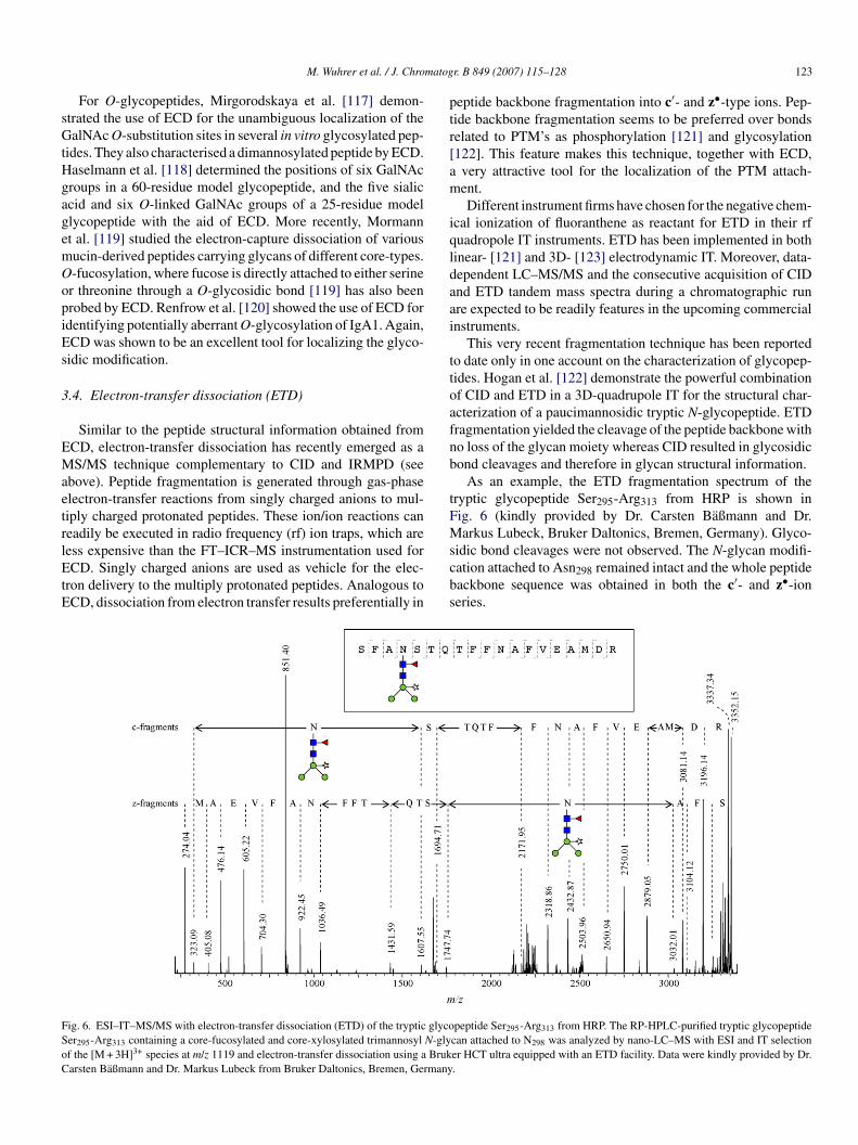
matog
sGtHgagemOopiEs
3
EMaetrlEtE
ptr[am
iqldaai
ttoafnb
tFM
FSoC
M. Wuhrer et al. / J. Chro
For O-glycopeptides, Mirgorodskaya et al. [117] demon-trated the use of ECD for the unambiguous localization of thealNAc O-substitution sites in several in vitro glycosylated pep-
ides. They also characterised a dimannosylated peptide by ECD.aselmann et al. [118] determined the positions of six GalNAcroups in a 60-residue model glycopeptide, and the five sialiccid and six O-linked GalNAc groups of a 25-residue modellycopeptide with the aid of ECD. More recently, Mormannt al. [119] studied the electron-capture dissociation of variousucin-derived peptides carrying glycans of different core-types.-fucosylation, where fucose is directly attached to either seriner threonine through a O-glycosidic bond [119] has also beenrobed by ECD. Renfrow et al. [120] showed the use of ECD fordentifying potentially aberrant O-glycosylation of IgA1. Again,CD was shown to be an excellent tool for localizing the glyco-idic modification.
.4. Electron-transfer dissociation (ETD)
Similar to the peptide structural information obtained fromCD, electron-transfer dissociation has recently emerged as aS/MS technique complementary to CID and IRMPD (see
bove). Peptide fragmentation is generated through gas-phaselectron-transfer reactions from singly charged anions to mul-iply charged protonated peptides. These ion/ion reactions caneadily be executed in radio frequency (rf) ion traps, which are
ess expensive than the FT–ICR–MS instrumentation used forCD. Singly charged anions are used as vehicle for the elec-ron delivery to the multiply protonated peptides. Analogous toCD, dissociation from electron transfer results preferentially in
scbs
ig. 6. ESI–IT–MS/MS with electron-transfer dissociation (ETD) of the tryptic glycoer295-Arg313 containing a core-fucosylated and core-xylosylated trimannosyl N-glyf the [M + 3H]3+ species at m/z 1119 and electron-transfer dissociation using a Brukarsten Baßmann and Dr. Markus Lubeck from Bruker Daltonics, Bremen, Germany
r. B 849 (2007) 115–128 123
eptide backbone fragmentation into c′- and z•-type ions. Pep-ide backbone fragmentation seems to be preferred over bondselated to PTM’s as phosphorylation [121] and glycosylation122]. This feature makes this technique, together with ECD,very attractive tool for the localization of the PTM attach-ent.Different instrument firms have chosen for the negative chem-
cal ionization of fluoranthene as reactant for ETD in their rfuadropole IT instruments. ETD has been implemented in bothinear- [121] and 3D- [123] electrodynamic IT. Moreover, data-ependent LC–MS/MS and the consecutive acquisition of CIDnd ETD tandem mass spectra during a chromatographic runre expected to be readily features in the upcoming commercialnstruments.
This very recent fragmentation technique has been reportedo date only in one account on the characterization of glycopep-ides. Hogan et al. [122] demonstrate the powerful combinationf CID and ETD in a 3D-quadrupole IT for the structural char-cterization of a paucimannosidic tryptic N-glycopeptide. ETDragmentation yielded the cleavage of the peptide backbone witho loss of the glycan moiety whereas CID resulted in glycosidicond cleavages and therefore in glycan structural information.
As an example, the ETD fragmentation spectrum of theryptic glycopeptide Ser295-Arg313 from HRP is shown inig. 6 (kindly provided by Dr. Carsten Baßmann and Dr.arkus Lubeck, Bruker Daltonics, Bremen, Germany). Glyco-
idic bond cleavages were not observed. The N-glycan modifi-ation attached to Asn298 remained intact and the whole peptideackbone sequence was obtained in both the c′- and z•-ioneries.
peptide Ser295-Arg313 from HRP. The RP-HPLC-purified tryptic glycopeptidecan attached to N298 was analyzed by nano-LC–MS with ESI and IT selectioner HCT ultra equipped with an ETD facility. Data were kindly provided by Dr..
124 M. Wuhrer et al. / J. Chromatogr. B 849 (2007) 115–128
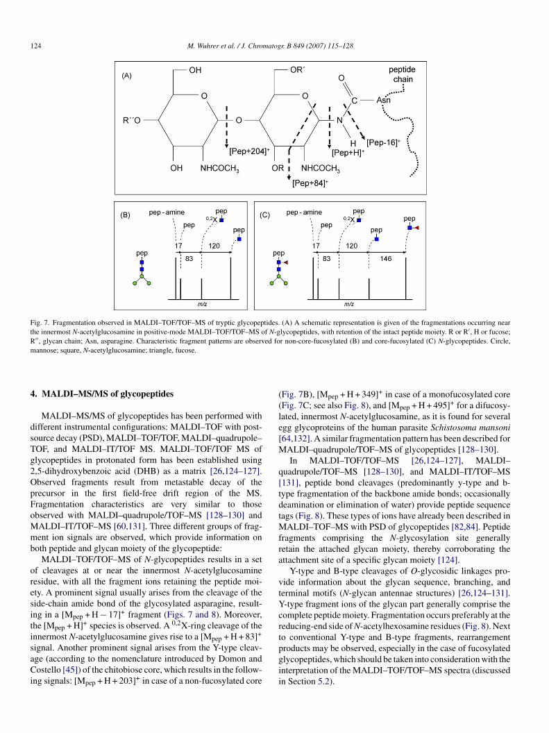
Fig. 7. Fragmentation observed in MALDI–TOF/TOF–MS of tryptic glycopeptides. (A) A schematic representation is given of the fragmentations occurring nearthe innermost N-acetylglucosamine in positive-mode MALDI–TOF/TOF–MS of N-glycopeptides, with retention of the intact peptide moiety. R or R′, H or fucose;R′′, glycan chain; Asn, asparagine. Characteristic fragment patterns are observed for non-core-fucosylated (B) and core-fucosylated (C) N-glycopeptides. Circle,m
4
dsTg2OpFoMmb
oresitisaCi
((le[M
q[tdtMfra
vtYcrt
annose; square, N-acetylglucosamine; triangle, fucose.
. MALDI–MS/MS of glycopeptides
MALDI–MS/MS of glycopeptides has been performed withifferent instrumental configurations: MALDI–TOF with post-ource decay (PSD), MALDI–TOF/TOF, MALDI–quadrupole–OF, and MALDI–IT/TOF MS. MALDI–TOF/TOF MS oflycopeptides in protonated form has been established using,5-dihydroxybenzoic acid (DHB) as a matrix [26,124–127].bserved fragments result from metastable decay of therecursor in the first field-free drift region of the MS.ragmentation characteristics are very similar to thosebserved with MALDI–quadrupole/TOF–MS [128–130] andALDI–IT/TOF–MS [60,131]. Three different groups of frag-ent ion signals are observed, which provide information on
oth peptide and glycan moiety of the glycopeptide:MALDI–TOF/TOF–MS of N-glycopeptides results in a set
f cleavages at or near the innermost N-acetylglucosamineesidue, with all the fragment ions retaining the peptide moi-ty. A prominent signal usually arises from the cleavage of theide-chain amide bond of the glycosylated asparagine, result-ng in a [Mpep + H − 17]+ fragment (Figs. 7 and 8). Moreover,he [Mpep + H]+ species is observed. A 0,2X-ring cleavage of thennermost N-acetylglucosamine gives rise to a [Mpep + H + 83]+
ignal. Another prominent signal arises from the Y-type cleav-ge (according to the nomenclature introduced by Domon andostello [45]) of the chitobiose core, which results in the follow-
ng signals: [Mpep + H + 203]+ in case of a non-fucosylated core
pgii
Fig. 7B), [Mpep + H + 349]+ in case of a monofucosylated coreFig. 7C; see also Fig. 8), and [Mpep + H + 495]+ for a difucosy-ated, innermost N-acetylglucosamine, as it is found for severalgg glycoproteins of the human parasite Schistosoma mansoni64,132]. A similar fragmentation pattern has been described for
ALDI–quadrupole/TOF–MS of glycopeptides [128–130].In MALDI–TOF/TOF–MS [26,124–127], MALDI–
uadrupole/TOF–MS [128–130], and MALDI–IT/TOF–MS131], peptide bond cleavages (predominantly y-type and b-ype fragmentation of the backbone amide bonds; occasionallyeamination or elimination of water) provide peptide sequenceags (Fig. 8). These types of ions have already been described in
ALDI–TOF–MS with PSD of glycopeptides [82,84]. Peptideragments comprising the N-glycosylation site generallyetain the attached glycan moiety, thereby corroborating thettachment site of a specific glycan moiety [124].
Y-type and B-type cleavages of O-glycosidic linkages pro-ide information about the glycan sequence, branching, anderminal motifs (N-glycan antennae structures) [26,124–131].-type fragment ions of the glycan part generally comprise theomplete peptide moiety. Fragmentation occurs preferably at theeducing-end side of N-acetylhexosamine residues (Fig. 8). Nexto conventional Y-type and B-type fragments, rearrangement
roducts may be observed, especially in the case of fucosylatedlycopeptides, which should be taken into consideration with thenterpretation of the MALDI–TOF/TOF–MS spectra (discussedn Section 5.2).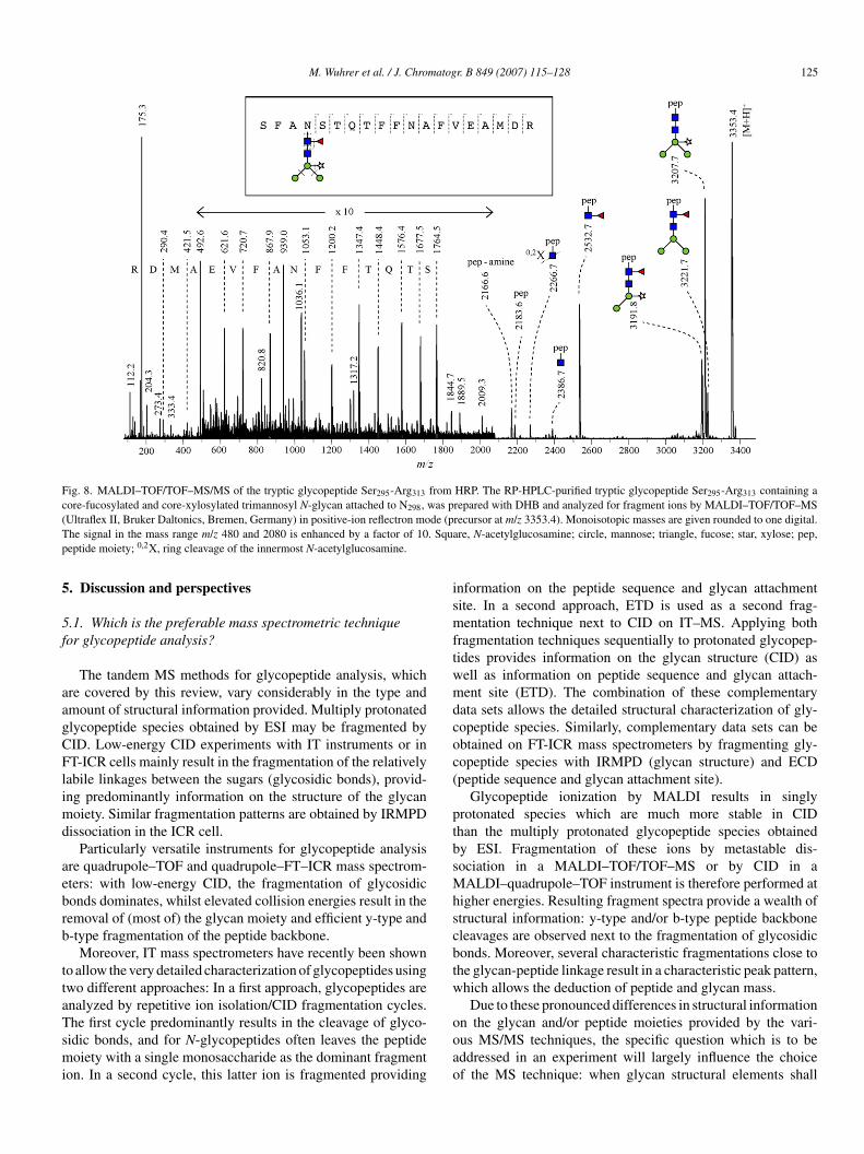
M. Wuhrer et al. / J. Chromatogr. B 849 (2007) 115–128 125
Fig. 8. MALDI–TOF/TOF–MS/MS of the tryptic glycopeptide Ser295-Arg313 from HRP. The RP-HPLC-purified tryptic glycopeptide Ser295-Arg313 containing acore-fucosylated and core-xylosylated trimannosyl N-glycan attached to N298, was prepared with DHB and analyzed for fragment ions by MALDI–TOF/TOF–MS( de (pT . Squap
5
5f
aagCFlimd
aebrb
ttaTsmi
ismftwmdcoc(
ptbsMhscbtw
Ultraflex II, Bruker Daltonics, Bremen, Germany) in positive-ion reflectron mohe signal in the mass range m/z 480 and 2080 is enhanced by a factor of 10eptide moiety; 0,2X, ring cleavage of the innermost N-acetylglucosamine.
. Discussion and perspectives
.1. Which is the preferable mass spectrometric techniqueor glycopeptide analysis?
The tandem MS methods for glycopeptide analysis, whichre covered by this review, vary considerably in the type andmount of structural information provided. Multiply protonatedlycopeptide species obtained by ESI may be fragmented byID. Low-energy CID experiments with IT instruments or inT-ICR cells mainly result in the fragmentation of the relatively
abile linkages between the sugars (glycosidic bonds), provid-ng predominantly information on the structure of the glycan
oiety. Similar fragmentation patterns are obtained by IRMPDissociation in the ICR cell.
Particularly versatile instruments for glycopeptide analysisre quadrupole–TOF and quadrupole–FT–ICR mass spectrom-ters: with low-energy CID, the fragmentation of glycosidiconds dominates, whilst elevated collision energies result in theemoval of (most of) the glycan moiety and efficient y-type and-type fragmentation of the peptide backbone.
Moreover, IT mass spectrometers have recently been showno allow the very detailed characterization of glycopeptides usingwo different approaches: In a first approach, glycopeptides arenalyzed by repetitive ion isolation/CID fragmentation cycles.
he first cycle predominantly results in the cleavage of glyco-idic bonds, and for N-glycopeptides often leaves the peptideoiety with a single monosaccharide as the dominant fragmenton. In a second cycle, this latter ion is fragmented providing
ooao
recursor at m/z 3353.4). Monoisotopic masses are given rounded to one digital.re, N-acetylglucosamine; circle, mannose; triangle, fucose; star, xylose; pep,
nformation on the peptide sequence and glycan attachmentite. In a second approach, ETD is used as a second frag-entation technique next to CID on IT–MS. Applying both
ragmentation techniques sequentially to protonated glycopep-ides provides information on the glycan structure (CID) asell as information on peptide sequence and glycan attach-ent site (ETD). The combination of these complementary
ata sets allows the detailed structural characterization of gly-opeptide species. Similarly, complementary data sets can bebtained on FT-ICR mass spectrometers by fragmenting gly-opeptide species with IRMPD (glycan structure) and ECDpeptide sequence and glycan attachment site).
Glycopeptide ionization by MALDI results in singlyrotonated species which are much more stable in CIDhan the multiply protonated glycopeptide species obtainedy ESI. Fragmentation of these ions by metastable dis-ociation in a MALDI–TOF/TOF–MS or by CID in a
ALDI–quadrupole–TOF instrument is therefore performed atigher energies. Resulting fragment spectra provide a wealth oftructural information: y-type and/or b-type peptide backboneleavages are observed next to the fragmentation of glycosidiconds. Moreover, several characteristic fragmentations close tohe glycan-peptide linkage result in a characteristic peak pattern,hich allows the deduction of peptide and glycan mass.Due to these pronounced differences in structural information
n the glycan and/or peptide moieties provided by the vari-us MS/MS techniques, the specific question which is to beddressed in an experiment will largely influence the choicef the MS technique: when glycan structural elements shall
1 matog
bnWfarf
5g
sciciat
msatedcwamNaasriftacis
5g
oambfeMsb
A
tFDk
R
26 M. Wuhrer et al. / J. Chro
e elucidated, low-energy CID, and IRMPD of multiply proto-ated glycopeptides will allow to specifically address this issue.hen (additional) peptide sequence information is required,
ragmentation should be performed at elevated energy withincollision cell or by MALDI–TOF/TOF–MS. Alternatively,
adical-formation and consecutive fragmentation may be per-ormed, i.e., ECD or ETD.
.2. Structural characterization of glycans by MS/MS oflycopeptides
First of all, caution has to be applied when deducing glycantructures from CID spectra of glycopeptides. A major compli-ation arises from fucose rearrangements, which are observedn MS/MS analysis of protonated glycans [133,134] and gly-opeptides [64]. Rearrangement products may erroneously benterpreted as conventional B-type and Y-type fragment ions,nd scientists may be tempted to postulate structures based onhese misleading fragment ions.
While CID MS/MS techniques do routinely provide infor-ation on the glycan moieties of glycopeptides, they do only
ometimes reveal information on peptide sequence and glycanttachment site(s). For a more detailed characterization of pro-ein glycosylation, these techniques may be combined with otherxperiments. Glycopeptides may be treated with exoglycosi-ases revealing the nature and anomericity of terminal monosac-haride residues. Alternatively, by treatment of glycopeptidesith peptide-N-glycosidase F or A, N-glycans can be released
nd deglycosylated peptide moieties are obtained. The peptidesay then be subjected to mass spectrometric characterization.otably, the conversion of the N-glycosylated asparagine into
n asparatate residue on enzymatic deglycosylation introducestag which allows the MS analysis of the glycan attachment
ite at the level of the deglycosylated peptide. Enzymaticallyeleased glycans may be analyzed by various techniques includ-ng mass spectrometric analysis in sodiated or deprotonatedorm and linkage analysis by GC–MS providing detailed struc-ural information, as reviewed by others [1–3,135,136]. Thesepproaches may in particular be necessary for the detailedharacterization of novel structural elements often observedn the analysis of glycoprotein samples from non-mammalianources.
.3. Towards a higher throughput in glycopeptide-basedlycoproteomics
Proteomics software tools for the fully automatic assignmentf the fragment spectra of unknown peptides are broadly used,nd similar tools for the characterization of glycans are gettingore and more popular [137–141]. Analysis of glycopeptides
y the various MS methods, however, still requires man power
or the assignment and interpretation of the spectra. Datavaluation is therefore the bottleneck in glycoproteomics viaS/MS of glycopeptides, and the development of suitableoftware tools is expected to make these techniques much moreroadly applicable.
r. B 849 (2007) 115–128
cknowledgements
We thank Carolien A.M. Koeleman for expert technical assis-ance. We appreciate the help of Dr. Bogdan Bogdanov with theT-ICR MS experiments. We thank Dr. Carten Baßmann andr. Markus Lubeck (Bruker Daltonics, Bremen, Germany) forindly providing the ETD fragmentation data.
eferences
[1] Y. Mechref, M.V. Novotny, Chem. Rev. 102 (2002) 321.[2] D.J. Harvey, Int. J. Mass Spectrom. 226 (2003) 1.[3] J. Zaia, Mass Spectrom. Rev. 23 (2004) 161.[4] W. Morelle, J.-C. Michalski, Curr. Anal. Chem. 1 (2005) 29.[5] K.F. Medzihradszky, Methods Enzymol. 405 (2005) 50.[6] K.F. Medzihradszky, Methods Enzymol. 402 (2005) 209.[7] B. Domon, R. Aebersold, Science 312 (2006) 212.[8] J. Hirabayashi, Glycoconj. J. 21 (2004) 35.[9] X. Fan, Y.M. She, R.D. Bagshaw, J.W. Callahan, H. Schachter, D.J. Mahu-
ran, Anal. Biochem. 332 (2004) 178.[10] J. Bunkenborg, B.J. Pilch, A.V. Podtelejnikov, J.R. Wisniewski, Pro-
teomics 4 (2004) 454.[11] L. Wang, F. Li, W. Sun, S. Wu, X. Wang, L. Zhang, D. Zheng, J. Wang,
Y. Gao, Mol. Cell. Proteomics 5 (2006) 560.[12] Z. Yang, W.S. Hancock, J. Chromatogr. A 1070 (2005) 57.[13] Z. Yang, W.S. Hancock, J. Chromatogr. A 1053 (2004) 79.[14] R. Qiu, F.E. Regnier, Anal. Chem. 77 (2005) 7225.[15] R. Qiu, F.E. Regnier, Anal. Chem. 77 (2005) 2802.[16] D.E. Sleat, H. Lackland, Y. Wang, I. Sohar, G. Xiao, H. Li, P. Lobel,
Proteomics 5 (2005) 1520.[17] D.E. Sleat, Y. Wang, I. Sohar, H. Lackland, Y. Li, H. Li, H. Zheng, P.
Lobel, Mol. Cell. Proteomics 5 (2006) 923.[18] G. Alvarez-Manilla, J. Atwood III, Y. Guo, N.L. Warren, R. Orlando, M.
Pierce, J. Proteome Res. 5 (2006) 701.[19] P. Hagglund, J. Bunkenborg, F. Elortza, O.N. Jensen, P. Roepstorff, J.
Proteome Res. 3 (2004) 556.[20] H. Zhang, X.J. Li, D.B. Martin, R. Aebersold, Nat. Biotechnol. 21 (2003)
660.[21] T. Liu, W.J. Qian, M.A. Gritsenko, D.G. Camp, M.E. Monroe, R.J. Moore,
R.D. Smith, J. Proteome Res. 4 (2005) 2070.[22] U. Lewandrowski, J. Moebius, U. Walter, A. Sickmann, Mol. Cell. Pro-
teomics 5 (2006) 226.[23] H. Zhang, E.C. Yi, X.J. Li, P. Mallick, K.S. Kelly-Spratt, C.D. Masselon,
D.G. Camp, R.D. Smith, C.J. Kemp, R. Aebersold, Mol. Cell. Proteomics4 (2005) 144.
[24] H. Kaji, H. Saito, Y. Yamauchi, T. Shinkawa, M. Taoka, J. Hirabayashi,K. Kasai, N. Takahashi, T. Isobe, Nat. Biotechnol. 21 (2003) 667.
[25] W. Morelle, S. Donadio, C. Ronin, J.C. Michalski, Rapid Commun. MassSpectrom. 20 (2006) 331.
[26] R. Uematsu, J. Furukawa, H. Nakagawa, Y. Shinohara, K. Deguchi, K.Monde, S. Nishimura, Mol. Cell. Proteomics 4 (2005) 1977.
[27] Y. Wang, S.L. Wu, W.S. Hancock, Glycobiology 16 (2006) 614.[28] Y. Takegawa, K. Deguchi, T. Keira, H. Ito, H. Nakagawa, S.I. Nishimura,
J. Chromatogr. A 1113 (2006) 177.[29] Y. Wada, M. Tajiri, S. Yoshida, Anal. Chem. 76 (2004) 6560.[30] M. Tajiri, S. Yoshida, Y. Wada, Glycobiology 15 (2005) 1332.[31] M.R. Larsen, P. Hojrup, P. Roepstorff, Mol. Cell. Proteomics 4 (2005)
107.[32] R.D. Cummings, S. Kornfeld, J. Biol. Chem. 257 (1982) 11235.[33] K. Tachibana, S. Nakamura, H. Wang, H. Iwasaki, K. Tachibana, K. Mae-
bara, L. Cheng, J. Hirabayashi, H. Narimatsu, Glycobiology 16 (2006)
46.[34] D.E. Sleat, H. Zheng, M. Qian, P. Lobel, Mol. Cell. Proteomics 5 (2006)686.
[35] J. Hirabayashi, T. Hashidate, K. Kasai, J. Biomol. Tech. 13 (2002) 205.[36] M. Wuhrer, A.M. Deelder, C.H. Hokke, J. Chromatogr. B 825 (2005) 124.

matog
M. Wuhrer et al. / J. Chro[37] M. Wuhrer, C.A.M. Koeleman, C.H. Hokke, A.M. Deelder, Anal. Chem.77 (2005) 886.
[38] K. Sparbier, S. Koch, I. Kessler, T. Wenzel, M. Kostrzewa, J. Biomol.Tech. 16 (2005) 407.
[39] J.D. Rawn, G.E. Lienhard, Biochemistry 13 (1974) 3124.[40] N.H. Packer, M.A. Lawson, D.R. Jardine, J.W. Redmond, Glycoconj. J.
15 (1998) 737.[41] C.W. Klampfl, Electrophoresis 27 (2006) 3.[42] A. Zamfir, J. Peter-Katalinic, Electrophoresis 22 (2001) 2448.[43] L. Bindila, J. Peter-Katalinic, A. Zamfir, Electrophoresis 26 (2005) 1488.[44] S. Amon, A. Plematl, A. Rizzi, Electrophoresis 27 (2006) 1209.[45] B. Domon, C. Costello, Glycoconj. J. 5 (1988) 253.[46] M.J. Huddleston, M.F. Bean, S.A. Carr, Anal. Chem. 65 (1993) 877.[47] S.A. Carr, M.J. Huddleston, M.F. Bean, Protein Sci. 2 (1993) 183.[48] P.A. Schindler, C.A. Settineri, X. Collet, C.J. Fielding, A.L. Burlingame,
Protein Sci. 4 (1995) 791.[49] K.F. Medzihradszky, M.J. Besman, A.L. Burlingame, Anal. Chem. 69
(1997) 3986.[50] J.C. Rogalski, J. Kast, Rapid Commun. Mass Spectrom. 19 (2005) 77.[51] B. Sullivan, T.A. Addona, S.A. Carr, Anal. Chem. 76 (2004) 3112.[52] M. Wuhrer, C.I. Balog, C.A. Koeleman, A.M. Deelder, C.H. Hokke,
Biochim. Biophys. Acta 1723 (2005) 229.[53] M.A. Ritchie, A.C. Gill, M.J. Deery, K. Lilley, J. Am. Soc. Mass Spec-
trom. 13 (2002) 1065.[54] J. Jebanathirajah, H. Steen, P. Roepstorff, J. Am. Soc. Mass Spectrom. 14
(2003) 777.[55] L.E. Ball, M.N. Berkaw, M.G. Buse, Mol. Cell. Proteomics 5 (2006) 313.[56] C.A. Settineri, K.F. Medzihradszky, F.R. Masiarz, A.L. Burlingame,
C. Chu, C. George-Nascimento, Biomed. Environ. Mass Spectrom. 19(1990) 665.
[57] K.F. Medzihradszky, B.L. Gillece-Castro, C.A. Settineri, R.R. Townsend,F.R. Masiarz, A.L. Burlingame, Biomed. Environ. Mass Spectrom. 19(1990) 777.
[58] K.F. Medzihradszky, B.L. Gillece-Castro, R.R. Townsend, A.L.Burlingame, M.R. Hardy, J. Am. Soc. Mass Spectrom. 7 (1996) 319.
[59] M.J. Kieliszewski, M. O’Neill, J. Leykam, R. Orlando, J. Biol. Chem.270 (1995) 2541.
[60] U.M. Demelbauer, M. Zehl, A. Plematl, G. Allmaier, A. Rizzi, Rapid.Commun. Mass Spectrom. 18 (2004) 1575.
[61] U.M. Demelbauer, A. Plematl, L. Kremser, G. Allmaier, D. Josic, A.Rizzi, Electrophoresis 25 (2004) 2026.
[62] F. Wang, A. Nakouzi, R.H. Angeletti, A. Casadevall, Anal. Biochem. 314(2003) 266.
[63] C. Gielens, K. Idakieva, V. Van den Bergh, N.I. Siddiqui, K. Parvanova,F. Compernolle, Biochem. Biophys. Res. Commun. 331 (2005) 562.
[64] M. Wuhrer, C.I.A. Balog, B. Catimel, F.M. Jones, G. Schramm, H. Haas,M.J. Doenhoff, D.W. Dunne, A.M. Deelder, C.H. Hokke, FEBS J. 273(2006) 2276.
[65] H. Jiang, H. Desaire, V.Y. Butnev, G.R. Bousfield, J. Am. Soc. MassSpectrom. 15 (2004) 750.
[66] T. Liu, J.-D. Li, R. Zeng, X.-X. Shao, K.-Y. Wang, Q.-C. Xia, Anal. Chem.73 (2001) 5875.
[67] K. Sandra, B. Devreese, J. Van Beeumen, I. Stals, M. Claeyssens, J. Am.Soc. Mass Spectrom. 15 (2004) 413.
[68] K. Biemann, Annu. Rev. Biochem. 61 (1992) 977.[69] S. Itoh, N. Kawasaki, A. Harazono, N. Hashii, Y. Matsuishi, T. Kawanishi,
T. Hayakawa, J. Chromatogr. A 1094 (2005) 105.[70] E. Stimson, J. Hope, A. Chong, A.L. Burlingame, Biochemistry 38 (1999)
4885.[71] Y. Satomi, Y. Shimonishi, T. Hase, T. Takao, Rapid Commun. Mass Spec-
trom. 18 (2004) 2983.[72] Y. Satomi, Y. Shimonishi, T. Takao, FEBS Lett. 576 (2004) 51.[73] T. Imre, G. Schlosser, G. Pocsfalvi, R. Siciliano, E. Molnar-Szollosi, T.
Kremmer, A. Malorni, K. Vekey, J. Mass Spectrom. 40 (2005) 1472.[74] A. Harazono, N. Kawasaki, S. Itoh, N. Hashii, A. Ishii-Watabe, T. Kawan-
ishi, T. Hayakawa, Anal. Biochem. 348 (2006) 259.[75] M. Nimtz, H.S. Conradt, K. Mann, Biochim. Biophys. Acta 1675 (2004)
71.
r. B 849 (2007) 115–128 127
[76] C. Zhang, A. Doherty-Kirby, R.R. Huystee, G. Lajoie, Phytochemistry65 (2004) 1575.
[77] J.F. Nemeth, G.P. Hochgesang Jr., L.J. Marnett, R.M. Caprioli, Biochem-istry 40 (2001) 3109.
[78] H. Henriksson, S.E. Denman, I.D. Campuzano, P. Ademark, E.R. Master,T.T. Teeri, H. Brumer III, Biochem. J. 375 (2003) 61.
[79] J.C. Marxen, M. Nimtz, W. Becker, K. Mann, Biochim. Biophys. Acta1650 (2003) 92.
[80] K. Deguchi, H. Ito, Y. Takegawa, N. Shinji, H. Nakagawa, S. Nishimura,Rapid Commun. Mass Spectrom. 20 (2006) 741.
[81] F.G. Hanisch, B.N. Green, R. Bateman, J. Peter-Katalinic, J. Mass Spec-trom. 33 (1998) 358.
[82] K. Alving, R. Korner, H. Paulsen, J. Peter-Katalinic, J. Mass Spectrom.33 (1998) 1124.
[83] K. Alving, H. Paulsen, J. Peter-Katalinic, J. Mass Spectrom. 34 (1999)395.
[84] S. Muller, S. Goletz, N. Packer, A. Gooley, A.M. Lawson, F.G. Hanisch,J. Biol. Chem. 272 (1997) 24780.
[85] S. Schmitt, D. Glebe, K. Alving, T.K. Tolle, M. Linder, H. Geyer, D.Linder, J. Peter-Katalinic, W.H. Gerlich, R. Geyer, J. Biol. Chem. 274(1999) 11945.
[86] S. Chalabi, M. Panico, M. Sutton-Smith, S.M. Haslam, M.S. Patankar,F.A. Lattanzio, H.R. Morris, G.F. Clark, A. Dell, Biochemistry 45 (2006)637.
[87] S. Muller, K. Alving, J. Peter-Katalinic, N. Zachara, A.A. Gooley, F.G.Hanisch, J. Biol. Chem. 274 (1999) 18165.
[88] B. Macek, J. Hofsteenge, J. Peter-Katalinic, Rapid Commun. Mass Spec-trom. 15 (2001) 771.
[89] R.J. Chalkley, A.L. Burlingame, J. Am. Soc. Mass Spectrom. 12 (2001)1106.
[90] R.J. Chalkley, A.L. Burlingame, Mol. Cell. Proteomics 2 (2003) 182.[91] K. Vosseller, J.C. Trinidad, R.J. Chalkley, C.G. Specht, A. Thalham-
mer, A.J. Lynn, J.H. Snedecor, S. Guan, K.F. Medzihradszky, D.A.Maltby, R. Schoepfer, A.L. Burlingame, Mol. Cell. Proteomics 5 (2006)923.
[92] K.D. Greis, B.K. Hayes, F.I. Comer, M. Kirk, S. Barnes, T.L. Lowary,G.W. Hart, Anal. Biochem. 234 (1996) 38.
[93] A. Gonzales de Peredo, D. Klein, B. Macek, D. Hess, J. Peter-Katalinic,J. Hofsteenge, Mol. Cell. Proteomics 1 (2002) 11.
[94] J. Hofsteenge, K.G. Huwiler, B. Macek, D. Hess, J. Lawler, D.F. Mosher,J. Peter-Katalinic, J. Biol. Chem. 276 (2001) 6485.
[95] J.W. Gauthier, T.R. Trautman, D.B. Jacobson, Anal. Chim. Acta 246(1991) 211.
[96] R.L. Woodin, D.S. Bomse, J.L. Eauchamp, J. Am. Chem. Soc. 100 (1978)3248.
[97] D.P. Little, J.P. Speir, M.W. Senko, P.B. Oconnor, F.W. McLafferty, Anal.Chem. 66 (1994) 2809.
[98] M. Froesch, L. Bindila, A. Zamfir, J. Peter-Katalinic, Rapid Commun.Mass Spectrom. 17 (2003) 2822.
[99] M. Froesch, L.M. Bindila, G. Baykut, M. Allen, J. Peter-Katalinic, A.D.Zamfir, Rapid Commun. Mass Spectrom. 18 (2004) 3084.
[100] K. Hakansson, H.J. Cooper, M.R. Emmett, C.E. Costello, A.G. Marshall,C.L. Nilsson, Anal. Chem. 73 (2001) 4530.
[101] K. Hakansson, M.J. Chalmers, J.P. Quinn, M.A. McFarland, C.L. Hen-drickson, A.G. Marshall, Anal. Chem. 75 (2003) 3256.
[102] J.T. Adamson, K. Hakansson, J. Proteome Res. 5 (2006) 493.[103] R.A. Zubarev, N.L. Kelleher, F.W. McLafferty, J. Am. Chem. Soc. 120
(1998) 3265.[104] S.C. Beu, M.W. Senko, J.P. Quinn, F.M. Wampler, F.W. McLafferty, J.
Am. Soc. Mass Spectrom. 4 (1993) 557.[105] H.J. Cooper, K. Hakansson, A.G. Marshall, Mass Spectrom. Rev. 24
(2005) 201.[106] T. Baba, Y. Hashimoto, H. Hasegawa, A. Hirabayashi, I. Waki, Anal.
Chem. 76 (2004) 4263.[107] K. Breuker, H.B. Oh, B.A. Cerda, D.M. Horn, F.W. McLafferty, Eur. J.
Mass Spectrom. 8 (2002) 177.[108] K. Hakansson, M.R. Emmett, C.L. Hendrickson, A.G. Marshall, Anal.
Chem. 73 (2001) 3605.
1 matog
28 M. Wuhrer et al. / J. Chro[109] F. Kjeldsen, K.F. Haselmann, B.A. Budnik, F. Jensen, R.A. Zubarev,Chem. Phys. Lett. 356 (2002) 201.
[110] R.A. Zubarev, K.F. Haselmann, B.A. Budnik, F. Kjeldsen, F. Jensen, Eur.J. Mass Spectrom. 8 (2002) 337.
[111] Y.O. Tsybin, P. Hakansson, B.A. Budnik, K.F. Haselmann, F. Kjeldsen,M. Gorshkov, R.A. Zubarev, Rapid Commun. Mass Spectrom. 15 (2001)1849.
[112] M. Palmblad, Y.O. Tsybin, M. Ramstrom, J. Bergquist, P. Hakansson,Rapid Commun. Mass Spectrom. 16 (2002) 988.
[113] Y.O. Tsybin, P. Hakansson, M. Wetterhall, K.E. Markides, J. Bergquist,Eur. J. Mass Spectrom. 8 (2002) 389–395.
[114] M.L. Nielsen, M.M. Savitski, R.A. Zubarev, Mol. Cell. Proteomics 4(2005) 835.
[115] H.J. Cooper, S. Akbarzadeh, J.K. Heath, M. Zeller, J. Proteome Res. 4(2005) 1538.
[116] F. Kjeldsen, K.F. Haselmann, B.A. Budnik, E.S. Sorensen, R.A. Zubarev,Anal. Chem. 75 (2003) 2355.
[117] E. Mirgorodskaya, P. Roepstorff, R.A. Zubarev, Anal. Chem. 71 (1999)4431.
[118] K.F. Haselmann, B.A. Budnik, J.V. Olsen, M.L. Nielsen, C.A. Reis, H.Clausen, A.H. Johnsen, R.A. Zubarev, Anal. Chem. 73 (2001) 2998.
[119] M. Mormann, H. Paulsen, J. Peter-Katalinic, Eur. J. Mass Spectrom. 11(2005) 497.
[120] M.B. Renfrow, H.J. Cooper, M. Tomana, R. Kulhavy, Y. Hiki, K. Toma,M.R. Emmett, J. Mestecky, A.G. Marshall, J. Novak, J. Biol. Chem. 280(2005) 19136.
[121] J.E.P. Syka, J.J. Coon, M.J. Schroeder, J. Shabanowitz, D.F. Hunt, Proc.
Natl. Acad. Sci. U.S.A. 101 (2004) 9528.[122] J.M. Hogan, S.J. Pitteri, P.A. Chrisman, S.A. McLuckey, J. Proteome Res.4 (2005) 628.
[123] S.J. Pitteri, P.A. Chrisman, J.M. Hogan, S.A. McLuckey, Anal. Chem. 77(2005) 1831.
r. B 849 (2007) 115–128
[124] M. Wuhrer, C.H. Hokke, A.M. Deelder, Rapid Commun. Mass Spectrom.18 (2004) 1741.
[125] S. Nishimura, K. Niikura, M. Kurogochi, T. Matsushita, M. Fumoto, H.Hinou, R. Kamitani, H. Nakagawa, K. Deguchi, N. Miura, K. Monde, H.Kondo, Angew. Chem. Int. Ed. Engl. 44 (2004) 91.
[126] M. Kurogochi, S. Nishimura, Anal. Chem. 76 (2004) 6097.[127] M. Kurogochi, T. Matsushita, S. Nishimura, Angew. Chem. Int. Ed. Engl.
43 (2004) 4071.[128] O. Krokhin, W. Ens, K.G. Standing, J. Wilkins, H. Perreault, Rapid Com-
mun. Mass Spectrom. 18 (2004) 2020.[129] O.V. Krokhin, W. Ens, K.G. Standing, J. Biomol. Tech. 16 (2005) 429.[130] N.V. Bykova, C. Rampitsch, O. Krokhin, K.G. Standing, W. Ens, Anal.
Chem. 78 (2006) 1093.[131] N. Takemori, N. Komori, H. Matsumoto, Electrophoresis 27 (2006)
1394.[132] K.H. Khoo, D. Chatterjee, J.P. Caulfield, H.R. Morris, A. Dell, Glycobi-
ology 7 (1997) 663.[133] D.J. Harvey, T.S. Mattu, M.R. Wormald, L. Royle, R.A. Dwek, P.M. Rudd,
Anal. Chem. 74 (2002) 734.[134] M. Wuhrer, C.A.M. Koeleman, C.H. Hokke, A.M. Deelder, Rapid Com-
mun. Mass Spectrom. 20 (2006) 1747.[135] H. Geyer, R. Geyer, Acta Anat. 161 (1998) 18.[136] R. Geyer, H. Geyer, Methods Enzymol. 230 (1994) 86.[137] H. Zhang, S. Singh, V.N. Reinhold, Anal. Chem. 77 (2005) 6263.[138] D. Ashline, S. Singh, A. Hanneman, V. Reinhold, Anal. Chem. 77 (2005)
6250.[139] D. Goldberg, M. Sutton-Smith, J. Paulson, A. Dell, Proteomics 5 (2005)
865.[140] A. Kameyama, S. Nakaya, H. Ito, N. Kikuchi, T. Angata, M. Nakamura,
H.K. Ishida, H. Narimatsu, J. Proteome Res. 5 (2006) 808.[141] A. Kameyama, N. Kikuchi, S. Nakaya, H. Ito, T. Sato, T. Shikanai, Y.
Takahashi, K. Takahashi, H. Narimatsu, Anal. Chem. 77 (2005) 4719.