glucose intolerance and reduced proliferation of pancreatic -cells in transgenic pigs with...
TRANSCRIPT

Glucose Intolerance and Reduced Proliferation ofPancreatic �-Cells in Transgenic Pigs With ImpairedGlucose-Dependent Insulinotropic Polypeptide FunctionSimone Renner,
1Christiane Fehlings,
1Nadja Herbach,
2Andreas Hofmann,
3
Dagmar C. von Waldthausen,1
Barbara Kessler,1
Karin Ulrichs,4
Irina Chodnevskaja,4
Vasiliy Moskalenko,4
Werner Amselgruber,5
Burkhard Goke,6
Alexander Pfeifer,3,7
Rudiger Wanke,2
and Eckhard Wolf1
OBJECTIVE—The insulinotropic action of the incretin glucose-dependent insulinotropic polypeptide (GIP) is impaired in type 2diabetes, while the effect of glucagon-like peptide-1 (GLP-1) ispreserved. To evaluate the role of impaired GIP function inglucose homeostasis and development of the endocrine pancreasin a large animal model, we generated transgenic pigs expressinga dominant-negative GIP receptor (GIPRdn) in pancreatic islets.
RESEARCH DESIGN AND METHODS—GIPRdn transgenicpigs were generated using lentiviral transgenesis. Metabolic testsand quantitative stereological analyses of the different endocrineislet cell populations were performed, and �-cell proliferationand apoptosis were quantified to characterize this novel animalmodel.
RESULTS—Eleven-week-old GIPRdn transgenic pigs exhibitedsignificantly reduced oral glucose tolerance due to delayedinsulin secretion, whereas intravenous glucose tolerance andpancreatic �-cell mass were not different from controls. Theinsulinotropic effect of GIP was significantly reduced, whereasinsulin secretion in response to the GLP-1 receptor agonistexendin-4 was enhanced in GIPRdn transgenic versus controlpigs. With increasing age, glucose control deteriorated in GIPRdn
transgenic pigs, as shown by reduced oral and intravenousglucose tolerance due to impaired insulin secretion. Importantly,�-cell proliferation was reduced by 60% in 11-week-old GIPRdn
transgenic pigs, leading to a reduction of �-cell mass by 35% and58% in 5-month-old and 1- to 1.4-year-old transgenic pigs com-pared with age-matched controls, respectively.
CONCLUSIONS—The first large animal model with impairedincretin function demonstrates an essential role of GIP forinsulin secretion, proliferation of �-cells, and physiological ex-pansion of �-cell mass. Diabetes 59:1228–1238, 2010
The incretin hormones glucose-dependent insuli-notropic polypeptide (GIP) and glucagon-likepeptide-1 (GLP-1) are secreted by enteroendo-crine cells in response to nutrients like fat and
glucose and enhance glucose-induced release of insulinfrom pancreatic �-cells (1). The effects of GIP and GLP-1are mediated through specific receptors, GIPR and GLP-1R, respectively. Both receptors belong to the family ofseven transmembrane-domain heterotrimeric G-protein–coupled receptors (2). Activation of the GIPR or GLP-1Rleads to enhanced exocytosis of insulin-containing gran-ules (3). Interestingly, variation in the GIPR gene influ-ences glucose and insulin responses to an oral glucosechallenge in humans (4). Furthermore, findings in insuli-noma cells (5–7) and rodent models (8,9) indicate thatactivation of incretin receptors promotes proliferation andsurvival of �-cells. Type 2 diabetic patients and �50% oftheir first-degree relatives show a reduced incretin effect,mainly due to an impaired insulinotropic action of GIP(10,11). Nearly sustained insulinotropic action of GLP-1(11) in type 2 diabetic patients revealed its therapeuticpotential and initiated the ongoing development of GLP-1Ragonists as well as inhibitors of dipeptidyl peptidase-4(1,12), which rapidly degrades incretin hormones in vivo.The reasons for the reduced response to GIP in type 2diabetes are unclear (1), but impaired GIP action might beinvolved in the early pathogenesis of type 2 diabetes (13).
To clarify this point, a mouse model lacking functionalGIPR expression was generated by gene targeting (14).Gipr�/� mice displayed only slightly impaired glucosetolerance and did not develop diabetes. Interestingly,double incretin receptor knockout mice exhibited a simi-lar phenotype. As possible explanations for this relativelymild phenotype (rev. in 15), compensatory regulation ofthe GLP-1 system or other compensatory mechanismswere discussed. In contrast, transgenic mice overexpress-ing a dominant-negative GIPR (GIPRdn) displayed a severephenotype (i.e., early-onset diabetes accompanied by amarked fasting hypoinsulinemia and severe reduction of�-cell mass associated with extensive structural alter-ations of the pancreatic islets) (16).
In light of these discrepant findings in mouse models,we generated a large animal model to address the questionwhether GIPR signaling plays a role in maintaining pan-creatic islet function and structure. Efficient lentiviralvectors (17) were used to generate transgenic pigs ex-pressing a GIPRdn under the control of the rat Ins2
From the 1Chair for Molecular Animal Breeding and Biotechnology andLaboratory for Functional Genome Analysis, Gene Center, Ludwig Maximil-ians University (LMU) Munich, Munich, Germany; the 2Institute of Veteri-nary Pathology, Faculty of Veterinary Medicine, LMU Munich, Munich,Germany; the 3Institute of Pharmacology and Toxicology, University ofBonn, Bonn, Germany; the 4Department of Experimental TransplantationImmunology, Surgical Clinic I, University Hospital of Wurzburg, Wurzburg,Germany; the 5Institute of Anatomy and Physiology, University of Stuttgart-Hohenheim, Stuttgart, Germany; the 6Medical Clinic II, Klinikum Grosshad-ern, LMU Munich, Munich, Germany; and the 7Pharma Center Bonn,University of Bonn, Bonn, Germany.
Corresponding author: Eckhard Wolf, [email protected] 8 April 2009 and accepted 10 February 2010. Published ahead of
print at http://diabetes.diabetesjournals.org on 25 February 2010. DOI:10.2337/db09-0519.
C.F. and N.H. contributed equally to this article.© 2010 by the American Diabetes Association. Readers may use this article as
long as the work is properly cited, the use is educational and not for profit,and the work is not altered. See http://creativecommons.org/licenses/by-nc-nd/3.0/ for details.
The costs of publication of this article were defrayed in part by the payment of page
charges. This article must therefore be hereby marked “advertisement” in accordance
with 18 U.S.C. Section 1734 solely to indicate this fact.
ORIGINAL ARTICLE
1228 DIABETES, VOL. 59, MAY 2010 diabetes.diabetesjournals.org

promoter in the pancreatic islets. This novel animal model,in contrast to GIPRdn transgenic mice (16), initially onlyexhibits a disturbed incretin effect but develops progres-sive deterioration of glucose control with increasing age,associated with reduced �-cell proliferation and an impair-ment of physiological age-related expansion of pancreatic�-cell mass.
RESEARCH DESIGN AND METHODS
Generation of RIP II-GIPRdn transgenic pigs. The expression cassetteconsisting of the rat Ins2 promoter (RIP II) and the cDNA of a human GIPRdn
(16) was cloned into the lentiviral vector LV-pGFP (18) (supplementary Fig. 1of the online appendix [available at http://diabetes.diabetesjournals.org/cgi/content/full/db09-0519/DC1]). Recombinant lentivirus was produced (18) andinjected into the perivitelline space of zygotes from superovulated gilts (17).Embryos were transferred into synchronized recipients (19). Offspring weregenotyped by PCR and Southern blot analyses using a probe directed towardthe RIP II promoter sequence. Expression of GIPRdn mRNA in the pancreaticislets was determined by RT-PCR. A total of 400 ng of total RNA were reversetranscribed into cDNA using SuperScriptII reverse transcriptase (Invitrogen)and random hexamer primers (Invitrogen) after digestion with DNaseI(Roche). For PCR, the following transgene specific primers were used: sense5�-TTT TTA TCC GCA TTC TTA CAC GG-3� and antisense 5�-ATC TTC CTCAGC TCC TTC CAG G-3�. All animal experiments were carried out accordingto the German animal protection law.Oral/intravenous glucose tolerance test and GIP/exendin-4 stimulation
test. For the oral glucose tolerance test (OGTT), one central venous catheter(Cavafix Certo; B. Braun) was inserted nonsurgically into the external jugularvein. After an 18-h overnight fast, animals were fed 2 g glucose/kg body weight(BW) (20) mixed with 50/100 g (11-week-old/5-month-old) commercial pigfodder. Blood samples were obtained from the jugular vein catheter at theindicated time points. For the intravenous glucose tolerance test (IVGTT) andGIP/exendin-4 stimulation test, two central venous catheters (Cavafix Certo)were surgically inserted into the external jugular vein under general anesthe-sia (21). For both tests, a bolus injection of 0.5 g glucose/kg BW (22) wasadministered through the central venous catheter after an 18-h fasting period.For the GIP/exendin-4 stimulation test, 80 pmol/kg BW of synthetic porcineGIP (Bachem) or 40 pmol/kg BW synthetic exendin-4 (Bachem) were admin-istered intravenously in addition to glucose. Blood samples were collected atthe indicated time points. Serum glucose levels were determined using an AU400 autoanalyzer (Olympus). Serum insulin levels were measured using aporcine insulin radioimmunoassay kit (Millipore).Pancreas preparation and islet isolation. Pancreatic islets were isolatedfrom three 12- to 13-month-old GIPRdn transgenic pigs and three littermatecontrol animals (23). After explantation of the pancreas in toto, the leftpancreatic lobe was separated from the rest of the organ (supplementary Fig.2). The left pancreatic lobe was digested using a modification of the half-automated digestion-filtration method as previously described (24). Purifica-tion of the isolated islets was performed with the discontinuous OptiPrepdensity gradient (Progen) in the COBE 2991 cell processor (COBE) (25). Isletnumbers were determined using dithizone-stained islet samples (26), whichwere counted under an Axiovert 25 microscope (Zeiss) with a calibrated gridin the eyepiece. For determination of islet vitality, fluorescein diacetate/propidium iodide (Sigma-Aldrich) staining was performed (27).Immunohistochemistry and quantitative stereological analyses. Afterprefixation, the pancreas was cut into 1-cm-thick slices. Slices were tilted totheir left side and covered by a 1-cm2 point-counting grid. Tissue blocks wereselected by systematic random sampling, fixed in 10% neutral bufferedformalin, routinely processed, and embedded in paraffin. The volume of thepancreas [V(Pan)] before embedding was calculated by the quotient of thepancreas weight and the specific weight of pig pancreas (1.07 g/cm3).The specific weight was determined by the submersion method (28). Paraffinsections were routinely prepared, and insulin, glucagon, somatostatin, andpancreatic polypeptide containing cells were stained, using the indirectimmunoperoxidase technique (16) and the antibodies described in the onlineappendix. The volume densities of �-, �-, �-, and pp-cells in the islets[Vv(�-cell/Islet), Vv(�-cell/Islet), Vv(�-cell/Islet), and Vv(pp-cell/Islet)], the total volumes of�-, �-, �-, and pp-cells in the islets [V(�-cell, Islet), V(�-cell, Islet), V(�-cell, Islet), andV
(pp-cell, Islet)] as well as the total volume of �-cells in the pancreas [referring to
�-cells in the islets and isolated �-cells, V(�-cell, Pan)], and the total volume ofisolated �-cells in the pancreas [V(iso�-cell, Pan)], a parameter indicative of isletneogenesis (29–31), were determined as described previously (32). Volumedensities of the various endocrine cell types in the islets refer to the volumefraction of the particular endocrine cell type in relation to the cumulative
volume of the various endocrine islet cells, thus excluding capillaries andother interstitial tissues in the islets.
Proliferation/apoptosis rates of �-cells were determined by double immu-nohistochemical staining for insulin and the proliferation marker Ki67 (33) orthe apoptosis marker cleaved caspase-3 (34) as detailed in the onlineappendix. A minimum of 104 �-cells per animal was included in the quantifi-cation of �-cell proliferation and apoptosis. Cell proliferation/apoptosis indexwas defined as the number of immunolabeled cell nuclei divided by the totalnumber of cell nuclei counted and expressed as the number of immunolabeled(Ki67�/Casp-3�) cell nuclei per 105 nuclei. GIPR and GLP-1R were detectedin pancreas sections using the streptavidin-biotin complex technique and theantibodies described in the online appendix.Statistics. All data are presented as means � SE. The results of glucosetolerance tests and incretin stimulation tests were statistically evaluated byANOVA (linear mixed models; SAS 8.2; PROC MIXED), taking the fixed effectsof group (wild type, transgenic), time (relative to glucose or hormoneapplication), and the interaction group time as well as the random effect ofanimal into account (35). Results of the linear mixed models analysis areshown in supplementary Table 1. The same model was used to compare bodyweight gain of GIPRdn transgenic and control pigs. Pancreas weight and theresults of quantitative stereological analyses were evaluated by the generallinear models procedure (SAS 8.2) taking the effects of group (wild type,transgenic), age (11 weeks, 5 months, or 1–1.4 years), and the interactiongroup age into account. Results of the general linear models analysis areshown in Table 1. Calculation of areas under the curve (AUCs) was performedusing Graph Pad Prism 4 software. Statistical significance of differencesbetween transgenic and wild-type pigs was tested using the Mann-Whitney U
test in combination with an exact test procedure (SPSS 16.0, Chicago, IL). P
values 0.05 were considered significant.
RESULTS
Generation of GIPRdn transgenic pigs. A lentiviralvector was cloned that expresses a dominant-negativeGIPR (GIPRdn) under the control of the rat insulin 2 genepromoter (RIP II) (Fig. 1A). The GIPRdn has an eight–amino acid deletion (positions 319–326) and an Ala3Gluexchange at amino acid position 340 in the third intracel-lular loop, which is essential for signal transduction (16).Lentiviral vectors were injected into the perivitelline spaceof pig zygotes (17). A total of 113 injected zygotes weretransferred laparoscopically into the oviducts of threecycle-synchronized recipient gilts. Nineteen piglets (17% ofthe transferred zygotes) were born. Southern blot analysisidentified nine founder animals (47% of the born animals)carrying one or two lentiviral integrants (Fig. 1B), confirm-ing the high efficiency of lentiviral transgenesis in largeanimals (17).
Two male founder animals (nos. 50 and 51) were matedto nontransgenic females (Fig. 1B). The resulting offspringdemonstrated germ line transmission and segregation ofthe integrants according to Mendelian rules (Fig. 1B). Toanalyze expression of GIPRdn mRNA, pancreatic isletswere isolated from transgenic and nontransgenic offspringand analyzed by RT-PCR. Expression of the GIPRdn wasdetected in the islets of all transgenic animals but not inthe islets of nontransgenic control animals (Fig. 1C).GIPRdn transgenic pigs developed normally and did notshow any deviation in body weight gain compared withcontrols (Fig. 2).Normal fasting glucose and fructosamine levels inGIPRdn transgenic pigs. To evaluate effects of GIPRdn
expression on glucose homeostasis, fasting blood glucoseand serum fructosamine levels were determined in regularintervals from 1 to 7 months of age. No significant differ-ences in blood glucose levels and serum fructosaminelevels were detected between GIPRdn transgenic and con-trol pigs (supplementary Fig. 3). Fasting blood glucoselevels, determined in irregular intervals up to an age of 2
S. RENNER AND ASSOCIATES
diabetes.diabetesjournals.org DIABETES, VOL. 59, MAY 2010 1229

years, were unaltered in GIPRdn transgenic pigs (data notshown).Reduced insulinotropic effect of GIP but enhancedinsulinotropic effect of exendin-4 in GIPRdn trans-genic pigs. To evaluate whether expression of a GIPRdn
specifically impairs the function of GIP, we performedstimulation tests with GIP and the GLP-1 receptor agonistexendin-4 (1). The insulinotropic effect of GIP, intrave-nously administered as a bolus, was significantly dimin-ished (Fig. 3A), while insulin secretion in response toexendin-4 was increased in GIPRdn transgenic versuscontrol pigs (Fig. 3B), leading to a faster decrease of
serum glucose levels (Fig. 3D). These findings demon-strate that expression of GIPRdn specifically reduces theinsulinotropic action of GIP and does not impair thefunction of a related G-protein–coupled receptor, namelythe GLP-1R. Further, the enhanced insulinotropic effect ofexendin-4 in GIPRdn transgenic versus control pigs indi-cates a compensatory hyperactivation of the GLP-1/GLP-1R system, which has also been observed in Gipr�/�
mice (rev. in 15). To clarify, whether compensatory mech-anisms involve altered expression of incretin receptors,we performed immunohistochemical staining of pancreassections for GIPR (Fig. 3E) and GLP-1R (Fig. 3F), whichrevealed no apparent difference in the abundance andspatial distribution of both receptors comparing GIPRdn
transgenic and control pigs.Disturbed incretin function in young GIPRdn trans-genic pigs. An OGTT (2 g glucose/kg BW) was performedin 11-week-old GIPRdn transgenic pigs (n � 5) and con-trols (n � 5) originating from founder boars nos. 50 and51. GIPRdn transgenic pigs exhibited elevated (P 0.05)serum glucose levels (Fig. 4A) as well as a distinct delay ininsulin secretion (Fig. 4B) after glucose challenge. Thearea under the insulin curve (AUC insulin) during the first45 min following glucose challenge was 31% (P 0.05)smaller in GIPRdn transgenic pigs than in age-matchedcontrols (Fig. 4B); however, the total amount of insulinsecreted during the experimental period (i.e., total AUCinsulin until 120 min following glucose load) was notdifferent between the two groups (5,155 � 763 vs. 5,698 �625; P � 0.351). These findings indicate that expression ofa GIPRdn in the pancreatic islets of transgenic pigs issufficient to interfere with the incretin effect but does notinitially affect the total AUC insulin. This assumption issupported by the fact that intravenous glucose tolerancewas not reduced in GIPRdn transgenic pigs (Fig. 5A), andthe time course and amount of insulin secreted in re-sponse to an intravenous glucose load were not differentbetween the two groups (Fig. 5B). Quantitative stereologi-cal investigations of the pancreas (32) revealed that thetotal volume of �-cells in the pancreas was not differentbetween GIPRdn transgenic pigs and controls (Fig. 6A).Further, the total volume of isolated �-cells (single insulin-positive cells and small clusters of insulin-positive cellsnot belonging to established islets) was equal in the twogroups (49 � 4 vs. 49 � 6 mm3; P � 0.695). These findings
FIG. 1. Lentiviral vector, Southern blot analyses, and transgene ex-pression. A: The lentiviral vector (LV-GIPRdn) carrying the cDNA ofthe dominant-negative GIPR (GIPRdn) under the control of the ratIns2 gene promoter (RIP II). ApaI, restriction site of ApaI; LTR, longterminal repeat; ppt, polypurine tract; SIN, self-inactivating mutation;W, woodchuck hepatitis posttranscriptional regulatory element; probe,probe used for Southern blot analyses; wavy lines, pig genome. B:Southern blot analyses of ApaI-digested genomic DNA isolated fromEDTA blood of piglets generated by subzonal injection of LV-GIPRdn
(transgenic [tg]) and two nontransgenic littermates (wild type [wt]).Pigs of the F0 generation show either one or two single-copy integra-tion sites of the transgene. Sires 50 and 51 (S 50/S 51) were selected toestablish two transgenic lines. Note that pigs of the F1 generation showsegregation of the integrants according to the Mendelian rules. C:Analysis of transgene expression (GIPRdn) in isolated porcine islets ofLangerhans of transgenic (tg) and nontransgenic littermates (wt) byRT-PCR. �-Actin RT-PCR used for confirmation of reverse transcrip-tion efficiency. Due to the use of intron-spanning primers to detect�-actin, two different-sized bands are visible differentiating cDNA andgenomic DNA. M: pUC Mix Marker; �RT wt: minus RT wild-type pigs;�RT tg: minus RT GIPRdn transgenic pigs (no signals were obtainedfrom islets of transgenic offspring after omission of the RT step,demonstrating that expressed rather than integrated sequences weredetected); �, genomic DNA of GIPRdn transgenic pig; �, aqua bidest.
0 50 100 150 200 250 300 3500
50
100
150
200
250 wt (n=5) tg (n=5)
Age (days)
Wei
ght (
kg)
FIG. 2. Body weight gain of GIPRdn transgenic pigs (tg) compared withcontrol pigs (wt). Data are means � SE. (A high-quality digitalrepresentation of this figure is available in the online issue.)
TRANSGENIC PIGS WITH IMPAIRED GIP FUNCTION
1230 DIABETES, VOL. 59, MAY 2010 diabetes.diabetesjournals.org

-10 0 10 20 30 40 50 600
20
40
60
80
100 Glc (tg; n=4) Glc (wt; n=4) Glc+GIP (tg; n=4) Glc+GIP (wt; n=4)
***
Time (minutes)
Insu
lin (µ
U/m
l)
-10 0 10 20 30 40 50 600
50
100
150
200
250
300
350Glc (wt; n=4)Glc (tg; n=4)
Glc+Exe-4 (wt; n=4)Glc+Exe-4 (tg; n=4)
** *
Time (minutes)
Glu
cose
(mg/
dl)
-10 0 10 20 30 40 50 600
50
100
150
200
250
300
350 Glc (wt; n=4) Glc (tg; n=4)
Glc+GIP (wt; n=4) Glc+GIP (tg; n=4)
Time (minutes)
Glu
cose
(mg/
dl)
-10 0 10 20 30 40 50 600
50
100
150
200
250
Glc (tg; n=4) Glc (wt; n=4)
Glc+Exe-4 (tg; n=4) Glc+Exe-4 (wt; n=4)
****
**
Time (minutes)
Insu
lin ( µ
U/m
l)
A
C
E
F
D
Time (minutes)Time (minutes)
GIPR 100 µm
wt tg
GIPR
100 µm GLP-1R
wt tg
GLP-1R 100 µm
100 µm
B
FIG. 3. Functional analysis of GIPRdn expression by GIP/exendin-4 stimulation test. Reduced insulinotropic action of GIP but enhancedinsulinotropic action of exendin-4 in 11-week-old GIPRdn transgenic pigs (tg) compared with nontransgenic control animals (wt). A: Seruminsulin levels of GIPRdn transgenic (tg) and control (wt) pigs after intravenous administration of glucose (Glc) � GIP. B: Serum insulin levelsof GIPRdn transgenic (tg) and control (wt) pigs after intravenous administration of glucose (Glc) � exendin-4 (Exe-4). C and D: Correspondingserum glucose levels for the GIP (C) and exendin-4 (D) stimulation test. 0 min � point of Glc/GIP/exendin-4 administration. Data are means �SE. *P < 0.05 vs. control; **P < 0.01 vs. control. E and F: Immunohistochemical staining of GIPR (E) and GLP-1R (F) in pancreas sections from11-week-old GIPRdn transgenic pigs (tg) and nontransgenic control animals (wt) does not provide evidence for differences in receptor abundance.(A high-quality digital representation of this figure is available in the online issue.)
S. RENNER AND ASSOCIATES
diabetes.diabetesjournals.org DIABETES, VOL. 59, MAY 2010 1231
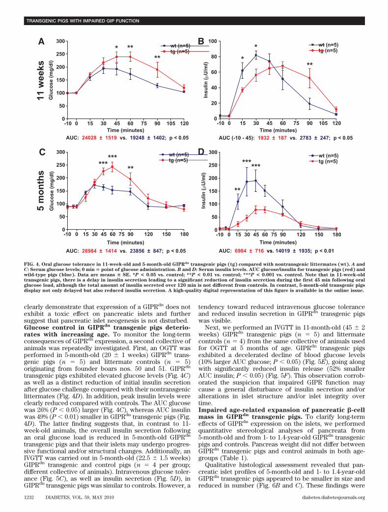
clearly demonstrate that expression of a GIPRdn does notexhibit a toxic effect on pancreatic islets and furthersuggest that pancreatic islet neogenesis is not disturbed.Glucose control in GIPRdn transgenic pigs deterio-rates with increasing age. To monitor the long-termconsequences of GIPRdn expression, a second collective ofanimals was repeatedly investigated. First, an OGTT wasperformed in 5-month-old (20 � 1 weeks) GIPRdn trans-genic pigs (n � 5) and littermate controls (n � 5)originating from founder boars nos. 50 and 51. GIPRdn
transgenic pigs exhibited elevated glucose levels (Fig. 4C)as well as a distinct reduction of initial insulin secretionafter glucose challenge compared with their nontransgeniclittermates (Fig. 4D). In addition, peak insulin levels wereclearly reduced compared with controls. The AUC glucosewas 26% (P 0.05) larger (Fig. 4C), whereas AUC insulinwas 49% (P 0.01) smaller in GIPRdn transgenic pigs (Fig.4D). The latter finding suggests that, in contrast to 11-week-old animals, the overall insulin secretion followingan oral glucose load is reduced in 5-month-old GIPRdn
transgenic pigs and that their islets may undergo progres-sive functional and/or structural changes. Additionally, anIVGTT was carried out in 5-month-old (22.5 � 1.5 weeks)GIPRdn transgenic and control pigs (n � 4 per group;different collective of animals). Intravenous glucose toler-ance (Fig. 5C), as well as insulin secretion (Fig. 5D), inGIPRdn transgenic pigs was similar to controls. However, a
tendency toward reduced intravenous glucose toleranceand reduced insulin secretion in GIPRdn transgenic pigswas visible.
Next, we performed an IVGTT in 11-month-old (45 � 2weeks) GIPRdn transgenic pigs (n � 5) and littermatecontrols (n � 4) from the same collective of animals usedfor OGTT at 5 months of age. GIPRdn transgenic pigsexhibited a decelerated decline of blood glucose levels(10% larger AUC glucose; P 0.05) (Fig. 5E), going alongwith significantly reduced insulin release (52% smallerAUC insulin; P 0.05) (Fig. 5F). This observation corrob-orated the suspicion that impaired GIPR function maycause a general disturbance of insulin secretion and/oralterations in islet structure and/or islet integrity overtime.Impaired age-related expansion of pancreatic �-cellmass in GIPRdn transgenic pigs. To clarify long-termeffects of GIPRdn expression on the islets, we performedquantitative stereological analyses of pancreata from5-month-old and from 1- to 1.4-year-old GIPRdn transgenicpigs and controls. Pancreas weight did not differ betweenGIPRdn transgenic pigs and control animals in both age-groups (Table 1).
Qualitative histological assessment revealed that pan-creatic islet profiles of 5-month-old and 1- to 1.4-year-oldGIPRdn transgenic pigs appeared to be smaller in size andreduced in number (Fig. 6B and C). These findings were
AUC: 24028 ± 1519 vs. 19248 ± 1402; p < 0.05
AUC: 28984 ± 1414 vs. 23856 ± 847; p < 0.05
AUC (-10 - 45): 1932 ± 187 vs. 2783 ± 247; p < 0.05
AUC: 6984 ± 716 vs. 14019 ± 1935; p < 0.01
C D
-10 0 15 30 45 60 75 90 105 1200
50
100
150
200
250
300 wt (n=5) tg (n=5)* **
**
Time (minutes)
Glu
cose
(mg/
dl)
A B
-10 0 15 30 45 60 75 90 105 1200
20
40
60
80
100 wt (n=5) tg (n=5)
**
**
Time (minutes)
Insu
lin ( µ
U/m
l)
-10 0 15 30 45 60 75 90 120 150 1800
50
100
150
200
250
300 wt (n=5) tg (n=5)***
*****
Time (minutes)
Glu
cose
(mg/
dl)
-10 0 15 30 45 60 75 90 120 150 1800
50
100
150
200
250
300 wt (n=5) tg (n=5)
**
******
Time (minutes)
Insu
lin (µ
U/m
l)
5 m
onth
s11
wee
ks
FIG. 4. Oral glucose tolerance in 11-week-old and 5-month-old GIPRdn transgenic pigs (tg) compared with nontransgenic littermates (wt). A andC: Serum glucose levels; 0 min � point of glucose administration. B and D: Serum insulin levels. AUC glucose/insulin for transgenic pigs (red) andwild-type pigs (blue). Data are means � SE. *P < 0.05 vs. control; **P < 0.01 vs. control; ***P < 0.001 vs. control. Note that in 11-week-oldtransgenic pigs, there is a delay in insulin secretion leading to a significant reduction of insulin secretion during the first 45 min following oralglucose load, although the total amount of insulin secreted over 120 min is not different from controls. In contrast, 5-month-old transgenic pigsdisplay not only delayed but also reduced insulin secretion. A high-quality digital representation of this figure is available in the online issue.
TRANSGENIC PIGS WITH IMPAIRED GIP FUNCTION
1232 DIABETES, VOL. 59, MAY 2010 diabetes.diabetesjournals.org

confirmed by quantitative stereological investigations. In5-month-old GIPRdn transgenic pigs (n � 4), the totalvolume of �-cells [V(�-cell, Pan)] was diminished by 35% (P 0.05) versus controls (n � 4) (Fig. 6B). In 1- to 1.4-year-oldGIPRdn transgenic pigs (n � 5), the reduction of total�-cell volume compared with controls (n � 5) was evenmore pronounced (58%; P 0.01) (Fig. 6C). Reduced�-cell mass of young adult GIPRdn transgenic pigs com-pared with controls was confirmed by islet isolation
experiments. The number of islet equivalents recoveredfrom pancreas samples of GIPRdn transgenic pigs (n � 3)was reduced by 93% (P 0.05) as compared with litter-mate controls (n � 3) (supplementary Table 2).
In contrast, volume density (data not shown) as well asthe total volume of isolated �-cells were not differentbetween GIPRdn transgenic and control pigs, neither at 5months of age (121 � 18 vs. 127 � 15 mm3; P � 0.883) norat 1–1.4 years of age (77 � 8 vs. 71 � 5 mm3; P � 0.844).
-15 0 10 30 60 90 120 150 1800
100
200
300
400
500
600
tg (n = 5) wt (n = 4)
* *
**
Time (minutes)
Glu
cose
(mg/
dl)
-10 0 10 20 30 40 50 60 70 80 900
50
100
150
200
250
300
350
400 wt (n=4) tg (n=4)
Time (minutes)
Glu
cose
(mg/
dl)
-10 0 10 20 30 40 50 60 70 80 900
50
100
150
200
250
300
350 wt (n=6) tg (n=5)
Time (minutes)
Glu
cose
(mg/
dl)
-10 0 10 20 30 40 50 60 70 80 900
10
20
30
40
50 wt (n=6) tg (n=5)
Time (minutes)
Insu
lin (µ
U/m
l)
FE
A B
C D
-10 0 10 20 30 40 50 60 70 80 900
20
40
60
80 wt (n=4) tg (n=4)
Time (minutes)
Insu
lin ( µ
U/m
l)
-15 0 10 30 60 90 120 150 1800
25
50
75
100
125
150
175
200
tg (n=5) wt (n=4)
********
******
Time (minutes)
Insu
lin ( µ
U/m
l)
11 m
onth
s11
wee
ks5
mon
ths
AUC: 13562 ± 359 vs. 14315 ± 651; p = 0.663
AUC: 14252 ± 1262 vs. 12733 ± 868; p = 0.165
AUC: 24802 ± 633 vs. 22536 ± 509; p < 0.05 AUC: 2782 ± 275 vs. 5806 ± 1272; p< 0.05
AUC: 1634 ± 191 vs. 1982 ± 243; p = 0.106
AUC: 1135 ± 111 vs. 1254 ± 269; p = 0.664
FIG. 5. Intravenous glucose tolerance in GIPRdn transgenic pigs (tg) compared with nontransgenic controls (wt). A, C, and E: Serum glucoselevels; 0 min � point of glucose administration. B, D, and F: Serum insulin levels. AUC glucose/insulin for transgenic pigs (red) and wild-type pigs(blue). Data are means � SE. *P < 0.05 vs. control; **P < 0.01 vs. control; ***P < 0.001 vs. control. Note that intravenous glucose tolerance (A)and insulin secretion (B) are not altered in 11-week-old transgenic pigs. In 5-month-old transgenic pigs, a tendency of reduced insulin secretion(D) is observed, while 11-month-old transgenic pigs display a significantly reduced intravenous glucose tolerance (E) due to a significantlyreduced insulin secretion (F). (A high-quality digital representation of this figure is available in the online issue.)
S. RENNER AND ASSOCIATES
diabetes.diabetesjournals.org DIABETES, VOL. 59, MAY 2010 1233

These data clearly demonstrate an age-related reduction ofpancreatic �-cell mass expansion in GIPRdn transgenicpigs, which is in line with previous evidence for a trophicaction of GIP on �-cells in vitro (5–7).Altered cellular composition of islets in GIPRdn
transgenic pigs. To evaluate effects of GIPRdn expressionon the volume densities of the various endocrine islet cellsand their total volumes, we performed detailed stereologi-cal analyses of the pancreatic islets in all three age classesinvestigated. In control animals, total volumes of �-, �-, �-,and pp-cells in established islets increased significantlywith age (Table 1). In GIPRdn transgenic pigs, a similarage-dependent increase was seen for the total volumes of�-, �-, and pp-cells. However, in comparison with controls,the increase of total �-cell volume of GIPRdn transgenicpigs was less pronounced from 11 weeks to 5 months ofage. Importantly, there was no further augmentation oftotal �-cell volume in 1- to 1.4-year-old GIPRdn transgenicpigs, demonstrating that impaired GIPR function inter-feres with the physiological expansion of pancreatic�-cells. In addition, the fractional volume of �-cells in theislets was decreased, while that of �- and �-cells was
increased in 1- to 1.4-year-old GIPRdn transgenic pigs.However, the total volumes of these non–�-cell popula-tions were not different from those of age-matched controlpigs (Table 1).Reduced proliferation rate of �-cells in GIPRdn trans-genic pigs. To clarify the mechanism of impaired �-cellexpansion in GIPRdn transgenic pigs, we determined �-cellproliferation by double immunohistochemical staining forinsulin and the proliferation marker Ki67 in all threeage-groups. Indeed, �-cell proliferation was significantlyreduced by 60% (P 0.05) in 11-week-old GIPRdn trans-genic pigs (Fig. 7A and B). In addition, we performeddouble immunohistochemical staining for insulin and theapoptosis marker cleaved caspase-3 to evaluate a potentialimpact of GIPRdn expression on cell death in the �-cellcompartment. Overall, the proportion of cleaved caspase-3positive cells was very low, with no significant differencebetween GIPRdn transgenic pigs and controls of all ageclasses. However, there was a trend (P � 0.075) of morecleaved caspase-3 positive �-cells in 1- to 1.4-year-oldGIPRdn transgenic pigs as compared with age-matchedcontrols (Fig. 7C and D).
1-1.
4 ye
ars
0
500
1000
1500
2000
2500
**
5 m
onth
s
0
200
400
600
800
1000
1200
1400
*
11 w
eeks
wt tg
wt tg
wt tg
0
50
100
150
200
250
300p=0.843
V (β
-cel
l, Pa
n) (m
m3 )
V (β
-cel
l, Pa
n) (m
m3 )
V (β
-cel
l, Pa
n) (m
m3 )
A
B
C
wt
wt
tg
tg
wt tg
FIG. 6. Immunohistochemistry for insulin and total �-cell volume in the pancreas [V(�-cell, Pan)] determined with quantitative stereologicalmethods. A–C: Representative histological sections of pancreatic tissue from a control (wt) and a GIPRdn transgenic pig (tg); scale bar � 200 �m.Unaltered total �-cell volume in 11-week-old GIPRdn transgenic pigs (n � 5 per group) (A) but reduction of the total �-cell volume in 5-month-old(n � 4 per group) (B) and young adult (1–1.4 years old) (n � 5 per group) GIPRdn transgenic pigs (C) compared with controls. Data are means �SE. *P < 0.05 vs. control; **P < 0.01 vs. control. A high-quality digital representation of this figure is available in the online issue.
TRANSGENIC PIGS WITH IMPAIRED GIP FUNCTION
1234 DIABETES, VOL. 59, MAY 2010 diabetes.diabetesjournals.org

DISCUSSION
This study established the first transgenic large animalmodel of impaired incretin function. The cDNA of thehuman GIPR was mutated at the third intracellular loop,where a deletion of eight amino acids (positions 319–326)and a point mutation at position 340 was introduced. Instably transfected Chinese hamster lymphoblast (CHL)cells, GIPRdn bound GIP with normal affinity but failed toincrease intracellular cAMP levels. Thus, the GIPRdn ex-pressed in transgenic pigs is capable of ligand binding butnot of signal transduction (16) and competes with theendogenous GIPR for GIP. Consequently, the insulino-tropic effect of GIP is highly reduced but not completelyeliminated, mirroring the situation in human type 2diabetes.
In view of the severe, early-onset diabetes of GIPRdn
transgenic mice (16), we tested the oral and intravenousglucose tolerance of young (11-week-old) GIPRdn trans-genic and control pigs and determined their pancreatic�-cell mass by quantitative stereological analyses. Thefinding of reduced oral glucose tolerance associated withdelayed insulin secretion is in line with a disturbance ofthe incretin effect. Reduced oral glucose tolerance inGIPRdn transgenic pigs is also consistent with previousobservations in Gipr�/� mice (14). Importantly, normalintravenous glucose tolerance, insulin secretion, and un-changed �-cell mass in 11-week-old GIPRdn transgenic pigsstrongly argue against a toxic effect of GIPRdn expression.The reasons for different outcomes in the GIPRdn trans-
genic pig and the GIPRdn transgenic mouse model remainunclear but may be related to different methods of trans-genesis (lentiviral transgenesis versus pronuclear DNAmicroinjection) or different copy numbers and/or integra-tion sites leading to different expression levels of thetransgene.
To evaluate long-term effects of GIPRdn expression inthe pancreatic islets, we performed a longitudinal study ofa collective of animals, involving OGTTs and IVGTTs.These revealed a progressive deterioration of glucosecontrol in GIPRdn transgenic pigs, although none of ourtransgenic pigs has developed fasting hyperglycemia up toan age of 2 years. Quantitative stereological investigationsof pancreata from 5-month-old and from 1- to 1.4-year-oldGIPRdn transgenic pigs showed a reduced pancreatic�-cell mass, which was confirmed by quantitative isletisolation experiments.
Quantitative stereological analyses of the pancreaticislets demonstrated a marked increase of total �-cellvolume from 11 weeks to 5 months (6.4-fold) and from 5months to 1–1.4 years (1.6-fold) of age in control pigs. Incontrast, the expansion of total �-cell volume in GIPRdn
transgenic pigs was less pronounced between 11 weeksand 5 months of age (4.3-fold), with no further increase in1- to 1.4-year-old animals. These findings are explained bya markedly reduced proliferation rate of �-cells in 11-week-old GIPRdn transgenic pigs, a developmental stagecharacterized by massive expansion of �-cells in pigs (36).Staining for cleaved caspase-3 did not show a significantly
TABLE 1Quantitative stereological analyses of the endocrine pancreas of GIPRdn transgenic pigs (tg) and wild-type control pigs (wt)
Parameter
11 weeks(n � 5 wt, 5 tg)
5 months(n � 4 wt, 4 tg)
1–1.4 years(n � 5 wt, 5 tg) ANOVA
Means � SE Means � SE Means � SE Group Age Group age
Pancreas weight (g)Wild type 34.5 � 4.2 115.3 � 5.6 183.4 � 13.8 NS 0.0001 NSTransgenic 32.7 � 3.1 125.7 � 6.1 206.1 � 4.9
VV(�-cell/islet) (%)Wild type 69.8 � 2.2 89.0 � 1.5 90.2 � 1.2 0.0066 0.0001 0.0024Transgenic 70.8 � 1.3 87.0 � 1.2 76.4 � 3.2*
VV(�-cell/islet) (%)Wild type 14.1 � 1.2 5.0 � 0.8 5.0 � 0.7 0.0122 0.0001 0.0008Transgenic 12.2 � 0.7 6.5 � 0.7 13.8 � 2.3*
VV(�-cell/islet) (%)Wild type 13.5 � 2.8 4.3 � 0.8 2.2 � 0.6 NS 0.0001 NSTransgenic 13.0 � 1.0 5.5 � 0.9 5.8 � 0.9†
VV(pp-cell/islet) (%)Wild type 2.7 � 0.7 1.8 � 0.3 2.8 � 0.8 NS 0.0355 NSTransgenic 3.9 � 0.5 1.3 � 0.3 3.8 � 1.1
V(�-cell/islet) (mm3)Wild type 168.7 � 29.5 1,088.2 � 82.0 1,694.6 � 251.7 0.0002 0.0001 0.0024Transgenic 152.1 � 17.1 664.1 � 74.5† 663.7 � 130.5*
V(�-cell/islet) (mm3)Wild type 32.6 � 8.7 58.4 � 6.3 95.7 � 17.4 NS 0.0001 NSTransgenic 24.5 � 1.7 47.7 � 4.5 112.8 � 14.5
V(�-cell/islet) (mm3)Wild type 26.6 � 3.9 49.5 � 6.1 36.8 � 6.3 NS 0.0014 NSTransgenic 25.9 � 1.8 39.6 � 3.5 47.5 � 5.2
V(pp-cell/islet) (mm3)Wild type 6.0 � 1.8 20.7 � 2.9 52.4 � 12.6 NS 0.0001 NSTransgenic 8.2 � 1.7 9.3 � 2.1 30.3 � 7.9
Data were analyzed by the general linear models procedure (SAS Institute) taking the effects of group (wild type, transgenic), age (11 weeks,5 months, 1–1.4 years), and the interaction group age into account. For significant effects of these factors P values are indicated in the lastthree columns. In addition, significant differences between groups within age classes are marked by: *P 0.01; †P 0.05; NS, not significant.
S. RENNER AND ASSOCIATES
diabetes.diabetesjournals.org DIABETES, VOL. 59, MAY 2010 1235

increased rate of cell death in the �-cell compartment ofGIPRdn transgenic pigs versus controls, although a trendof higher numbers of cleaved caspase-3 positive �-cellswas visible in 1- to 1.4-year-old GIPRdn transgenic pigs.This may suggest a contribution of apoptosis to thereduction of total �-cell volume in mature GIPRdn trans-genic pigs.
Due to the reduced volume fraction of �-cells in theislets of 1- to 1.4-year-old GIPRdn transgenic pigs, therelative volumes of �- and �-cells in the islets wereincreased. However, since the islet volume was concomi-tantly reduced, the absolute volumes of �- and �-cells werenot different between GIPRdn transgenic and control pigs.
The numbers of animals investigated in our study are, inpart, smaller than in some rodent studies. However, due tothe large size of the pig, several blood-based parameterscould be measured repeatedly with short time intervals inthe same animals, providing an important advantage forstatistical analysis. Further, the stereological data of allanimals (n � 28) were evaluated together by ANOVA,demonstrating significant group effects (Table 1) with Pvalues 0.01 for many data/differences supporting ourcore statements.
Interestingly, Gipr�/�mice provided no evidence thatGIPR action is required for the maintenance of islet and�-cell integrity in vivo (15,37). These mice exhibited anincrease in relative �-cell area referring to pancreas area(37), leading to the conclusion that in vivo the function ofGIP is primarily restricted to that of an incretin (15).However, the relatively mild phenotype of Gipr�/� micemay result from compensatory mechanisms (15). Althoughmice lacking both GIPR and GLP-1R exhibited moresevere glucose intolerance than the individual mutants(38,39) these double mutant animals did not developdiabetes, raising the suspicion of the existence of compen-satory mechanisms other than the GIP/GLP-1 system (38).The findings in GIPRdn transgenic pigs suggest that, inaddition to its role as an incretin hormone, GIP is neces-sary for the expansion of �-cell mass and that its partialloss of function cannot be fully compensated by hyperac-tivation of the GLP-1/GLP-1R system.
In conclusion, GIPRdn transgenic pigs resemble charac-teristic features of human type 2 diabetic patients veryclosely in the following ways: disturbed GIP function,glucose intolerance, and reduced pancreatic �-cell mass.Moreover, GIPRdn transgenic pigs may be an attractive
5 months 1-1.4 years11 weeks 5 months 1-1.4 years11 weeks0
1000
2000
3000
4000
5000
6000
7000
8000
9000
10000
*p=0.549
p=0.352Ki6
7+ n
ucle
i/105
nuc
lear
pro
files
0
10
20
30
40p=0.313
p=0.336 p=0.075C
asp3
+ nu
clei
/105
nuc
lear
pro
files
A
B
C
D
FIG. 7. �-Cell proliferation and apoptosis. A and C: Representative histological sections doublestained for insulin (blue) and Ki67 (brown) (A)or for insulin (light brown) plus cleaved caspase-3 (dark blue; see arrow) (C). B and D: Determination of the number of Ki67� (B) and cleavedcaspase-3–positive �-cells (D). Wild type: blue bars, transgenic: red bars. Wild type: n � 5, transgenic: n � 5 for 11-week-old and 1- to 1.4-year-oldpigs; wild type: n � 4, transgenic: n � 4 for 5-month-old pigs. Data are means � SE. *P < 0.05 vs. control; scale bar � 20 �m. Note the significantly(P < 0.05) reduced �-cell proliferation rate in 11-week-old GIPRdn transgenic pigs. (A high-quality digital representation of this figure is availablein the online issue.)
TRANSGENIC PIGS WITH IMPAIRED GIP FUNCTION
1236 DIABETES, VOL. 59, MAY 2010 diabetes.diabetesjournals.org

animal model for the development and preclinical evalua-tion of incretin-based therapeutic strategies (40). Anotherpotential application of GIPRdn transgenic pigs is thedevelopment of novel techniques for dynamic in vivomonitoring of pancreatic islet mass (41). Due to their sizeand close physiological and anatomical similarities tohumans (42), pigs represent attractive animal models fortranslating novel therapeutic and diagnostic principlesinto clinical practice.
ACKNOWLEDGMENTS
This study was supported by the Deutsche Forschungsge-meinschaft (GRK 1029), the Bayerische Forschungsstiftung(492/02), and the Diabetes Hilfs- und ForschungsfondsDeutschland (DHFD).
No potential conflicts of interest relevant to this articlewere reported.
Parts of this work were presented in abstract form at the51st Annual Meeting of the German Society of Endocrinol-ogy, Salzburg, Austria, 7–10 March 2007; the 42nd AnnualMeeting of the German Diabetes Society, Hamburg, Ger-many, 16–19 May 2007; the 34th Annual Conference of theInternational Embryo Transfer Society, Denver, Colorado,5–9 January 2008; the 44th Annual Meeting of the GermanDiabetes Society, Leipzig, Germany, 20–23 May 2008; the68th Scientific Sessions of the American Diabetes Associ-ation, San Francisco, California, 6–10 June 2008; and the69th Scientific Sessions of the American Diabetes Associ-ation, New Orleans, Louisiana, 5–9 June 2009.
The authors thank Prof. Dr. Karl Heinritzi, Prof. Dr.Holm Zerbe, and Dr. Birgit Rathkolb for the generoussupport of this study; Prof. Dr. Helmut Kuechenhoff (Sta-BLab, Ludwig Maximilians University Munich) for experthelp with the statistical analysis of longitudinal data; andTamara Holy, Lisa Pichl, Bianca Schneiker, Elfi Holupirek,Christian Erdle, and Siegfried Elsner for excellent techni-cal assistance and animal management.
REFERENCES
1. Baggio LL, Drucker DJ. Biology of incretins: GLP-1 and GIP. Gastroenter-ology 2007;132:2131–2157
2. Mayo KE, Miller LJ, Bataille D, Dalle S, Goke B, Thorens B, DruckerDJ. International Union of Pharmacology. XXXV. The glucagon receptorfamily. Pharmacol Rev 2003;55:167–194
3. Holst JJ, Gromada J. Role of incretin hormones in the regulation of insulinsecretion in diabetic and nondiabetic humans. Am J Physiol EndocrinolMetab 2004;287:E199–E206
4. Saxena R, Hivert MF, Langenberg C, Tanaka T, Pankow JS, VollenweiderP, Lyssenko V, Bouatia-Naji N, Dupuis J, Jackson AU, Kao WH, Li M, GlazerNL, Manning AK, Luan J, Stringham HM, Prokopenko I, Johnson T, GrarupN, Boesgaard TW, Lecoeur C, Shrader P, O’Connell J, Ingelsson E, CouperDJ, Rice K, Song K, Andreasen CH, Dina C, Kottgen A, Le Bacquer O,Pattou F, Taneera J, Steinthorsdottir V, Rybin D, Ardlie K, Sampson M, QiL, van Hoek M, Weedon MN, Aulchenko YS, Voight BF, Grallert H, BalkauB, Bergman RN, Bielinski SJ, Bonnefond A, Bonnycastle LL, Borch-Johnsen K, Bottcher Y, Brunner E, Buchanan TA, Bumpstead SJ, Cavalca-nti-Proenca C, Charpentier G, Chen YD, Chines PS, Collins FS, Cornelis M,G JC, Delplanque J, Doney A, Egan JM, Erdos MR, Firmann M, Forouhi NG,Fox CS, Goodarzi MO, Graessler J, Hingorani A, Isomaa B, Jorgensen T,Kivimaki M, Kovacs P, Krohn K, Kumari M, Lauritzen T, Levy-Marchal C,Mayor V, McAteer JB, Meyre D, Mitchell BD, Mohlke KL, Morken MA,Narisu N, Palmer CN, Pakyz R, Pascoe L, Payne F, Pearson D, RathmannW, Sandbaek A, Sayer AA, Scott LJ, Sharp SJ, Sijbrands E, Singleton A,Siscovick DS, Smith NL, Sparso T, Swift AJ, Syddall H, Thorleifsson G,Tonjes A, Tuomi T, Tuomilehto J, Valle TT, Waeber G, Walley A, Water-worth DM, Zeggini E, Zhao JH, Illig T, Wichmann HE, Wilson JF, van DuijnC, Hu FB, Morris AD, Frayling TM, Hattersley AT, Thorsteinsdottir U,Stefansson K, Nilsson P, Syvanen AC, Shuldiner AR, Walker M, BornsteinSR, Schwarz P, Williams GH, Nathan DM, Kuusisto J, Laakso M, Cooper C,
Marmot M, Ferrucci L, Mooser V, Stumvoll M, Loos RJ, Altshuler D, PsatyBM, Rotter JI, Boerwinkle E, Hansen T, Pedersen O, Florez JC, McCarthyMI, Boehnke M, Barroso I, Sladek R, Froguel P, Meigs JB, Groop L,Wareham NJ, Watanabe RM: Genetic variation in GIPR influences theglucose and insulin responses to an oral glucose challenge. Nat Genet2010;42:142–148
5. Ehses JA, Casilla VR, Doty T, Pospisilik JA, Winter KD, Demuth HU,Pederson RA, McIntosh CH. Glucose-dependent insulinotropic polypeptidepromotes beta-(INS-1) cell survival via cyclic adenosine monophosphate-mediated caspase-3 inhibition and regulation of p38 mitogen-activatedprotein kinase. Endocrinology 2003;144:4433–4445
6. Trumper A, Trumper K, Horsch D. Mechanisms of mitogenic and anti-apoptotic signaling by glucose-dependent insulinotropic polypeptide inbeta(INS-1)-cells. J Endocrinol 2002;174:233–246
7. Trumper A, Trumper K, Trusheim H, Arnold R, Goke B, Horsch D.Glucose-dependent insulinotropic polypeptide is a growth factor for beta(INS-1) cells by pleiotropic signaling. Mol Endocrinol 2001;15:1559–1570
8. Kim SJ, Winter K, Nian C, Tsuneoka M, Koda Y, McIntosh CH. Glucose-dependent insulinotropic polypeptide (GIP) stimulation of pancreaticbeta-cell survival is dependent upon phosphatidylinositol 3-kinase (PI3K)/protein kinase B (PKB) signaling, inactivation of the forkhead transcrip-tion factor Foxo1, and down-regulation of bax expression. J Biol Chem2005;280:22297–22307
9. Yusta B, Baggio LL, Estall JL, Koehler JA, Holland DP, Li H, Pipeleers D,Ling Z, Drucker DJ. GLP-1 receptor activation improves beta cell functionand survival following induction of endoplasmic reticulum stress. CellMetab 2006;4:391–406
10. Meier JJ, Hucking K, Holst JJ, Deacon CF, Schmiegel WH, Nauck MA.Reduced insulinotropic effect of gastric inhibitory polypeptide in first-degree relatives of patients with type 2 diabetes. Diabetes 2001;50:2497–2504
11. Nauck MA, Heimesaat MM, Orskov C, Holst JJ, Ebert R, Creutzfeldt W.Preserved incretin activity of glucagon-like peptide 1 [7–36 amide] but notof synthetic human gastric inhibitory polypeptide in patients with type-2diabetes mellitus. J Clin Invest 1993;91:301–307
12. Drucker DJ, Nauck MA. The incretin system: glucagon-like peptide-1receptor agonists and dipeptidyl peptidase-4 inhibitors in type 2 diabetes.Lancet 2006;368:1696–1705
13. Nauck MA, Baller B, Meier JJ. Gastric inhibitory polypeptide and glucagon-like peptide-1 in the pathogenesis of type 2 diabetes. Diabetes 2004;53(Suppl. 3):S190–S196
14. Miyawaki K, Yamada Y, Yano H, Niwa H, Ban N, Ihara Y, Kubota A,Fujimoto S, Kajikawa M, Kuroe A, Tsuda K, Hashimoto H, Yamashita T,Jomori T, Tashiro F, Miyazaki J, Seino Y. Glucose intolerance caused by adefect in the entero-insular axis: a study in gastric inhibitory polypeptidereceptor knockout mice. Proc Natl Acad Sci U S A 1999;96:14843–14847
15. Hansotia T, Drucker DJ. GIP and GLP-1 as incretin hormones: lessons fromsingle and double incretin receptor knockout mice. Regul Pept 2005;128:125–134
16. Herbach N, Goeke B, Schneider M, Hermanns W, Wolf E, Wanke R.Overexpression of a dominant negative GIP receptor in transgenic miceresults in disturbed postnatal pancreatic islet and beta-cell development.Regul Pept 2005;125:103–117
17. Hofmann A, Kessler B, Ewerling S, Weppert M, Vogg B, Ludwig H,Stojkovic M, Boelhauve M, Brem G, Wolf E, Pfeifer A. Efficient transgen-esis in farm animals by lentiviral vectors. EMBO Rep 2003;4:1054–1060
18. Pfeifer A, Ikawa M, Dayn Y, Verma IM. Transgenesis by lentiviral vectors:lack of gene silencing in mammalian embryonic stem cells and preimplan-tation embryos. Proc Natl Acad Sci U S A 2002;99:2140–2145
19. Klose R, Kemter E, Bedke T, Bittmann I, Kelsser B, Endres R, Pfeffer K,Schwinzer R, Wolf E. Expression of biologically active human TRAIL intransgenic pigs. Transplantation 2005;80:222–230
20. Larsen MO, Rolin B, Ribel U, Wilken M, Deacon CF, Svendsen O,Gotfredsen CF, Carr RD. Valine pyrrolidide preserves intact glucose-dependent insulinotropic peptide and improves abnormal glucose toler-ance in minipigs with reduced beta-cell mass. Exp Diabesity Res 2003;4:93–105
21. Moritz MW, Dawe EJ, Holliday JF, Elliott S, Mattei JA, Thomas AL. Chroniccentral vein catheterization for intraoperative and long-term venous accessin swine. Lab Anim Sci 1989;39:153–155
22. Kobayashi K, Kobayashi N, Okitsu T, Yong C, Fukazawa T, Ikeda H, KosakaY, Narushima M, Arata T, Tanaka N. Development of a porcine model oftype 1 diabetes by total pancreatectomy and establishment of a glucosetolerance evaluation method. Artif Organs 2004;28:1035–1042
23. Krickhahn M, Meyer T, Buhler C, Thiede A, Ulrichs K. Highly efficientisolation of porcine islets of Langerhans for xenotransplantation: numbers,purity, yield and in vitro function. Ann Transplant 2001;6:48–54
S. RENNER AND ASSOCIATES
diabetes.diabetesjournals.org DIABETES, VOL. 59, MAY 2010 1237

24. Ricordi C, Socci C, Davalli AM, Staudacher C, Baro P, Vertova A, Sassi I,Gavazzi F, Pozza G, Di Carlo V. Isolation of the elusive pig islet. Surgery1990;107:688–694
25. van der Burg MP, Graham JM. Iodixanol density gradient preparation inuniversity of wisconsin solution for porcine islet purification. Scientific-WorldJournal 2003;3:1154–1159
26. Latif ZA, Noel J, Alejandro R. A simple method of staining fresh andcultured islets. Transplantation 1988;45:827–830
27. Jones KH, Senft JA. An improved method to determine cell viability bysimultaneous staining with fluorescein diacetate-propidium iodide. J His-tochem Cytochem 1985;33:77–79
28. Scherle W. A simple method for volumetry of organs in quantitativestereology. Mikroskopie 1970;26:57–60
29. Bonner-Weir S, Baxter LA, Schuppin GT, Smith FE. A second pathway forregeneration of adult exocrine and endocrine pancreas: a possible reca-pitulation of embryonic development. Diabetes 1993;42:1715–1720
30. Petrik J, Reusens B, Arany E, Remacle C, Coelho C, Hoet JJ, Hill DJ. A lowprotein diet alters the balance of islet cell replication and apoptosis in thefetal and neonatal rat and is associated with a reduced pancreaticexpression of insulin-like growth factor-II. Endocrinology 1999;140:4861–4873
31. Xu G, Stoffers DA, Habener JF, Bonner-Weir S. Exendin-4 stimulates both�-cell replication and neogenesis, resulting in increased �-cell mass andimproved glucose tolerance in diabetic rats. Diabetes 1999;48:2270–2276
32. Herbach N, Rathkolb B, Kemter E, Pichl L, Klaften M, de Angelis MH,Halban PA, Wolf E, Aigner B, Wanke R. Dominant-negative effects of anovel mutated Ins2 allele causes early-onset diabetes and severe �-cell lossin Munich Ins2C95S mutant mice. Diabetes 2007;56:1268–1276
33. Iatropoulos MJ, Williams GM. Proliferation markers. Exp Toxicol Pathol1996;48:175–181
34. Hui H, Dotta F, Di Mario U, Perfetti R. Role of caspases in the regulationof apoptotic pancreatic islet beta-cells death. J Cell Physiol 2004;200:177–200
35. Verbeke G, Molenberghs G. Linear Mixed Models for Longitudinal Data.New York, Springer, 2001
36. Bock T, Kyhnel A, Pakkenberg B, Buschard K. The postnatal growth of thebeta-cell mass in pigs. J Endocrinol 2003;179:245–252
37. Pamir N, Lynn FC, Buchan AM, Ehses J, Hinke SA, Pospisilik JA, MiyawakiK, Yamada Y, Seino Y, McIntosh CH, Pederson RA. Glucose-dependentinsulinotropic polypeptide receptor null mice exhibit compensatorychanges in the enteroinsular axis. Am J Physiol Endocrinol Metab 2003;284:E931–E939
38. Hansotia T, Baggio LL, Delmeire D, Hinke SA, Yamada Y, Tsukiyama K,Seino Y, Holst JJ, Schuit F, Drucker DJ. Double incretin receptor knockout(DIRKO) mice reveal an essential role for the enteroinsular axis intransducing the glucoregulatory actions of DPP-IV inhibitors. Diabetes2004;53:1326–1335
39. Preitner F, Ibberson M, Franklin I, Binnert C, Pende M, Gjinovci A,Hansotia T, Drucker DJ, Wollheim C, Burcelin R, Thorens B. Gluco-incretins control insulin secretion at multiple levels as revealed in micelacking GLP-1 and GIP receptors. J Clin Invest 2004;113:635–645
40. Shaffer C. Incretin mimetics vie for slice of type 2 diabetes market. NatBiotechnol 2007;25:263
41. Medarova Z, Moore A. MRI as a tool to monitor islet transplantation. NatRev Endocrinol 2009;5:444–452
42. Larsen MO, Rolin B. Use of the Gottingen minipig as a model of diabetes,with special focus on type 1 diabetes research. ILAR J 2004;45:303–313
TRANSGENIC PIGS WITH IMPAIRED GIP FUNCTION
1238 DIABETES, VOL. 59, MAY 2010 diabetes.diabetesjournals.org