genotoxicity studies with paracetamol
TRANSCRIPT

Mutation Research, 138 (1984) 21-32 21 Elsevier
MTR 0919
Genotoxicity studies with paracetamol
Er ik D y b i n g 1, J o r n A. H o l m e 1, W. P e r r y G o r d o n 2, Er ik J. S o d e r l u n d 1,
D a v i d C. D a h l i n 2 a n d S i d n e y D. N e l s o n 2
I Department of Toxicology, National Institute of Public llealth, Oslo 1 (Norway) and 2 Department of Medicinal Chemistry, University of Washington, Seattle, WA 98195 (U.S.A.)
(Received 5 April 1984) (Accepted 27 June 1984)
Summa~
Paracetamol and its major ultimate reactive metabolite, N-acetyl-p-benzoquinone imine (NAPQI) were studied for their genotoxic potential. Neither paracetamol nor NAPQI were found to cause mutations in Salmonella typhimurium, whereas NAPQI was severely cytotoxic to the bacteria. Radiolabelled paracetamol was found to bind covalently to DNA added to mouse-liver microsomal incubations at a rate of 2.6 pmoles/mg D'NA/min. Paracetamol also bound covalently to hepatic DNA at a level of 15 pmoles/mg DNA after a hepatotoxic dose of paracetamol to mice. NAPQI caused extensive DNA single-strand breaks as evidenced by alkaline elution of DNA from treated Reuber hepatoma cells. This effect occurred at concentrations which later resulted in cytotoxicity. Paracetamol was shown to induce increased DNA-re- pair synthesis in isolated mouse-liver cells in monolayer culture, at concentrations where also cytotoxicity was evident. Increased DNA-repair synthesis occurred at lower paracetamol concentrations in cells isolated from mice pretreated with phenobarbital. Taken together, these data show that paracetamol can cause DNA interaction leading to damage at levels which are cytotoxic.
The widely used over-the-counter analgesic paracetamol is known at higher doses to cause acute liver necrosis in man (Prescott et al., 1971) and experimental animals (Mitchell et al., 1973a), whereas it is believed to be remarkably safe at therapeutic doses. This assumption, however, has been questioned because of the recent finding that paracetamol caused liver-cell tumors in mice after long-term feeding (Flaks and Flaks, 1983). The acute liver damage caused by paracetamol is corre-
Abbreviations: AAF, 2-acetylaminofluorene; B6 mice, C57/BLI6/BOM mice; CBI, covalent binding index; DMSO, dimethyl sulfoxide; HA, hydroxyapatite; LDH, lactate dehy- drogenase; MC, 3-methylcholanthrene; NAPQI, N-acetyl-p- benzoquinone imine; N-OH-AAF, N-hydroxy-2-acetylamino- fluorene; PB, phenobarbital; $9 fraction, 9000x g supernatant fraction; TdR, thymidine.
lated with the formation of an arylating metabolite which binds covalently to tissue nucleophilic sites (Jollow et al., 1973). At low doses, the electrophile preferentially interacts with low molecular sulf- hydryl compounds such as glutathione and is thus detoxified, whereas at higher doses vital cellular macromolecules are damaged after tissue stores of glutathione become depleted (Mitchell et al., 1973b). Initially, it was believed that the electro- philic paracetamol intermediate was generated via the formation of N-hydroxyparacetamol (Potter et al., 1973; Mitchell and Jollow, 1975). This was later found not to be the case (Hinson et al., 1979; Nelson et al., 1980). The major ultimate reactive hepatotoxic metabolite has recently been shown to be N-acetyl-p-benzoquinone imine (NAPQI) (Miner and Kissinger, 1979; Dahlin et al., 1984).
The finding that paracetamol only causes liver
0165-1218/84/$03.00 © 1984 Elsevier Science Publishers B.V.

22
tumors in mice at doses which were acutely hepatotoxic (Flaks and Flaks, 1983) raises the question whether paracetamol is carcinogenic through a genotoxic or an epigenetic mechanism (Williams, 1980). Paracetamol and N-hydroxy- paracetamol, which spontaneously forms NAPQI (Corcoran et al., 1980), have not been shown to be mutagenic in Salmonella typhimurium (Wirth et al., 1980). To address the question of the mechanism for paracetamol hepatocarcinogenicity, we have performed studies on bacterial mutagenicity, cova- lent macromolecular binding, DNA damage as well as DNA repair with paracetamol and NAPQI.
Materials and methods
Chemicals [3H-G]Paracetamol (spec. act. 3.0 Ci/mmole,
> 98.5% pure), [14C-9]AAF (spec. act. 52 mCi /m- mole, > 98% pure) and [Me-3H]thymidine (spec. act. 47 Ci/mmole, > 96% pure) were obtained from New England Nuclear, Boston, MA (U.S.A.). NAPQI was synthesized as described (Dahlin and Nelson, 1982), and kept under argon at - 2 0 ° C until use. Immediately before use it was weighed and dissolved in DMSO. Other chemicals were obtained from the following sources: Mycostatin from Squibb, Twickenham (U.K.), fetal calf serum, calf serum and Dulbecco's modified Eagle medium from Gibco, Grand Island, NY (U.S.A.); horse serum from the National Institute of Public Health, Oslo (Norway); dexamethazone, insulin, collagenase (type IV), bovine serum albumin (type V), ~-aminolevulinic acid, Nonidet P-40, hydroxy- urea, mitomycin C and deoxyribonucleic acid (type I) from Sigma, St. Louis, MO (U.S.A.); proteinase K from E. Merck, Darmstadt (F.R.G.); AAF from Koch-Light Laboratories, Colnbrook (U.K.); paracetamol and sodium phenobarbital from The Norwegian Medicinal Depot, Oslo (Norway); hy- droxyapatite from Bio-Rad Laboratories, Munich (F.R.G.). N-OH:AAF was a generous gift from Dr. S.S. Thorgeirsson, National Cancer Institute, Bethesda, MD (U.S.A.).
Animals and pretreatments Male B6 mice, weighing 20-27 g, were obtained
from the Norwegian Research Animal Centre, Na- tional Institute of Public Health, Oslo (Norway).
They were given a pelleted diet (Norwegian Stan- dard) and water ad libitum. For induction purpo- ses phenobarbital 75 mg/kg in saline was given i.p. 72, 48 and 24 h before isolation of liver cells or in vivo treatments. For determination of in vivo macromolecular covalent binding mice were given 1.5-1.8 mCi of [3H]paracetamol, 500 mg/kg, in 0.9% saline i.p. The animals were killed after 4 h.
Preparation of liver subfractions Livers were homogenized in a Teflon-glass ho-
mogenizer in 2 vol. of sterile, ice-cold 1.15% KC1 containing 20 mM Tris-buffer, pH 7.4, and 10% glycerol. $9 and microsomal fractions were pre- pared as described (Dybing and Thorgeirsson, 1977). The $9 fraction was stored as such, whereas the microsomes were stored in the Tris-KCl buffer containing 30% glycerol at - 7 0 ° C until use. Pro- tein concentrations were determined according to Lowry et al. (1951) using bovine serum albumin as standard.
Mutagenicity asssays Mutagenic activity was assayed with Salmonella
typhimurium TA98, TA100 and TA102. In some experiments the regular plate assay (Ames et al., 1975) as described (Dybing and Thorgeirsson, 1977), or a quantitative modification thereof (Soderlund et al., 1979), were used in the absence or presence of mouse liver subfractions and cofac- tors. Freshly dissolved NAPQI in DMSO was added last in the mutagenicity assays, bacteria were incubated with NAPQI for 30 min before plating in the quantitative experiments. In other experiments substances were tested in cocultures of S. typhimurium and mouse hepatocytes (Holme et al., 1983a). Hepatocytes plated on 60-mm dishes were rinsed, 2 h after plating, with Hanks ' -Hepes buffer, pH 7.4, supplemented with 1% bovine serum albumin, and then added the same buffer containing test substance and 0.1 ml of an over- night culture of S. typhimurium. After 2 h of co-incubation, the bacteria was collected by centrifugation and plated to determine the number of revertants. The genotype of colonies on the revertant plates were checked according to Ames et al. (1975). The S. typhimurium strains were generously provided by Dr. B.N. Ames, University of California, Berkeley, CA (U.S.A.).

Macromolecular covalent binding In vitro. Ice-cold reaction vessels contained
(final concentrations): 2.0 mg/ml microsomal pro- tein, 0.5 mM [3H]paracetamol (1.6 x 10³ c p m / n m o l e for protein binding, 1.7 X 10 4
cpm/nmole for DNA binding) or 0.05 mM 14C- AAF (1.5 × 10³ cpm/nmole for protein binding or 1.3 × 10⁴ cpm/nmole for DNA binding) in 25 ~1 DMSO, a NADPH-generating system with or without 0.67 mg/ml DNA in a total volumc of 3.0 ml. Incubations were carried out at 37°C in a shaking water-bath incubator. Reactions for de- termination of protein binding were stopped after 15 min by adding 1.0 ml of 30% trichloroacetic acid (TCA). For determination of DNA binding, the reactions were stopped after 15 rain with 1.0 ml 0.17 M Na-dodecylsulfate and 0.64 ml 4 M NaC1 (Neal et al., 1979). Covalent protein binding was assayed as described (Saderlund et al., 1982). DNA was extracted from microsomal incubations with 9.9 ml phenol reagent (50 g phenol, 50 ml chloroform, and 1 ml isoamyl alcohol) according to Neal et al. (1979), and washed subsequently with acetone, chloroform/ethanol (1 : 1) and ether. DNA was dissolved in Tris-KCl buffer, pH 7.4 (0.1 M). After the 100-~1 addition of a proteinase K solution (1 mg/ml) the DNA was precipitated in the presencc of 30% TCA (1.0 ml) and 2.5% bovine serum albumin (0.5 ml). The precipitate was collected by centrifugation and DNA was hydrolysed by heating in 5% TCA at 90°C for 20 min. The amounts of radioactivity incorporated into the DNA were determined by liquid-scintilla- tion counting and DNA concentrations were mea- sured according to Burton (1956).
In vivo. In the in vivo experiments, livers and femoral muscles were minced and homogenized in 4 vol. of 75 mM NaCI, 10 mM EDTA, and 10 mM Tris pH 7.8 in a teflon Potter-Elvehjem-type ho- mogenizer. 100/tl of whole homogenate was used for protein binding. Purification of DNA was per- formed essentially as described by Sagelsdorff et al. (1983) with some modifications. The rest of the homogenate was centrifuged at 9000 rpm for 20 min. The supernatant was decanted and the pellet was resuspended in Tris-buffcr (4 mi). A 10% solution (v /v) of the non-ionic detergent Nonidet P-40 up to a final concentration of 1.0% was added to the samples. The samples were incubated
23
at 4°C for 15 min after vortexing and the vortex- ing was repeated. The crude chromatin was pel- leted at 2000 rpm for 5 min and the pellet was resuspended in 0.25 M sucrose buffer (50 mM Tris-HC1, 25 mM KC1, 15 mM MgCI2, pH 8.0). After this, 0.88 M sucrose buffer was added to the bottom of the tube and the tubes were centrifuged at 3000 rpm for 20 min. The pellet was washed once with Tris buffer and centrifugated again at 3000 rpm for 20 min. The pellet was then sus- pended in 10 ml of lysing medium (8 M urea, 0.24 M sodium phosphate buffer, pH 6.8, 10 mM EDTA, 1% (w/v) SDS) and homogenized. Pro- teins were extracted under intensive shaking for 10 min with CIP (chloroform : isoamylalcohol : phe- nol 25 :1 :25) . The resulting suspension was sep- arated into layers by centrifugation at 14000 rpm for 20 min. The aqueous layer was washed again with CIP. The aqueous solution was extracted twice with 25 ml of ether to remove trace amounts of phenol. The aqueous solution was thereafter left standing overnight at room temperature and was applied to an HA column. Dry HA (2 g / g liver) was suspended in 0.014 M sodium phosphate buffer, pH 6.8 (6 vol.). The slurry was swirled gently and heated at 85°C for 15 min then was left untouched for 10 min. The fine particles were decanted. The remaining slurry was resuspended in MUP (8 M urea, 0.16 M sodium phosphate buffer, pH 6.8). The suspension was allowed to settle. The slurry was then added to glass columns (1 cm x 25 cm). The column was allowed to equi- librate and MUP was let run off. Thc aqueous solution containing nucleic acids was loaded on the column and the elution was monitored at 260 nm. Proteins and RNA were washed from the column with MUP at a flow rate of 0.5 ml /min until the transmission had returned to background values. Two bed volumes of 14 mM sodium phos- phate buffer, pH 6.8, was used to purge off the ui'ea. DNA was eluted with 0.48 M sodium phos- phate buffer, pH 6.8, and 25-30 ml of the DNA solution was collected. The samples were dialyzed at 4°C against 8 1 of 0.2 M NaCI (24 h). DNA was precipitated by adding 2 vol. ethanol and storing at - 2 0 ° C for at least 12 h. The DNA was centri- fuged for 20 min at 1500 rpm, the supernatant decanted and DNA washed with acetone. The DNA was hydrolysed in 10% HC104 and the ra-
24
< F-,
b,
Z
< z r, z <
©
< [-
< ,¢ <
z
[ -
z
E ~ l O
i <
,q.
.-!.
o
I
d
I ," .o
0
~E o
~ ; > .
$ < < ~ < .=
~o,-, :~ ~ z+

dioactivity determined by scintillation counting. The amount of DNA was determined by diphenyl- amine assay (Burton, 1956). 100 ~1 of whole ho- mogenate was added to 1.0 mi of cold 5% TCA, vortexed and was let standing for 10 min. Cova- lent protein binding was assayed as described (Soderlund et al., 1982). The amount of protein was measured by the Lowry assay (1951).
DNA damage in hepatorna cells DNA damage of Reuber H4-II -E rat hepatoma
cells was determined by alkaline elution (Kohn et al., 1981). The cells were grown in Dulbecco's minimal essential medium supplemented with 10% calf serum and 10% fetal calf serum and 60 U / m l mycostatin. DNA was labelled by growing 2.5 × 10⁵ cells in 0.05 /~Ci 3 H - T d R / m l for 24 h, fol- lowed by growth in non-radioactive medium for an additional 24 h. The cells were deposited on a 2.0-/~m pore size polyvinyl chloride filter (Milli- pore Corp., Bedford, MA, U.S.A.) using mild suc- tion, and lysed with 5 ml of a 2% sodium lauryl sulfate solution containing 0.02 M Na2EDTA, 0.1 M glycine, pH 10.0, which was allowed to drip through the filter by gravity. After lysis, 2 ml of a 0.5 m g / m l solution of proteinase K in the 2% sodium lauryl sulfate lysing solution was pumped through the filter at 0.035 ml /min , followed by a 0.02 M EDTA solution adjusted to pH 12.3 with tetrapropyl ammonium hydroxide pumped at the same rate for a total of 15 h.
For chemical exposure 100-/~1 stock solutions of paracetamol and N-OH-AAF in DMSO were ad- ded per 10-ml fresh medium. NAPQI was dis- solved immediately before exposure of the cells in DMSO and added directly to the cell cultures. After exposure of cells for 1 h, medium with test chemicals was removed and the cultures rinsed with cold growth medium. The cells were removed with a calcium-free Hanks ' balanced salt solution containing 0.1% trypsin and 0.2% EDTA.
3-h fractions were collected and processed for liquid-scintillation counting, as described by Kohn et al. (1981). The elution rate constant, k, was determined as described by Sina et al. (1983). Cytotoxicity was measured either immediately after 1-h exposure with the test chemicals or after in- cubating the cultures in fresh medium for another 23 h, by determining trypan blue uptake (Laishes et al., 1978).
25
DNA repair in hepatocytes The two-step technique for isolation of rat
hepatocytes (Seglen, 1975) as described elsewhere (Holme et al., 1983b), was modified for isolation of mouse hepatocytes. The mice were anesthetized with ether and the portal vein was cannulated with a 20 gauge intravenous needle. The liver was per- fused in situ with a Ca 2 * -free Hanks ' bicarbonate buffer containing 0.25 mM EGTA, for 3-5 min, then with Hanks ' bicarbonate buffer containing 5 mM Ca 2 ~ and 0.6% collagenase for 8-10 min. A perfusion rate of 10 m l / m i n was maintained for both perfusates using non-recirculating conditions by a cut in the inferior caval vein. Before disper- sion of the hepatocytes the gall bladder was re- moved. Livers from 4-5 mice were pooled and preincubated for 20 min at 37 °C in Hanks ' -Hepes buffer containing 1% bovine serum albumin, and then centrifuged 3 times at 50 × g for 30 sec. In 4 Expts. with pooled hepatocytes from 4-5 mice, the mean cell yield was 44 + 6 (S.D.) million cells per mouse. The mean viability of the cells was 91 + 3 (S.D.) per cent as determined by trypan blue ex- clusion. Hepatocytes were incubated 37°C as sta- tionary monolayers (7 × 10⁴ cel ls /cm 2) in 35-mm or 100-mm Falcon tissue-culture dishes for de- termination of cytotoxicity and DNA repair, re- spectively. Viability of the hepatocytes was de- termined by trypan blue exclusion (Laishes et al., 1978) and by the release of LDH (Auforo et al., 1978) into the culture medium. Unscheduled DNA-repair synthesis was measured by liquid- scintillation counting of 3H-TdR incorporated into nuclear DNA (Althaus et al., 1982) as described (Holme et al., 1984). Monolayers of hepatocytes were incubated with 10 mM hydroxyurea for 1 h before exposure with test chemicals. Medium con- taining hydroxyurea and 0.62 ~tCi/ml, 3H-TdR and test substance in 0.5% DMSO was added to the hepatocytes 3 h after plating and the experi- ments were terminated after an incubation time of 18-19 h. Some cultures were exposed to UV light for 30 sec at a distance of 6 cm using a Model UVSL-25 Mineralight lamp (Ultra-Violet Prod- ucts, San Gabriel, CA, U.S.A.) with a multiband 254-366 nm at 220V, 50 Hz and 0.12 A. In each experiment nuclear DNA was isolated from 3 dis- hes, as described elsewhere (Holme et al., 1984). Nuclear D N A content was measured according to Burton (1956).

26
Resul ts
Mutagenicity in Salmonella typhimurium Paracetamol was not mutagenic in S. typhimu-
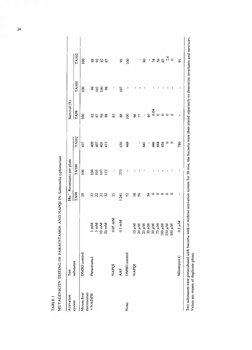
rium TA98, TA100 or TA102, tested in the pres- ence of mouse-liver subfractions ($9 or micro- somes) and cofactors, either in the regular plate assay (data not shown) or in a quantitative assay where revertants and survivors were plated sep- arately (Table 1), or in coculteres with mouse hepatocytes (Table 2). No marked bacterial cyto- toxicity of paracetamol was evident under the conditions employed. On the other hand, NAPQI was markedly cytotoxic for the bacteria, especially in the absence of an activation system (Table 1). Of interest to note was that the strain TAI02 appeared to be somewhat more resistant to the cytotoxic effects of NAPQI than TA98. Under extreme cytotoxic conditions (less than 1% survival) some of the colonies on the revertant plates showed an altered genotype so that a calculation of re- vertants per 10 6 survivors appeared to increase in relation to the spontaneous reversion frequency. However, this was an inconsistent finding after having performed many experiments, a statistical significance could therefore not be ascribed to this phenomenon.
Macromolecular covalent binding Paracetamol was readily converted to inter-
TABLE 2
MUTAGENICITY TESTING OF PARACETAMOL IN Salmonella typhimurium TA98 WITH H E P A T O C Y T E ACTIVATION
Test substance Hi s ' Revertants
per plate
DMSO (control)
Paracetarnol
N-OH-AAF
15+12
0.5 mM 30+ 7 1.0 mM 22+ 3 5.0 mM 20+ 3
10.0 mM 26+ 3 20.0 mM 22+ 5
0.1 mM 908_+25
Test substances were added to co-culture.s of S. typhimurium TA98 and isolated hepatocytes pooled from 5 untreated mice. The values are mean + S.D. of 3 incubations.
mediates which bound covalently to mouse liver microsomal protein in the presence of NADPH (Table 3). This occurred at a rate which was higher than that of the hepatocarcinogen AAF. When DNA was added to microsomal incubations, paracetamol also bound to this macromolecule, although at a rate appreciably slower than that of AAF. In preliminary experiments, intrapcritoneal injections of 500 mg/kg paracetamol to PB- pretreated B6 mice caused extensive liver necrosis. When the mice were given radiolabelled para- cetamol (1.5-1.8 mCi per mouse), 1201 + 150 pmoles [3H]paracetamol bound covalently per mg liver protein when measured 4 h after treatment (Table 3). In this experiment, only 23 pmoles [3H]paracetamol bound covalently per mg muscle protein. Hepatic DNA, purified by hydroxyapatite chromatography, was found to contain 15 + 9 pmoles bound paracetamol per mg DNA. Liver DNA extracted and purified from untreated mice gave a value of 4 pmoles/mg DNA. The level of
1.0
I -
I - z o a
| F-
z Q z _o I - U
0.8
0.7
0.6
0.5
o14
0.3
0.2
CONTROL
pHAA 10"2M
NAPOI 5 10 -5M
NAPOl 10-4M
N-OH-AAF 10-4M
I ~ N A P Q I 2,5 10-4M
0 .1 I I i I I 3 6 9 12 15
E L U T I O N T I M E (HOURS)
Fig. 1. DNA damage in Reuber hepatoma cells after exposure to paracetamol, NAPQI and N-OH-AAF. The cells were in- cubated for 60 rain in presence of various concentrations of the compounds. The data shown are from one representative ex- periment.

27
TABLE 3
COVALENT BINDING OF [3H]PAP, ACETAMOL AND [14C]2-ACETYLAMINOFLUORENE TO MOUSE-LIVER MACRO- MOLECULES IN VITRO AND IN VIVO
Test substance Covalent binding in vitro Covalent binding in vivo
Protein DNA Protein DNA (pmoles/mg (pmoles/mg (pmoles/mg (pmoles/mg protein/min) l)NA/min) protein) DNA)
[3H]Paracetamol 100.4+4.1 2.6+_ 0.8 1 201 + 150 15 + 9
I14C]AAF 76.0 + 4.8 7.6 + 1.0 N.D. N.I).
N.D., not determined. In vitro studies were performed by incubating 0.5 mM of radiolabelled substrates with 2 mg/ml mouse-liver microsomes, a NADPH-generating system with or without 0.67 mg/ml DNA. For determination of in vivo macromolecular covalent binding phenobarbital-pretreated mice were given 500 mg/kg [3l--l]paracetamol 4 h before the animals were killed. Values are means+ S.D. of 4 incubations or 4 animals, respectively.
p a r a c e t a m o l b i n d i n g to D N A in t rea ted mice was
ca l cu l a t ed to give a CBI (Lutz , 1979) of 1.2.
DNA damage in hepatoma cells A l k a l i n e e lu t ion of ce l lu la r D N A is a sens i t ive
assay for the d e t e r m i n a t i o n of D N A d a m a g e (Sina
et al., 1983). W h e n p a r a c e t a m o l was a d d e d to
R e u b e r rat h e p a t o m a cel ls g r o w n in cul ture , no
inc rease in the e lu t ion ra te o f D N A was o b s e r v e d
(Fig . 1, T a b l e 4). O n the o t h e r hand , N A P Q I
caused ex tens ive D N A d a m a g e at c o n c e n t r a t i o n s
o f 0 .05 -0 .25 m M , resu l t ing in the f o r m a t i o n o f
s ing le - s t r and breaks . Th i s was no t due to a gene ra l
c y t o t o x i c ef fec t of N A P Q I u n d e r these e x p e r i m e n -
tal c o n d i t i o n s as d e t e r m i n e d by ce l lu la r t r ypan
b l u e up take , s ince N A P Q I d id no t cause cy to tox i c -
ity m e a s u r e d a f te r an i n c u b a t i o n pe r iod o f 1 h
( T a b l e 4). H o w e v e r , N A P Q I caused a c o n c e n t r a -
t ion d e p e n d e n t c y t o t o x i c effect w h e n assayed 24 h
a f te r exposure . T h e p r o x i m a t e c a r c i n o g e n N - O H -
A A F also caused c o n s i d e r a b l e DNA d a m a g e in
the R e u b e r cells as m e a s u r e d by a lka l ine e lu t ion
(Fig . 1, T a b l e 4).
DNA repair in hepatocytes D a m a g e to ce l lu la r D N A can a lso be assessed
by the d e t e r m i n a t i o n o f an increase in unsched -
u led D N A synthes i s in i so la ted h e p a t o c y t e s g r o w n
"FABLE 4
EFFEC~I" OF PARACETAMOL, NAPQI AND N-OH-AAF EXPOSURE ON ALKALINE ELUTION OF DNA AND CYTO- TOXICITY IN REUBFR IIEPATOMA CELLS
Test substance Elution rate constant Cytotoxicity (% of control)
1 h 24h
DMSO (control) 0.010 + 0.002 0 + 0 0 + 0 Paracetamol 10 mM 0.012 + 0.004 0 + 0 0 + 0
NAPQI 0.05 mM 0.041 +0.019 0+0 36+ l l 0.10 mM 0.075 + 0.036 2 + 2 81 + 3 0.25 mM 0.173+0.068 5+1 100+ 0
N-OH-AAF 0.10 mM 0.155+0.029 0~-0 0+ 0
Reuber cells, prelabelled with 3H-TdR, were exposed to test substances for 1 h. The cultures were then rinsed and prepared for the alkaline elution assay or cytotoxicity was determined immediately or after incubation for further 23 h. Means+ S.D. of 4-8 determinations.

28
<
< ,..1
<
Z
<
< ;.I.1
Z
<
< Z C,
Z
[,.
,.-1
Z <
< Z
©
a.l
<
©
< +,
~ ~'°'~<[.z. I
~" I ~<~'~'~
~m ~ ~ _~' ~ ~' m~ I 1 I I
<
4-I
7
++' ~ ~+ _ +=+' ~+ _ _ ~ + ~ ~ ~+'
4-I +I +I -H +I +I ~ -H + +I +I +I
-H -H +I 4-I -H +I -I-I i I +I
-H +I JH +I +I +I +I -H +I 4-I -H -H
+I +I +I -H "H -H +I +I -H
o

29
in monolayer culture (Althaus et al., 1982). Experi- ments with paracetamol showed that the drug increased DNA-repair synthesis in control mouse hepatocytes at concentrations of 5.0 mM and higher (Table 5). At these concentrations cellular toxicity, as determined by an increased leakage of L D H (Table 5) or by trypan blue exclusion (not shown), also became evident. No significant in- crease in unscheduled DNA synthesis was found with concentrations of NAPQI up to 0.25 mM. When using hepatocytes from mice pretreated with PB, a lowering of the concentration threshold for both increased DNA-repair synthesis as well as cytotoxicity was noted. A brief exposure of the cells to UV-light was used as a positive control for D N A damage in these experiments.
Discussion
The present investigation was carried out in an at tempt to address some of the questions per- taining to the mechanism(s) for paracetamol hepatocarcinogenicity (Flaks and Flaks, 1983) in mice. Whether or not paracetamol could be shown to display genotoxic effects should be of impor- tance when assessing the risk to humans of para- cetamol intake. These studies were in part made possible through establishment of a procedure (Dahlin and Nelson, 1982) for the synthesis of the proposed ultimate reactive metabolite of para- cetamol, NAPQI.
For many carcinogens, mutagenicity in Salmo- nella typhimurium is a very good predictor of carcinogenic activity (McCann et al., 1975). How- ever, for some chemical classes there is not a good correlation between mutagenicity and carcinogen- icity (Rinkus and Legator, 1979), in part this can be due to a nongenotoxic mechanism of carcino- genic action. Paracetamol did not cause mutations in S. typhirnurium, neither in the presence of mouse liver subfractions or with mouse hepatocytes as activating system. Interestingly paracetamol was not mutagenic for the new strain TA102 which is sensitive to active oxygen mutagens (Levin et al., 1982). Testing of the major ultimate reactive metabolite of paracetamol, NAPQI (Dahlin et al., 1984), also did not shown mutagenic activity in the presence of metabolic activation systems. In the absence of activation, concentrations of NAPQI
above 25 /xM were extremely cytotoxic. Thus, the liver subfractions protect the bacteria from NAPQI cytotoxicity, which in part may be explained by a reduction of N A P Q I back to paracetamol (Corcoran et al., 1980). Under severe cytotoxic conditions, a moderate increase in the reversion rate with NAPQI was found. However, this was an inconsistent finding. NAPQI, which is very short- lived and reactive (Dahlin et al., 1984), presuma- bly interacts preferentially with vital bacterial macromolecules so that the organism dies even if its DNA also should have premutational lesions from NAPQI exposure. Similar to the present find- ings were those reported earlier for N-hydroxy- paracetamol (Wirth et al., 1980), which sponta- neously breaks down to NAPQI (Corcoran et al., 1980).
In addition to its avid binding to protein, it could be shown that paracetamol bound cova- lently to exogenously added DNA in a microsomal incubation system. The DNA-binding rate was slower than that of the hepatocarcinogen AAF, whereas the reverse was true with respect to pro- tein binding. This presumably reflects differences in the chemical reactivity and stability of the re- spective electrophiles of the two carcinogens. In addition to the well-known fact that paracetamol binds covalently to liver proteins in vivo (Jollow ct al., 1973), it could also be demonstrated that paracetamol binds covalently to hepatic DNA. However, the level of binding was two orders of magnitude lower than that reported for AAF, as normalized by determination of the CBI of Lutz (1979). In this study it was found that paracetamol had a CB! of 1.2, whereas the moderately strong hepatocarcinogen AAF has a CBI of 560 in rats (Lutz, 1979). Further, it is important to note that the covalent binding of paracetamol was demon- strated at a hepatotoxic dose. If this covalent binding only occurred in cells which would later die, such a DNA interaction would not lead to mutation, an event which most probably is in- volved in initiation of carcinogenesis.
For many compounds, detection of DNA single-strand breaks by alkaline elution (Kohn et al., 1981) correlates well with mutagenic and carcinogenic activity (Sina et al., 1983). In the present study, rat hepatoma cells with a low capac- ity for cytochrome P-450 metabolism (Owens and
30
Nebert, 1975) was used. It was thus not surprising that paracetamol did not cause DNA damage in this system. However, the ultimate reactive metabolite NAPQI was sufficiently stable and lipophilic to traverse cellular membranes and cause DNA single-strand breaks at a time where NAPQI showed no cytotoxic effect. Thus, these single- strand breaks were probably not caused by an immediate, general cytotoxicity. However, after an additional incubation time of the cells for 23 h NAPQI cytotoxicity became manifest, in agree- ment with findings using isolated rat hepatocytes (Holme et al., 1983b).
Induction of unscheduled DNA synthesis, a measure of DNA repair, has also been shown to be a good indicator for genotoxic carcinogens (Alt- haus et al., 1982). When mouse-liver hepatocytes were incubated in the presence of paracetamol at concentrations which caused cytotoxicity, an in- crease in DNA-repair synthesis was demonstrated. This finding was presumably related to metabolic activation of paracetamol, since cells from mice pretreated with PB showed a lower concentration threshold for DNA-repair synthesis induction than did cells from untreated mice. In contrast to the experiments with the hepatoma cells, addition of NAPQ! to the hepatocyte culture medium did not elicit a response indicative of D N A damage in this system. This difference in cytotoxicity vs. geno- toxicity of NAPQI in the two cell systems may be related to differences in the cell membranes, cellu- lar shape and size, in cellular reduction capacity a n d / o r in number of cellular nucleophilic sites between the mouse hepatocytes and the rat hepatoma cells.
In summary, paracetamol or its ultimate reac- tive metabolite has been found to show genotoxic effects in 3 out of 4 test systems, only bacterial mutagenicity did not give a significant positive response. However, the covalent binding of para- cetamol to DNA and the increased DNA alkaline elution and DNA-repair synthesis only show that paracetamol may cause DNA damage. This is not a surprising finding taken together with the fact that paracetamol is known to be metabolized to an electrophilic intermediate which binds covalently to nuclcophilic sites. Whether such an initial DNA damage will be properly repaired or the cells will die before a mutation becomes fixed is an open
question. The finding that the genotoxic effects of paracetamol were observed at levels which caused delayed cytotoxic effects, indicates that cytotoxic- ity of paracetamol may predominate compared to genotoxicity. Thus, the necrogenic effects of para- cetamol, which would be expected to cause a strong proliferative stimulus to an initiated liver cell, are probably of major importance for paracetamol-in- duced hepatocarcinogenicity in mice (Flaks and Flaks, 1983). In this connection, it is important to note the apparent dose-thresholds for both acute liver damage (Mitchell et al., 1973a) and liver- tumor formation (Flaks and Flaks, 1983) corre- lates with the non-linearity of the relationship between paracetamol dose and covalent protein binding (Mitchell et al., 1973b; Mudge et al., 1978). However, the present findings taken to- gether with the report that paracetamol may cause chromosome aberrations in Chinese hamster cells in vitro (Ishidate and Yoshikawa, 1980) indicate that it is important to further examine paraceta- mol for possible mutagenic effects.
Acknowledgements
This study was supported by The Royal Norwegian Council for Scientific and Industrial Research and by grant GM-25418 from The Na- tional Institutes of Health (U.S.A.).
References
Althaus, R.F., S.D. Lawrence, G.L. Sattler, D.G. Longfellow and H.C. Pitot (1982) Chemical quantification of unsched- uled DNA synthesis in cultured hepatocytes as an assay for the rapid screening of potential chemic',d carcinogens, Cancer Res., 42, 3010-3015.
Ames, B.N., J. McCann and E. Yamas',tki (1975) Methods for detecting carcinogens and mutagens with the Salmonella/ mammalian-microsome mutagenicity test, Mutation Res., 31,347-362.
Anuforo, D.C., D. Acosta and R.V. Smith (1978) Hepatotoxic- ity studies with primary cultures of rat liver cells, In Vitro, 14, 981-987.
Burton, K. (1956) Determination of DNA concentration with diphenylamine, Biochem. J., 62, 315-323.
Corcoran, G.B., J.R. Mitchell, Y.N. Vaishnav and E.C. Horn- ing (1980) Evidence that acetaminophen and N-hydroxy- acetaminophen form a common arylating intermediate, N- acetyl-p-benzoquinoeimine, Mol. Pharmacol., 18, 536 542.
Dahlin, D.C., and S.D. Nelson (1982) Synthesis, decomposition kinetics, and preliminary toxicological studies of pure N-

31
acetyl-p-benzoquinonc imine, a proposed toxic metabolite of acetaminophen, J. Med. Chem., 25, 885-886.
Dahlin, D.C., G.T. Miwa, ANAl . Lu and S.D. Nelson (1984) N-Acetyl-p-benzoquinone imine: A cytochrome P-450 mediated oxidation product of acetaminophen, Proc. Natl. Acad. Sci. (U.S.A.), 81, 1327-1331.
Dybing, E., and S.S. Thorgeirsson (1977) Metabolic activation of 2,4-diaminoanisole, a hair dye component, I. Role of cytoehrome P-450 metabolism in mutagenicity in vitro, Biochem. Pharmacol., 26, 729 734.
Flaks, A., and B. Flaks (1983) Induction of liver cell tumors in IF mice by paracetamol, Carcinogenesis, 4, 363-368.
llinson, J.A., 1,.R. Pohl and J.R. Gillette (1979) N-Hydroxy- acetaminophen: A microsomal metabolite of N-hydroxy- phenacetin but apparently not of acetaminophen, Life Sci., 24, 2133-2138.
Holme, J.A., D.C. Dahlin, S.D. Nelson and E. Dybing (1983a) Cytotoxic effects of N-acetyl-p-benz(xtuinone imine, a com- mon arylating intermediate of paracetamol and N-hydroxy- paracetamol, Biochem. Pharmacol., 33, 401-406.
tlolme, J.A., L.T. Haug and E. Dybing (1983b) Modulation of aromatic amine mutagenicity in Salmonella typhimurium with rat-liver 9000 g supernatant or monolayers of rat hcpatocytes as an activation system, Mutation Res., 117, 113-125.
Holme, J.A., E.J. Soderlund, J.K. Hongslo, S.D. Nelson and E. Dybing (1984) Comparative genotoxicity studies of the flame retardant tris(2,3-dibromopropyl)phosphate and pos- sible metabolites, Mutation Res., 124, 213-224.
Ishidate Jr., M., and K. lshikawa (1980) Chromosome aberra- tion tests with Chinese hamster cells in vitro with and without metabolic activation .... a comparative study on mutagens and carcinogens, Arch. Toxicol., Suppl. 4, 41-44.
Jollow, D.J., J.R. Mitchell, W.Z. Potter, D.C. Davis, J.R. Gillette and B.B. Brodie (1973) Acetaminophen-induced hepatic necrosis, 1I. Role of covalent binding in vivo, J. Pharmacol. Exp. Ther., 187, 195-202.
Kohn, K.W., L.C. Erickson, R.A.G. Ewig and L.A. Qwelling (1981) Measurement of strand breaks and cross-links by alkaline elution, in: E.C. Friedberg and P.C. Hanawalt (Eds.), DNA Repair: A Laboratory Manual of Recent Procedures, Marcel Dekker, New York, pp. 379-401.
Laishcs, B.A., E. Roberts and E. Farber (1978) In vitro mea- surement of carcinogen-resistant liver cells during hepato- carcinogenesis, Int. J. Cancer, 21, 186-193.
Levin, D.E., M. Hollstein, M.F. Christman, E.A. Schwiers and B.N. Ames (1982) A new Salmonella tester strain (TA102) with A.T- base pairs at the site of mutation detects oxida- tive mutagens, Proe. Natl. Acad. Sci. (U.S.A.), 79, 7445-7449.
Lowry, O.H., N.J. Rosebrough, A.L. Farr and R.J. Randall (1951) Protein measurement with the Folin phenol reagent, J. Biol. Chem., 193, 265-275.
Lutz, W.K. (1979) In vivo covalent binding of organic chemi- cals to DNA as a quantitative indicator in the process of chemical carcinogenesis, Mutation Res., 65, 289-356.
McCann, J., E. Choi, E. Yamasaki and B.N. Ames (1975) Detection of carcinogens as mutagens in the Salmonella/
microsome test: Assay of 300 chemicals, Proe. Natl. Acad. Sci. (U.S.A.), 72, 5135-5139.
Miner, D.J., and P.T. Kissinger (1979) Evidence for the in- volvement of N-acetyl-p-quinoneimine in acetaminophen metabolism, Biochem. Pharmacol., 28, 3285-3290.
Mitchell, J.R., and D.J. Jollow (1975) Metabolic activation of drugs to toxic substances, Gastroenterology, 68, 392--410.
Mitchell, J.R., D.J. Jollow, W.Z. Potter, D.C. Davis, J.R. Gillette and B.B. Brodie (1973a) Acetaminophen-induced hepatic necrosis, I. Role of drug metabolism, J. Pharmacol. Exp. Ther., 187, 185 194.
Mitchell, J.R., I).J. Jollow, X.Z. Potter, J.R. Gillette and B.B. Brodie (1973b) Acetaminophen-induced hepatic necrosis, IV. Protective role of glutathionc, J. Pharmacol. Exp. Ther., 187, 211-217.
Mudge, G.H., M.W. Gemborys and G.G. l)uggin (1978) Cova- lent binding of metabolites of acetaminophen to kidney protein and depletion of renal glutathione, J. Pharmacol. Exp. Ther., 206, 218-226.
Ncal, G.E., A.R. Mattocks and D.J. Judah (1979) The micro- som',d activation of aflatoxin B 1 and 2-(N-ethylcarbamoyl- oxymethyl)furan in vitro using a novel diffusion apparatus, Bioehim. Biophys. Acta, 585, 134-142.
Nelson, S.D., A.J. Forte and D.C. l)ahlin (1980) Lack of evidence for N-hydroxyacetaminophen as a reactive metabolite of acetaminophen in vitro, Biochem. Pharmacol., 29, 1617-1620.
Owens, I.S., and D.W. Nebert (1975) Arylhydrocarbon hydrox- ylase induction in mammalian liver-derived cell cultures, Stimulation of 'cytoehrome P1450-associated' enzyme activ- ity by many inducing compounds, Mol. Pharmacol., 11, 94-104.
Potter, W.Z., D.C. Davis, J.R. Mitchell, I).J. Jollow, J.R. Gillette and B.B. Brodie (1973) Acetaminophen-induced hepatic necrosis, II1. Cytochrome P-450-mediated covalent binding in vitro, J. Pharmacol. Exp. Ther., 187, 203-210.
Prescott, L.F., N. Wright, P. Roscoe and S.S. Brown (1971) Plasma-paracetamol half-life and hepatic necrosis in pa- tients with paracetamol overdosage, Lancet, 1,519 522.
Rinkus, S.J., and M.S. Legator (1979) Chemical characteriza- tion of 465 known or suspected carcinogens and their correlation with mutagenic activity in the Salmonella typhimurium system, Cancer Res., 39, 3289-3318.
Sagelsdorff, P., W.K. Lutz and C. Schlatter (1983) The rele- vance of covalent binding to mouse liver DNA to the carcinogenic action of hexachlorocyclohexane isomers, Carcinogenesis, 4, 1267-1273.
Seglen , P. (1975) Preparation of isolated liver cells, in: D.M. Prescott (Fxt.), Methods in Cell Biology, 13, Academic Press, New York, pp. 29-83.
Sina, J.F., C.L. Bean, G.R. Dysart, V.I. Taylor and M.O. Bradley (1983) Evaluation of the ',alkaline elution/rat hepatoeyte assay as a predictor of carcinogenic/mutagenic potential, Mutation Res., 113, 357-391.
Soderlund, E.J., S.D. Nelson, C. von Bahr and E. Dybing (1982) Species differences in kidney toxicity and metabolic activation of tris(2,3-dibromopropyl)phosphate, Fund. Appl. Toxicol., 2, 187-194.
32
Soderlund, E.J., S.D. Nelson and E. Dybing (1979) Mutagenic activation of tris(2,3-dibromopropyl)phosphate: the role of microsomal oxidative metabolism, Acta Pharmacol. Toxi- col., 56, 171-181.
Williams, G.M. (1980) Classification of genotoxic and epi- genetic hepatocarcinogens using liver culture assays, Ann. N.Y. Acad. Sci., 349, 273-282.
Wirth, P.J., E. Dybing, C. von Bahr and S.S. Thorgeirsson (1980) Mechanism of N-hydroxyacetyla~laminc mutagcnic- ity in the Salmonella test system: Metabolic activation of N-hydroxyphenacetin by liver and kidney fractions from rat, mouse, hamster and man, Mol. Pharmacol., 18, 117-127.