genotoxic stress causes the accumulation of the splicing regulator sam68 in nuclear foci of...
TRANSCRIPT
Genotoxic stress causes the accumulation of thesplicing regulator Sam68 in nuclear foci oftranscriptionally active chromatinRoberta Busa1,2, Raffaele Geremia1 and Claudio Sette1,2,*
1Department of Public Health and Cell Biology, University of Rome Tor Vergata, Via Montpellier 1, 00133 Romeand 2Laboratory of Neuroembryology, Fondazione Santa Lucia, Via del Fosso di Fiorano 64, 00143 Rome, Italy
Received July 31, 2009; Revised December 18, 2009; Accepted January 5, 2010
ABSTRACT
DNA-damaging agents cause a multifaceted cellularstress response. Cells set in motion either repairmechanisms or programmed cell death pathways,depending on the extent of the damage and ontheir ability to withstand it. The RNA-bindingprotein (RBP) Sam68, which is up-regulated inprostate carcinoma, promotes prostate cancer cellsurvival to genotoxic stress. Herein, we haveinvestigated the function of Sam68 in this cellularresponse. Mitoxantrone (MTX), a topoisomerase IIinhibitor, induced relocalization of Sam68 from thenucleoplasm to nuclear granules, together withseveral other RBPs involved in alternative splicing,such as TIA-1, hnRNP A1 and the SR proteins SC35and ASF/SF2. Sam68 accumulation in nuclear stressgranules was independent of signal transductionpathways activated by DNA damage. Using BrUlabelling and immunofluorescence, we demonstratethat MTX-induced nuclear stress granules aretranscriptionally active foci where Sam68 and thephosphorylated form of RNA polymerase II accu-mulate. Finally, we show that MTX-inducedrelocalization of Sam68 correlates with changes inalternative splicing of its mRNA target CD44, andthat MTX-induced CD44 splicing depends onSam68 expression. These results strongly suggestthat Sam68 is part of a RNA-mediated stressresponse of the cell that modulates alternativesplicing in response to DNA damage.
INTRODUCTION
Cells have developed several mechanisms to cope withexternal sources of stress, like heat shock and oxidativestress, or with insults that affect the integrity of the
genome, such as ultraviolet (UV) irradiation and DNAalkylating agents. Depending on the nature and the per-sistence of the stress, cells will adopt a ‘safety’ mechanismto limit and eventually overcome the damage, undergoingcell cycle arrest and DNA repair, or they will succumb byactivating programmed cell death. A complex andwell-studied stress response is that imposed by DNAdamage (1), which has strong clinical implications in che-motherapy of human cancers. Most chemotherapeuticdrugs induce breaks in the genome by targeting DNAprocessing enzymes, such as the topoisomerase inhibitors,or DNA directly, such as the alkylating agents. Althoughmost cells are highly sensitive to these drugs and undergoapoptosis, cancer cells often escape this response andadopt mechanisms to withstand and repair the damage,thereby surviving to treatments. Thus, understanding themolecular mechanisms that allow cancer cells to survive togenotoxic stresses is a crucial step in the development ofimproved and more efficacious therapies.Genotoxic stress causes a general suppression of the
transcriptional activity, through degradation of theRNA polymerase II (RNAPII) (2), which allows to saveenergy and readapt the protein repertoire of the cell to thenew tasks. In addition to changes in transcription, recentevidence demonstrated that genotoxic stress induces largespectrum modifications in alternative splicing (AS),thereby altering the isoforms produced by several genes(3). AS affects most human genes and allows to expandthe cell proteome through differential assembly of exons inthe mRNAs. AS is operated by the spliceosome, amacromolecular machinery composed by small nuclearribonucleuprotein particles (snRNPs, U1, U2, U4, U5and U6) and many constitutive and ancillary proteinsthat regulate the assembly of the spliceosome at theexon-intron junctions (4). The main regulators of consti-tutive and alternative splicing are RNA-binding proteins(RBPs) belonging to the serine–arginine (SR) richproteins and the heterogeneous ribonucleoproteins(hnRNPs), which often play antagonistic roles (5).
*To whom correspondence should be addressed. Tel: +3906 72596260; Fax: +3906 72596268; Email: [email protected]
Published online 27 January 2010 Nucleic Acids Research, 2010, Vol. 38, No. 9 3005–3018doi:10.1093/nar/gkq004
� The Author(s) 2010. Published by Oxford University Press.This is an Open Access article distributed under the terms of the Creative Commons Attribution Non-Commercial License (http://creativecommons.org/licenses/by-nc/2.5), which permits unrestricted non-commercial use, distribution, and reproduction in any medium, provided the original work is properly cited.

In addition to DNA damage, changes in AS have beenreported in cellular responses to many other sources ofstress (6), indicating that it is a crucial regulatory mecha-nism in cell adaptation to external insults. Moreover,recent observations have highlighted the specific differ-ences in AS regulation in cancer cells (7–10), suggestingthat this step of RNA processing plays a role also in celltransformation.In line with its crucial role in the DNA damage
response, several changes in AS of specific transcriptshave been observed in cancer cells treated with cisplatinor etoposide. Remarkably, some of these transcriptsencode for proteins regulating apoptosis, such asCaspase 2 (11) and Bcl-2 related genes (3), cell motility,like CD44 (12) and cell proliferation, like the p53 negativemodulators MDM2 and MDM4 (13) or cyclin D1b, asplicing variant aberrantly expressed in prostate andbreast cancer cells that confers resistance to therapies(14,15). Thus, it is likely that regulation of these ASevents represents a novel mechanism by which cancercells gain drug resistance and survive to chemotherapy.The mechanisms underlying stress-induced changes in
AS are just beginning to be understood (6). A recentreport indicated that UV irradiation alters a substantialnumber of splicing events in hepatocarcinoma cells (3).The AS events were predominantly modulated bychanges in the rate of pre-mRNA transcription,elicited through phosphorylation of the RNAPII (3).Nevertheless, additional mechanisms to modulate AS reg-ulation exist. For instance, in p53-deficient cells, genotoxicstress caused up-regulation of SRp55, thereby promotingthe inclusions of CD44 variable exons involved intumorigenesis (12) and changes in AS of othercancer-related genes (16). DNA damage and otherstresses can also alter the subcellular distribution ofseveral RBPs involved in post-transcriptional events.Cisplatin and a mild heat shock caused accumulation inthe nucleoli of the RNA recognition motif (RRM)-con-taining protein RDM1, which modulates the cellularresponse to genotoxic agents (17). In addition, variousstresses triggered the cytoplasmic translocation of thesplicing regulator hnRNP A1 (18), suggesting that theymight alter nuclear RNA processing events regulated bythis RBP (6). On the other hand, UV irradiation and heatshock induced accumulation of other RBPs in nuclearstress bodies (19,20), whose function is still largelyunknown.An interesting RBP involved in regulation of cell pro-
liferation and survival is Sam68, which belongs to thesignal transduction and activation of RNA (STAR)family (21). Sam68 is implicated in several steps of pro-cessing of target genes. In the cytoplasm, it regulatestranslation of viral and cellular mRNAs through its asso-ciation with the polyribosomes or the stress granules(22–25). In the nucleus, Sam68 integrates signaltransduction pathways with pre-mRNA AS in responseto extracellular cues (26–28). Notably, we have previouslydemonstrated that Sam68 is up-regulated in prostatecancer (PCa) cells, where it sustains their resistance totreatments with chemotherapeutic agents (29). Theseresults suggested that Sam68 plays a role in the cellular
response to genotoxic stress. Herein, we have investigatedthe function of Sam68 in PCa cells exposed to genotoxicstress induced by mitoxantrone (MTX), a topoisomeraseII inhibitor used in chemotherapy. Our results demon-strate that Sam68 takes part to the cellular response togenotoxic stress by altering its subcellular localizationand contributing to modulation of AS. These resultssuggest a novel role for Sam68 in the DNA damageresponse of cancer cells.
MATERIALS AND METHODS
Cell cultures and treatments
PC3 and HeLa cells were cultured in Dulbecco’s modifiedEagle’s medium (Gibco BRL), LNCaP cells were culturedin RPMI 1640 medium (Lonza), media were supplementedwith 10% fetal bovine serum (FBS) (Lonza), gentamycin,penicillin and streptomycin. For genotoxic treatments,cells at 80% of confluence were treated with 80 mMcisplatin (Sigma-Aldrich) for 16 h, or with 5 mM MTX(Sigma-Aldrich) for increasing times (2–24 h). At the endof the incubation, cells were fixed and stained forimmunoflorescence analysis. For studies involving inhibi-tors, PC3 cells were pre-incubated for 15min with 10 mMJNK inhibitor 1 (JNK-inh.) (Alexis Biochemicals), 10 mMSB202190 (Calbiochem) for p-38 kinase inhibition or10 mM MNK inhibitor 4-amino-5-(4-fluoroanilino)-pyrazolo[3,4-d]pyrimidine (MNK-inh.) (Calbiochem), or1 h with 10 mM 2-Morpholin-4-yl-6-thianthren-1-yl-pyran-4-one (ATM-inh.) (Calbiochem), and thenincubated with 5 mM MTX for 8–24 h. For RNASatIIIinduction experiments, PC3 cells were treated either withheat shock as previously described (30) or with 5 mMMTX (15min to 24 h).
Cell proliferation assay, cell viability and cell cycleanalysis
The CellTiter A96 MTS (Promega) assay was used, aspreviously described (29), to monitor PC3 cell prolifera-tion after treatment with MTX (0.5–5 mM, Sigma-Aldrich)for increasing times (24–72 h) or for 2 h and released for24–72 h after stress. For cell viability analyses, treated PC3cells were incubated for 5min at room temperature (RT)with 0.4% Trypan blue solution 1 : 1 (v/v) (Sigma-Aldrich)and analysed on a hemocytometer. The percentage ofdead cells (stained) versus viable cells (unstained) wascalculated. Cell cycle analysis with propidium iodide(10 mg/ml) was performed with a FACSCalibur FlowCytometer (Becton Dickinson) as described (29).
Stable knockdown of Sam68 in PC3 cells
PC3 cells were transfected either with pLKO.1puro(control pLKO) or PLKO.1-KHDRBS1_527 (pLKO-si-Sam68) (MISSION shRNA, Sigma Aldrich) in a 12multi-well plate using Lipofectamine 2000 reagent(Invitrogen) according to manufacturer’s instructions.The puromycin (Sigma-Aldrich) resistance marker wasadded at a concentration of 1 mg/ml in fresh mediumevery 2 days for stable selection and maintenance of
3006 Nucleic Acids Research, 2010, Vol. 38, No. 9

PC3 clones. Sam68 knockdown was verified by RT–PCRand western blot analyses.
Plasmid vectors
For the pHsp70.2EGFP-Sam68 expression vector, theCMV promoter was excised from pEGFPC1-Sam68(28), substituted with Hsp70.2 promoter, amplifiedwith Pwo SuperYield DNA polymerase (Roche) fromHspa2-pblue (generous gift of Prof. E.M. Eddy), andintroduced in pEGFP-C1-Sam68. The cDNA encodingmurine Sam68 was amplified by PCR using cDNA frommurine embrional fibroblast (MEF) as template and Pfupolymerase (Roche), cloned in frame with myc intopCDNA3myc (28). The expression vector pEGFP-GSG(GFP-Sam68GSG) was previously described (29). Allexpression vectors were analysed by sequencing.
Cell transfections and CD44v5-luciferase (v5-Luc)splicing assay
For GFP-tagged proteins expression, PC3 cells wereseeded the day before transfection (3.5� 105) in 35mmplates and then transfected with 1 mg of pEGFPhsp70.2-Sam68 wt or pEGFP-GSG using Lipofectamine 2000(Invitrogen) according to manufacturer’s instructions.Twenty-four hours after transfection, cells were treatedwith 5 mM MTX for 24 h, incubated 15min at 37�C with40,6-diamidino-2-phenylindole (DAPI), washed inphosphate-buffered saline (PBS) and fixed in 4%paraformaldehyde (PFA) 10min on ice. Cells were thenwashed with PBS, mounted with Mowiol (Calbiochem)and analysed by confocal microscope (28). ForLuciferase assays, control pLKO and pLKO-si-Sam68PC3 cells were cultured in 12-well plates(�10� 105/well). Cells were treated with 5 mM MTX for2 h, rinsed with PBS and let recover with fresh mediumfor 2 h and then transfected with 0.4mg of minigenepETv5luc (31), 0.25 mg of pCDNA3myc orpCDNA3mycSam68 (mycSam68) and the Renillaluciferase reporter gene (1 ng) as an internal controlusing Lipofectamine 2000 (Invitrogen), according to themanufacturer’s instructions. Twenty-four hours aftertransfection, cells were harvested, lysed and analysedusing a biocounter luminometer using the dual-luciferasereporter assay system (Promega). Data from three exper-iments were normalized for transfection efficiency usingthe ratio between firefly and Renilla luciferase activity.
RNA extraction, real-time PCRand RT–PCR
For endogenous CD44 v5 AS analysis after genotoxicstress, control pLKO and pLKO-si-Sam68 PC3 stableclones were treated with 0.5 or 5 mM MTX for 2 h, letrecover after stress for 24 h and then harvested inTRIzol reagent (Invitrogen) for total RNA extractionaccording to the manufacturer’s instructions. After diges-tion with RNase-free DNase (Roche), 1 mg of total RNAwas used for RT using M-MLV reverse transcriptase(Invitrogen). Quantitative real-time PCRs were carriedoutwith Sybr Green mix (for Light-Cycler480 - Roche)according to manufacturer’s instructions. Primers usedfor CD44 v5 were the followings: CD44 v5 Fw 50TGTA
GACAGAAATGGCACCACTG30; CD44 v5 Rev 50TGGTGCTTGTAGAATGTGGGGTC30. Samples werenormalized on CD44 constitutive isoforms (with respectto the constitutive exons C6 and C7) using the followingprimers: CD44 c Fw 50AGACACATTCCACCCCAGTG30; CD44 c Rev 50CCAGAGGTTGTGTTTGCTCCACC30. The value given for the amount of CD44 v5 PCRproduct present in untreated cells was set as 1. Data arereported as the mean±SD of three independent experi-ments. For SatIII RNAs expression experiments after heatshock or MTX treatments, RT–PCRs specific for G-richRNA SatIII were performed as described (30). Sampleswere normalized on hypoxantine-phosphoribosyl-transferase (HPRT) expression levels.
Western blot analysis
Western blot analysis of cell extracts was performed aspreviously described (29,32). Protein concentration wasdetermined by using Bradford reagent (Bio-Rad).Primary antibodies used (at 1 : 1000 dilution; overnightat 4�C): rabbit anti-Sam68, anti-Erk2, anti-TFIIH andanti-cyclin A, goat anti-lamin b, and mouse anti-cyclinB1 (Santa Cruz Biotechnology); rabbit p-Chk2 andp-p38 (Cell Signalling); mouse c-Jun (BD biosciences);rabbit anti-pJNK1/2 and p-eIF4E (Biosource); mouseanti-hnRNP A1 (Sigma-Aldrich); rabbit anti-cyclin D1and anti-Histone 3, unmodified (Abcam). Secondaryanti-mouse, anti-rabbit or anti-goat IgGs conjugated tohorseradish peroxidase (Amersham) were incubated for1 h at RT (1 : 10 000 dilution in PBS containing 0.1%Tween-20). Immunostained bands were detected bychemiluminescent method (Santa Cruz Biotecnology).
Nuclei isolation and chromatin fractionation
PC3 cells were treated with 0.5 or 5 mM MTX for 24 h.Nuclei were isolated, resuspended in solution G (300mMsucrose, 50mM triethanolamine, 25mM KCl, 4mMMgCl2, 1mM CaCl2 and 1mM phenylmethylsulfonylfluoride, pH 7.4) and digested with micrococcal nuclease(Sigma-Aldrich) at 37�C as previously described (33). Forchromatin fractionation, digested nuclei were cooled onice for 10min and centrifuged at 12 800� g for 10min at4�C. The supernatant (S1) was removed, the pellet wassuspended in 2mM EDTA pH 7.4 and centrifuged toseparate supernatant S2 and pellet (P) as previouslydescribed (34). For protein analysis, samples wereadjusted to 10mM MgCl2 and precipitated with twovolumes of ethanol at �20�C. Protein samples wereanalysed by western blot.
Immunofluorescence and bromo-uridine incorporation
Cells were fixed and stained for immunofluorescenceanalysis as previously described (28). Primary antibodieswere the followings: rabbit anti-Sam68 1 : 1000 and goatanti-TIA-1 1 : 200 (Santa Cruz, Biotechnology); mouseanti-gH2AX 1 : 10000 (Cell signaling); mouse monoclonalantibody PG-M3 (anti-PML) 1 : 100 (generous gift fromProf. F. Lo Coco); mouse anti-hnRNP A1 1 : 500 andanti-SC35 1 : 500 (Sigma-Aldrich); mouse anti-SF2/ASF1:200 (USBiological) andmouse monoclonal anti-RNA
Nucleic Acids Research, 2010, Vol. 38, No. 9 3007

polymerase II (phospho S2) H5 IgM 1 : 200 (Abcam).DAPI was used for DNA staining and the secondaryantibodies were: Alexa Fluor 488, 568 goat anti-rabbitand Alexa Fluor 488 goat anti-mouse (1 : 500)(Invitrogen); FITC-coniugated donkey anti-rabbit;cy3-coniugated donkey anti-goat 1 : 500 and cy3-coniugated donkey anti-mouse IgM 1 : 500 (JacksonImmunoresearch). For BrU incorporation, PC3 cellswere incubated in the last 15min to 1 h of culture with1mM BrU (Sigma-Aldrich). Cells treated with 5 mMMTX (2–24 h) were incubated with BrU in the last hourof treatment. For MTX-release experiments, PC3 cellswere treated with 5 mM MTX for 2 h and let recover for24 h and incubated with BrU in the last hour of recovery.Cells were fixed and stained as previously described (35).Briefly, cells were washed twice with sterile cold PBS sup-plemented with 1mM MgCl2 and 0.1mM CaCl2 (PBS*)and fixed in PFA 2% for 20min on ice. Afterpermeabilization with PBS* +0.1% Triton X-100(PBS*T) for 5min, samples were quenched with 50mMNH4Cl in PBS*T 10min, blocked with PBS*T+1%BSA for 1 h at RT, and incubated with the followingprimary antibodies: mouse anti-BrdU (1 : 40) (Becton-Dickinson) and rabbit anti-Sam68 (1 : 500) inPBS*T +0.2 % BSA 1h at RT. Cells were washed threetimes in PBS*T and incubated 1 h at RT with the followingsecondary antibodies diluted in PBS*T +0.2 % BSA:cy3-coniugated donkey anti-mouse 1 : 100 or cy3-coniugated donkey anti-mouse 1 : 100 and FITC donkeyanti-rabbit 1 : 500 (Jackson Immunoresearch) forco-stainings. Finally, cells were washed, mounted withMowiol (Calbiochem) and analysed by confocal micro-scope. All the solutions were prepared in RNAase-freewater. Immunofluorescence analyses were performedusing a Leica confocal microscope and a Plan-NeofluarHCX 40.0� /1.25 oil UV objective and acquired usingIAS AF Lite software (Leica Microsystems). Imageswere saved as TIFF files, and Photoshop (Adobe) andPowerPoint (Microsoft) were used for composing thepanels.
RESULTS
Genotoxic stress induces changes in the subcellularlocalization of Sam68
Depletion of Sam68 renders PCa cells more susceptible toapoptosis induced by genotoxic drugs (29). To furtherinvestigate this protective function of Sam68, we moni-tored the intracellular localization of the protein inresponse to MTX, a drug used in treatment ofandrogen-resistant PCa patients (36). Sam68 was diffuselylocalized in the nucleoplasm of control cells, whereastreatment with MTX for 2–24 h induced profoundchanges in the localization of the protein (Figure 1A andB). After 2–4 h, Sam68 accumulated in ring-shaped struc-tures that surrounded the nucleoli, whereas at later times itwas enriched in large granules that were faintly stained byDAPI (Figure 1A and B). Notably, treatment with MTXcaused morphological changes in PC3 cells, which at
phase contrast appeared more flattened on the cul-ture plate with larger nuclei than untreated cells(Figure 1C). Western blot analysis showed that MTXdid not affect Sam68 protein levels during the time-course-of the treament (Figure 1D). Accumulation of Sam68in nuclear granules was also observed in another PCacell line, LNCaP, and in HeLa cells (SupplementaryFigure S1), indicating that it is not a PC3 cell-specificresponse. Moreover, similar changes in the subcellularlocalization of Sam68 were also elicited by cisplatin(Figure 1E), an alkylating agent that causes DNAdamage by a different mechanism than MTX, suggestingthat various genotoxic stresses trigger this event in PCacells.
To test whether the Sam68-containing granules werefoci of DNA lesions induced by MTX, we co-stainedPC3 cells with Sam68 and the phosphorylated form ofH2AX (gH2AX), a substrate of the ATM kinase thatmarks foci of double-strand breaks (37). However,confocal microscopy analysis showed that Sam68nuclear granules and double-strand breaks foci are dif-ferent structures (Supplementary Figure S2A). Othernuclear structures affected by genotoxic stress are thepromyelocytic leukemia (PML) nuclear bodies, whichare sites of post-transcriptional modifications of nuclearproteins such as HIPK2 and p53 (38). PML bodies aredynamic compartments that act as DNA damagesensors, they are regulated by ATM and its target Chk2,and their number in the cell increases under genotoxicstress (39). Nevertheless, also in this case confocal micros-copy demonstrated that MTX-induced Sam68 nucleargranules and PML bodies are different entities(Supplementary Figure S2B).
Sam68 colocalizes with other stress-responsive RBPs bothin nuclear and cytoplasmic granules
In addition to the nuclear relocalization of Sam68, 16–24 hof treatment with MTX (Figure 1F) and cisplatin (datanot shown) caused the translocation of Sam68 in the cyto-plasm in �5–10% of cells, where it accumulated incytoplasmic granules. These structures resembled thecytoplasmic stress granules (SGs) in which splicingfactors, such as hnRNP A1 and TIA-1, re-localize as astress-adaptation response of the cell (40). Thus, wetested whether genotoxic stress caused recruitment ofSam68 to the same granules marked by thesestress-response RBPs. Confocal immunofluorescenceanalyses showed that MTX-induced genotoxic stresscaused subcellular re-distribution of both hnRNP A1and TIA-1 in the nucleus and in the cytoplasm similarto that observed for Sam68 (Figure 2).Moreover, Sam68colocalized, with identical time-course, to the same struc-tures as hnRNP A1 and TIA-1 both in the nucleus(Figure 2A and B, and Supplementary Figure S3A) and,albeit only in a small percentage of cells, in the cytoplasm(Supplementary Figure S3B and C).
Colocalization of Sam68 with TIA-1 in cytoplasmicstress granules has been recently reported in cells underoxidative stress (41). However, no colocalization of these
3008 Nucleic Acids Research, 2010, Vol. 38, No. 9

proteins in nuclear granules was observed under thesecircumstances, suggesting that these structures are specif-ically induced by genotoxic stress. Nuclear stress bodies(nSBs) where RBPs are actively recruited have beenpreviously described in response to heat shock.However, although these structures contained Sam68,SAFB and the SR protein ASF/SF2, other RBPsinvolved in splicing like hnRNP A1 or the SR proteinSC35 were not recruited (20). To further characterize thenature of the MTX-induced stress granules, we analysedthe subcellular localization of SC35 and ASF/SF2 inresponse to this genotoxic drug in PC3 cells. Confocalmicroscopy showed that SC35 and ASF/SF2 localizedboth to nuclear speckles and in the nucleoplasm inuntreated cells, whereas MTX caused their accumulationin large granules in a time-dependent manner. Notably,Sam68, which is mostly diffused in the nucleoplasm ofuntreated PC3 cells, colocalized with these SR proteinsin the large nuclear granules only after 8–24 h of MTXtreatment (Figure 3A and B). These results indicate thatMTX-induced nuclear stress granules are novel structureswhere both hnRNPs and SR proteins accumulate togetherwith Sam68.
The GSG domain of Sam68 is sufficient for relocalizationafter genotoxic stress
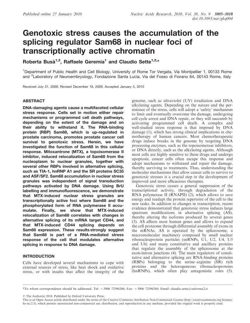
We have previously demonstrated that Sam68 interactswith hnRNP A1 in a functional splicing complex (28).This interaction required the C-terminal 93 residues ofSam68, downstream of the GSG domain that mediateshomodimerization and RNA binding. Similarly, it wasrecently shown that the region comprised by residues269–321, mostly outside of the GSG, was required forthe interaction with TIA-1 and the recruitment of Sam68to cytoplasmic SGs (41). Since Sam68 colocalized withhnRNP A1 and TIA-1 in MTX-induced nucleargranules, we set out to test whether its relocalizationrequired the interaction with these RBPs. PC3 cells weretransiently transfected with full-length GFP-Sam68 orwith GFP-Sam68GSG (residues 91–276), which lacks theregions required for the interaction with hnRNP A1 andTIA-1. We observed that full-length GFP-Sam68 behavedlike the endogenous protein and relocalized into nucleargranules after MTX-induced stress (Figure 4A). TheGFP-Sam68GSG protein showed a diffused localizationboth in the cytoplasm and in the nucleus in untreatedcells. However, this deletion mutant lacking most of the
Figure 1. Sam68 relocalizes in subcellular compartments after genotoxic stress. (A) Time course treatment of PC3 cells with 5 mM MTX. Treatedcells were stained with an anti-Sam68-specific antibody and analysed by immunofluorescence by confocal microscopy. The localization of Sam68around the nucleoli (2 h MTX) and in large nuclear granules (8–24 h MTX) is indicated by white arrows. Scale bar=15 mm. (B) Graph of thepercentage of PC3 cells treated showing ring-shaped structures and granules after MTX treatment (5 mM, 2–24 h). (C) Phase contrast images ofcontrol and MTX-treated cells (5 mM). (D) Cell extracts (10 mg) from PC3 treated as in panel A were analysed by SDS–PAGE and western blot withthe indicated antibodies. (E) PC3 cells were treated for 16 h with 80 mM cisplatin, fixed and stained with anti-Sam68 antibody. The white arrowindicates a Sam68 granule in treated cells. Scale bar=15 mm. (F) Confocal images of PC3 cells after prolonged treatment (24 h) with 5 mM MTXindicating the translocation of Sam68 in the cytoplasm. Scale bar=15 mm.
Nucleic Acids Research, 2010, Vol. 38, No. 9 3009

Figure 2. Sam68 colocalizes with stress-response RNA-binding proteins after genotoxic stress. PC3 cells were treated with 5mM MTX (2–24 h) andco-stained with Sam68 and hnRNP A1 (A) or TIA-1 (B) specific antibodies and analysed by confocal microscopy as in Figure 1. The merge panelsshow colocalization of Sam68 and hnRNP A1 or TIA-1 to the same nuclear granules. Scale bar=10 mm.
Figure 3. SR proteins colocalize with Sam68 in nuclear granules after genotoxic stress. Confocal analyses of PC3 cells treated with 5mM MTX(2–24 h) and co-stained with Sam68 and SC35 (A) or ASF/SF2 (B). Sam68 colocalized to the same stress-induced nuclear granules as SC35 and ASF/SF2 after 8–24 h of MTX treatment. Stress-induced nuclear granules are pointed by white arrows. Scale bar=10 mm.
3010 Nucleic Acids Research, 2010, Vol. 38, No. 9
regions required for protein–protein interactions andpost-translational modifications (21) was still capable torelocalize into nuclear granules after MTX treatment(Figure 4B), indicating that the homodimerization andRNA-binding activity of Sam68 were sufficient for thisevent. On the other hand, depletion of Sam68 by RNAiin PC3 cells did not affect the recruitment of hnRNP A1and TIA-1 to the nuclear granules after MTX treatment(data not shown), indicating that these RBPs areindependently recruited to these structures duringgenotoxic stress.
Signal transduction pathways activated after genotoxicstress in PCa cells do not affect Sam68 accumulation innuclear granules
Sam68 is known to link signal transduction pathways toRNA metabolism (21). We asked whether theMTX-induced subcellular relocalization of Sam68 wasdetermined by activation of specific signal transductionpathways. PC3 cells were treated with MTX in a time-and dose-dependent manner and extracts were analysedby western blot. The DNA-damage response involves acti-vation of the ATM/Chk2 signal transduction pathway
(42), which recruits DNA repair proteins to the lesions,and of mitogen activated protein kinases (MAPKs)responsive to various stresses, such as JNK1/2 and p38(43,44). We observed that MTX induced phosphorylationof the ATM substrate Chk2 at both doses, whereas fullactivation of JNK1/2 and p38 required the higher dose(Supplementary Figure S4A). Both doses of MTX weresufficient to elicit growth arrest, albeit at different cellcycle phases (Supplementary Figure S5), and to affectthe localization of Sam68 in the nucleus. However, weobserved that 0.5 mM MTX, which was sufficientonly for complete activation of the ATM/Chk2(Supplementary Figure S4A), caused relocalization ofSam68 to the ring-shaped structures surrounding thenucleoli, typical of the early phase of the response tohigher MTX doses. On the other hand, the accumulationof Sam68 in the nuclear granules was induced only by thehigher dose of MTX (5mM) (Supplementary Figure S4B)and correlated more with the activation of MAPKs(Supplementary Figure S4A). Nevertheless, we testedwhether any of these signal transduction pathways wasrequired for Sam68 accumulation in nuclear granules.Cells were treated with inhibitors of ATM (ATM-inh.),the p38 downstream kinase MNK1 (MNK-inh.), whichis known to regulate hnRNPA1 nucleocytoplasmicexport after stresses (18), p38 (SB202190) and JNK1/2(JNK-inh.). Remarkably, none of the inhibitors testedsuppressed relocalization of Sam68 to nuclear granulesinduced by 5 mM MTX (Supplementary Figure S6A andB). Western blot analysis confirmed the inhibitory effectsof these drugs on the corresponding signal transductionpathways, Chk2 phosphorylation for the ATM-inh.(Supplementary Figure S6C), eIF4E phosphorylation forMNK-inh. and SB202190 (Supplementary Figure S6Dand E), and c-Jun mobility shift for the JNK-inh.(Supplementary Figure S6E). Moreover, MTX treatmentdid not affect the levels of serine/threonine or tyrosinephosphorylation of Sam68 immunoprecipitated fromPC3 cells (data not shown). These results indicate thatpost-translational modifications by stress-induced signaltransduction pathways are not required forMTX-induced relocalization of Sam68 in nuclear stressgranules.
Sam68 nuclear granules induced by genotoxic stress aretranscriptionally active foci
It has been recently shown that heat shock induced thetranscriptional activation of tandem arrays of repetitiveSatellite III (SatIII) sequences, resulting in expression ofnon-coding RNA molecules of various length (45,46).SatIII RNAs remain associated with sites of transcription(47) and are bound by several RBPs, like SAFB and ASF/SF2, thus leading to the formation of nSBs (20). A milderinduction of SatIII RNA transcription was also observedwith other stresses, including that elicited by etoposide(30), a topoisomerase II inhibitors like MTX. However,we observed that, unlike heat shock (SupplementaryFigure S7A), MTX did not stimulate transcription ofSatIII RNAs in PC3 cells (Supplementary Figure S7B
Figure 4. The Sam68 GSG homodimerization and RNA-bindingdomain is sufficient for relocalization after genotoxic stress. PC3cells were transiently transfected with full-length GFP-Sam68 (A) orwith GFP-Sam68GSG (B) and treated with 5 mM MTX. After 24 h,the cells were fixed and analysed by confocal microscope. Scalebar=10 mm.
Nucleic Acids Research, 2010, Vol. 38, No. 9 3011
and C), suggesting that MTX-induced nuclear granuleswere different structures than heat shock-induced nSBs.Next, we hypothesized that MTX induced the
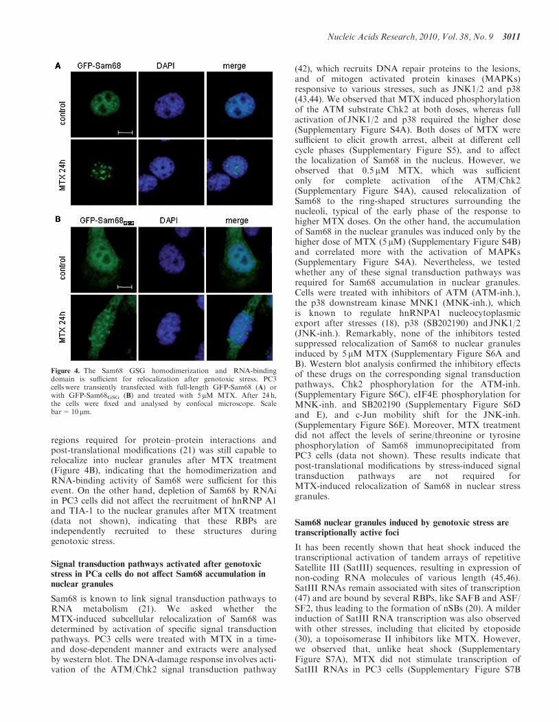
recruitment of Sam68 to specific domains of chromatinstructure and/or function. To test this possibility, weseparated chromatin components using micrococcalnuclease treatment followed by divalent ions and EDTAextractions as previously described (33,34). This procedureyields fractions enriched (S1) or depleted (S2) intranscriptionally active genes, which also remainassociated to the insoluble chromatin and nuclear matrix(P) (34). Chromatin separation from control or treatedPC3 cells showed that MTX caused an enrichment ofSam68 and hnRNP A1 in the S1 fraction at both doses,similarly to what observed for the transcription factorTFIIH p89 (Figure 5A and B). The effect of MTXappeared specific because it did not affect localization ofLamin B in the nuclear matrix fraction P (Figure 5A). Thisexperiment suggested that genotoxic stress induces achange in distribution of chromatin components, withan enrichment of the splicing factors Sam68 and hnRNPA1 in the transcriptionally active fractions.Since many splicing factors are recruited to the
transcriptionally active chromatin by phosphorylatedRNAPII, we investigated its localization in response toMTX treatment. We performed a time course treatment
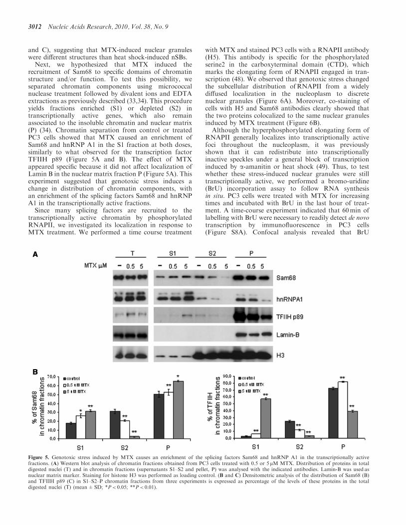
with MTX and stained PC3 cells with a RNAPII antibody(H5). This antibody is specific for the phosphorylatedserine2 in the carboxyterminal domain (CTD), whichmarks the elongating form of RNAPII engaged in tran-scription (48). We observed that genotoxic stress changedthe subcellular distribution ofRNAPII from a widelydiffused localization in the nucleoplasm to discretenuclear granules (Figure 6A). Moreover, co-staining ofcells with H5 and Sam68 antibodies clearly showed thatthe two proteins colocalized to the same nuclear granulesinduced by MTX treatment (Figure 6B).
Although the hyperphosphorylated elongating form ofRNAPII generally localizes into transcriptionally activefoci throughout the nucleoplasm, it was previouslyshown that it can redistribute into transcriptionallyinactive speckles under a general block of transcriptioninduced by a-amanitin or heat shock (49). Thus, to testwhether these stress-induced nuclear granules were stilltranscriptionally active, we performed a bromo-uridine(BrU) incorporation assay to follow RNA synthesisin situ. PC3 cells were treated with MTX for increasingtimes and incubated with BrU in the last hour of treat-ment. A time-course experiment indicated that 60min oflabelling with BrU were necessary to readily detect de novotranscription by immunofluorescence in PC3 cells(Figure S8A). Confocal analysis revealed that BrU
Figure 5. Genotoxic stress induced by MTX causes an enrichment of the splicing factors Sam68 and hnRNP A1 in the transcriptionally activefractions. (A) Western blot analysis of chromatin fractions obtained from PC3 cells treated with 0.5 or 5 mM MTX. Distribution of proteins in totaldigested nuclei (T) and in chromatin fractions (supernatants S1–S2 and pellet, P) was analysed with the indicated antibodies. Lamin-B was used asnuclear matrix marker. Staining for histone H3 was performed as loading control. (B and C) Densitometric analysis of the distribution of Sam68 (B)and TFIIH p89 (C) in S1–S2–P chromatin fractions from three experiments is expressed as percentage of the levels of these proteins in the totaldigested nuclei (T) (mean±SD; *P< 0.05; **P< 0.01).
3012 Nucleic Acids Research, 2010, Vol. 38, No. 9
staining, which was at first diffused into the nucleoplasmand partly in the cytoplasm, accumulated into specificnuclear foci under genotoxic stress (SupplementaryFigure S8B). To understand if these foci were the samestress-induced sites of Sam68 accumulation, MTX-treatedPC3 cells were stained for both BrU and Sam68. Asclearly shown in Figure 7A, the stress-induced Sam68nuclear granules incorporated BrU, indicating that theyare transcriptionally active foci.
Genotoxic stress induced by MTX affects CD44 ASprofile in a Sam68-dependent manner
Since we observed that Sam68 is recruited from thenucleoplasm to foci of residual transcription undergenotoxic stress, we asked whether its function affectsMTX-modulated AS of target mRNAs. A well describedsplicing event regulated by Sam68 is the inclusion of thevariable v5 exon in CD44 (26,27,50), a membrane receptorinvolved in cell proliferation and migration (51). TheCD44 gene contains 10 constitutive and variable exonsthat can be differentially assembled in the maturemRNA (52). Inclusion of several variable exons (v5, v6,v8 and v10) correlates with increased cell motility,invasion of neighboring tissues and malignancy and ithas been correlated to poor prognosis in patients(50–53). Moreover, recent data have implicated a changein CD44 splicing isoforms as an adaptive response of somecancer cells to genotoxic stress (12). Thus, we investigatedwhether MTX caused Sam68-dependent modulation ofthe CD44 v5 exon inclusion. Control PC3 cells (pLKO),
or PC3 cells depleted of Sam68 by stable transfection of asiRNA (pLKO si-Sam68), were treated for 2 h with 0.5 or5 mM MTX. Cells were then allowed to recover afterdamage and harvested after 24 h (3). Under these condi-tions, Sam68 and BrU labelling accumulated in nuclearstress granules like under continuous MTX treatment(Figure 7B), and cells underwent a similar proliferationarrest (Supplementary Figure S5E and F). This protocolwas used to allow cells to recover, thereby allowing highertransfection efficiency and transcription of new mRNAswithout the interference of MTX, a genotoxic agent thatcould damage the transfected minigene. Real-time PCRanalysis of CD44v5 endogenous levels, with respect tothe constitutive exons c6 and c7, showed that MTXinduced an increase in v5 inclusion in control PC3 cells(Figure 8A). Importantly, this effect required Sam68expression, because its down-regulation by RNAicompletely suppressed the effect of MTX (Figure 8Aand B). In order to understand if the increase ofCD44v5 isoform levels was directly due to AS modulation,we used a splice-reporter minigene in which inclusion ofexon v5 leads to expression of firefly luciferase (pETv5luc)(26,31). pLKOand pLKO si-Sam68 PC3 cells were treatedfor 2 h with 5 mM MTX. After recovery from the stress,cells were then transiently transfected with thesplice-reporter minigene and analysed for luciferaseactivity 24 h later. Remarkably, also in this case, weobserved that MTX treatment stimulated the inclusionof the variable CD44 exon v5 in a Sam68-dependentmanner (Figure 8C). Moreover, inclusion of exon v5 in
Figure 6. RNAPII and Sam68 colocalize in genotoxic stress-induced nuclear granules. (A) PC3 cells untreated (control) or treated with 5 mM MTX(2–24 h), were stained with a RNAPII antibody (H5) specific for the phosphorylated serine2 and analysed by confocal microscopy. (B) Co-staining ofSam68 and phosphorylated RNAPII (H5) in PC3 cells treated as in panel A. Scale bar=10 mm.
Nucleic Acids Research, 2010, Vol. 38, No. 9 3013

Sam68-depleted cells could be restored by overexpressionof a non-degradable form of Sam68 (murine mycSam68 inFigure 8C and D). Myc-Sam68 similarly induced v5 inclu-sion also in pLKO control cells (data not shown), aspreviously shown for parental PC3 cells (29). Westernblot analysis of cell extracts confirmed down-regulationof Sam68 levels in the pLKO si-Sam68 PC3 cells usedfor both experiments described above and the recoveryin expression after transfection of the murine mycSam68(Figure 8B and D).
DISCUSSION
In this report, we have investigated the subcellular local-ization and the function of Sam68 during theMTX-induced DNA damage response of PCa cells.These studies stemmed from our previous observationthat Sam68 played a protective role in PCa cells subjectedto genotoxic stress induced by chemotherapeutic drugs(29). We now show that genotoxic stress stimulates theaccumulation of Sam68 in nuclear granules, togetherwith other splicing regulators like TIA-1, SR proteinsand hnRNP A1. Moreover, we provide evidence thatthese MTX-induced granules are transcriptionally activeand contain the phosphorylated form of RNAPII. Finally,our results indicate that the subnuclear relocalization ofSam68 correlates with changes in AS of its mRNA targetCD44 and that Sam68 function is necessary for such ASmodulation. Thus, our study identifies Sam68 as a novel
stress-regulated RBP involved in the response to genotoxicstress in PCa cells.
As a first approach, we attempted a characterization ofthe Sam68 nuclear granules induced by MTX usingmarkers of previously described nuclear structuresaffected by genotoxic stress. Topoisomerase inhibitorsand alkylating agents cause double-strand breaks in theDNA, which set in motion a well-characterized DNAdamage response (1,42). The ATM kinase is recruited tothe lesions and phosphorylates H2AX, which acts as asignal to recruit proteins involved in DNA repair. Thus,the phosphorylated form of this histone (gH2AX) is usedas a marker of DNA repair foci. By using this marker, wewere able to show that Sam68 is not recruited todouble-strand break foci in the cell. By using a similarapproach, we also demonstrated that Sam68 nucleargranules were different from the PML bodies, which alsoenlarge after DNA damage and participate to thegenotoxic stress response (39). Moreover, inhibition ofthe ATM signalling pathway did not affect recruitmentof Sam68 to the nuclear foci. Thus, although Sam68relocalization is induced by lesions in the DNA, the sitesof its accumulation are not chromatin foci involved inDNA repair.
The most characterized nuclear structures induced bystress are the nSBs that form in cells exposed to heatshock (20). These bodies present several features incommon with the MTX-induced nuclear granules. First,both structures are characterized by accumulation ofSam68 (19 and this work). Moreover, nSBs also recruit
Figure 7. Sam68 nuclear granules induced by genotoxic stress are transcriptionally active foci. (A) BrU-incorporation assay in PC3 cells untreated(control) or treated with 5 mM MTX (2–24 h). PC3 cells were stained with anti-Sam68 and BrU specific antibodies. Confocal analysis revealed thatSam68 MTX-induced nuclear granules incorporate BrU. (B). PC3 cells were treated for 2 h with 5mM MTX, and allowed to recover 24 h afterdamage. At the end of incubation, cells were fixed and stained with anti-Sam68 and BrU specific antibodies. Scale bar=10 mm.
3014 Nucleic Acids Research, 2010, Vol. 38, No. 9

RNAPII and are transcriptionally active (54) like thestress granules induced by MTX. However, the activityof these structures appears somewhat different, becausenSBs are engaged in transcription of SatIII repetitivesequences after heat shock (46), whereas these non-codingRNAs are not transcribed in response to MTX treatment.Moreover, the repertoire of RBPs recruited to nSBs andMTX-induced nuclear granules is only partiallyoverlapping. For instance, the latter structures recruitalso hnRNP A1 and SC35, which were excluded fromnSBs (20). Thus, although they share some componentsand activities, they likely play different functions in thecell, which might be instructed by the differential needsimposed by heat shock and DNA lesions.
Another type of RNA-containing structure thatresembles MTX-induced stress granules are thewell-characterized cytoplasmic SGs (40). SGs areinvolved in the stress-adaptation response of the cell
induced by several poisoning treatments, such as oxidativestress, osmotic and heat shock. Cytoplasmic SGs arecharacterized by accumulation of the splicing regulatorsTIA-1 and hnRNP A1. We observed that Sam68 alsoaccumulated in cytoplasmic granules in a subset ofMTX-treated cells. Since these structures also containedhnRNP A1 and TIA-1, they can be regarded as bona-fideSGs. However, although Sam68 interacts with hnRNP A1through its C-terminal region, a GFP-Sam68GSG deletionmutant lacking the entire C-terminal domain was stillcapable to relocalize both in the nuclear and cytoplasmicstress granules after MTX treatment, suggesting thatinteraction with hnRNP A1 was not necessary for theseevents. Interestingly, our results are in line with therecently described accumulation of Sam68 in cytoplasmicSGs after oxidative stress (41), and suggest that genotoxicstress also causes formation of Sam68-containingSGs. These data indicate that Sam68 is part of a
Figure 8. Genotoxic stress induced by MTX affects CD44 alternative splicing in a Sam68-dependent manner. (A) control pLKO andpLKO-si-Sam68 PC3 cells were treated for 2 h with 0.5 (white columns) or 5 mM MTX (dim grey columns), allowed to recover from stress for24 h and analysed for the levels of endogenous CD44 v5 exon inclusion by real-time PCR. CD44 v5 levels in untreated cells (dark grey columns) wereset as 1 and MTX-induced v5 inclusion was calculated as fold increase. Samples were normalized for CD44 constitutive isoforms levels (see detailsunder ‘Materials and Methods’ section). Data are expressed as mean-fold induction ±SD from three separate experiments. (B) Western blot analysisof cell extracts (15 mg) from the PC3 clones confirmed down-regulation of Sam68 levels in pLKO-si-Sam68 cells. (C) pLKO and pLKO-si-Sam68 PC3cells were treated with 5 mM MTX for 2 h, allowed to recover and transiently transfected with pETv5Luc minigene in the presence (+) or absence (�)of murine mycSam68. The v5-Luc activity in untreated cells was set as 100% (dark grey columns) and v5-Luc activity in MTX-treated cells without(dim grey columns) or with (white column) murine mycSam68 was expressed as percentage respect to untreated cells. Data are expressed as mean-foldinduction ±SD from three separate experiments. (D) Western blot analysis of the pLKO clones cell extracts 15 mg) confirmed down-regulationof Sam68 levels in pLKO-si-Sam68 cells and the overexpression of mycSam68. Protein loading was normalized on hnRNP A1 levels both inpanel B an D.
Nucleic Acids Research, 2010, Vol. 38, No. 9 3015

RNA-mediated genotoxic stress response of the cell andthat it may exert an active role through changes in itssubcellular localization.Sam68 is a prototype of the STAR proteins, which are
known to link signal transduction pathways to changes inRNA metabolism (21). For instance, growth factors stim-ulate the MAPKs ERK1/2, which in turn phosphorylateSam68 and stimulate its splicing activity (26). Since MTXtriggered the activation of stress-response MAPKs, weasked whether these pathways were involved in Sam68relocalization. However, our results, obtained withspecific inhibitors of the p38 and JNK1/2 pathways,strongly suggest that phosphorylation of Sam68 by thesekinases is not required for its recruitment toMTX-induced nuclear stress granules.Chromatin fractionation experiments and
colocalization with the phosphorylated RNAPII andnascent RNAs indicated that MTX-induced nucleargranules were transcriptionally active. We labelled thecells in the last hour of treatment, thus, the BrU-positivegranules are likely sites of de novo synthesis of RNA ratherthan sites of accumulation of RNAs that had beensynthesized before the MTX treatment. Since severalRBPs involved in AS and other steps of post-transcriptional regulation of mRNAs were recruited tothese MTX-induced nuclear granules, we asked whetherSam68 was involved in processing of target mRNAsduring the genotoxic stress. A particularly interestingsplicing target of Sam68 is CD44. This gene encodes forseveral isoforms that differ for the inclusion of variableexons (v1–v10), which confer oncogenic potential to thecorresponding proteins (51–53). Remarkably, a recentstudy showed that CD44 undergoes profound changes inAS during genotoxic stress caused by cisplatin in cancercells (12). Thus, we tested whether MTX induced changesin the inclusion of v5, the target of Sam68 activity (26),and whether or not these changes required Sam68.Remarkably, our experiments, which monitored both theendogenous CD44 mRNA and a reporter minigene,clearly showed that v5 exon inclusion correlated with for-mation of the nuclear stress granules and that this splicingevent was dependent on Sam68 expression. Thus, it ispossible that compartmentalization of Sam68, and ofother splicing factors, in the transcriptionally activenuclear stress granules redirects the splicing machineryduring the response to genotoxic stress, thereby affectingspecific splicing events. This response might allow the cellto produce protein isoforms that allow to withstand thestress and to favor repair of the damage caused. Indeed,expression of CD44v5 isoforms was shown to have prog-nostic value in human thymic tumors (53). In this regard,it will be interesting to determine the genes affected byMTX at the level of AS on a large scale, similarly towhat recently described for the response to UV irradiation(3). Since Sam68 protects PCa cells from genotoxic stress(29), it is possible that its splicing targets contribute toenhance cell survival in response to DNA damage. Thus,future studies will aim at clarifying how Sam68 modulatesAS in response to genotoxic stress and at the identificationof mRNAs whose AS is regulated in a Sam68-dependentmanner after DNA damage in cancer cells.
SUPPLEMENTARY DATA
Supplementary Data are available at NAR Online.
ACKNOWLEDGEMENTS
The authors wish to thank Dr Maria Paola Paronetto forthe establishment of Sam68-depleted PC3 clones and sug-gestions throughout this study, Dr Harald Konig for thegift of the pET-v5Luc plasmid, Prof. Edward Eddy for thegift of pHspa.2-blue, Prof. Francesco Lo Coco for the giftof the PML antibody, Dr Pamela Bielli and Dr MariaLoiarro for assistance with luciferase reporter assays, DrEnrica Bianchi for assistance with FACS cell cycleanalyses and Dr Federica Barbagallo for the pHspa-2-GFP plasmid.
FUNDING
‘Associazione Italiana Ricerca sul Cancro’ (AIRC); the‘Association for International Cancer Research’; (AICR)and the Istituto Superiore della Sanita (ISS Project n.527/B/3A/5). Funding for open access charges: AssociazioneItaliana Ricerca sul Cancro’ (AIRC); and ‘Association forInternational Cancer Research’ (AICR).
Conflict of interest statement. None declared.
REFERENCES
1. Crighton,D. and Ryan,K.M. (2004) Splicing DNA-damageresponses to tumour cell death. Biochim. Biophys. Acta, 1705,3–15.
2. Kleiman,F.E., Wu-Baer,F., Fonseca,D., Kaneko,S., Baer,R. andManley,J.L. (2005) BRCA1/BARD1 inhibition of mRNA 30
processing involves targeted degradation of RNA polymerase II.Genes Dev., 19, 1227–1237.
3. Munoz,M.J., Perez Santangelo,M.S., Paronetto,M.P., de laMata,M., Pelisch,F., Boireau,S., Glover-Cutter,K., Ben-Dov,C.,Blaustein,M., Lozano,J.J. et al. (2009) DNA damage regulatesalternative splicing through inhibition of RNA polymerase IIelongation. Cell, 137, 708–720.
4. Black,D.L. (2003) Mechanisms of alternative pre-messenger RNAsplicing. Annu. Rev. Biochem., 72, 291–336.
5. Matlin,A.J., Clark,F. and Smith,C.W. (2005) Understandingalternative splicing: towards a cellular code. Nat. Rev. Mol. Cell.Biol., 6, 386–398.
6. Biamonti,G. and Caceres,J.F. (2009) Cellular stress and RNAsplicing. Trends Biochem. Sci., 34, 146–153.
7. Srebrow,A. and Kornblihtt,A.R. (2006) The connection betweensplicing and cancer. J. Cell Sci., 119, 2635–2641.
8. Venables,J.P. (2006) Unbalanced alternative splicing and itssignificance in cancer. Bioessays, 28, 378–386.
9. Ghigna,C., Valacca,C. and Biamonti,G. (2008) Alternativesplicing and tumor progression. Curr. Genomics, 9, 556–570.
10. Venables,J.P., Klinck,R., Koh,C., Gervais-Bird,J., Bramard,A.,Inkel,L., Durand,M., Couture,S., Froehlich,U., Lapointe,E. et al.(2009) Cancer-associated regulation of alternative splicing. Nat.Struct. Mol. Biol., 16, 670–676.
11. Solier,S., Lansiaux,A., Logette,E., Wu,J., Soret,J., Tazi,J.,Bailly,C., Desoche,L., Solary,E. and Corcos,L. (2004)Topoisomerase I and II inhibitors control caspase-2pre-messenger RNA splicing in human cells. Mol. Cancer Res., 2,53–61.
12. Filippov,V., Filippova,M. and Duerksen-Hughes,P.J. (2007) Theearly response to DNA damage can lead to activation ofalternative splicing activity resulting in CD44 splice patternchanges. Cancer Res., 67, 7621–7630.
3016 Nucleic Acids Research, 2010, Vol. 38, No. 9

13. Chandler,D.S., Singh,R.K., Caldwell,L.C., Bitler,J.L. andLozano,G. (2006) Genotoxic stress induces coordinately regulatedalternative splicing of the p53 modulators MDM2 and MDM4.Cancer Res., 66, 9502–9508.
14. Burd,C.J., Petre,C.E., Morey,L.M., Wang,Y., Revelo,M.P.,Haiman,C.A., Lu,S., Fenoglio-Preiser,C.M., Li,J., Knudsen,E.S.et al. (2006) Cyclin D1b variant influences prostate cancer growththrough aberrant androgen receptor regulation. Proc. Natl Acad.Sci. USA, 103, 2190–2195.
15. Wang,Y., Dean,J.L., Millar,E.K., Tran,T.H., McNeil,C.M.,Burd,C.J., Henshall,S.M., Utama,F.E., Witkiewicz,A., Rui,H.et al. (2008) Cyclin D1b is aberrantly regulated in response totherapeutic challenge and promotes resistance to estrogenantagonists. Cancer Res., 68, 5628–5638.
16. Filippov,V., Schmidt,E.L., Filippova,M. andDuerksen-Hughes,P.J. (2008) Splicing and splice factor SRp55participate in the response to DNA damage by changing isoformratios of target genes. Gene, 420, 34–41.
17. Messaoudi,L., Yang,Y.G., Kinomura,A., Stavreva,D.A., Yan,G.,Bortolin-Cavaille,M.L., Arakawa,H., Buerstedde,J.M., Hainaut,P.,Cavaille,J. et al. (2007) Subcellular distribution of human RDM1protein isoforms and their nucleolar accumulation in response toheat shock and proteotoxic stress. Nucleic Acids Res., 35,6571–6587.
18. Guil,S., Long,J.C. and Caceres,J.F. (2006) hnRNP A1relocalization to the stress granules reflects a role in the stressresponse. Mol. Cell. Biol., 26, 5744–5758.
19. Denegri,M., Chiodi,I., Corioni,M., Cobianchi,F., Riva,S. andBiamonti,G. (2001) Stress-induced nuclear bodies are sites ofaccumulation of pre-mRNA processing factors. Mol. Biol. Cell.,12, 3502–3514.
20. Biamonti,G. (2004) Nuclear stress bodies: a heterochromatinaffair? Nat. Rev. Mol. Cell. Biol., 6, 93–98.
21. Lukong,K.E. and Richard,S. (2003) Sam68, the KHdomain-containing superSTAR. Bioch. Biophys. Acta, 1653,73–86.
22. Coyle,J.H., Guzik,B.W., Bor,Y.C., Jin,L., Eisner-Smerage,L.,Taylor,S.J., Rekosh,D. and Hammarskjold,M.L. (2003) Sam68enhances the cytoplasmic utilization of intron-containing RNAand is functionally regulated by the nuclear kinase Sik/BRK.Mol. Cell. Biol., 23, 92–103.
23. Paronetto,M.P., Zalfa,F., Botti,F., Geremia,R., Bagni,C. andSette,C. (2006) The nuclear RNA-binding protein Sam68translocates to the cytoplasm and associates with the polysomesin mouse spermatocytes. Mol. Biol. Cell, 17, 14–24.
24. Paronetto,M.P., Messina,V., Bianchi,E., Barchi,M., Vogel,G.,Moretti,C., Palombi,F., Stefanini,M., Geremia,R., Richard,S.et al. (2009) Sam68 regulates translation of target mRNAs inmale germ cells, necessary for mouse spermatogenesis.J. Cell Biol., 185, 235–249.
25. Henao-Mejia,J., Liu,Y., Park,I.W., Zhang,J., Sanford,J. andHe,J.J. (2009) Suppression of HIV-1 Nef translation by Sam68mutant-induced stress granules and nef mRNA sequestration.Mol. Cell, 33, 87–96.
26. Matter,N., Herrlich,P. and Konig,H. (2002) Signal-dependentregulation of splicing via phosphorylation of Sam68. Nature, 420,691–695.
27. Batsche,E., Ianiv,M. and Muchardt,C. (2006) The human SWI/SNF subunit Brm is a regulator of alternative splicing.Nat. Struct. Mol. Biol., 13, 22–29.
28. Paronetto,M.P., Achsel,T., Massiello,A., Chalfant,C.E. andSette,C. (2007) The RNA-binding protein Sam68modulates the alternative splicing of Bcl-x. J. Cell Biol., 176,929–939.
29. Busa,R., Paronetto,M.P., Farini,D., Pierantozzi,E., Botti,F.,Angelini,D.F., Attisani,F., Vespasiani,G. and Sette,C. (2007)The RNA-binding protein Sam68 contributes to proliferationand survival of human prostate cancer cells. Oncogene, 26,4372–4382.
30. Valgardsdottir,R., Chiodi,I., Giordano,M., Rossi,A., Bazzini,S.,Ghigna,C., Riva,S. and Biamonti,G. (2008) Transcription ofSatellite III non-coding RNAs is a general stress response inhuman cells. Nucleic Acids Res., 36, 423–434.
31. Weg-Remers,S., Ponta,H., Herrlich,P. and Konig,H. (2001)Regulation of alternative pre-mRNA splicing by the ERKMAP-kinase pathway. EMBO J., 20, 4194–4203.
32. Paronetto,M.P., Venables,J.P., Elliott,D.J., Geremia,R., Rossi,P.and Sette,C. (2003) Tr-kit promotes the formation of amultimolecular complex composed by Fyn, PLCgamma1 andSam68. Oncogene, 22, 8707–8715.
33. Bafus,N.L., Albright,S.C., Todd,R.D. and Garrard,W.T. (1978) Amethod for mapping the distributions of modified and varianthistones among mono- and polynucleosomes. J. Biol. Chem., 253,2568–2574.
34. Rose,S.M. and Garrard,W.T. (1984) Differentiation-dependentchromatin alterations precede and accompany transcription ofimmunoglobulin light chain genes. J. Biol. Chem., 259,8534–8544.
35. Herrmann,A., Sommer,U., Pranada,A.L., Giese,B., Kuster,A.,Haan,S., Becker,W., Heinrich,P.C. and Muller-Newen,G. (2004)STAT3 is enriched in nuclear bodies. J. Cell. Sci., 117, 339–349.
36. Rosenberg,J.E., Ryan,C.J., Weinberg,V.K., Smith,D.C.,Hussain,M., Beer,T.M., Ryan,C.W., Mathew,P., Pagliaro,L.C.,Harzstark,A.L. et al. (2009) Phase I study of ixabepilone,mitoxantrone, and prednisone in patients with metastaticcastration-resistant prostate cancer previously treated withdocetaxel-based therapy: a study of the department of defenseprostate cancer clinical trials consortium. J. Clin. Oncol., 27,2772–2778.
37. Huang,X., Traganos,F. and Darzynkiewicz,Z. (2003) DNAdamage induced by DNA topoisomerase I- and topoisomeraseII-inhibitors detected by histone H2AX phosphorylation inrelation to the cell cycle phase and apoptosis. Cell Cycle, 2,614–619.
38. D’Orazi,G., Cecchinelli,B., Bruno,T., Manni,I., Higashimoto,Y.,Saito,S., Gostissa,M., Coen,S., Marchetti,A., Del Sal,G. et al.(2002) Homeodomain-interacting protein kinase-2 phosphorylatesp53 at Ser 46 and mediates apoptosis.Nat. Cell. Biol., 4, 11–19.
39. Dellaire,G., Ching,R.W., Ahmed,K., Jalali,F., Tse,K.C.,Bristow,R.G. and Bazett-Jones,D.P. (2006) Promyelocyticleukemia nuclear bodies behave as DNA damage sensorswhose response to DNA double-strand breaks is regulatedby NBS1 and the kinases ATM, Chk2, and ATR.J. Cell. Biol., 175, 55–66.
40. Kedersha,N. and Anderson,P. (2002) Stress granules: sites ofmRNA triage that regulate mRNA stability and translatability.Biochem. Soc. Trans., 30, 963–969.
41. Henao-Mejia,J. and He,J.J. (2009) Sam68 relocalization into stressgranules in response to oxidative stress through complexing withTIA-1. Exp. Cell Res., 315, 3381–3395.
42. Shiloh,Y. (2003) ATM and related protein kinases: safeguardinggenome integrity. Nat. Rev. Cancer, 3, 155–168.
43. Stadheim,T.A. and Kucera,G.L. (2002) c-Jun N-terminal kinase/stress-activated protein kinase (JNK/SAPK) is required formitoxantrone- and anisomycin-induced apoptosis in HL-60 cells.Leuk. Res., 26, 55–65.
44. Roux,P.P. and Blenis,J. (2004) ERK and p38MAPK-activated protein kinases: a family of protein kinaseswith diverse biological functions. Microbiol. Mol. Biol. Rev., 68,320–344.
45. Jolly,C., Metz,A., Govin,J., Vigneron,M., Turner,B.M.,Khochbin,S. and Vourc’h,C. (2004) Stress-induced transcription ofsatellite III repeats. J. Cell Biol., 164, 25–33.
46. Rizzi,N., Denegri,M., Chiodi,I., Corioni,M., Valgardsdottir,R.,Cobianchi,F., Riva,S. and Biamonti,G. (2004) Transcriptionalactivation of a constitutive heterochromatic domain of thehuman genome in response to heat shock. Mol. Biol. Cell., 15,543–551.
47. Valgardsdottir,R., Chiodi,I., Giordano,M., Cobianchi,F., Riva,S.and Biamonti,G. (2005) Structural and functional characterizationof noncoding repetitive RNAs transcribed in stressed human cells.Mol. Biol.Cell, 16, 2597–2604.
48. Phatnani,H.P. and Greenleaf,A.L. (2006) Phosphorylation andfunctions of the RNA polymerase II CTD. Genes Dev., 20,2922–2936.
Nucleic Acids Research, 2010, Vol. 38, No. 9 3017

49. Zeng,C., Kim,E., Warren,S.L. and Berget,S.M. (1997) Dynamicrelocation of transcription and splicing factors dependent upontranscriptional activity. EMBO J., 16, 1401–1412.
50. Cheng,C. and Sharp,P.A. (2006) Regulation of CD44 alternativesplicing by SRm160 and its potential role in tumor cell invasion.Mol. Cell. Biol., 26, 362–370.
51. Ponta,H., Sherman,L. and Herrlich,P.A. (2003) CD44: fromadhesion molecules to signalling regulators. Nat. Rev. Mol. Cell.Biol., 4, 33–45.
52. Naor,D., Sionov,R.V. and Ish-Shalom,D. (1997) CD44: structure,function, and association with the malignant process. Adv. CancerRes., 71, 241–319.
53. Lee,S.C., Harn,H.J., Lin,T.S., Yeh,K.T., Liu,Y.C., Tsai,C.S. andCheng,Y.L. (2003) Prognostic significance of CD44v5 expressionin human thymic epithelial neoplasms. Ann. Thorac. Surg., 76,213–218.
54. He,B., Meng,Y.H. and Mivechi,N.F. (1998) Glycogen synthasekinase 3beta and extracellular signal-regulated kinase inactivateheat shock transcription factor 1 by facilitating the disappearanceof transcriptionally active granules after heat shock. Mol. Cell.Biol., 18, 6624–6633.
3018 Nucleic Acids Research, 2010, Vol. 38, No. 9