genomic sequence and embryonic expression of the zebrafish homeobox gene hox-3.4
TRANSCRIPT
Seediscussions,stats,andauthorprofilesforthispublicationat:https://www.researchgate.net/publication/15095712
Genomicsequenceandembryonicexpressionofthezebrafishhomeoboxgenehox-3.4
ARTICLEinTHEINTERNATIONALJOURNALOFDEVELOPMENTALBIOLOGY·JULY1993
ImpactFactor:1.9·Source:PubMed
CITATIONS
13
READS
36
3AUTHORS,INCLUDING:
JohannaUEricsonSollid
UniversityofTromsoe
40PUBLICATIONS1,724CITATIONS
SEEPROFILE
AndersFjose
UniversityofBergen
66PUBLICATIONS3,570CITATIONS
SEEPROFILE
Availablefrom:AndersFjose
Retrievedon:03February2016
Int..J. [)t"". Riol. .'7: 263-272 (199.1) 263
Original Arlie/"
Genomic sequence and embryonic expression of thezebrafish homeobox gene hox-3.4
JOHANNA U. ERICSON", STEFAN KRAUSS' and ANDERS FJOSE3
IOepartment of Molecular Genetics, Institute of Medical Biology, University of Tromso, Tromso, Norway.20ivision of Biomedical and Biomolecular Sciences, King's Coflege London. University of London, London, England and
JOepartment of Biochemistry, University of Bergen, Bergen, Norway
ABSTRACT We have analyzed the genomic DNA sequence and embryonic expression pattern of thezebrafish hox-3.4 gene. Two exons, encoding a protein with a total size of 232 amino acids, wereidentified within a 3.5 kbp genomic region. Besides the homeodomain, which is identical to that of thehuman HOX3D and mouse Hox-3.4 genes, the first 58 residues of the N-terminal domain in thepredicted Hox-3.4 protein share 48% sequence identity with the gene product of the human cognate.Some of the N.terminal sequence elements are also conserved relative to the two other members ofthe Hox-1.3/Hox-2.1/Hox-3.4 paralogy group. In addition, the paralogous genes share a significantdegree of sequence identity in non-coding regions. This conservation is particularly evident in thepromoter regions of the cognates hox.3.4, Hox-3.4, and HOX3D, where a 180 bp TATA-box.containingelement with a 60% identity is located. This is in agreement with the previous finding that the HOX3Dpromoter region contains response elements for other Hox proteins and retinoids, Also with respectto embryonic expression, the zebrafish hox-3.4 gene is very similar to its mammalian counterparts.Within the central nervous system of 16, 24, and 48 h embryos, hox-3,4 transcripts were detectedthroughout most of the spinal cord from a boundary at the posterior end of the hindbrain, In 16 hembryos the hox-3.4 gene is also active within a restricted region of the tailbud,
KEY WORDS: :rhmjish. homr,,!Jox gent'. 110.\"-3.--1.genomir .\rqllencr, rmh,)onir r.\"lnrssiol/
Introduction
Following the first identification of homeobox sequences in theANT-C and BX-Chomeotic gene clusters of Orosophila melanogaster(McGinnis et af.. 1984a,c), a large number of related homeoboxgenes (Hox)have been characterized in different vertebrate species,including Xenopus laevis (Carrasco et al.. 1984), chicken (Ranginiet a/.. 1989), mice (McGinnis et al.. 1984b), human (Levine et al.,1984), and zebrafish (Eiken et al.. 1987). The strong conservationof both DNA sequence and gene organization among Hoxgenes fromdifferent species implies that the clusters had a common ancestor.HOM.C (homologous to the Drosophila ANT.C and BX-C clusters injuxtaposition; Beeman, 1987), from which they all have evolved.Relatively high levels of Hox gene expression are detected in thecentral nervous system (CNS) of vertebrates. in which the gene
organization reflects the expression pattern. i.e. 3'-anterior-earlyj5'.posterior.late (Dolle et al.. 1989; Izpisua.Belmonte et al..1991).
Gene paralogues display similar anteroposterior (A-P) but differentdorsoventral (D-V) expression patterns. indicating that duplicationsofthe Hoxcluster (up to four copies in higher vertebrates; Acamporaet al.. 1989; Graham et al.. 1989) might have provided a possibility
to direct development of a more complex CNS. not only by thepositional information along the A-P axis but also in the ~Vorientation (Graham et al.. 1991), The correlation between Hoxexpression and the segmental organization of the hindbrain sug-gests that Hox proteins participate in the determination of segmen-tal identities rather than establishing the individual segments(Wilkinson et al.. 1989; Hunt and Krumlauf, 1991). Experimentalmanipulation of Hox gene expression in Xenopus and mouseembryos have provided more direct evidence for this assumption.Ectopic expression of Hox-l.l in transgenic mice leads to posteriortransformations in tissues anterior of the regions normally express-ing Hox.l.l (Puschel et a/.. 1991). Conversely. loss of function.either by Hox-3.1 disruption in mouse (Le Mouellic et al.. 1992) oranti-Xlhboxl antibody injections in Xenopus (Wright et al.. 1989).displayed anteriorization of a subset of embryonic cells. resulting intransformed vertebrae (Hox-3.1) or hindbrain structures (Xfhbox1).
.-\hhrl'viatiol/s IBid ill IM\ Imp,,.: C:\"S. ("('IIIr;1I IH:"f\OILS systt'm; .-\-1'.
;1IJ1('ropoo;tt'rior; 0-\', ,lor'IJ\"t'ntral: R.-\. H'linoic acid: ORF, opt"1I n';uIiIlH
tramt'o
-Address for reprints: Department of MolecularGenetics.lnstitute of Medical Biology, University ofTromsQ, N-9037 TromsQ, Norway. FAX: 47.83.45350.
0214.62X21Y3/S03.00Ci l:BC Pre"I'rintcdinSp'ln

264 .l.V. Ericson et a!.
A
HE E
..~...-
.....
..
B rE
c
---
D 1111111111111111111111111111111111111111111111111111111111111111111111111111111111111
Fig. 1. Genomic organization of the hox.3.4 gene. (Part AI shows the restriction enzyme map of the two genomiC clones carrying the zebraflsh hox-
3. 4 homeobox. (Part 81 Below, represents the totafsubcfonedregion. The Size-bar corresponds to 1000bpinparr A and 250 bp in part 8. The rwopredictedexons of rhe hox-3,4 gene are indicated below the map In B. (Part C) shows the 3.5 kbp sequenced region, and {Part DI the restriction fragment usedas a probe for in situ hybridization expenments. Abbreviations: B, BamH/; C, Clal; E. EeaRI; H, Hind/If: K, Kpn/; X, XbaJ.
Furthermore. these data confirmed that each Hoxparalogue has aunique function that cannot be replaced by another gene from thesame subfamily.
Establishment of the -Hox code-, i.e. the combination of func-tionally active Hox genes that will specify the identity of a bodyregion, can be influenced by endogenous molecules like retinoicacid (RA) and peptide growthfactors. RA,a posteriorizingmorphogen,influences the expression of Hexgenes accordingtotheir individualsensitivity, both in whole embryos (Kessel and Gruss. 1991;Morriss-Kay et al..1991) and in tissue cell culture (Boncinelli et al.,1991; Arcioniet at.. 1992). It was suggested by Kessel and Gruss(1991) that during gastrulation, ingressing cells are exposed to a RAsignal generated from midline embryonic structures, and will respondwith a sequential activation of more and more Hox genes, leadingto nonidentical, overlapping expression domains of Hoxgenes alongthe anteroposterior axis.
Some of the Drosophila homeoproteins have been shown to workas transcription factors (Biggin and Tjian. 1989; Han et al., 1989:Winslow et al., 1989; Jaynes and O'Farrell, 1991). In vertebrates,little is known about the repressing/derepressing activities of theHox gene products. However, the characterization of the humanHOX3D promoter sequence revealed potential targets for multipleregulatory mechanisms acting independently (Arcioni et al., 1992),among them trans-activation by three. more posteriorly expressed,Hox gene products. i.e. HOX3C. HOX4D and HOX4C proteins. inaddition to the mouse Hox-4.3 protein (Arcioni et at., 1992).Alignment of the promoter regions from the HOX3D gene and itsmouse homologue (Hox-3.4) showed conservation in all identified
regulatory targets (Arcioni et al.. 1992). Thus, the vertebratehomeoproteins display evolutionarily conserved regulatory functionsmediated by sequence-specific DNA-binding.
In zebrafish (Brachydanio rerio), strong similarities to the mouseHex genes have been observed, both with respect to the geneorganization and DNA sequence (Eiken et al..1987; Njolstad et al..1988a.b. 1990: Molven et al.. 1990. 1992). Evidently there are atleast two Hox gene clusters in zebrafish. corresponding to themammalian HOX-2 and-3 complexes (Njolstad et al.. 1988b. 1990).In this paper we describe the genomic DNA sequence of thezebrafish hox-3.4 gene. which encodes a homeoprotein homologousto the human HOX3D gene product. Additional sequence conser-vations were identified in the non-coding regions including thepromoter. Results obtained by in situ hybridization analysis of theembryonic zebrafish hox-3.4 expression pattern are also consistentwith the spatial distribution of transcripts reported for the murineHox-3.4 gene (Gaunt et al.. 1990).
Results
Identification of the zebrafish hox-3.4 protein-encoding sequence
Two recombinant lambda-EMBL3 clones, C25 and C26. contain-ing genomic zebrafish DNA. were previously shown to carry the hox-3.4homeoboxsequence(Eiken. et al..1987: Njolstad et al..1990).The unique Kpn I-site in lambda C25. found in the homeobox regionof hox-3.4 (Eiken et al.. 1987). was used to identify the hox-3.4-containing subclones (Fig. 1). DNA sequencing of a 3.5 kbp region,including 1.7 kbp downstream and 1.8 kbp upstream of the Kpn I
Zebrafish 110-,-3.4 sequcnce and cxpression 265
TTIG TCA T AA T AreG T AICGCA.1TG1TTC1TGTCCA TCA tCTTGTTTGCACATG TCTGTGT A TGTG.A.GAGAG TeCGe! T tG'I'CCAG 'I'
AT! AGA03GCCAGA TGACGCTC'!1'GTCT MCGG
121 ICI ACGGCCC'I"I' 'I"!1 AGCCAAGACCC'TTGC A ':'GA '!TTCCA T ACCGAGAAG IT A T(;GGACACGTTTCCCTG TeI ATCAA! AACCTCCTGGGGA Ie MGCCAA1TT ATGACTGGCCAGGAGC
1.4 1 TGCACG!GA!!CT ATTT AMC;.. TIceA TA T7tGGGCATT ACACG T CG TATCAAGAAAAAAAGAAJ\ATG.".I T TCCTCCACcr ATAM TCCTGCtC1TTTTT AGGACJo.A!j(JCCt MCCTeer
361 ('1 AGAAAT ACAAA T AJ>.MXCM T AAACAAA T ACAACTT C1 AAACMCTT A TGT J..T ACTTTAT'! AGCIGATTTCGTTTCTGCAGTGACCGAGTGTT A TTTGTGAGTCTCTTTAG.JGIGA T A
(8 1 TTG!1TGGGAAA:rAGCA:rGA::7CTCA TACGT!GGGAAG Ie! T T neT AAGCNJACGCA1\GACCCC'ICCtCT!GT AGAATGCACACTTTtGACMCT A TGGAGCTCACAGTGAGTTCCACGA
1 M $ S Y V G K F S K Q T 0 D ASS C R M H T D N Y G A H S E F H E
601 GTCCM IT ACGCGT ACGMGGGCTtGA T C1 CGGCGGATCCT rCAGTTCTC AA tCCCCACCAACTCTTTGAGGCGGGAGGCGA T J..AACACAACCGACCGTGCAAGGAGCAGTGCAGCAGT
36 S NY},':: :: G L D L G G S F S S
QI P '! N 5 L R REA I N T T D R A R 5 S A A V
711 TCJ-GCGMCACAGTCTTG'! TC AGC'! CIGGG:''1'CTCGT AGCTTTGT AASCAC'! CACGGGT ACAACCCCCTCAGTCACGGACTGTTGAGCCAAAAlIGCCGAGGGGAA T A TGGAAGTT A TGGA
76 Q R T Q5 C 5 A L G R S F v 5 I H G Y N P L 5 H G L L 5 Q
K A E G N MEV M E
6 4 1 GAN3CCCJl.Gl:GJCAAGAGCA.GCJI,G CGA I A ICAAJ<.ATGGN:;ACT ACITe GCGA'! AN3CAACJ..AACT AA ITeT ACTCJlGCGTCJlGAACCAG T CGCAGCCGCA.GA TAT ATCCG TG(;A TGAr:
116 K PSG K 5 5 R R Y Q~ G D Y F D K Q Q T N 5 T Q R Q N
Q5
QP
QI Y P W M T
9 61 AJJ>,J;CT ACACA TGA.GCCACc1 I AAA.G I I)'CG HTC ..v.GTTTGTT IT .".GAAA.".GAGGAAACCTGCACGO. TCTCA TCA TCG T CA'! TGCATCI CCI I TA.GACTG I nTG TCGCTCCA TA Tn156 K L H M 5 H
1 0 6 1 nCCTCnGAG TTTI AI AGGCC'.".AACGCJ>..GGNvTAATAAAACI Jl.Gl:GGCCGT AAATTTT A TGACAAAGGCA TCT ATTGCICG I AAACCTGTCCTG '!T ACTG TGAAGAGGTGATCCCCGG
1101 TCAAATTTATGGAGAAAIAATIGTIAAG~CAAAAAATGk~"'CGT.~"'TTTGTTGeTTTTATGAATAAAAGGGAGIIIACAIGIIAIICAAGTAAAAGGGAATTTTAACCGAAIAI
1321 TTGCA TACAT ACA I AA I CIGGCAGGACTT ATGTTTCCGG II AG I '!GCACICTGCACCGCT AC';AGCAGT AAAGCAAAG I ACA.GCACACCT ACATTGTCAGTT TGGT!CATCTT AAGMG TG
14 4 1 CM T AGCAGI T'! cn '! '! AT '!TG TTCAr..GTCTT ACA TT A T T TGJIG '! C'!GACATGTT AAGAAGCTCA TGGCCnG I CAT A T T TAGGn TATCATGTTCC ATCAC'GTC1 A T '!TGTGCATGACA
1561
1661162
1601169
1921209
CCTTCT AA TTTC JIG.v.. T M.AJ..AA T ATT ,\GCG":CT ACCACCIG I CCACTG '!TTTCATTTTGGJ.. 'iTTC A I AA TGCI TGA TGT !TCC A TTGCA T1 IT AAGGAC1GTGTTTGGTGGCAACT TCA
TCA TTT A IT AI AAAAA T T AA T TG T AT }JXAAT A TGCA TGAGGACGACA.GTCGA T ACGTTTtT ACA TAT AGGT AACCCA.G TATCT T TCTGATGCCATCTCA.GkTCTGACGG I AAAAGGTC
E 5 D G K R 5ACGAACCJlGTT ACACCCGG T ACCl\G;"CTCTGGIIGTTGGAGJJoAGAG I ICe TTTCMCCGA T J..CCTCACACG TCGCAGACGT ATCGA.GA TIGec M T MCC1CTGCT1GAACGA.GCGCC A
R T 5 Y TRY Q T L E L EKE F H F N R Y L T R R R R I E I ANN L C L N E R QAATTAAJ<.A'!ATGGITCCA.GAACCGTCGCATGAAGTGGAAG}AGGACICAAA.GTTGAAAGTAAAAGGAGGACTATAAATAATG'!GTTGCAOGACTTCAATTACMCAGCCTGAAAATAAAG
Ie I '0'/ FQ
:-: R R 1-1 K W Kj(
:J 5 K L K V K G G L 232 -
2 0 4 1 GCAATGIT AACCCTTGAACT MMCAACGAA TGA ITT AAAA I IGA T ITTGIT;.. 7T AACMCTGTTGA '!G T CT A TCATT IG ITI ACM T A TT A TGTTGTT ATGTGM T A TG TTGA T ATT T I
2 1 6 1 CM T AT T T'! ATTGAT JlGTGCTCTGTCCCeAG T ACGGCAGATGc'r MTTTGAA T M TGT A TGAGCI I MAC A T
TG>'TT A TGCTn ACT MACTCTCTTGT A TO. TCCT ACA T M TAT A T AC
2;: 81 CAAGGCCIGAAI ACAGTT AAGTTGACA TM'"
'!G T ACG TGCAAAAATGTGGTGCTTTGCTTGA T CAT TC ATC MTCTGTTCA TTGCA TTTTTGA TGCT ACTTGTCnT A TTTTTCA TTGT A
2 ~ 0 1 CTT TCIGeCT MAT ACA TCT A T AJ..T ACCI Ae C.".AGM'!GGT AAATCCCM TTCA TGA I AI AM ICGCAAAAACT -'GTCTCA TAIT AAACGTTGTGT A TT A TGTTGTCCGTCAGTCAGCA
2521 GCCAATGTTATMTTTTCTC}ACAIGAIIGTGTTTATTGMTTACTGTGCAATCAGAAIAITTTGATeTTTAAAAAGTTTTTAGTTTTTTTITCTTAAGTGGGCTCTTGTTTTTACACAC
2641 CCCACTCTCGM;TJlGCCTATATTT~AAICCTGT AAGAACTATAAC;GAGTGGCITATTGAI ACTICCCAATTCCCJl.TACCAAACATAC CG CCTACITTTGTACAAAACGCATTTGT
2 7 61 ICTGCGA TCC ATA TT ACCI I I AI I I CA CCGG T1(;1TTT(GA::;A TTTCGT AGA I AAG':CACA TCG). T M TTTTCCT A TCM T AAAGA.".G I AI I I k...CAAGA TT MCT TeeM TAATGCAC
2861 CGTTCGGMGGTTTTGTCAGCTGIGGGII~".GCCATCTGTeTCCAATAAAAGIGAIATCAITGTT~TAAACGATTAMCGTATGAT'!MCACCCTTTTGTTAACGTTTTTCTCTGTT
3 CO 1 AAGN:;CAAOG1IGTCTTTCACTACAAACACIGCATTTTTI AAATTCAAC:;TCCTATTTGCCACCCGGAGGGTTAGGJ..A}'}.AGAAJ.~ATTTGCCCCAAITMTATGACTATAAATATTC
31 21 GTT MTGCA TCAAACGT AGCI IGCACG T IT AT ACTTCCAGTGATGA::;ACJ.. TGA TTGMCT T I AGGGCTCCGGTCIICTT A TTT ACAG T A.GCCCTT AMCGGAGGCACA T AAAACTTT ATGA
3 2 4 1 CCCCM T A.".ACTTTT AC;.GCJ..CI T CCICTCACCCATCA.GACACTG;"CAAeTGTTTCCCCI T ITAACAT A TCCGCGGAAGTT T ACACM.ACA'! T '! CAAAG TGACG I TTTGG TAT ACMACC
3361 CTTTAAAACAJ..TAGGATAGT'!TGCACTTTIAATIAAATAG7T7GT:JGCACTAATTACGMTA'!MTCATTATTACGTCCTTAAAAIGCATATGCATIATGCTTCIAAAACGTTATTMTC
3(61 GTCCTJIGC1GGTAAGCTT 34~8
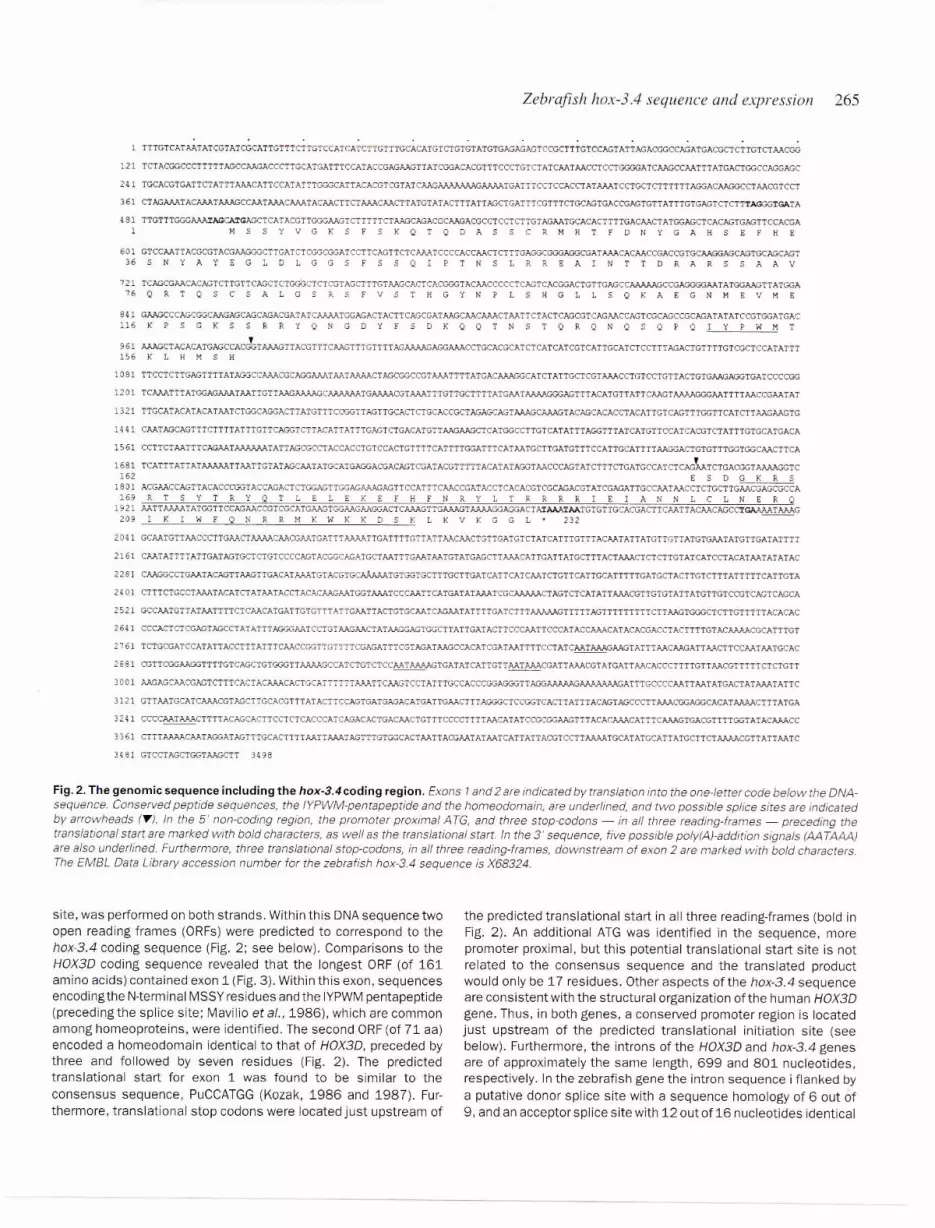
Fig. 2. The genomic sequence including the hox-3.4coding region. Exons 1and 2 are rndicated by translation into the one-letter code below the DNA-sequence. Conserved peptide sequences. the IYPWM-pentapeptide and the homeodomain, are underlined, and rwo possible splice sites are indicatedby arrowheads (~). In rhe S' non-coding region, rhe promoter proximal ATG, and three stop-codons
- in all three reading-frames- preceding the
translational srart are marked with bold characters, as welt as the translarional start. In the 3' sequence, five possible poly{A)-addition signals (AA TAAA)are also underlined. Furthermore, three translational stop-codons, in all three reading-frames, downstream of exon 2 are marked with bold characters.The EMBL Data Library accession number for the zebra fish hox-3A sequence is X68324.
site, was performed on both strands. Within this DNA sequence twoopen reading frames (ORFs) were predicted to correspond to thehox-3.4 coding sequence (Fig. 2; see below). Comparisons to theHOX30 coding sequence revealed that the longest ORF (of 161amino acids) contained exon 1 (Fig. 3). Within this exon, sequencesencoding the N-terminal MSSY residues and the IYPWM penta peptide(preceding the splice site; Mavilio et aI" 1986), which are commonamong homeoproteins, were identified. The second ORF (of 71 aa)encoded a homeodomain identical to that of HOX3D, preceded bythree and followed by seven residues (Fig. 2). The predictedtranslational start for exon 1 was found to be similar to theconsensus sequence, PuCCATGG (Kozak, 1986 and 1987). Fur-thermore, translational stop codons were located just upstream of
the predicted translational start in all three reading-frames (bold inFig. 2). An additional ATG was identified in the sequence, morepromoter proximal, but this potential translational start site is notrelated to the consensus sequence and the translated productwould only be 17 residues. Other aspects of the hox-3.4 sequenceare consistent with the structural organization of the human HOX3Dgene. Thus, in both genes, a conserved promoter region is locatedjust upstream of the predicted translational initiation site (seebelow). Furthermore, the introns of the HOX3D and hox-3.4 genesare of approximately the same length, 699 and 801 nucleotides,respectively. In the zebrafish gene the intron sequence i flanked by
a putative donor splice site with a sequence homology of 6 out of9, and an acceptor splice site with 12 out of 16 nucleotides identical
EXON 2
ho.>:-3.4
HOX-3D
HOX-2.1
Hox-2. !
H~x-l. )
.'1,:,x-3."
Xlfibc.d
266 .I.U. Ericson et al.
EXON 1
.. .NYAYEGLCLGGSf$SQIPT~S!.P_EE.r..WTT[\.. FA?$SAAVQF.T':::!SCSAL
.':::1);-2.1 , f.m GRYFNSP~YQ~L. . . ---!S-SAJ-:''�~SYRDSG!MHSc;.SYG''N=~,"~--~VNR-!STGhFGA'/GDIlSP.VfQ. . SFAFFTRFRQ?5=.~L;"
Hex-i.] -=~~F\~--CSRYFNGPDYQ~-. ..=.-D--SVS=.QFRDSASY.HSGRYG-~-~&~=-~V~R-QSGHfGSGERARSYAAGASAAPAEFRYSQPATSTHS
G. ....-....
HQX- 3D
ho.";-2 . 1 SF£ PLPCSNSES fGTQRLF",P-D;S~T",.: t:- Ui- :J'fHfTE I DEAS';SSE;Tr:EASHPJo.NI1SAPRTCQKQETTp.rSTTSATSrJIjQA---!:=='"? !-..
Ho.v. -2.1 ~PEsLPC'rNGDS. . . . HGAKP~ASSPSDQATPASSSAtlFT::IDEASASSEFE.EAASQLSSPSLA. . . PAQP['VATSTP..AP::SQT €-~=R---l-"
Hox-i. 3 PPPDPLPCSAVAPSFGSr'SHH:;GKNSLG%SGASAKAGSTHISSREG\'GTASJ..AEEDAPASS EQAGAQSEPSPAPPA ==Ru-l--
ESD G"~$RTSYTRYQ~L!'.LEKE FEN~YLTP.RRRIEIANNLCLNERQI KI'..:IfQtlRR.V;K\.-IKKDSK LI\'VKGGL
rnTGP- ---!':--6=== w~~== === HA =S == />= sHSLA':'AGSAFQP
[~Gf- ---6~"6=-~ ==== ===~*---~M~S==S~ "'~=== N= "-SMSL;TAGSAFQF
D!:IGGff 6-~6= ==~~ z=~ ~~=~rS = ~~-~- --s~s~~c~rRP
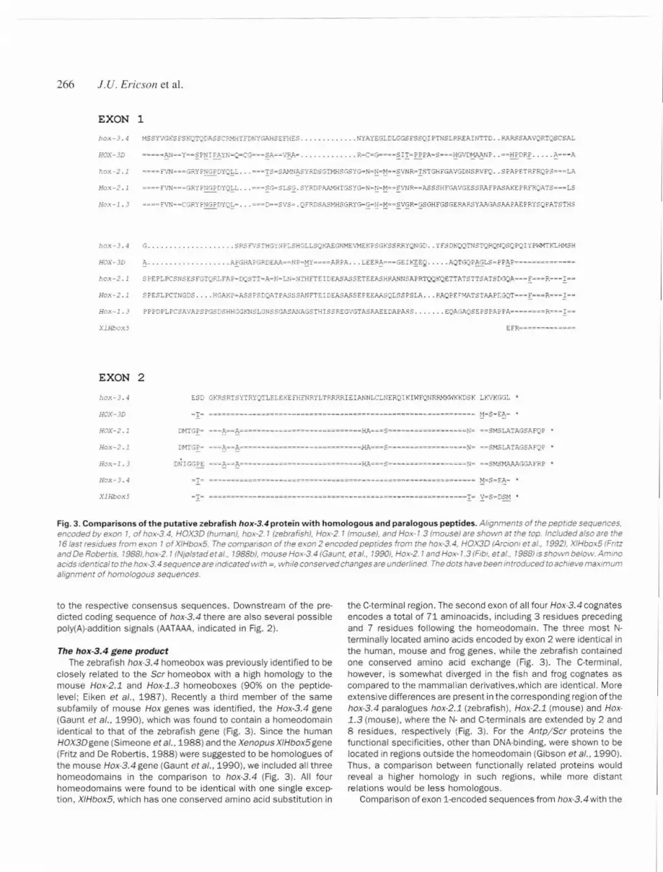
Fig. 3. Comparisons of the putative zebrafish hox-3.4 protein with homologous and paralogous peptides. Alignments of the peptide sequences.encoded by exon ,. of hox-3.4. HOX3D (human). hox-2_ 1 (zebra fish), Hox-2.1 (mouse). and Hox-l.3 (mouse) are shown at the top. Included also are the161ast residues from exon 1 of XIHbox5. The comparison of the e\on 2 encoded peptides from the hox~3.4, HOX3D (Arciont et al.. 1992), XIHbo:o.5(Fnrzand De Robertis, 1988),hox-2. 1 (NJolstad et al., 1988b), mouse Hox-3.4 (Gaunt, et al., 1990), Hox-2. 1and Hox-1.3 (Flbl. et at., 1988) 15shown below. Ammoacids Identical to the hox-3.4 sequence are indicated with =, while conserved changes are underlined The dots have been intrOduced toach/eve maximumalignment of homologous sequences.
to the respective consensus sequences. Downstream of the pre-dicted coding sequence of hox-3.4 there are also several possiblepoly(A~addition signals (AATAAA,indicated in Fig. 2).
The hox-3.4 gene productThe zebrafish hox-3.4 homeobox was previously identified to be
closely related to the Scr homeobox with a high homology to themouse Hox-2.1 and Hox-l.3 homeoboxes (90% on the peptide-level; Eiken et al., 1987). Recently a third member of the samesubfamjjy of mouse Hox genes was identified, the Hox-3.4 gene(Gaunt et al.. 1990). which was found to contain a homeodomainidentical to that of the zebrafish gene (Fig. 3). Since the humanHOX3Dgene (Simeone et al., 1988) and the Xenopus XIHbox5 gene(Fritz and De Robertis, 1988) were suggested to be homologues ofthe mouse Hox-3.4 gene (Gaunt et al., 1990), we included all threehomeodomains in the comparison to hox-3.4 (Fig. 3). All fourhomeodomains were found to be identical with one single excep-tion, XIHbox5. which has one conserved amino acid substitution in
the C-terminal region. The second exon of all four Hox-3.4 cognatesencodes a total of 71 aminoacids, including 3 residues precedingand 7 residues following the homeodomain. The three most N-terminally located amino acids encoded by exon 2 were identical inthe human. mouse and frog genes. while the zebrafish containedone conserved amino acid exchange (Fig. 3). The C.terminal.however, is somewhat diverged in the fish and frog cognates ascompared to the mammalian derivatives,which are identical. Moreextensive differences are present in the corresponding region of thehox.3.4 paralogues hox-2.1 (zebrafish), Hox-2.1 (mouse) and Hox-1.3 (mouse), where the N- and C-terminals are extended by 2 and8 residues, respectively (Fig. 3). For the AntpjScr proteins thefunctional specificities, other than DNA-binding, were shown to belocated in regions outside the homeodomain (Gibson et ar.. 1990).Thus. a comparison between functionally related proteins wouldreveal a higher homology in such regions. while more distantrelations would be less homologous.
Comparison of exon i-encoded sequences from hox-3.4 with the

A
PROMOTOR ELEMENT(region 6; hox-3.4 / HOX3D)
Zebrafish llOx-3.4 Si''�lIellce alld express!oll 267
hox.-3.4!IQX3D
Ho~:-3.4
1862121-158
BOX B BOX P.CCCTGTCT A TCAA.r AACC'::'CCTGGGG. . . ATCAAGCCA. ATTTATGACTGGCCAGGAGCTGCACGTGATTCTATTT AAACATree-GGCT- -c - - - -c- - - - - - - -c- - } -GTC- -- - -- - - -A- - - - -- --G-- - - -GCTC-AGT-- - - -- -c- --- - - - - -GGC-C---GGCT--C c c--A-.rc A G GCTCCAGT c GGC-C--
2662205-75
hox-3.4EOX3DHox-3.4
2672206
-74
BOX CATATTTGGGCATTACACGTCGT}\TCA.n...G,.A~A':>J'..SF.AAA TGAl'TTCCTCCACCTATAAATCCTGCTCTTTTTT AGGACAAGGCCT A-GAG-G-A-A-G--A G-G .-AT TG-C-A GG--A AAAAT A-GAG-G-A-A-'3--; G-G-, ..-AT TG---A GG--A ~AC
3512280
+l
B
INTRON ELEMENT(region 15; hw:-J.4 I H0XJD)
h8X-3.4 1082 TCCTCTTGAGTTTTA~AGGCC.~CGCAGGAA;T .AATAAAACTAGCGGCCGTAAATTTTATGACA~GGCATCT 1155HOXJD 2B42 ---G-C--G TG--GG-C AAASA--A CT A C--TCC A 2920
hox-3.~ 1156 ATTGCTCGTAAACCTGTCCTGT.TACTGTr,~~GAGGTGATCCCCGGTCAA~TTTATGGAGAAATAATTG 1<23HOX3D 2921 --G A AC-AA-AG-CTT CTG-GTG--CC~ C-AC--C 2989
Fig. 4. Conserved DNA sequence-elements in the two hox-3.4 cognates. Sequence comparisons of the two conserved DNA elements Identified bycomputer analysis between hox-3.4 and the human HOX3D (Arcloni et ai" 1992). (A) The 180 bp promoter element with the three defined regularorytarget sites marked: BOX A. BOX B. and BOX C (see text for details). The mouse Hox-3.4 sequence, homologous to the promoter region, was includedaccording ro Arcioni et al. (1992). (B) The 141 bp intron element homologous in the two cognate genes
corresponding parts of HOX3D and the paralogues (zebrafish hox-2.1, mouse Hox-2.1 and Hox-1.3) revealed extensive differences(Fig. 3). On the basis of the variable exon length, the peptides canbe divided into two categories. First the hox-3.4cognates, encoding161 and 151 amino acids-long exon l-peptides, and secondly thethree longer paralogues of 193 residues for the zebrafish, and 187for the mouse derivatives. The protein sequence alignment (Fig. 3)also showed that the protein sequences encoded by the two endsof exon 1 are the most conserved. In the middle part of this proteinregion the sequence conservation is quite low, especially when theparalogues are compared. Thus, the overall identity between thehox-3.4 and HOX3D homeoproteins is only 56%, and this homologyis mainly concentrated in the N-terminal and the -extendedhomeodomain- - includingthe 14 last residues encoded by exon1 in addition to exon 2. Only 48% of the first 58 N-terminat aminoacids are identical between the two cognates. The correspondingcomparison to the paralogous peptides gave an identity not exceed-ing32%. The .extended homeodomain-, on the other hand, showeda 94% identity between the two cognates and 81% to the paralogues.
Identification of conserved sequence elements in non-codingregions of hox-3.4 cognates and paralogues
Computer comparisons of the hox-3.4 genomic sequence withHOX3Dand hox-2.1 sequences revealed many regions of homology.Outside the coding sequence, two conserved DNAelements wereidentified in the hox-3.4jHOX3Dcognates. The first corresponds toa 180 bp promoter element of 60% sequence identity (Fig. 4A; see
discussion). The second element of 141 bp which is 73% identicalis located within the intron sequence in the two cognates (Fig. 48).The remaining sequence elements, which vary in length between 15and 40 bp and share a 60-90% sequence identity, are scatteredthroughout the gene (data not shown), but only a few of theseelements are located in similar positions.
In the comparison between the two paralogous zebrafish genes(hox-2.1 and hox-3.4), a similar result was obtained as for the twohox-3.4 cognates (data not shown). However, the number ofconserved elements as well as the average length and the level ofsequence conservation are reduced.
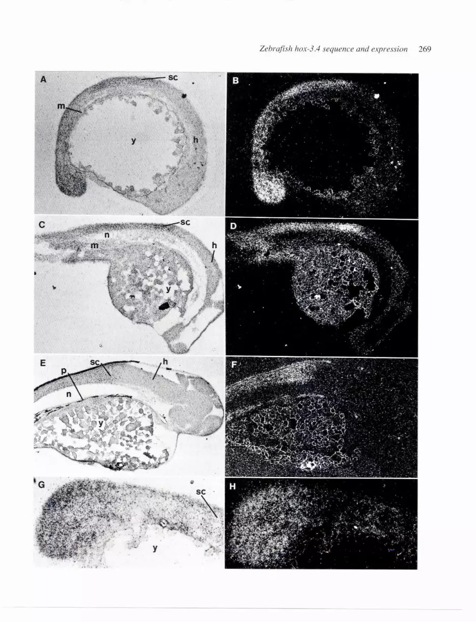
Expression of the hox-3.4 gene during early developmentTo analyze the hox-3.4 expression pattern, in situ hybridization
to parasagittal tissue sections from zebrafish embryos was per-formed. Using a probe that covered the complete hox-3.4 gene(indicated in Fig. 1), embryos of three different developmentalstages were studied (Fig. 5). The hox-3.4 mRNA level was found tobe relatively low at all three stages, and the detection of expressionwas limited (at least at the 24 and 48 hour stages) to the centralnervous system (CNS) with an anterior border located within thecaudal hindbrain. This expression extends posteriorly throughoutthe spinal cord. Reconstruction of the embryos by camera lucidadrawings showed that the hybridization signal had its anteriorborder in the caudal-most (ninth) hindbrain segment, Ca3 (Hannemanet al., 1988), which is located rostral of the first myotome (data notshown). Thisexpression pattern was maintained fromthe 16 to the

268 i.V. Ericsoll et al.
48 h stage. The relative movement of the anterior border duringdevelopment (cf. Fig. 58 to E) corresponds to the length reduction.or compression, of the hindbrain (Trevarrow et al.. 1990). One ex-ception from the CNS-limited expression was observed at the 16 h
stage. In the tailbud a restricted area showed an enhanced hox-3.4expression (Fig. 5G and H) that was not observed in the olderembryos.
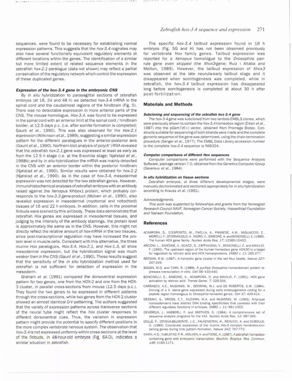
Cross-sections of the hatching embryo in the rostral spinal cord(Fig. 6A and B) and at the level 01 the linbuds (Fig. 6C and D) alsorevealed the hox-3.4 expression in the CNS. Hybridization signalswere not observed in muscle tissue (Fig. 68 and D). However, thehox-3.4 expression, possibly present in this tissue, might havebeen below the detection level. Mesodermal expression of Hoxgenes in vertebrates has previously been identified in regionslocated posterior relative tothe CNS expression {Gaunt et al., 1990;Molven et al.. 1990). The mRNA seemed to be more or lessuniformly distributed across the neural tube in the rostral parts ofthe CNS (Fig. 68), while the expression became more ventrallylocalized in the sections at the level of the finbuds (Fig. 60). Furtherposterior, the hybridization signal was weaker, as observed on thesagittal sections (Fig. 50 and F), and to distinguish between uniformor ventral distribution of hox-3.4 mRNA became impossible. Finally,no hox-3.4 mRNA could be detected in the finbuds (data not shown).
Discussion
Conservation of sequence and functional specificity in proteinproducts of the hox-3.4 cognates and paralogues
From comparisons to other recently described vertebratehomeobox-containing genes, we concluded that hox-3.4 is thezebrafish homologue of the human HOX30, mouse Hox-3.4and frogXIHbox5 genes (Fig. 3), which were previously suggested to becognates, by Gaunt and co-workers (1990). All four genes containidentical homeodomains, except for one amino acid substitution inthe X/Hbox5 protein. Comparisons of the complete gene productswas not possible for the mouse and frog derivatives, since only theC-terminal sequences have been published (Fritz and De Robertis,1988; Gaunt et al., 1990). However, direct alignment ofthe aminoacid sequences derived from hox-3.4 and the human HOX3Dgenesrevealed a total identity of 56%. In the N-terminal region, includingthe first 58 amino acids, 48% of the amino acid residues areidentical. Less conservation is present in the middle part of theprotein (position 59-147), where the sequence identity is only 18%.Thus, the level of conservation between these Hox-3 cognates isconsiderably lower than for the corresponding cognates in the Hox-2 complex (Nj0lstad et a/.. 1988b).
An interesting aspect concerns the relationship between theevolutionary divergence of cognate genes and the conservation offunctional specificity of the corresponding protein products. Theresults obtained from analyses of the functional specificity ofhomeodomain proteins from Drosophila are somewhat contradic-tory. Studies on hybrid proteins of Dfd and Ubx indicated that thehomeodomain, due to its target-specificity, was responsible for the
regulatory specificity (Kuzoira and McGinnis, 1990; Dessain et al..1992). However, similar studies on hybrid proteins of Antpand Scrindicated a requirement for additional sequences to completelydefine the functional specificity (Gibson et al.. 1990). On the basisofthese results, also a model forthe functional structure ofthe Antpprotein in Drosophila was suggested. According to this model, theAntp-peptide can be divided into two major regions, the potentiatingportion, which included the most N-terminal two thirds, and thehomeodomain-containing specifying portion, at the C-terminal oftheprotein (Gibson et al.. 1990). It was proposed that the potentiatingportion could be further subdivided into an N-terminal generalpotentiating region, determining the overall levels of Antp activity,and a specific potentiating region, located closer to the middle ofthe protein, which could be involved in adjusting the level of activityin specific cells. Consistent with this model, the C-terminal speci-fying portion of the hox-3.4 and HOX30 proteins are almost identi-cal. In the potentiating region, however, only the general portion atthe N-terminal is strongly conserved in the two cognates. This maysuggest that the mechanisms regulating the overall activity of hox-3.4 and HOX3D have been preserved, while the circuits for specificadjustments in certain cells have been modified through evolution.Similarly, the proteins derived from the zebrafish and mouse Hox-2.1 cognates share the highest level of sequence identity in theregions corresponding to the location of the specifying portion (C-terminal) and the general potentiating region (Nj01stad et a/.,1988b). These domains are also partially conserved relative to theparalogues hox-3.4 and HOX30, while the sequences in the pro-posed specific potentiating region are almost completely diverged.
Since regions with an elevated level of polar, non-acidic aminoacids - serine and threonine or proline - have been shown to beinvolved in transcriptional activation (Tanaka and Herr, 1990), thedistribution of ser/thr and pro residues within the differenthomeoproteins were calculated. In the predicted hox-3.4 protein,24% of the exon i-encoded amino acids are serine or threonine,while proline residues constitute only 3%. A somewhat differentdistribution is present in the human cognate, which has equalamounts of the two categories, 13% of each. The paralogous genesencode peptides similar to that of hox-3.4 with a ser/thr content of21-28%, while the proline levels vary from 5% (zebrafish hox-2.1) to9-10% for the murine Hox~2.3 and Hox-l.3 genes. Overall, it seemsas if the preference for polar amino acids has been altered towardsserine/threonine residues in the zebrafish proteins and moreproline residues in mammals.
Multiple non-coding sequence elements are conserved amongcognates and paralogues of the hox-3.4 gene
In the DNA-sequence comparison between hox-3.4 and HOX3Dadditional regions of homology were found outside the codingregions. One of the most interesting conservations was the promoterelementol about 180 bp (Fig. 4A) with a 60% homology between thetwo species. The human promoter was found to be, at leastto someextent, cell- or tissue-specific in the expression systems tested forthe human derivative, and to contain one retinoic acid-responsive
Fig. 5. Detection of hox-3.4transcripts in the CNS of different embryonic stages. In situ hybridization to parasagittal sections of zebra fish embryos.shown in bright-field lA, C, and E) and dark-field IB, D, and F). Three different developmental stages were used; 16 h (A and B). 24 h (C and OJ, and 48h (E and F). The restricted expression identified on parasagittal sections in the tail-bud of 16-h-old embryos. (G and H) show the tail-bud in bright-fieldand in dark-field. respectively. Abbreviations used: h. hindbrain; sc. spinal cord; y, yolk; m, mesoderm; n, notochord; and P. pigmentation
Zebrafisl1 l1ox-3.../ sequence and expression 269

270 .I.U. Ericson et al.
A-.: .".>'..::
..~:I:}:j \.;:-:'<r<'~'.. ':'~''''.'f 'p, '. ::,..
: : _:~~ ',~ .~1,::~~~r~~~:-5fii.~;~)if~~~~' ,"s;:"~' '.:-'
",',,,Jr~"'!;;\>';j .",~J.,":,!\~?*1.k~"~
""
. ,/ '. .,-,' ,,"'fi,
;:"::::':,:,":,::< ~
,:-,,!;\~L>I.:<~:'
Fig. 6. Analysis of dorsoventral distribution of hox-3.4 transcripts in the CNS. In situ hybridization to cross-sections of zebra fish embryos at rhe 48h stage, shown in bnght-field(A and C) anddark-field(B and DJ. The section shown in A and B comes from the rostralspinal cord/hindbrain region, whilerhesecrion In Cand 0 comes from the level of the finbud. Abbreviations used: SC,spinal cord: m, myotome; n, notochord. and p, pigmentation.
element and two homeoprotein binding sites (Arcioni et al., 1992).When compared to the murine Hox-3.4j-2.1j-1.3 para logy group,the HOX3D promoter was found to be almost identical to its cognate(Hox-3.4), while the paralogous promoters (Hox-2.1 and Hox-1.3)contained more divergent sequences in all three regulatoryelements(Arcioni et al., 1992). Alignment of the hox-3.4 upstream sequence
to the human promoter showed that the two homeodomain-bindingsites (box Band C; Fig. 4A) were highly conserved, implying afunctional conservation of the regulatory elements. The RA-respon-sive element, on the other hand, contained less conservation andthe 10 base-pairs long palindromic sequence - suggested to be
essential for the RA-induced binding (boxA; Fig. 4A; Arcioni et al.,1992) - was found to be reduced to only 6 bp in the hox-3.4sequence. However. this may reflect alternative or a more degener-ate binding-specificity involved in Hox gene regulation in zebrafish.The protein responsible for RA-induced binding in the embryonal
carcinoma cells was not identified but previously identified RAreceptors were unable to activate HOX3D transcription (Arcioni etal" 1992),
Besides the promoter, one surprisingly large region of 142 bp,located within the intron (Fig. 48), was found to be 73% identical inthe two cognates. Additional, shorter stretches of homology (60-90%) are also scattered throughout the genes. The majority of theseelements have different locations within the two cognates. Also nocommon binding sites for known transcription factors were identi-fied in a computer search for such consensus sequences. There-fore, it remains unclear whether it is regulatory or structuraldemands that have preserved these elements. However, this widelyscattered distribution of potential cis-acting elements may relate tothe results obtained from transgenic analyses of Hox-l.l and Hox-2,6 regulation (Puschel et ai" 1991; Whiting et a/" 1991), In both
cases, large DNA regions, including the introns and downstream
sequences, were found to be necessary for establishing normalexpression patterns. This suggests that the hox-3.4 cognates mayalso have several functionally eQuivalent regulatory elements atdifferent locations within the genes. The identification of a similarbut more limited extent of related sequence elements in thezebrafish hox-2.1 paralogue (data not shown) may reflect a partialconservation of the regulatory network which control the expressionof these duplicated genes.
Expression of the hox-3.4 gene in the embryonic CNSBy in situ hybridization to parasagittal sections of zebrafish
embryos (at 16, 24 and 48 h) we detected hox.3.4 mRNA in thespinal cord and the caudal most regions of the hindbrain (Fig. 5).There was no detectable expression in more anterior parts of the
CNS. The mouse homologue. Hox-3.4, was found to be expressedin the spinal cord with an anterior limit at the spinal cord / hindbrainborder, at 12.5 days p.c. (i.e. after somite formation is completed;Gaunt et al., 1990), This was also observed for the Hox11expression (Wilkinson et af..1989), suggesting a similar expressionpattern for the different paralogues in the anterior-posterior axis(Gaunt et a1..1990). Northern.blotanalysis ofpoly(A+) RNA revealed
that the zebrafish hox-2.1 gene was expressed at least as early asfrom the 12.5 h stage (i.e. at the 6.som;te stage; Njolstad et a1..1988b) and by in situ hybridization the mRNA was mainly detectedin the CNS with an anterior border within the posterior hindbrain(Njclstad et al.. 1990). Similar results were obtained for hox-2.2(Njolstad et al., 1990). As in the case of hox.3.4, mesodermalexpression was not detected for the two zebrafish genes. However.immunohistochemical analyses of zebrafish embryos with an antibodyraised against the Xenopus Xlhboxl protein, which probably cor-responds to the hox.3.3 gene.product (Molven et a1.. 1990), alsorevealed expression in mesodermal (myotomal and notochord)tissues of 16 and 22 h embryos. In addition, cells in the proximalfinbuds were stained by this antibody. These data demonstrate thatzebrafish Hox genes are expressed in mesodermal tissues, andjudging by the intensity of the antibody stainings, the protein levelis approximately the same as in the CNS. However, this might notdirectly reflect the relative amount of hox-mRNA in the two tissues,since post-transcriptional regulation may have increased the pro-tein level in muscle cells. Consistent with this alternative, the threemurine Hox paralogues, Hox-3.4, Hox-2.1, and Hox-l.3. all showmesodermal expression, but the hybridization signal was muchweaker then in the CNS (Gaunt et af..1990). These results suggestthat the sensitivity of the in situ hybridization method used forzebrafish is not sufficient for detection of expression in themesoderm.
Graham et al_ (1991) compared the dorsoventral expressionpattern for two genes. one from the HOX-2 and one from the HOX-3 cluster. in parallel cross-sections from mouse (12.5 days p.c.).They found the two genes to be expressed in different patternsthrough the cross-sections, while two genes from the HOX-2 clustershowed an almost identical D-V patterning. The authors suggestedthat the variety of expression patterns across transverse sectionsof the neural tube might reflect the hox cluster responses todifferent dorsoventral cues. Thus. the variation in expressionpattern might provide the potential to specify different positions inthe more complex vertebrate nervous system. The observation thathox-3.4 is not expressed uniformly within cross-sections at the levelof the finbuds, in 48.hour-old embryos (Fig. 6A.D), indicates asimilar situation in zebrafish.
Zebra fish IIOX-3.-Isequence and expfl'ssioll 271
The specific hox-3.4 tailbud expression found in 16 hembryos (Fig. 5G and H) has not been observed previouslyfor vertebrate Hox family genes. Tailbud expression wasreported for a Xenopus homologue to the Drosophila pair-rule gene even skipped (the Xhox3-gene; Ruiz i Altaba andMelton. 1989). However. the tai/bud expression ot Xhox3was observed at the late neurula-early tailbud stage and itdisappeared when somitogenesis was completed. while inzebrafish, the hox-3.4 tailbud expression has disappearedlong before somitogenesis is completed at about 30 h afterpost-fertil ization.
Materials and Methods
Subcloning and sequencing of the zebrafish hox-3.4 geneThe hox-3.4 gene was subcloned from two lambda EMBL3 clones, which
were previously shown to contain the hox-3.4 homeobox region (Eiken et al..1987) into the pGem7zf(+) vector. obtained from Promega Biotec. Con-structs suitable for sequencing of both strands were made and the completegenomic sequence of the gene was determined. using the chain terminationprocedure (Sanger et al..1977). The EMBLData library accession numberto the complete hox-3.4 sequence is X68324.
Computer comparisons of different Hox sequencesComputer comparisons were performed with the Sequence Analysis
Software, package version 7.0. obtained from the Genetics Computer Group(Devereux et al" 1984).
In situ hybridization on tissue sectionsZebrafish embryos, at three different developmental stages. were
manually dechorionated and sectioned appropriately for in situ hybridizationaccording to Krauss et al. (1991).
AcknowledgmentsThis work was supported by fellowships and grants from the Norwegian
Research Council NAVF. Norwegian Cancer Society, Hasselblad Foundationand Nansen Foundation,
References
ACAMPORA. D., D'ESPOSITO, M.. FAIELLA.A., PANNESE. A.M., MIGLIACCIO, E..MORELU,F.. STORNAIUOLO.A..NIGRO,v.. SIMEONE,A.and BONCINElll. E. (1989).The human HOXgene family. Nucleic ACIds Res. 17: 10385-10402.
ARCIONI. L. SIMEONE. A.. GUAZZI, S.. ZAPPAVIGNA. V.. BONCINElli. E. and MAVILJO,
F. (1992). The upstream region of the human homeobo. gene HOX3D is a targetfor regulation by retinoic acid and HOXhomeoproteins. fAtBD J. 11: 265-277.
BEEMAN.R.W. (1987). A homeotic gene cluster in the red flour beetle. Nature 327:247-249.
BIGGIN, M.D. and TJIAN, R. (1989). A purified Drosophila homeodomain protein re-presses transcription in vItro. Cell 58: 433-440.
BONCINELlI, E.. SIMEONE, A., ACAMPORA. D. and MAVILJO, F. (1991). HOX gene
acti\'ation by retmolc acid. Trends Genet. 7: 329-334.
CARRASCO. A.E.. McGINNIS. w., GEHRING. w.J. and DE ROBERTIS.E.M. (1984).Cloning of it x. laevis gene eJ\pressed during earty embryogenesis coding for apeptide region homologous to Drosophila homeotic genes. Cell 37: 409-414.
DESSAlrJ. S.. GROSS. C.T.. KUZIORA. M.A. and McGINNIS, w. (1992). Antp-typehomeodomains have distinct DNA binding specificities that correlate with theirdifferent regulatory functions in embryos. fMaD J. 11: 991.1002.
DEVEREUX. J.. HAEBERLJ, P. and SMITHIES. O. (1984). A comprehensive set ofsequence analysis programs for the VAX. Nucleic Acids Res. 12: 387.395.
DOllt.. P..IZPISUA-BElMONTE, J.-C.. FALKENSTEIN. H.. AENUCCI, A. and DUBOUlE.D. (19891. Coord mate eJ\pression of the murine HOJ\.5 comple. homeoboJl-<:on-taining genes during limb pattern formation. Nature 342: 767-772.
EIKEN, H.G.. NJOLST AO, P.R., MOlVEN. A. and FJOSE, A. (1987). A zebrafish homeobo.r.-
containing gene with embryonic transcription. Biochim. Blophys. Res. Commun.149: 1165.1171.

272 .t.V. Ericson et al.
ABI, M.. liNK. B.. KESSEL. M., COLBERG-POLEY,A.M.. LABEIT, S., LEHRACH, H.andGRUSS. P. (198B). Coding sequence and CJ.pression of the homeobo)( gene Hox-1.3. Development 102: 349-359.
FRITZ, M. and DE ROBERTIS, E.M. (1988). Xenopus homeoboJl-(:ontalning cDNAse)(pressed in earry development. Nucleic Acids Res. 16: 1453-1469.
GAUNT.S.J.. COLETTA,P.L. PRAVTCHEVA, D. and SHARPE, P.T. (1990). Mouse Hex.-3.4: homeobo sequence and embryonic expression patterns compared with othermembers of the HOil gene network. Development 109: 329-339.
GIBSON. G.. SCHIER, A.. LEMOTTE.P. and GEHRING. W. (1990). The specificities ofSex combs reduced and Antennapedia are defined by a distinct portion of eachprotein that includes the homeodomain. Cell 62: 1087.1103,
GRAHAM. A.. MADEN. M. and KRUMLAUF. R. (1991). The murine HOJl..2genes displaydynamic dorsoventral patterns of expression during central nervous system
development. De~-elopment 112:255-264.
GRAHAM, A., PAPAlOPUlU, N. and KRUMLAUF,R. (1989). The murine and DrosophilahomeoboJl. gene compleJl.es have common features of organization and e:s.pression.Ce1/57: 367.378.
HAN, K..lEVINE, M.S. and MANLEY, J.l. (1989). Synergistic activation and repression
of transcription by Drosophila homeobox-proteins. Ce1/56: 573.583.
HANNEMAN. E., TREVARROW, B., METCALFE, WK, KIMMEL, C.B. and WESTERFIELD,
M. (1988). Segmental pattems of development of the hindbrain and spinal cord ofthe zebraflsh embryo. Development 103: 49-58.
HUNT, P. and KRUMLAUF,R. (1991). Deciphering the HOHode: clues to patterningbranchial regions of the head. Ce1/66: 1075-1078.
IZPISOA-BElMONTE, J-C., FALKENSTEIN. H.. DOlltp.. RENUCCI, A. and DUBOULE, D.(1991). Murme genes related to the DrosophilaAbdBhomeoticgene are sequentially
expressed during development of the posterior part of the body. EMBD J. 10: 2279-
2289.
JAYNES, J.B. and O'FARRElL, P.H. (1991). Acti\le repression of transcription by theEngrailed homeodomain protein. EMaD J. 10: 1427-1433.
KESSEl. M. and GRUSS, P. (1991). Homeotictransformations of murine vertebrae and
concomitant alteration of Hox codes induced by retinoic acid. Ce1/57: 89-104.
KOZAK,M. (1986). Point mutations define a sequence flanking the AUGinitiator codonthat modulates translation by eukaryotic ribosomes. Ce1/44: 283-292.
KOZAK, M. (1987). An analysis of 5'-f1oncodlng sequences from 699 ...ertebratemessenger RNAs. Nucleic Acids Res. 15: 8125-8148-
KRAUSS, S., JOHANSEN. T., KORZH, V.. MOENS, U" ERICSON. lU. and FJOSE, A.(1991). Zebraflsh pax(zf-a}: a paired box-containing gene expression in the neural
tube. EMBO J. 10: 3609-3619.
KUZIORA. M.A. and McGINNIS, W. (1990). Altering the regulatory targets of theDeformed protein in Drosophila embryos by substituting the Abdomlnal-B
homeodomain. Alech. Dev. 33: 83-94.
LE MOUELUC, H.. LALlEMAND, Y. and BROlH, P. (1992). Homeosis in the mouseinduced by a null mutation in the Hox-3.1 gene. Ce1/69: 251.264.
LEVINE, M., RUBIN, G.M. and TJIAN, R. (1984). Human DNA seQuences homologousto a protein coding region conserved between homeotic genes of Drosophila. Cell38: 667-673.
MAVIUO,F.. SIMEONE.A.. GIAMPAOLO,A., FAIELLA,A., ZAPPAVIGNA, V., ACAMPORA.
D.. POIANA,G"
RUSSO. G., PESCHLE. L. and BONCINELU. E. (1986). Differentialand stage.related expression in embryonic tissues of a new human homeoboJl.gene. Nature 324: 664-668.
McGINNIS, W., GARBER, R.L., WIRZ, J., KUROIWA. A. and GEHRING. W.J. (198403). Ahomologous protein-<:odlng sequence in Drosophila homeotic genes and Itsconservation in other metazoans. Cel/ 37: 403-408.
McGINNIS, W., HART, C.P.. GEHRING. W.J. and RUDDLE, F.H. (1984b). Molecularcloning and chromosome mapping of a mouse DNA sequence homologous tohomeotic genes in Drosophila. Cell 38: 675-680.
McGINNIS. W., LEVINE, M.S.. HAFEN. E., KUROIWA. A. and GEHRING, W.J. (1984c). Aconserved DNA sequence in homeotic genes of the Drosophila Antennapedia andBlthOraJl.complexes. Nature 308: 428-433.
MOLVEN, A., HORDVIK.I., NJOLSTAD, P.R.. VAN GHELUE, M. and FJOSE.A.11992). ThezebrafJsh homeobox gene hox[zf.114}: primary structure. expression pattern ande'IQlutionary aspects. Int. J. Dev. 8/0/. 36: 229-237.
MOLVEN,A., WRIGHT,C.V.E.. BREMILlER, R.. DE ROBERTlS,LM. and KIMMEL, C,B.(1990). Expression of a homeobox gene product in normal and mutant
zebrafish embryos: evolution of the tetrapod body plan. Development 109: 279.288.
MORRISS-KAY, G.M., MURPHY, P., HilL, R.E. and DAVIDSON. D.R. (1991). Effects ofretinolc acid eJl.cess on eJl.pression of Hox.2.9 and Krox.20 and on morphologicalsegmentation in the hindbrain of mouse embryos. EMBO J. 10: 2985-2995.
NmLSTAD. P.R.. MOlVEN. A. and FJOSE.A. (198803).A zebrafish homologue of themurine HOJl.-2.1gene. FEBSLerr. 230: 25-30.
NJOLSTAD. P.R., MOlVEN. A., APOlD. J. and FJOSE. A. (1990). The zebraflshhomeobo\ gene hox.2.2:transcription Unit. potential regulatory regIons and in Situlocalization of transcripts. EMBO J. 9: 515-524.
NJ0LSTAD. P.R., MOLVEN. A.. HORDVIK. I., APOLD, J. and F.JOSE. A. (198Sb). Primarystructure, developmentally regulated expression and potential duplication of the
zebrafish homeoboJl. gene ZF.21. Nucleic ACids Res. 16: 9097-9111.
PUSCHEl. A.W., BALLING, R. and GRUSS, P. (1991). Separate elements cause lineagerestriction and specify boundaries of Hox-1.1 expression. Development 112: 279.287.
RANGINI, Z., FRUMKIN, A., SHAN!. G.. GUTTMAN. M., EYAL-GILADI, H.. GRUBENBAUM,Y. and FAINSOD, A. (1989). The chicken homeoboxgene Chox1and Cho.d:cloning,seQuencing and expression dunng embryogenesis. Gene 76: 61.74.
RUIZ I ALTABA, A., and MELTON, D.A. (1989). Bimodlal and graded expression of theXenopus homeobox gene Xhox3 during embryonic development. Development
106: 173-183.
SANGER, F., NICKlEN, S. AND COULSON, A.R. (1977). DNA sequencing with chain-
terminating inhibitors. PrQC. Natl. Acad. Sci. USA 74: 5463-5464.
SIMEONE, A.. PANNESE. M.. ACAMPORA. D., D'ESPOSnO. M. and BONCINELU, E.(1988). At least three human homeoboxes on Chromosome 12 belong to the sametranscription unit. Nucleic Acids Res. 16: 5379-5390.
TANAKA, M.. and HERR, W. (1990). Differential transcriptional activation by Oct.l and
Oct-2: Interdependent activation domains induce Oct-2 phosphorylation. Cell 60:375-386.
TREVARROW. B., MARKS. D.L. and KIMMEL. C.B. (1990). Organization of hindbrainsegments in the zebraflsh embryo. Neuron 4: 669-679.
WHITING, J.. MARSHAll, H., COOK, M., KRUMLAUF, R., RIGBY, P.W.J.. SCOTT,D.and AllEMANN, R.K. (1991). Multiple spatially specific enhancers are reqUired
to reconstruct the pattem of Hox.2.6 gene expression. Genes Dev. 5: 2048-2059.
WILKINSON, D.G., BHATT, S., COOK, M., BONCINElU, E. and KRUMLAUF. R. (1989).Segmental expression of Hox.2 homeobox-(:ontalning genes In the developing
mouse hindbrain. Nature 341: 405-409.
WINSLOW, G.M., HAYASHI, S.. KRASNOW, M., HOGNESS. 0.5. and SCOTT,M.P.(1989). Transcriptional activation by the Antennapedia and Fushi tarazu proteins
in cultivated Drosophila cells. Cell 57: 1017-1030.
WRIGHT,C.V.E.. CHO, K.W.Y., HARDWICKE, J..COLUNS. R.H. and DEROBERTIS,E.M.(1989). Interference Ith function of a homeobox gene in Xenopus embryosproduces malformations of the anterior spinal cord. Cell 59: 81-93.