genome duplication and mutations in ace2 cause multicellular, fast-sedimenting phenotypes in evolved...
TRANSCRIPT
Genome duplication and mutations in ACE2 causemulticellular, fast-sedimenting phenotypes inevolved Saccharomyces cerevisiaeBart Ouda,b, Victor Guadalupe-Medinaa,b, Jurgen F. Nijkampb,c, Dick de Ridderb,c,d, Jack T. Pronka,b,d,Antonius J. A. van Marisa,b, and Jean-Marc Darana,b,d,1
aDepartment of Biotechnology, Delft University of Technology, 2628 BC, Delft, The Netherlands; bKluyver Centre for Genomics of Industrial Fermentation,2600 GA, Delft, The Netherlands; cThe Delft Bioinformatics Lab, Department of Intelligent Systems, Delft University of Technology, 2628 CD, Delft, TheNetherlands; and dPlatform Green Synthetic Biology, 2600 GA, Delft, The Netherlands
Edited by Arnold L. Demain, Drew University, Madison, NJ, and approved October 1, 2013 (received for review March 28, 2013)
Laboratory evolution of the yeast Saccharomyces cerevisiae in bio-reactor batch cultures yielded variants that grow as multicellular,fast-sedimenting clusters. Knowledge of the molecular basis ofthis phenomenon may contribute to the understanding of naturalevolution of multicellularity and to manipulating cell sedimenta-tion in laboratory and industrial applications of S. cerevisiae. Mul-ticellular, fast-sedimenting lineages obtained from a haploidS. cerevisiae strain in two independent evolution experimentswere analyzed by whole genome resequencing. The twoevolved cell lines showed different frameshift mutations ina stretch of eight adenosines in ACE2, which encodes a tran-scriptional regulator involved in cell cycle control and mother-daughter cell separation. Introduction of the two ace2 mutantalleles into the haploid parental strain led to slow-sedimentingcell clusters that consisted of just a few cells, thus representingonly a partial reconstruction of the evolved phenotype. In ad-dition to single-nucleotide mutations, a whole-genome duplica-tion event had occurred in both evolved multicellular strains.Construction of a diploid reference strain with two mutant ace2alleles led to complete reconstruction of the multicellular-fastsedimenting phenotype. This study shows that whole-genomeduplication and a frameshift mutation in ACE2 are sufficient togenerate a fast-sedimenting, multicellular phenotype in S. cerevi-siae. The nature of the ace2 mutations and their occurrence in twoindependent evolution experiments encompassing fewer than 500generations of selective growth suggest that switching betweenunicellular and multicellular phenotypes may be relevant for com-petitiveness of S. cerevisiae in natural environments.
whole genome sequencing | reverse engineering
Ease of cultivation and genome analysis, short generationtimes, and large population sizes have contributed to the
popularity of microorganisms as model systems in experimentalevolution. In addition to providing insights into evolutionaryadaptation mechanisms and strategies, laboratory evolution ofmicroorganisms provides a powerful tool to improve character-istics that are relevant to microbial biotechnology. The latterapplication of laboratory evolution, known as evolutionary en-gineering (1) has, for example, contributed to expanding sub-strate range (2–5), functional implementation of alternativeproduct pathways (6, 7), and increased tolerance to inhibitors (4,8) in various production organisms (9). Recent advances in DNAsequencing and genetic modification facilitate characterizationand reconstruction of the genetic changes that underlie evolvedphenotypes obtained in laboratory evolution. This progress con-tributes to identification of the molecular mechanisms that underliespecific phenotypes and enables experimental testing of hypotheseson evolutionary strategies (10). Laboratory evolution has generatednew insights into mutation rates (11, 12), genetic drift (12, 13),epistasis (14), clonal interference (15), and other important aspectsof evolution by natural selection (16). In microbial biotechnology,
reverse engineering of evolved phenotypes, known as inverse met-abolic engineering (17), has similarly benefited from the availabilityof these genomic methodologies (18). In this applied researchcontext, knowledge of the genetic basis of an industrially relevantphenotype not only increases understanding, but also enables itsreconstruction and improvement in other microbial strains andspecies (18–20).In unicellular organisms such as the yeast Saccharomyces cer-
evisiae, laboratory evolution is facilitated by the ease with whichsingle-cell lines can be isolated from evolving cultures. Recently,however, Ratcliff et al. described evolution of multicellularity inS. cerevisiae within a single long-term cultivation experiment(21). The multicellular variant, in which daughter cells did notseparate from the mother cell on cell division, dominated thepopulation within a few generations when fast sedimentation wasselected for in test tubes. Evolution of these multicellular clus-ters of S. cerevisiae, which even showed signs of cellular differ-entiation, was proposed to be a laboratory model for the originof multicellularity in eukaryotes (21).At least 25 occurrences of the shift from unicellular to mul-
ticellular life forms have been recognized in the evolution of lifeon Earth (22–24). It has been proposed that multicellularity cancontribute to phenotypes as diverse as stress tolerance (25, 26),affinity for substrates (27), and relief of predatory pressure (28).However, knowledge on the selective pressures resulting in the
Significance
The shift from unicellular to multicellular life forms representsa key innovation step in the evolution of life on Earth. How-ever, knowledge on the evolutionary pressures resulting in theselection of multicellular life forms and the underlying molec-ular mechanisms is far from complete. Our study providesa complete identification of the specific genetic changes bywhich the unicellular eukaryote S. cerevisiae can acquire amulticellular, fast-sedimenting phenotype. We demonstratedthat a minimal evolutionary mechanism encompassed a de-regulation of the late step of the cell cycle through mutation inACE2 followed by whole genome duplication.
Author contributions: J.T.P., A.J.A.v.M., and J.-M.D. designed research; B.O., V.G.-M., J.F.N.,and J.-M.D. performed research; B.O., J.F.N., and J.-M.D. contributed new reagents/analytictools; B.O., J.F.N., D.d.R., and J.-M.D. analyzed data; and B.O., D.d.R., J.T.P., A.J.A.v.M., andJ.-M.D. wrote the paper.
The authors declare no conflict of interest.
This article is a PNAS Direct Submission.
Freely available online through the PNAS open access option.
Data deposition: The raw sequencing data were deposited as Sequence Read Archive(SRA) at NCBI (BIOproject ID code PRJNA193417).1To whom correspondence should be addressed. E-mail: [email protected].
This article contains supporting information online at www.pnas.org/lookup/suppl/doi:10.1073/pnas.1305949110/-/DCSupplemental.
www.pnas.org/cgi/doi/10.1073/pnas.1305949110 PNAS | Published online October 21, 2013 | E4223–E4231
EVOLU
TION
PNASPL
US
evolution of multicellular life forms and on the underlying mo-lecular mechanisms is far from complete.Knowledge of the mutations that cause the switch from uni-
cellular to multicellular growth in yeast may contribute to un-derstanding of the events leading to the transition to multicellularlives. Moreover, such knowledge can contribute to a better mod-ulation of biomass sedimentation in laboratory research and in-dustrial application of S. cerevisiae. In our research on evolutionaryengineering of S. cerevisiae, we frequently observed multicellular,fast-sedimenting clusters that, on microscopic examination, re-semble the phenotype described by Ratcliff et al. (21). The goal ofthe present study was to elucidate mutations that are responsiblefor the generation of multicellular variants. To this end, wemonitored the formation of multicellular variants in two in-dependent laboratory evolution experiments with a haploid labo-ratory strain of S. cerevisiae. Subsequently, representative mutantsfrom the two evolution experiments were characterized. Geneticchanges identified by whole-genome resequencing were reverseengineered in the unicellular parental strain, enabling the identi-fication of two changes that, together, were sufficient to reproducethe multicellular, fast-sedimenting phenotype.
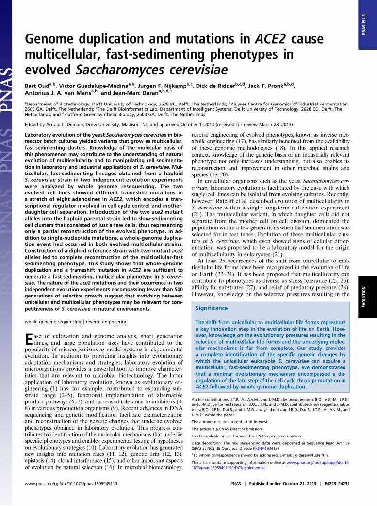
ResultsSelection of Multicellular Clusters in Sequential Bioreactor BatchCultures. Where previous reports studied evolution of S. cer-evisiae in serial shake flask cultures (29–33), we reproduciblyobserved the occurrence of large multicellular clusters duringprolonged anaerobic cultivation of the haploid S. cerevisiae strainCEN.PK113-7D (34) in sequential bioreactor batch cultures. Thephenotype of these clusters was similar to the “snowflake yeast”previously described by Ratcliff and coworkers (21, 35). The de-sign of the “fill and draw” system used in our bioreactors providedan unintended selective advantage to fast-sedimenting cell lines.The vertical pipe used to empty the bioreactor after each culti-vation cycle did not reach the bottom of the vessel. Consequently,fast-sedimenting cells were enriched in the small remaining vol-ume used as inoculum for the next batch cultivation cycle.To facilitate identification of mutations contributing to the
multicellular phenotype (18, 33), two identical independent an-aerobic evolution experiments were started on a mixture of20 g·L−1 glucose and 20 g·L−1 galactose. Although the specificgrowth rate on galactose doubled during both evolution experi-ments (from 0.11 to 0.22 and 0.20 h−1; Fig. 1A and Fig. S1A) andthe length of the batch cultivation cycles decreased by at least 35%(Fig. S1 H and I), the morphology of S. cerevisiae changed dra-matically as large, multicellular clusters became dominant in bothevolution experiments (Fig. 1 B–F and Fig. S1 B–G). The sedi-mentation index, calculated from the time-dependent decrease ofthe optical density of statically incubated cell suspensions, stronglyincreased, in parallel with the increasing abundance of multicel-lular clusters (Fig. 1 B–F and Fig. S1 B–G). Culture samples takenat the end of the two evolution runs [after 4,200 (∼900 gen-erations) or 2,880 h (∼500 generations)] showed almost completesedimentation after 5 min of static incubation (Fig. 1G).In S. cerevisiae, reversible aggregation of individual cells into fast-
sedimenting clusters can occur via flocculation, which involvesa Ca2+-dependent interaction of yeast cell wall proteins and car-bohydrates (36). However, the multicellular clusters observed in theevolved cultures could not be reverted to a single-cell morphologyby incubation with well-known antiflocculent agents such as EDTA(0.5 M) (37), mannose (38), or protease (trypsin 1,500 units·mL−1)(39). This observation indicated that the phenotype did not resultfrom interaction of unicellular yeasts, but rather from an incom-plete cell division (36).
Whole Genome Sequence Analysis of Two EvolvedMulticellular Isolates.To investigate the molecular basis of the evolved multicellularphenotype, fast-sedimenting strains IMS0267 and IMS0386 were
isolated from evolution experiments 1 and 2, respectively. Toverify the genetic stability of the mutations responsible for multi-cellularity, the evolved strains were grown for at least 50 gen-erations on glucose in shake flask cultures. This test did not resultin observable changes in multicellularity or sedimentation be-havior, confirming that these phenotypes were independent onthe bioreactor context in which they had been evolved and thatthey were caused by stable mutations. Genomic DNA of strainsIMS0267 and IMS0386 was sequenced at high genome coverage(81.6- and 38.5-fold coverage for IMS0267 and IMS0386, re-spectively) and compared with the reference genome of the pa-rental strain CEN.PK113-7D (34). The high coverage enabledaccurate analysis of genomewide copy number variation (CNV)by coassembly (40), as well as identification of single-nucleotidevariations (SNV) and indels.To estimate the ploidy of the evolved strains we de novo
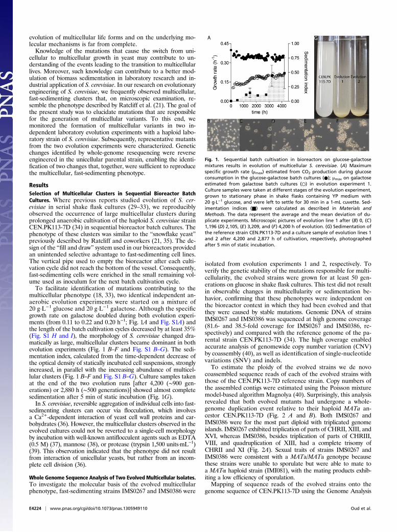
coassembled sequence reads of each of the evolved strains withthose of the CEN.PK113-7D reference strain. Copy numbers ofthe assembled contigs were estimated using the Poisson mixturemodel-based algorithm Magnolya (40). Surprisingly, this analysisrevealed that both evolved mutants had undergone a whole-genome duplication event relative to their haploid MATa an-cestor CEN.PK113-7D (Fig. 2 A and B). Both IMS0267 andIMS0386 were for the most part diploid with triplicated genomeislands. IMS0267 exhibited triplication of parts of CHRII, XIII, andXVI, whereas IMS0386, besides triplication of parts of CHRIII,VIII, and quadruplication of XIII, had a complete trisomy ofCHRII and XI (Fig. 2A). Sexual traits of strains IMS0267 andIMS0386 were consistent with a MATa/MATa genotype becausethese strains were unable to sporulate but were able to mate toa MATα haploid strain (IMI081), with the mating products exhib-iting a low efficiency of sporulation.Mapping of sequence reads of the evolved strains onto the
genome sequence of CEN.PK113-7D using the Genome Analysis
Fig. 1. Sequential batch cultivation in bioreactors on glucose-galactosemixtures results in evolution of multicellular S. cerevisiae. (A) Maximumspecific growth rate (μmax) estimated from CO2 production during glucoseconsumption in the glucose-galactose batch cultures (●); μmax on galactoseestimated from galactose batch cultures (○) in evolution experiment 1.Culture samples were taken at different stages of the evolution experiment,grown to stationary phase in shake flasks containing YP medium with20 g·L−1 glucose, and were left to settle for 30 min in a 1-mL cuvette. Sed-imentation indices (■) were calculated as described in Materials andMethods. The data represent the average and the mean deviation of du-plicate experiments. Microscopic pictures of evolution line 1 after (B) 0, (C)1,196 (D) 2,105, (E) 3,209, and (F) 4,200 h of evolution. (G) Sedimentation ofthe reference strain CEN.PK113-7D and a culture sample of evolution lines 1and 2 after 4,200 and 2,877 h of cultivation, respectively, photographedafter 5 min of static incubation.
E4224 | www.pnas.org/cgi/doi/10.1073/pnas.1305949110 Oud et al.
Toolkit (GATK) software package (41) and assuming a ploidy of2n revealed 60 mutated positions (SNVs and indels) of which 3were homozygous and 57 were heterozygous (Table S1). Strik-ingly, a single gene, ACE2, was affected in both strains by twohigh-probability homozygous indels (Table S1). ACE2 encodes atranscriptional regulator of, among others, CTS1, a gene involvedin the final phase of the cell cycle, more specifically required forseptum destruction after cytokinesis (42–44). Interestingly, al-though differently mutated ACE2 alleles were identified in theevolved isolates, the mutations were found in the same regionof ACE2: in IMS0267 an adenosine was introduced at position1,112, whereas in IMS0386 an adenosine was deleted at the sameposition. The resulting alleles were named ace2-1 and ace2-2.Both mutations caused the introduction of a premature stop co-don, at position 1,165 or position 1,114 in IMS0267 and IMS0386,respectively (Fig. S2). Based on its occurrence in both evolvedstrains and its known role in the yeast cell cycle, we hypothesizedthat the mutations in ACE2 contributed to the evolved multicel-lular phenotype.
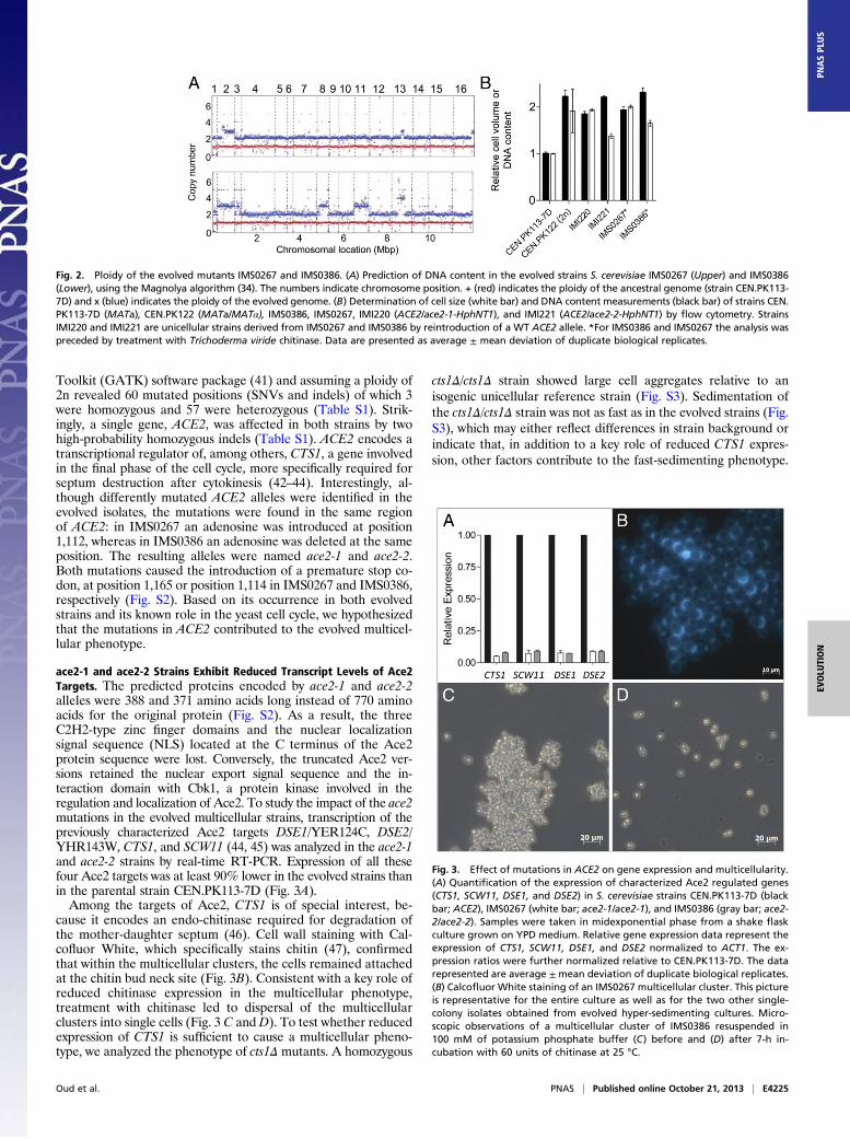
ace2-1 and ace2-2 Strains Exhibit Reduced Transcript Levels of Ace2Targets. The predicted proteins encoded by ace2-1 and ace2-2alleles were 388 and 371 amino acids long instead of 770 aminoacids for the original protein (Fig. S2). As a result, the threeC2H2-type zinc finger domains and the nuclear localizationsignal sequence (NLS) located at the C terminus of the Ace2protein sequence were lost. Conversely, the truncated Ace2 ver-sions retained the nuclear export signal sequence and the in-teraction domain with Cbk1, a protein kinase involved in theregulation and localization of Ace2. To study the impact of the ace2mutations in the evolved multicellular strains, transcription of thepreviously characterized Ace2 targets DSE1/YER124C, DSE2/YHR143W, CTS1, and SCW11 (44, 45) was analyzed in the ace2-1and ace2-2 strains by real-time RT-PCR. Expression of all thesefour Ace2 targets was at least 90% lower in the evolved strains thanin the parental strain CEN.PK113-7D (Fig. 3A).Among the targets of Ace2, CTS1 is of special interest, be-
cause it encodes an endo-chitinase required for degradation ofthe mother-daughter septum (46). Cell wall staining with Cal-cofluor White, which specifically stains chitin (47), confirmedthat within the multicellular clusters, the cells remained attachedat the chitin bud neck site (Fig. 3B). Consistent with a key role ofreduced chitinase expression in the multicellular phenotype,treatment with chitinase led to dispersal of the multicellularclusters into single cells (Fig. 3 C and D). To test whether reducedexpression of CTS1 is sufficient to cause a multicellular pheno-type, we analyzed the phenotype of cts1Δ mutants. A homozygous
cts1Δ/cts1Δ strain showed large cell aggregates relative to anisogenic unicellular reference strain (Fig. S3). Sedimentation ofthe cts1Δ/cts1Δ strain was not as fast as in the evolved strains (Fig.S3), which may either reflect differences in strain background orindicate that, in addition to a key role of reduced CTS1 expres-sion, other factors contribute to the fast-sedimenting phenotype.
Fig. 2. Ploidy of the evolved mutants IMS0267 and IMS0386. (A) Prediction of DNA content in the evolved strains S. cerevisiae IMS0267 (Upper) and IMS0386(Lower), using the Magnolya algorithm (34). The numbers indicate chromosome position. + (red) indicates the ploidy of the ancestral genome (strain CEN.PK113-7D) and x (blue) indicates the ploidy of the evolved genome. (B) Determination of cell size (white bar) and DNA content measurements (black bar) of strains CEN.PK113-7D (MATa), CEN.PK122 (MATa/MATα), IMS0386, IMS0267, IMI220 (ACE2/ace2-1-HphNT1), and IMI221 (ACE2/ace2-2-HphNT1) by flow cytometry. StrainsIMI220 and IMI221 are unicellular strains derived from IMS0267 and IMS0386 by reintroduction of a WT ACE2 allele. *For IMS0386 and IMS0267 the analysis waspreceded by treatment with Trichoderma viride chitinase. Data are presented as average ± mean deviation of duplicate biological replicates.
Fig. 3. Effect of mutations in ACE2 on gene expression and multicellularity.(A) Quantification of the expression of characterized Ace2 regulated genes(CTS1, SCW11, DSE1, and DSE2) in S. cerevisiae strains CEN.PK113-7D (blackbar; ACE2), IMS0267 (white bar; ace2-1/ace2-1), and IMS0386 (gray bar; ace2-2/ace2-2). Samples were taken in midexponential phase from a shake flaskculture grown on YPD medium. Relative gene expression data represent theexpression of CTS1, SCW11, DSE1, and DSE2 normalized to ACT1. The ex-pression ratios were further normalized relative to CEN.PK113-7D. The datarepresented are average ±mean deviation of duplicate biological replicates.(B) Calcofluor White staining of an IMS0267 multicellular cluster. This pictureis representative for the entire culture as well as for the two other single-colony isolates obtained from evolved hyper-sedimenting cultures. Micro-scopic observations of a multicellular cluster of IMS0386 resuspended in100 mM of potassium phosphate buffer (C) before and (D) after 7-h in-cubation with 60 units of chitinase at 25 °C.
Oud et al. PNAS | Published online October 21, 2013 | E4225
EVOLU
TION
PNASPL
US
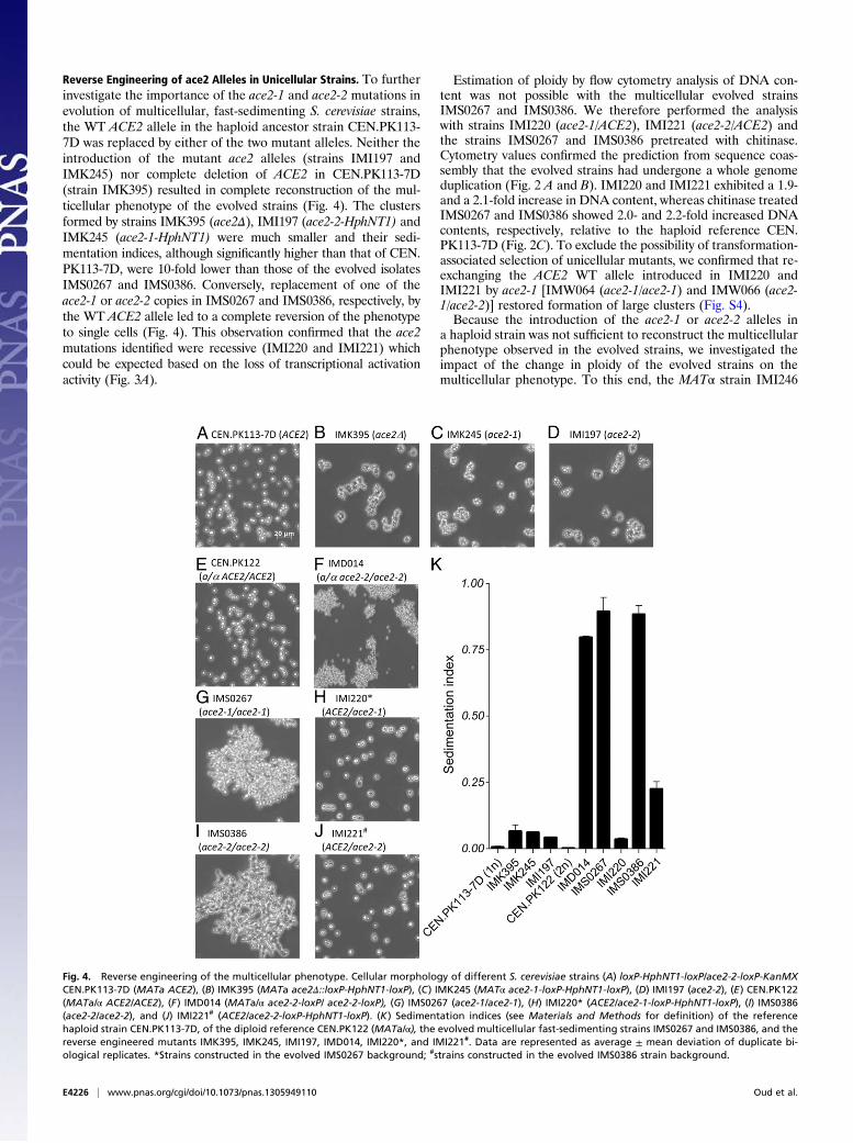
Reverse Engineering of ace2 Alleles in Unicellular Strains. To furtherinvestigate the importance of the ace2-1 and ace2-2 mutations inevolution of multicellular, fast-sedimenting S. cerevisiae strains,the WT ACE2 allele in the haploid ancestor strain CEN.PK113-7D was replaced by either of the two mutant alleles. Neither theintroduction of the mutant ace2 alleles (strains IMI197 andIMK245) nor complete deletion of ACE2 in CEN.PK113-7D(strain IMK395) resulted in complete reconstruction of the mul-ticellular phenotype of the evolved strains (Fig. 4). The clustersformed by strains IMK395 (ace2Δ), IMI197 (ace2-2-HphNT1) andIMK245 (ace2-1-HphNT1) were much smaller and their sedi-mentation indices, although significantly higher than that of CEN.PK113-7D, were 10-fold lower than those of the evolved isolatesIMS0267 and IMS0386. Conversely, replacement of one of theace2-1 or ace2-2 copies in IMS0267 and IMS0386, respectively, bythe WT ACE2 allele led to a complete reversion of the phenotypeto single cells (Fig. 4). This observation confirmed that the ace2mutations identified were recessive (IMI220 and IMI221) whichcould be expected based on the loss of transcriptional activationactivity (Fig. 3A).
Estimation of ploidy by flow cytometry analysis of DNA con-tent was not possible with the multicellular evolved strainsIMS0267 and IMS0386. We therefore performed the analysiswith strains IMI220 (ace2-1/ACE2), IMI221 (ace2-2/ACE2) andthe strains IMS0267 and IMS0386 pretreated with chitinase.Cytometry values confirmed the prediction from sequence coas-sembly that the evolved strains had undergone a whole genomeduplication (Fig. 2 A and B). IMI220 and IMI221 exhibited a 1.9-and a 2.1-fold increase in DNA content, whereas chitinase treatedIMS0267 and IMS0386 showed 2.0- and 2.2-fold increased DNAcontents, respectively, relative to the haploid reference CEN.PK113-7D (Fig. 2C). To exclude the possibility of transformation-associated selection of unicellular mutants, we confirmed that re-exchanging the ACE2 WT allele introduced in IMI220 andIMI221 by ace2-1 [IMW064 (ace2-1/ace2-1) and IMW066 (ace2-1/ace2-2)] restored formation of large clusters (Fig. S4).Because the introduction of the ace2-1 or ace2-2 alleles in
a haploid strain was not sufficient to reconstruct the multicellularphenotype observed in the evolved strains, we investigated theimpact of the change in ploidy of the evolved strains on themulticellular phenotype. To this end, the MATα strain IMI246
Fig. 4. Reverse engineering of the multicellular phenotype. Cellular morphology of different S. cerevisiae strains (A) loxP-HphNT1-loxP/ace2-2-loxP-KanMXCEN.PK113-7D (MATa ACE2), (B) IMK395 (MATa ace2Δ::loxP-HphNT1-loxP), (C) IMK245 (MATα ace2-1-loxP-HphNT1-loxP), (D) IMI197 (ace2-2), (E) CEN.PK122(MATa/α ACE2/ACE2), (F) IMD014 (MATa/α ace2-2-loxP/ ace2-2-loxP), (G) IMS0267 (ace2-1/ace2-1), (H) IMI220* (ACE2/ace2-1-loxP-HphNT1-loxP), (I) IMS0386(ace2-2/ace2-2), and (J) IMI221# (ACE2/ace2-2-loxP-HphNT1-loxP). (K) Sedimentation indices (see Materials and Methods for definition) of the referencehaploid strain CEN.PK113-7D, of the diploid reference CEN.PK122 (MATa/α), the evolved multicellular fast-sedimenting strains IMS0267 and IMS0386, and thereverse engineered mutants IMK395, IMK245, IMI197, IMD014, IMI220*, and IMI221#. Data are represented as average ± mean deviation of duplicate bi-ological replicates. *Strains constructed in the evolved IMS0267 background; #strains constructed in the evolved IMS0386 strain background.
E4226 | www.pnas.org/cgi/doi/10.1073/pnas.1305949110 Oud et al.
(ace2-2-KanMX) was constructed by replacing ACE2 in CEN.PK113-13D and crossed with the MATa strain IMI197 (ace2-2-HphNT1). The resulting diploid strain IMD014 (ace2-2-KanMX/ace2-2-HphNT1) formed large multicellular clusters (Fig. 4) andexhibited a sedimentation index similar to that of the evolvedstrains IMS0267 and IMS0386 (Fig. 4). Similarly, the homozy-gous diploid strains IMD015 (ace2-1-KanMX/ace2-1-HphNT1)and IMD017 (ace2::loxP-HphNT1-loxP/ace2::loxP-KanMX-loxP),as well as the heterozygous diploid strain IMD016 (ace2-2-KanMX/ace2-1-HphNT1) exhibited a multicellular, fast-sedimentingphenotype comparable to that of the two evolved strains IMS0267and IMS0386 (Fig. S5).These results demonstrate complete reverse engineering of an
evolved multicellular, fast sedimenting phenotype by introduction,in diploid S. cerevisiae, of specific recessive mutations in ACE2 thatdrastically reduce or eliminate transcriptional activation of Ace2target genes. Consistent with the ploidy-dependent phenotype oface2 null mutants, deletion of the Ace2 target gene CTS1 ina haploid strain background did not result in the multicellularphenotype observed in diploid cts1Δ/cts1Δ strain (Fig. S3).
DiscussionThis study provides the first identification of a defined set ofgenetic changes by which the unicellular eukaryote S. cerevisiaecan evolve into a multicellular, fast-sedimenting phenotype.Considering the impact of multicellularity in evolution, the mo-lecular events underlying the transformation of unicellular yeastto multicellular clusters were surprisingly simple, requiring onlya mutation in a single gene and a whole genome duplication. Therecessive characteristic of the ace2-1 and ace2-2 mutationsstrongly suggests that they preceded or even facilitated the originof the genome duplication event that occurred during laboratoryevolution of strains IMS0267 and IMS0386. Although generationof multicellular clusters is easily observable, numerous shakeflask–based laboratory evolution studies with S. cerevisiae strains,including the strain used in our study, do not report this phe-notype (29–33). The fast and reproducible selection of multi-cellular mutants in the present study was, in all likelihood,a consequence of the design of the effluent-removal system inour bioreactor setups. We thereby inadvertently mimicked theexperimental design of Ratcliff and coworkers (21) who in-tentionally selected for a fast-sedimenting snowflake phenotypeby including a biomass settling phase in their serial-batch labo-ratory evolution experiments.The accelerated diauxic consumption of glucose-galactose
mixtures (Fig. 1A and Fig. S1) by the evolved cultures cannot becompletely attributed to the mutations that caused multicellu-larity (Fig. S6), suggesting that additional mutations contributedto this characteristic. Analysis of several of these mutations,which is outside the scope of this study, was complicated by theirheterozygous nature.The observed ploidy dependency of the phenotype caused by
the ace2 alleles identified in the evolved strains is probably atleast partly due to the different bud-site selection preferences ofhaploid and diploid S. cerevisiae strains (48, 49). Haploid cellsexhibit axial budding, during which a new bud is formed directlyadjacent to the bud scar. Conversely, diploid cells exhibit a polarbudding pattern, in which daughter cells bud distally (48). Dif-ferent bud-site selection strategies will inevitably affect themorphology of multicellular aggregates in mutants with com-promised cell division. For example, polar budding should resultin less steric hindrance, thereby facilitating generation of largerstructures, consistent with the larger size of multicellular clustersin diploid ace2/ace2 strains. Additionally, ploidy may affectseparation of mother and daughter cells even in unicellularstrains. Of a set of only 17 S. cerevisiae genes whose expression isaffected by ploidy (50), two (CTS1 and DSE4, of which only theendo-chitinase–encoding CTS1 gene is a known Ace2 target) are
associated with mother-daughter cell separation. The strongpositive correlation of ploidy and CTS1 gene expression suggeststhat, in diploid cells, separation of mother and daughter cellsrequires more endo-chitinase than in haploids. This assumptionwould be consistent with the observed stronger phenotype ofreduced CTS1 expression in diploids. The strong ploidy de-pendence of ace2 phenotypes underlines the importance of an-alyzing whole or partial genome duplication in the analysis ofevolved strains (51–53). In addition to facilitating the identifica-tion of key mutations, research on genome duplication and sub-sequent further evolution in laboratory experiments may lead tofurther insight in the evolutionary past of S. cerevisiae, in whicha whole genome duplication played a pivotal role (54).Lack of degradation of the chitin septum between the
mother and the daughter cells appears to be the predominantmechanism underlying the formation of the multicellularclusters observed in the present study. This mechanism mayhave played a role in the transition from unicellular fungi todimorphic and filamentous organisms, because these organ-isms share a conserved role for chitin in cell wall architecture.Inactivation of the ACE2 ortholog in the pathogenic yeastCandida glabrata led to cell clusters and hypervirulence ina murine model (55, 56). Similarly, C. albicans strains with anace2Δ/Δ genotype showed altered separation and morphologyand, moreover, resistance to azole antifungal drugs (56). However,outbreaks of hypervirulent and/or antibiotic-resistant mutants ofthese pathogens have hitherto not been reported.Although mutations in the endo-chitinase-encoding CTS1
gene and/or in other components of the regulation of Ace2 andmorphogenesis (RAM) pathway can be expected to have similarimpacts on sedimentation characteristics, only mutations inACE2 were found in two independent evolution experiments.Moreover, the ace2-1 and ace2-2 mutations occurred in the samehomopolymer of eight adenosine residues (Fig. S2). Poly-(dA:dT) tracts occur frequently in S. cerevisiae genome (57, 58), andthese regions may participate in the yeast genome evolution bycreating mutagenesis hot-spots (57). Poly-(dA:dT) tracts are,however, less abundant in coding regions than in intergenicregions (Table S2), presumably because a resulting evolvabilityconfers a selective disadvantage in most protein-encoding DNA.In contrast, acquisition of a fast-sedimentation phenotype mayoffer selective advantages in nutrient-rich environments wheresingle cells are easily washed away, such as flowers or fruitssubjected to frequent bursts of intensive rainfall. Close in-spection of the nucleotide sequences of Candida ACE2 ortho-logs, and S. cerevisiae genes of the RAM pathway did not revealhomopolymers longer than five residues. In pathogenic Candidastrains, this might limit the frequency with which hypervirulenceoccurs as a consequence of loss of function mutations in ACE2.Knowledge of the mutations responsible for a multicellular, fast-
sedimenting phenotype in S. cerevisiae allows modulation of thisproperty by genetic engineering. The results presented in this studyindicate that stable, fast-sedimenting yeast strains for use in cellretention systems can be constructed by inactivation of both copiesof ACE2 in diploid strains. Formation of multicellular clusters, asobserved in the evolved strains investigated in this study, does nothinder cell growth. In fact, the evolved strains IMS0267 andIMS0386 showed higher growth rates than their ancestor CEN.PK113-7D in chemically defined medium with glucose and galac-tose (Fig. 1 and Fig. S1). Additionally, it may be possible to pre-vent or delay occurrence of multicellular phenotypes in adaptiveevolution experiments, where it is not always a desirable feature,by ectopic integration of multiple ACE2 genes.
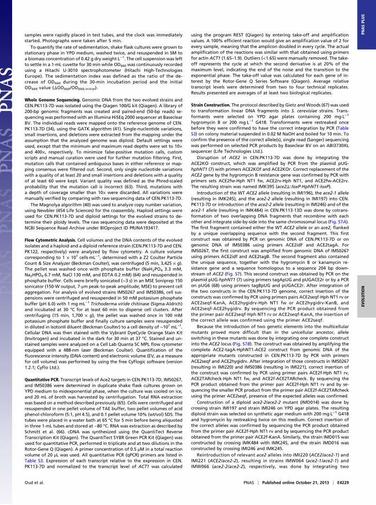
Materials and MethodsStrain Maintenance. S. cerevisiae strains used in this study (Table 1) werederived from the CEN.PK family (59) and from the BY lineage (60). Strainswere maintained on YP medium [demineralized water; 10 g·L−1 Yeast extract
Oud et al. PNAS | Published online October 21, 2013 | E4227
EVOLU
TION
PNASPL
US
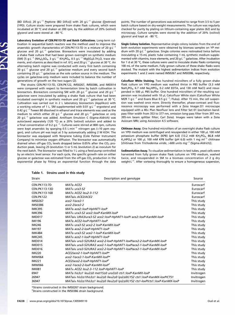
(BD Difco); 20 g·L−1 Peptone (BD Difco)] with 20 g·L−1 glucose (Dextrose)(YPD). Culture stocks were prepared from shake flask cultures, which wereincubated at 30 °C and shaken at 200 rpm, by the addition of 20% (vol/vol)glycerol and were stored at −80 °C.
Laboratory Evolution of CEN.PK113-7D and Batch Cultivations. Long-term cul-tivation in sequential batch reactors was the method used to improve theanaerobic growth characteristics of CEN.PK113-7D in a mixture of 20 g·L−1
glucose and 20 g·L−1 galactose. Bioreactors were inoculated by addinga shake flask culture that had been grown overnight on synthetic medium(SM) [5 g·L−1 (NH4)2SO4, 3 g·L−1 KH2PO4, 0.5 g·L−1 MgSO40.7H2O, trace ele-ments, and vitamins as described in ref. 61], and 20 g·L−1 glucose at 30 °C. Analternating batch regime was conducted with every first batch containing20 g·L−1 glucose and 20 g·L−1 galactose medium and every second batchcontaining 20 g·L−1 galactose as the sole carbon source in the medium. Thecycles on galactose-only medium were included to balance the number ofgenerations of growth on the two sugars (2).
The strains CEN.PK113-7D, CEN.PK122, IMS0267, IMS0386, and IMD014were compared with respect to fermentation time by batch cultivation inbioreactors. Bioreactors containing SM with 20 g·L−1 glucose and 20 g·L−1
galactose were inoculated by adding a shake flask culture that had beenincubated overnight in synthetic medium and 20 g·L−1 galactose at 30 °C.Cultivation was carried out in 2 L laboratory bioreactors (Applikon) witha working volume of 1 L. SM supplemented with 0.01 g·L−1 ergosterol and0.42 g·L−1 Tween 80 dissolved in ethanol and trace elements was used as themedium to which either 20 g·L−1 glucose and 20 g·L−1 galactose or only20 g·L−1 galactose was added. Antifoam Emulsion C (Sigma-Aldrich) wasautoclaved separately (120 °C) as a 20% (wt/vol) solution and added toa final concentration of 0.2 g·L−1. Cultures were stirred at 800 rpm, cultureswere kept anaerobic by sparging 0.5 L·min−1 nitrogen gas (<10 ppm oxy-gen), and culture pH was kept at 5 by automatically adding 2 M KOH. Thebioreactor was equipped with Norprene tubing (Cole Palmer InstrumentCompany) to minimize oxygen diffusion. The bioreactor was automaticallydrained when off-gas CO2 levels dropped below 0.05% after the CO2 pro-duction peak, leaving 25 (evolution 1) or 5 mL (evolution 2) as inoculum forthe next batch. The bioreactor was filled to 1 L using a feed pump controlledby an electric level sensor. For each cycle, the specific growth rate on eitherglucose or galactose was estimated from the off-gas CO2 production in theexponential phase by fitting an exponential function through the data
points. The number of generations was estimated to range from 3.5 to 5 perbatch culture based on dry-weight measurements. The culture was regularlychecked for purity by plating on lithium-containing agar plates (62) and bymicroscopy. Culture samples were stored by the addition of 20% (vol/vol)glycerol and kept at −80 °C.
Single Colony Isolation. Representative single colony isolates from the end ofboth evolution experiments were obtained by biomass samples on YP me-dium with 20 g·L−1 galactose. Single colonies were restreaked twice beforeinoculating a 15-mL plastic tube containing 1 mL synthetic medium supple-mented with vitamins, trace elements, and 20 g·L−1 galactose. After incubationfor 1 d at 30 °C, these cultures were used to inoculate shake flasks containing100 mL of the same medium. Fully grown cultures of these shake flasks werestocked. The mutant with the highest sedimentation indices from evolutionexperiments 1 and 2 were named IMS0267 and IMS0386, respectively.
Calcofluor White Staining. Two hundred microliters of a fully grown shakeflask culture on YPD medium was washed thrice in PBS buffer (3.3 mMNaH2PO4, 6.7 mM Na2HPO4, 0.2 mM EDTA, and 130 mM NaCl) and resus-pended in 500 μL PBS buffer. One hundred microliters of the resulting sus-pension was incubated with 10 μL Calcofluor White stain (Calcofluor WhiteM2R 1 g·L−1 and Evans Blue 0.5 g·L−1; Fluka). After 15 min, the cell suspen-sion was washed once more. Directly thereafter, phase-contrast and fluo-rescence microscopy was performed with a Zeiss Imager.D1 microscopeequipped with a 40× Plan Neofluor lens and Filter Set 01 (excitation band-pass filter width from 353 to 377 nm, emission long-pass filter from 397 nm,395-nm beam splitter filter; Carl Zeiss). Images were taken with a ZeissAxiocam MRc using Axiovision 4.5 software.
Chitinase Assay. One hundred microliters of an overnight shake flask cultureon YPD medium was centrifuged and resuspended in either 100 μL 100 mMpotassium phosphate buffer (KPB) (pH 6.0) (13.2 mM KH2PO4, 86.8 mMK2HPO4) or 100 μL 100 mM KPB buffer (pH 6.0) with 1 mg ml−1 chitinase[chitinase from Trichoderma viride, >600 units mg−1 (Sigma-Aldrich)].
Sedimentation Assay. To visualize sedimentation in test tubes, yeast cells wereharvested from fully grown shake flask cultures on YPD medium, washedtwice, and resuspended in SM to a biomass concentration of 2 g dryweight·L−1. After vortexing thoroughly to ensure a homogeneous suspension,
Table 1. Strains used in this study
Strain Description and genotype Source
CEN.PK113-7D MATa ACE2 EuroscarfCEN.PK113-13D MATα ura3-52 EuroscarfCEN.PK113-16B MATα ACE2 leu2-3–112 EuroscarfCEN.PK122 MATa/α ACE2/ACE2 EuroscarfIMS0267 ace2-1/ace2-1 This studyIMS0386 ace2-2/ace2-2 This studyIMK395 MATa ace2::loxP-HphNT1-loxP This studyIMK396 MATα ura3-52 ace2::loxP-KanMX-loxP This studyIMD017 MATa/α URA3/ura3-52 ace2::loxP-HphNT1-loxP/ ace2::loxP-KanMX-loxP This studyIMI196 MATa ACE2-loxP-HphNT1-loxP This studyIMI246 MATα ura3-52 ace2-2 loxP-KanMX-loxP This studyIMI197 MATa ace2-2-loxP-HphNT1-loxP This studyIMK484 MATa ura3-52 ace2-1-loxP-KanMX-loxP This studyIMK245 MATα ace2-1-loxP-HphNT1-loxP This studyIMD014 MATa/α ura3-52/URA3 ace2-2-loxP-HphNT1-loxP/ace2-2-loxP-KanMX-loxP This studyIMD015 MATa/α ura3-52/URA3 ace2-1-loxP-HphNT1-loxP/ace2-1-loxP-KanMX-loxP This studyIMD016 MATa/α ura3-52/URA3 ace2-2-loxP-HphNT1-loxP/ace2-1-loxP-KanMX-loxP This studyIMI220 ACE2/ace2-1-loxP-HphNT1-loxP* This studyIMW064 ace2-1/ace2-1-loxP-KanMX-loxP* This studyIMI221 ACE2/ace2-2-loxP-HphNT1-loxP† This studyIMW066 ace2-1/ace2-2-loxP-KanMX-loxP† This studyIMI081 MATα ACE2 leu2-3–112 loxP-HphNT1-loxP This study6947 MATa his3Δ1 leu2Δ0 met15Δ0 ura3Δ0 cts1::loxP-KanMX-loxP Invitrogen26947 MATa/α his3Δ1/his3Δ1 leu2Δ0 /leu2Δ0 lys2Δ0/LYS2 cts1::loxP-KanMX-loxP/CTS1 Invitrogen36947 MATa/α his3Δ1/his3Δ1 leu2Δ0 /leu2Δ0 lys2Δ0/LYS2 cts1-loxP/cts1::loxP-KanMX-loxP Invitrogen
*Strains constructed in the IMS0267 strain background.†Strains constructed in the IMS0386 strain background.
E4228 | www.pnas.org/cgi/doi/10.1073/pnas.1305949110 Oud et al.
samples were rapidly placed in test tubes, and the clock was immediatelystarted. Photographs were taken after 5 min.
To quantify the rate of sedimentation, shake flask cultures were grown tostationary phase in YPD medium, washed twice, and resuspended in SM toa biomass concentration of 0.42 g dry weight·L−1. The cell suspension was leftto settle in a 1-mL cuvette for 30 min while OD660 was continuously recordedusing a Hitachi U-3010 spectrophotometer (Hitachi High-TechnologiesEurope). The sedimentation index was defined as the ratio of the de-crease of OD660 during the 30-min incubation period and the initialOD660 value (ΔOD660/OD660,initial).
Whole Genome Sequencing. Genomic DNA from the two evolved strains andCEN.PK113-7D was isolated using the Qiagen 100/G kit (Qiagen). A library of200-bp genomic fragments was created and paired-end (50-bp reads) se-quencing was performed with an Illumina HiSEq 2000 sequencer at BaseclearBV. The individual reads were mapped onto the reference genome of CEN.PK113-7D (34), using the GATK algorithm (41). Single-nucleotide variations,small insertions, and deletions were extracted from the mapping under theassumption that the analyzed genome was diploid. Default settings wereused, except that the minimum and maximum read depths were set to 10×and 400×, respectively. To minimize false-positive mutation calls, customscripts and manual curation were used for further mutation filtering. First,mutation calls that contained ambiguous bases in either reference or map-ping consensus were filtered out. Second, only single nucleotide variationswith a quality of at least 20 and small insertions and deletions with a qualityof at least 60 were kept. Variant quality was defined as the Phred-scaledprobability that the mutation call is incorrect (63). Third, mutations witha depth of coverage smaller than 10× were discarded. All variations weremanually verified by comparing with raw sequencing data of CEN.PK113-7D.
The Magnolya algorithm (40) was used to analyze copy number variation,using Newbler (454 Life Sciences) for the coassembly. Haploid settings wereused for CEN.PK113-7D and diploid settings for the evolved strains to de-termine their ploidy levels. The raw sequencing data were deposited at theNCBI Sequence Read Archive under BIOproject ID PRJNA193417.
Flow Cytometric Analysis. Cell volumes and the DNA contents of the evolvedisolates and a haploid and a diploid reference strain (CEN.PK113-7D and CEN.PK122, respectively) were analyzed by flow cytometry. A culture volumecorresponding to 1 × 10⁷ cells·mL−1, determined with a Z2 Coulter ParticleCount & Size Analyzer (Beckman Coulter), was centrifuged (5 min, 3,425 × g).The pellet was washed once with phosphate buffer (NaH2PO4 3.3 mM,Na2HPO4 6.7 mM, NaCl 130 mM, and EDTA 0.2 mM) (64) and resuspended inphosphate buffer. Cells were briefly sonicated (∼3 s) in an MSE Soniprep 150sonicator (150-W output, 7-μm peak-to-peak amplitude; MSE) to prevent cellaggregation. For analysis of evolved strains IMS0267 and IMS0386, cell sus-pensions were centrifuged and resuspended in 50 mM potassium phosphatebuffer (pH 6.0) with 1 mg·mL−1 Trichoderma viride chitinase (Sigma-Aldrich)and incubated at 30 °C for at least 60 min to disperse cell clusters. Aftercentrifuging (15 min, 1,700 × g), the pellet was washed once in 100 mMpotassium phosphate buffer and finally culture samples were resuspendedin diluted in IsotonII diluent (Beckman Coulter) to a cell density of ∼107 mL−1.Cellular DNA was then stained with the Vybrant DyeCycle Orange Stain Kit(Invitrogen) and incubated in the dark for 30 min at 37 °C. Stained and un-stained samples were analyzed on a Cell Lab Quanta SC MPL flow cytometerequipped with a 488-nm laser (Beckman Coulter). Quantification of thefluorescence intensity (DNA content) and electronic volume (EV, as a measurefor cell volume) was performed by using the free CyFlogic software (version1.2.1; CyFlo Ltd.).
Quantitative PCR. Transcript levels of Ace2 targets in CEN.PK113-7D, IMS0267,and IMS0386 were determined in duplicate shake flask cultures grown onYPD medium to midexponential phase, when the culture was cooled on ice,and 20 mL of broth was harvested by centrifugation. Total RNA extractionwas based on a method described previously (65). Cells were centrifuged andresuspended in one pellet volume of TAE buffer, two pellet volumes of acidphenol-chloroform (5:1, pH 4.5), and 0.1 pellet volume 10% (wt/vol) SDS. Thetubes were placed in a water bath at 65 °C for 5 min before being aliquotedin three 1-mL tubes and stored at −80 °C. RNA was extraction as described bySchmitt et al. (66). cDNA was synthesized using the QuantiTect ReverseTranscription Kit (Qiagen). The QuantiTect SYBR Green PCR Kit (Qiagen) wasused for quantitative PCR, performed in triplicate and at two dilutions in theRotor-Gene Q (Qiagen). A primer concentration of 0.5 μM in a total reactionvolume of 20 μL was used. All quantitative PCR (qPCR) primers are listed inTable S3. Expression of each transcript relative to the expression in CEN.PK113-7D and normalized to the transcript level of ACT1 was calculated
using the program REST (Qiagen) by entering take-off and amplificationvalues. A 100% efficient reaction would give an amplification value of 2 forevery sample, meaning that the amplicon doubled in every cycle. The actualamplification of the reactions was similar with that obtained using primersfor actin ACT1 (1.65–1.9). Outliers (<1.65) were manually removed. The take-off represents the cycle at which the second derivative is at 20% of themaximum level, indicating the end of the noise and the transition to theexponential phase. The take-off value was calculated for each gene of in-terest by the Rotor-Gene Q Series Software (Qiagen). Average relativetranscript levels were determined from two to four technical replicates.Results presented are averages of at least two biological replicates.
Strain Construction. The protocol described by Gietz andWoods (67) was usedto transformation linear DNA fragments into S. cerevisiae strains. Trans-formants were selected on YPD agar plates containing 200 mg·L−1
hygromycin B or 200 mg·L−1 G418. Transformants were restreaked oncebefore they were confirmed to have the correct integration by PCR (TableS3) on colony material suspended in 0.02 M NaOH and boiled for 10 min. Toconfirm the presence of the correct allele(s), single read (Sanger) sequencingwas performed on selected PCR products by Baseclear BV on an ABI3730XLsequencer (Life Technologies Ltd.).
Disruption of ACE2 in CEN.PK113-7D was done by integrating theACE2KO construct, which was amplified by PCR from the plasmid pUG-hphNT1 (7) with primers ACE2KOf and ACE2KOr. Correct replacement of theACE2 gene by the hygromycin B resistance gene was confirmed by PCR withprimers sets ACE2fw-Hph NT1 fw, ACE2rv-Hph NT1, and ACE2fw-ACE2rv.The resulting strain was named IMK395 (ace2Δ::loxP-HphNT1-loxP).
Introduction of the WT ACE2 allele (resulting in IMI196), the ace2-1 allele(resulting in IMK245), and the ace2-2 allele (resulting in IMI197) into CEN.PK113-7D or introduction of the ace2-2 allele (resulting in IMI246) and of theace2-1 allele (resulting in IMK484) in CEN.PK113-13D was done by cotrans-formation of two overlapping DNA fragments that recombine with eachother and integrate side-by-side into the same chromosomal locus (Fig. S7A).The first fragment contained either the WT ACE2 allele or an ace2, flankedby a unique overlapping sequence with the second fragment. This firstconstruct was obtained by PCR on genomic DNA of CEN.PK113-7D or ongenomic DNA of IMS0386 using primers ACE2idF and ACE2tagA. ForIMS0267, the first construct was amplified from genomic DNA of IMS0267using primers ACE2idf and ACE2tagB. The second fragment also containedthe unique sequence, together with the hygromycin B or kanamycin re-sistance gene and a sequence homologous to a sequence 204 bp down-stream of ACE2 (Fig. S7). This second construct was obtained by PCR on theplasmid pUG-hphNT1 (7) using primers tagApUG and pUGACE2r or by a PCRon pUG6 (68) using primers tagBpUG and pUGACE2r. After integration ofthe two constructs in the CEN.PK113-7D genome, correct insertion of theconstructs was confirmed by PCR using primers pairs ACE2seqf-Hph NT1 rv orACE2seqf-KanA, ACE2hygidrv-Hph NT1 fw or ACE2hygidrv-KanB, andACE2seqf-ACE2hygidrv. By sequencing the PCR product obtained fromthe primer pair ACE2seqf-Hph NT1 rv or ACE2seqf-KanA, the insertion ofthe correct allele was confirmed using the primer ACE2seqf.
Because the introduction of two genetic elements into the multicellularmutants proved more difficult than in the unicellular ancestor, alleleswitching in these mutants was done by integrating one complete constructinto the ACE2 locus (Fig. S7B). The construct was obtained by amplifying thecomplete ACE2-tagA-HphNT1-ACE2 construct from genomic DNA of theappropriate mutants constructed in CEN.PK113-7D by PCR with primersACE2seqf and ACE2hygidrv. After integration of those constructs in IMS0267(resulting in IMI220) and IMS0386 (resulting in IMI221), correct insertion ofthe construct was confirmed by PCR using primer pairs ACE2f-Hph NT1 rv,ACE2TARcheck-Hph NT1 fw, and ACE2f-ACE2TARcheck. By sequencing thePCR product obtained from the primer pair ACE2f-Hph NT1 rv and by se-quencing the smaller PCR product from the primer pair ACE2f-ACE2TARcheckusing the primer ACE2seqf, presence of the expected alleles was confirmed.
Construction of a diploid ace2-2/ace2-2 mutant (IMD014) was done bycrossing strain IMI197 and strain IMI246 on YPD agar plates. The resultingdiploid strain was selected on synthetic agar medium with 200 mg·L−1 G418and hygromycin by restreaking twice on this medium. Correct insertion ofthe correct alleles was confirmed by sequencing the PCR product obtainedfrom the primer pair ACE2f-Hph NT1 rv and by sequencing the PCR productobtained from the primer pair ACE2f-KanA. Similarly, the strain IMD015 wasconstructed by crossing IMK484 with IMK245, and the strain IMD016 wasconstructed by crossing IMI246 and IMK245.
Reintroduction of relevant ace2 alleles into IMI220 (ACE2/ace2-1) andIMI221 (ACE2/ace2-2), resulting in strains IMW064 (ace2-1/ace2-1) andIMW066 (ace2-2/ace2-2), respectively, was done by integrating two
Oud et al. PNAS | Published online October 21, 2013 | E4229
EVOLU
TION
PNASPL
US
overlapping constructs into the ACE2 locus, thereby replacing the ACE2-tagA-HphNTI-ACE2 construct (Fig. S7A). The first construct contained anace2-1 or ace2-2 allele, flanked by a unique overlapping sequence withthe second construct. The first construct was obtained by PCR on geno-mic DNA of IMS0267 or IMS0386, using primers ACE2idf and ACE2tagB.The second construct also contained the unique sequence, together with thekanamycin resistance gene and a sequence homologous to a sequence 204 bpdownstream of ACE2. This second construct was obtained by PCR on theplasmid pUG6 (68) using primers tagBpUG and pUGACE2r. After transforma-tion of the two constructs to the appropriate strain, correct insertion wasconfirmed by PCR using primer pairs ACE2f-KanA, ACE2TARcheck-KanB, andACE2f-ACE2TARcheck, as well as by demonstrating resistance to G418 platesbut not to hygromycin. Presence of the desired alleles was confirmed bysequencing the PCR product obtained from the primer pair ACE2f-Hph NT1rv and by sequencing the smaller PCR product from the primer pair ACE2f-ACE2TARcheck using the primer ACE2seqf. Introduction of a hygromycin re-sistance gene into the MATα CEN.PK113-16B strain was done by transforminga genetic construct obtained by PCR from the plasmid pUG-hphNT1 (7) usingprimers MTH1markfw and MTH1markrv. The resulting strain was namedIMI081 (ACE2 loxP-HphNT1-loxP).
Constructs were made by PCR amplification on genomic DNA by usingExpand high fidelity Polymerase (Roche) according to the manufacturer’sinstructions in a Biometra TGradient Thermocycler (Biometra). Isolation offragments from gel was done with the Zymoclean Gel DNA Recovery kit(Zymo Research). PCR amplification on colony material was done usingFastStart Taq DNA Polymerase (Roche) according to the manufacturer’sinstructions on colony material suspended in 0.02 M NaOH and heated for 10min at 100 °C.
Mating and Sporulation. Strains IMS0267 and IMS0386 were mated withIMI081 by streaking both strains on YPD plates. After overnight incubationat 30 °C, the strains were streaked over each other. After another 4 h ofincubation at 30 °C, diploids were selected by streaking on selective medium
(SM medium with 20 g·L−1 glucose and 200 mg·L−1 hygromycin). Resultingsingle colonies were restreaked twice on the same medium.
Sporulation was performed by incubating a culture in YP medium sup-plemented with 10 g·L−1 potassium acetate for 2 d at 23 °C. Subse-quently, the entire culture was washed twice, resuspended in 20 g·L−1
potassium acetate, and incubated for 3–4 d at 23 °C. Spores were segregatedon YPD plates using a micromanipulator (Singer Instruments) and incubatedat 30 °C.
Homopolymer Distribution. The S. cerevisiae reference genome and its an-notation (release 64-1-1, February 3, 2011) were downloaded from theSaccharomyces Genome Database (www.yeastgenome.org/) (69). A file“domains.tab,” containing domains predicted using InterProScan (70), wasdownloaded from the same site (March 10, 2013). The number of occur-rences of dA:dT homopolymers of eight or more residues was counted in theoverall genome, in genes (i.e., sequences annotated as gene in the referencegenome), in coding sequences within genes, in introns, and in domains.Homopolymers were considered present when all bases fell inside the ge-nomic feature. For each of these features, a Fisher exact test (two-tailed) wasthen performed under the null hypothesis that the occurrence of homo-polymeric stretches is independent of the underlying genomic feature(genes, coding sequences, introns, and domains).
ACKNOWLEDGMENTS. We thank Mark Bisschops and Marijke Luttik forhelp with fluorescence microscopy, Marit Hebly for help with flowcytometry, Erik de Hulster for expert advice and support on bioreactoroperation, and Marcel van den Broek for bioinformatics support. CarstenBlom, Edwin van der Pol, and Vito Meulenberg are acknowledged fortheir contributions via student projects. We thank Tim Vos for criticalreading of the manuscript and the other members of our research groupfor constructive discussions. The PhD project of B.O. is part of theresearch program of the Kluyver Centre for Genomics of IndustrialFermentation, which is subsidized by the Netherlands Genomics Initia-tive/Netherlands Organization for Scientific Research.
1. Sauer U (2001) Evolutionary engineering of industrially important microbial pheno-types. Adv Biochem Eng Biotechnol 73:129–169.
2. Wisselink HW, Toirkens MJ, Wu Q, Pronk JT, van Maris AJA (2009) Novel evolutionaryengineering approach for accelerated utilization of glucose, xylose, and arabinosemixtures by engineered Saccharomyces cerevisiae strains. Appl Environ Microbiol75(4):907–914.
3. Kuyper M, et al. (2005) Evolutionary engineering of mixed-sugar utilization by a xy-lose-fermenting Saccharomyces cerevisiae strain. FEMS Yeast Res 5(10):925–934.
4. Guadalupe Medina V, Almering MJH, van Maris AJA, Pronk JT (2010) Elimination ofglycerol production in anaerobic cultures of a Saccharomyces cerevisiae strain en-gineered to use acetic acid as an electron acceptor. Appl Environ Microbiol 76(1):190–195.
5. Koppram R, Albers E, Olsson L (2012) Evolutionary engineering strategies to enhancetolerance of xylose utilizing recombinant yeast to inhibitors derived from sprucebiomass. Biotechnol Biofuels 5(1):32.
6. Zelle RM, Harrison JC, Pronk JT, van Maris AJA (2011) Anaplerotic role for cytosolicmalic enzyme in engineered Saccharomyces cerevisiae strains. Appl Environ Microbiol77(3):732–738.
7. de Kok S, et al. (2012) Laboratory evolution of new lactate transporter genes in a jen1delta mutant of Saccharomyces cerevisiae and their identification as ADY2 alleles bywhole-genome resequencing and transcriptome analysis. FEMS Yeast Res 12(3):359–374.
8. Adamo GM, Brocca S, Passolunghi S, Salvato B, Lotti M (2012) Laboratory evolution ofcopper tolerant yeast strains. Microb Cell Fact 11:1.
9. Portnoy VA, Bezdan D, Zengler K (2011) Adaptive laboratory evolution—harnessingthe power of biology for metabolic engineering. Curr Opin Biotechnol 22(4):590–594.
10. Wagner A (2008) Neutralism and selectionism: A network-based reconciliation. NatRev Genet 9(12):965–974.
11. Loh E, Salk JJ, Loeb LA (2010) Optimization of DNA polymerase mutation rates duringbacterial evolution. Proc Natl Acad Sci USA 107(3):1154–1159.
12. Elena SF, Lenski RE (2003) Evolution experiments with microorganisms: The dynamicsand genetic bases of adaptation. Nat Rev Genet 4(6):457–469.
13. Stanek MT, Cooper TF, Lenski RE (2009) Identification and dynamics of a beneficialmutation in a long-term evolution experiment with Escherichia coli. BMC Evol Biol 9:302.
14. Kvitek DJ, Sherlock G (2011) Reciprocal sign epistasis between frequently experi-mentally evolved adaptive mutations causes a rugged fitness landscape. PLoS Genet7(4):e1002056.
15. Kao KC, Sherlock G (2008) Molecular characterization of clonal interference duringadaptive evolution in asexual populations of Saccharomyces cerevisiae. Nat Genet40(12):1499–1504.
16. Conrad TM, Lewis NE, Palsson BO (2011) Microbial laboratory evolution in the era ofgenome-scale science. Mol Syst Biol 7:509.
17. Bailey JE, et al. (1996) Inverse metabolic engineering: A strategy for directed geneticengineering of useful phenotypes. Biotechnol Bioeng 52(1):109–121.
18. Oud B, van Maris AJA, Daran JM, Pronk JT (2012) Genome-wide analytical approaches
for reverse metabolic engineering of industrially relevant phenotypes in yeast. FEMS
Yeast Res 12(2):183–196.19. Bro C, Nielsen J (2004) Impact of ‘ome’ analyses on inverse metabolic engineering.
Metab Eng 6(3):204–211.20. Teusink B, Bachmann H, Molenaar D (2011) Systems biology of lactic acid bacteria: A
critical review. Microb Cell Fact 10(Suppl 1):S11.21. Ratcliff WC, Denison RF, Borrello M, Travisano M (2012) Experimental evolution of
multicellularity. Proc Natl Acad Sci USA 109(5):1595–1600.22. Bonner JT (1998) The origins of multicellularity. Integr Biol Issues News Rev 1(1):27–36.23. Rokas A (2008) The origins of multicellularity and the early history of the genetic
toolkit for animal development. Annu Rev Genet 42:235–251.24. Knoll AH (2011) The multiple origins of complex multicellularity. Annu Rev Earth
Planet Sci 39:217–239.25. Weitao T (2009) Multicellularity of a unicellular organism in response to DNA repli-
cation stress. Res Microbiol 160(1):87–88.26. Lindén T, Peetre J, Hahn-Hägerdal B (1992) Isolation and characterization of acetic
acid-tolerant galactose-fermenting strains of Saccharomyces cerevisiae from a spent
sulfite liquor fermentation plant. Appl Environ Microbiol 58(5):1661–1669.27. Koschwanez JH, Foster KR, Murray AW (2011) Sucrose utilization in budding yeast as
a model for the origin of undifferentiated multicellularity. PLoS Biol 9(8):e1001122.28. Boraas ME, Seale DB, Boxhorn JE (1998) Phagotrophy by a flagellate selects for co-
lonial prey: A possible origin of multicellularity. Evol Ecol 12(2):153–164.29. Jasmin JN, Zeyl C (2012) Life-history evolution and density-dependent growth in ex-
perimental populations of yeast. Evolution 66(12):3789–3802.30. Jasmin JN, Zeyl C (2013) Evolution of pleiotropic costs in experimental populations.
J Evol Biol 26(6):1363–1369.31. Samani P, Bell G (2010) Adaptation of experimental yeast populations to stressful
conditions in relation to population size. J Evol Biol 23(4):791–796.32. Raynes Y, Gazzara MR, Sniegowski PD (2011) Mutator dynamics in sexual and asexual
experimental populations of yeast. BMC Evol Biol 11:158.33. Hong KK, Vongsangnak W, Vemuri GN, Nielsen J (2011) Unravelling evolutionary
strategies of yeast for improving galactose utilization through integrated systems
level analysis. Proc Natl Acad Sci USA 108(29):12179–12184.34. Nijkamp JF, et al. (2012) De novo sequencing, assembly and analysis of the genome of
the laboratory strain Saccharomyces cerevisiae CEN.PK113-7D, a model for modern
industrial biotechnology. Microb Cell Fact 11:36.35. Ratcliff WC, Pentz JT, Travisano M (2013) Tempo and mode of multicellular adapta-
tion in experimentally evolved Saccharomyces cerevisiae. Evolution 67(6):1573–1581.36. Soares EV (2011) Flocculation in Saccharomyces cerevisiae: A review. J Appl Microbiol
110(1):1–18.37. Soares EV, Vroman A (2003) Effect of different starvation conditions on the floccu-
lation of Saccharomyces cerevisiae. J Appl Microbiol 95(2):325–330.38. Stratford M (1992) Yeast flocculation: A new perspective. Adv Microb Physiol 33:2–71.
E4230 | www.pnas.org/cgi/doi/10.1073/pnas.1305949110 Oud et al.
39. Stratford M, Assinder S (1991) Yeast flocculation: Flo1 and NewFlo phenotypes and
receptor structure. Yeast 7(6):559–574.40. Nijkamp JF, et al. (2012) De novo detection of copy number variation by co-assembly.
Bioinformatics 28(24):3195–3202.41. McKenna A, et al. (2010) The Genome Analysis Toolkit: A MapReduce framework for
analyzing next-generation DNA sequencing data. Genome Res 20(9):1297–1303.42. Voth WP, Olsen AE, Sbia M, Freedman KH, Stillman DJ (2005) ACE2, CBK1, and BUD4
in budding and cell separation. Eukaryot Cell 4(6):1018–1028.43. Saputo S, Chabrier-Rosello Y, Luca FC, Kumar A, Krysan DJ (2012) The RAM network in
pathogenic fungi. Eukaryot Cell 11(6):708–717.44. Doolin MT, Johnson AL, Johnston LH, Butler G (2001) Overlapping and distinct roles of
the duplicated yeast transcription factors Ace2p and Swi5p. Mol Microbiol 40(2):
422–432.45. Sbia M, et al. (2008) Regulation of the yeast Ace2 transcription factor during the cell
cycle. J Biol Chem 283(17):11135–11145.46. Kuranda MJ, Robbins PW (1991) Chitinase is required for cell separation during
growth of Saccharomyces cerevisiae. J Biol Chem 266(29):19758–19767.47. Herth W, Schnepf E (1980) The fluorochrome, calcofluor white, binds oriented to
structural polysaccharide fibrils. Protoplasma 105(1-2):129–133.48. Casamayor A, Snyder M (2002) Bud-site selection and cell polarity in budding yeast.
Curr Opin Microbiol 5(2):179–186.49. Chant J, Pringle JR (1995) Patterns of bud-site selection in the yeast Saccharomyces
cerevisiae. J Cell Biol 129(3):751–765.50. Galitski T, Saldanha AJ, Styles CA, Lander ES, Fink GR (1999) Ploidy regulation of gene
expression. Science 285(5425):251–254.51. Yona AH, et al. (2012) Chromosomal duplication is a transient evolutionary solution
to stress. Proc Natl Acad Sci USA 109(51):21010–21015.52. Chen GB, Rubinstein B, Li R (2012) Whole chromosome aneuploidy: Big mutations
drive adaptation by phenotypic leap. Bioessays 34(10):893–900.53. Koszul R, Caburet S, Dujon B, Fischer G (2004) Eucaryotic genome evolution through
the spontaneous duplication of large chromosomal segments. EMBO J 23(1):234–243.54. Wolfe KH, Shields DC (1997) Molecular evidence for an ancient duplication of the
entire yeast genome. Nature 387(6634):708–713.55. Kamran M, et al. (2004) Inactivation of transcription factor gene ACE2 in the fungal
pathogen Candida glabrata results in hypervirulence. Eukaryot Cell 3(2):546–552.
56. MacCallum DM, et al. (2006) Different consequences of ACE2 and SWI5 gene dis-ruptions for virulence of pathogenic and nonpathogenic yeasts. Infect Immun 74(9):5244–5248.
57. Ma X, et al. (2012) Mutation hot spots in yeast caused by long-range clustering ofhomopolymeric sequences. Cell Rep 1(1):36–42.
58. Dechering KJ, Cuelenaere K, Konings RN, Leunissen JA (1998) Distinct frequency-distributions of homopolymeric DNA tracts in different genomes. Nucleic Acids Res26(17):4056–4062.
59. Entian KD, Kötter P (2007) Yeast genetic strain and plasmid collections. MethodMicrobiol 36:629–666.
60. Brachmann CB, et al. (1998) Designer deletion strains derived from Saccharomycescerevisiae S288C: A useful set of strains and plasmids for PCR-mediated gene dis-ruption and other applications. Yeast 14(2):115–132.
61. Verduyn C, Postma E, Scheffers WA, Van Dijken JP (1992) Effect of benzoic acid onmetabolic fluxes in yeasts: A continuous-culture study on the regulation of respirationand alcoholic fermentation. Yeast 8(7):501–517.
62. Daran-Lapujade P, et al. (2009) An atypical PMR2 locus is responsible for hypersen-sitivity to sodium and lithium cations in the laboratory strain Saccharomyces cer-evisiae CEN.PK113-7D. FEMS Yeast Res 9(5):789–792.
63. Ewing B, Green P (1998) Base-calling of automated sequencer traces using phred. II.Error probabilities. Genome Res 8(3):186–194.
64. Porro D, Brambilla L, Alberghina L (2003) Glucose metabolism and cell size in con-tinuous cultures of Saccharomyces cerevisiae. FEMS Microbiol Lett 229(2):165–171.
65. Daran-Lapujade P, Daran JM, van Maris AJA, de Winde JH, Pronk JT (2009) Chemo-stat-based micro-array analysis in baker’s yeast. Adv Microb Physiol 54:257–311.
66. Schmitt ME, Brown TA, Trumpower BL (1990) A rapid and simple method for prep-aration of RNA from Saccharomyces cerevisiae. Nucleic Acids Res 18(10):3091–3092.
67. Gietz RD, Woods RA (2002) Transformation of yeast by lithium acetate/single-stranded carrier DNA/polyethylene glycol method. Methods Enzymol 350:87–96.
68. Gueldener U, Heinisch J, Koehler GJ, Voss D, Hegemann JH (2002) A second set of loxPmarker cassettes for Cre-mediated multiple gene knockouts in budding yeast. NucleicAcids Res 30(6):e23.
69. Engel SR, Cherry JM (2013) The new modern era of yeast genomics: Community se-quencing and the resulting annotation of multiple Saccharomyces cerevisiae strains atthe Saccharomyces Genome Database. Database (Oxford) 2013:bat012.
70. Mulder N, Apweiler R (2007) InterPro and InterProScan: Tools for protein sequenceclassification and comparison. Methods Mol Biol 396:59–70.
Oud et al. PNAS | Published online October 21, 2013 | E4231
EVOLU
TION
PNASPL
US