genetic deletion in uncoupling protein 3 augments 18 f-fluorodeoxyglucose cardiac uptake in the...
TRANSCRIPT
Gargiulo et al. BMC Cardiovascular Disorders 2014, 14:98http://www.biomedcentral.com/1471-2261/14/98
RESEARCH ARTICLE Open Access
Genetic deletion in uncoupling protein 3augments 18F-fluorodeoxyglucose cardiac uptakein the ischemic heartSara Gargiulo1,2,3†, Maria Piera Petretta1†, Adelaide Greco1,2, Mariarosaria Panico3, Michele Larobina3,Matteo Gramanzini1,2,3, Gabriele G Schiattarella1, Giovanni Esposito1, Mario Petretta4, Arturo Brunetti1,2
and Alberto Cuocolo1*
Abstract
Background: We investigated the effects of uncoupling protein 3 (UCP3) genetic deletion on 18F-fluorodeoxyglucose(FDG) cardiac uptake by positron emission tomography (PET)/computed tomography (CT) dedicated animal systemafter permanent coronary artery ligation.
Methods: Cardiac 18F-FDG PET/CT was performed in UCP3 knockout (UCP3−/−) and wild-type (WT) mice one week afterinduction of myocardial infarction or sham procedure.
Results: In sham-operated mice no difference in left ventricular (LV) volume was detectable between WT and UCP3−/−.After myocardial infarction, LV volume was higher in both WT and UCP3−/− compared to sham animals, with asignificant interaction (p < 0.05) between genotype and myocardial infarction. In sham-operated animals no differencein FDG standardized uptake value (SUV) was detectable between WT (1.8 ± 0.6) and UCP3−/− (1.8 ± 0.6). After myocardialinfarction SUV was significantly higher in remote areas than in infarcted territories in both UCP3−/− and WT mice(both p < 0.01). Moreover, in remote areas, SUV was significantly higher (p < 0.001) in UCP3−/− as compared to WT,while in the infarcted territory SUV was comparable (p = 0.29). A significant relationship (r = 0.68, p < 0.001) between LVvolume and SUV was found.
Conclusions: In a mice model of permanent coronary occlusion, UCP3 deficiency results in a metabolic shift thatfavored glycolytic metabolism and increased FDG uptake in remote areas.
Keywords: Uncoupling protein, Myocardial infarction, Glucose metabolism, Positron emission tomography
BackgroundThe family of mitochondrial uncoupling proteins (UCP)has been recognized as being important in the regulationof mitochondrial function and reactive oxygen species(ROS) production [1]. In mitochondria from skeletalmuscle, UCP3 was found to be necessary for the fasting-induced enhancement of fatty acid oxidation rate andcapacity, possibly via mitigated mitochondrial oxidativestress [2]. Moderate physiological induction of UCP3protein expression in muscle cells results in increased
* Correspondence: [email protected]†Equal contributors1Department of Advanced Biomedical Sciences, University Federico II, ViaPansini 5, 80131 Naples, ItalyFull list of author information is available at the end of the article
© 2014 Gargiulo et al.; licensee BioMed CentraCommons Attribution License (http://creativecreproduction in any medium, provided the orDedication waiver (http://creativecommons.orunless otherwise stated.
fatty acid oxidation in the absence of uncoupling, leadingto the possibility that it may be involved in protectionfrom lipotoxicity in muscle [3].In the mammalian heart, UCP2 and UCP3 are the
predominant isoforms [4] and have a protective effectin ischemia-reperfusion injury [5,6]. However, the roleof UCP3 in cardiac muscle remains relatively unex-plored and it is still controversial [7,8]. Animal modelsover- or under-expressing UCP3, evaluated by ex vivogenomic or proteomic analysis, did not provide uniform an-swers. Essop et al. [9] demonstrated a marked reduction inleft ventricular (LV) UCP3 mitochondrial gene expressionfollowing experimental chronic hypoxia in association withmetabolic switch from fatty acid to glucose utilization,resulting in an increased reliance on anaerobic glycolysis
l Ltd. This is an Open Access article distributed under the terms of the Creativeommons.org/licenses/by/4.0), which permits unrestricted use, distribution, andiginal work is properly credited. The Creative Commons Public Domaing/publicdomain/zero/1.0/) applies to the data made available in this article,

Gargiulo et al. BMC Cardiovascular Disorders 2014, 14:98 Page 2 of 7http://www.biomedcentral.com/1471-2261/14/98
by cardiomyocytes. More recent evidences suggest thatUCP3 genetic deletion promotes mitochondrial dysfunc-tion, and increases ROS production and apoptotic celldeath after myocardial infarction in mice, enlarging infarctsize and accelerating heart failure [10]. However, the effectsof UCP3 deletion on glucose metabolism after permanentcoronary artery ligation have never been tested. In thisstudy we measured in vivo 18F-fluorodeoxyglucose (FDG)cardiac uptake by high-resolution positron emissiontomography (PET)/computed tomography (CT) in amouse model lacking UCP3 after permanent coronaryartery ligation to highlight possible alterations in myocar-dial energetic metabolism.
MethodsAnimal studiesAnimal experiments conformed to the “Guide for theCare and Use of Laboratory Animals” published by theUS National Institutes of Health (NIH Publication No.85–23, revised 1996) and were approved by the animalwelfare regulation of University Federico II of Naples,Italy. Mice were purchased from the Jackson Laboratory(genetic background-strain: 129S4/SvJae). The UCP3 knock-out (UCP3−/−) mice were obtained as previously described[11]. Male adult UCP3−/− (aged 8 to 9 weeks, n = 17)and wild-type (WT) mice (aged 8 to 9 weeks, n = 14)were included in the study and maintained under iden-tical conditions of temperature (21 ± 1°C), humidity(60 ± 5%), and light–dark cycle and had free access tonormal mouse chow.
Mouse model of myocardial infarctionMyocardial infarction was induced in UCP3−/− (n = 8) andWT mice (n = 8) by permanent ligation of the left coron-ary artery. Sham-operated animals underwent the sameprocedure without ligation of the coronary artery at thesame time (sham: UCP3−/−, n = 9 and WT, n = 6). Perman-ent ligation of left coronary artery was performed as previ-ously described [12] using a surgical microscope to clearlydetect and ligate the small vessel, dedicated microsurgicalinstruments, thin sutures and needles, and a customizedmouse ventilator (Harvard Apparatus, March-Hugstetten,Germany). Briefly, mice were anesthetized with 2.4%sevofluorane plus oxygen, fixed in a supine position ona heating table to prevent hypothermia, intubated andventilated with a tidal volume of 200 μl and a respira-tory rate of about 110 breaths/min. The thoracotomywas performed by a transverse 5 mm incision of theleft fifth intercostal space, 2 mm away from the leftsternal border, then the pericardial sac was opened andthe left anterior descending coronary artery was oc-cluded 2–3 mm distal to the tip of the left auricle usinga 7.0 silk suture.
Trans-thoracic echocardiographyTrans-thoracic echocardiography was performed 7 days(range 5–8) after surgery in both myocardial infarction andsham groups using the Vevo 770 high-resolution imagingsystem (VisualSonics), as previously described [13]. Briefly,the mice were anesthetized with an intramuscular injectionof ketamine 100 mg/kg and xylazine 2.5 mg/kg, and echo-cardiograms were performed with a 30-MHz RMV-707Bscanning head. Cardiac function was evaluated by measur-ing LV fractional shortening [13].
PET/CT imagingThe same day of echocardiography, PET/CT was per-formed in all mice using a dedicated animal scanner(eXplore Vista, GE Healthcare, Milwaukee, WI, USA).The scanner has a PET spatial resolution of 1.6 mmfull-width at half maximum and a CT spatial resolutionof 200 μm. The animals had unrestricted access to waterand their normal food before scanning. Prior to imaging,mice were warmed for 15 minutes with an infrared lampto induce vasodilatation of the lateral tail vein. Mice werethen anesthetized with a mixture of isoflurane 4% andoxygen 1 L/minute for 5 minutes and positioned in themouse restrainer. A dose of 300 MBq/kg of 18F-FDGwas administrated as a bolus in the lateral tail vein by a30-gauge needle (injection volume, 100 μL). Animalswere maintained at a temperature of 23°C during thebiodistribution of FDG. This standardized protocolsafeguarded animal welfare and optimized the PET scanwith FDG, i.e., avoiding stress for restrain or cold to reduceinterscapular brown fat uptake and improve the uptake inthe target structure. In addition, it minimizes the risk ofmotion artifacts during acquisition. After 40 minutes, micewere anesthetized with ketamine 100 mg/kg and xylazine10 mg/kg (injection volume, 100 μL/10 g). Thereafter, themice, with the heart centered in the tomograph, were sym-metrically positioned on a warm bed with micropore tape,and a 15-minute static PET (single bed position with anaxial field-of-view of 4.8 cm; energy window 250–700 keV)scan was performed, followed by a 7-minute CT scan.PET/CT images were processed as previously described
[14]. PET data were reconstructed using a 3D-FORE/2D-OSEM iterative algorithm (16 subsets, 2 iterations, matrixsize 175 × 175, voxel size of 0.3875 × 0.3875 × 0.7750 mm3)including random, scatter correction, dead time, decay, andattenuation correction using CT data. Reconstructed im-ages were reoriented to obtain axial sections perpendicularto the LV long axis and the whole ventricle wall wasmanually segmented tracing a region of interest in eachslice (eXplore Vista Software). FDG uptake was measuredin the LV wall volume and expressed as standardizeduptake value (SUV): tissue activity (MBq/cc)/[injecteddose (MBq)/body weight (g)]). Ellipsoidal regions ofinterest were also drawn on the right lobe of the liver
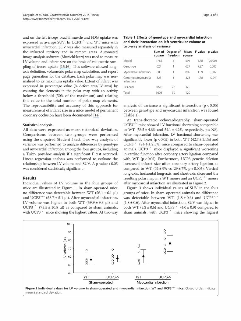
Table 1 Effects of genotype and myocardial infarctionand their interaction on left ventricular volume attwo-way analysis of variance
Sum ofsquare
Degree offreedom
Meansquare
F-value p-value
Model 1782 3 594 8.78 0.0003
Genotype 627 1 627 9.27 0.005
Myocardial infarction 805 1 805 11.9 0.002
Genotype/myocardialinfarction
323 1 323 4.78 0.04
Residual 1826 27 68
Total 3608 30 120
Gargiulo et al. BMC Cardiovascular Disorders 2014, 14:98 Page 3 of 7http://www.biomedcentral.com/1471-2261/14/98
and on the left triceps brachii muscle and FDG uptake wasexpressed as average SUV. In UCP3−/− and WT mice withmyocardial infarction, SUV was also measured separately inthe infarcted territory and in remote areas. Automatedimage analysis software (MunichHeart) was used to measureLV volume and infarct size on the basis of volumetric sam-pling of tracer uptake [15,16]. This software allowed long-axis definition, volumetric polar map calculation, and reportpage generation for the database. Each polar map was nor-malized to its maximum uptake value. Extent of infarct wasexpressed in percentage value (% defect area/LV area) bycounting the elements in the polar map with an activitybelow a threshold (50% of the maximum) and relatingthis value to the total number of polar map elements.The reproducibility and accuracy of this approach formeasurement of infarct size in a mice model of permanentcoronary occlusion have been documented [14].
Statistical analysisAll data were expressed as mean ± standard deviation.Comparisons between two groups were performedusing the unpaired Student t test. Two-way analysis ofvariance was performed to analyze differences by genotypeand myocardial infarction among the four groups, includinga Tukey post-hoc analysis if a significant F test occurred.Linear regression analysis was performed to evaluate therelationship between LV volume and SUV. A p value < 0.05was considered statistically significant.
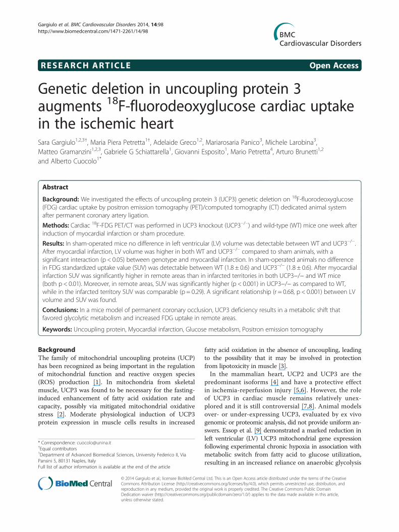
ResultsIndividual values of LV volume in the four groups ofmice are illustrated in Figure 1. In sham-operated miceno difference was detectable between WT (56.1 ± 6.1 μl)and UCP3−/− (58.7 ± 5.1 μl). After myocardial infarction,LV volume was higher in both WT (59.9 ± 9.3 μl) andUCP3−/− (75.5 ± 10.8 μl) as compared to sham animals,with UCP3−/− mice showing the highest values. At two-way
Figure 1 Individual values for LV volume in sham-operated and mymean ± standard deviation.
analysis of variance a significant interaction (p < 0.05)between genotype and myocardial infarction was found(Table 1).At trans-thoracic echocardiography, sham-operated
UCP3−/− mice showed LV fractional shortening comparableto WT (56.5 ± 4.6% and 54.1 ± 4.2%, respectively, p =NS).After myocardial infarction, LV fractional shortening wassignificantly lower (p < 0.05) in both WT (42.7 ± 3.1%) andUCP3−/− (24.4 ± 2.5%) mice compared to sham-operatedanimals. UCP3−/− mice displayed a significant worseningin cardiac function after coronary artery ligation comparedwith WT (p < 0.05). Furthermore, UCP3 genetic deletionincreased infarct size after coronary artery ligation ascompared to WT (44 ± 9% vs. 29 ± 7%, p < 0.005). Verticallong-axis, horizontal long-axis, and short-axis slices and theresulting polar map in a WT mouse and an UCP3−/− mouseafter myocardial infarction are illustrated in Figure 2.Figure 3 shows individual values of SUV in the four
groups of mice. In sham-operated animals no differencewas detectable between WT (1.8 ± 0.6) and UCP3−/−
(1.8 ± 0.6). After myocardial infarction, SUV was higher inboth WT (2.2 ± 0.6) and UCP3−/− (4.0 ± 0.9) compared tosham animals, with UCP3−/− mice showing the highest
ocardial infarction WT and UCP3−/− mice. Closed circles indicate
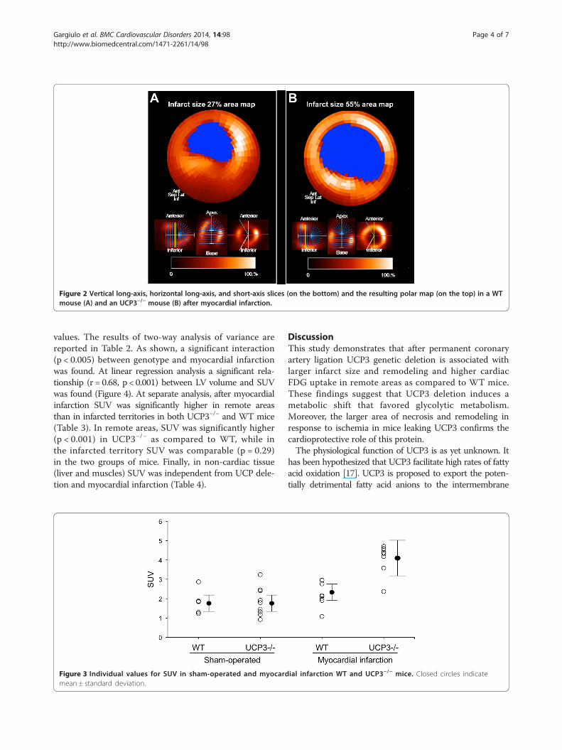
Figure 2 Vertical long-axis, horizontal long-axis, and short-axis slices (on the bottom) and the resulting polar map (on the top) in a WTmouse (A) and an UCP3−/− mouse (B) after myocardial infarction.
Gargiulo et al. BMC Cardiovascular Disorders 2014, 14:98 Page 4 of 7http://www.biomedcentral.com/1471-2261/14/98
values. The results of two-way analysis of variance arereported in Table 2. As shown, a significant interaction(p < 0.005) between genotype and myocardial infarctionwas found. At linear regression analysis a significant rela-tionship (r = 0.68, p < 0.001) between LV volume and SUVwas found (Figure 4). At separate analysis, after myocardialinfarction SUV was significantly higher in remote areasthan in infarcted territories in both UCP3−/− and WT mice(Table 3). In remote areas, SUV was significantly higher(p < 0.001) in UCP3−/− as compared to WT, while inthe infarcted territory SUV was comparable (p = 0.29)in the two groups of mice. Finally, in non-cardiac tissue(liver and muscles) SUV was independent from UCP dele-tion and myocardial infarction (Table 4).
Figure 3 Individual values for SUV in sham-operated and myocardmean ± standard deviation.
DiscussionThis study demonstrates that after permanent coronaryartery ligation UCP3 genetic deletion is associated withlarger infarct size and remodeling and higher cardiacFDG uptake in remote areas as compared to WT mice.These findings suggest that UCP3 deletion induces ametabolic shift that favored glycolytic metabolism.Moreover, the larger area of necrosis and remodeling inresponse to ischemia in mice leaking UCP3 confirms thecardioprotective role of this protein.The physiological function of UCP3 is as yet unknown. It
has been hypothesized that UCP3 facilitate high rates of fattyacid oxidation [17]. UCP3 is proposed to export the poten-tially detrimental fatty acid anions to the intermembrane
ial infarction WT and UCP3−/− mice. Closed circles indicate
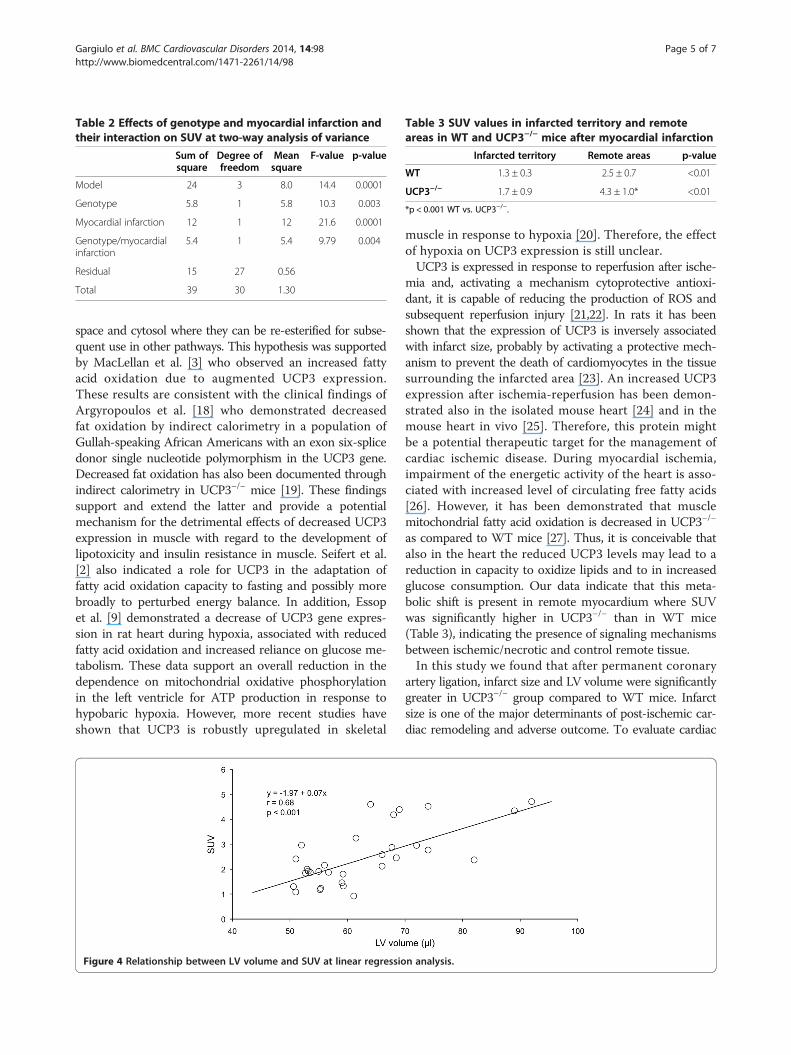
Table 2 Effects of genotype and myocardial infarction andtheir interaction on SUV at two-way analysis of variance
Sum ofsquare
Degree offreedom
Meansquare
F-value p-value
Model 24 3 8.0 14.4 0.0001
Genotype 5.8 1 5.8 10.3 0.003
Myocardial infarction 12 1 12 21.6 0.0001
Genotype/myocardialinfarction
5.4 1 5.4 9.79 0.004
Residual 15 27 0.56
Total 39 30 1.30
Table 3 SUV values in infarcted territory and remoteareas in WT and UCP3−/− mice after myocardial infarction
Infarcted territory Remote areas p-value
WT 1.3 ± 0.3 2.5 ± 0.7 <0.01
UCP3−/− 1.7 ± 0.9 4.3 ± 1.0* <0.01
*p < 0.001 WT vs. UCP3−/−.
Gargiulo et al. BMC Cardiovascular Disorders 2014, 14:98 Page 5 of 7http://www.biomedcentral.com/1471-2261/14/98
space and cytosol where they can be re-esterified for subse-quent use in other pathways. This hypothesis was supportedby MacLellan et al. [3] who observed an increased fattyacid oxidation due to augmented UCP3 expression.These results are consistent with the clinical findings ofArgyropoulos et al. [18] who demonstrated decreasedfat oxidation by indirect calorimetry in a population ofGullah-speaking African Americans with an exon six-splicedonor single nucleotide polymorphism in the UCP3 gene.Decreased fat oxidation has also been documented throughindirect calorimetry in UCP3−/− mice [19]. These findingssupport and extend the latter and provide a potentialmechanism for the detrimental effects of decreased UCP3expression in muscle with regard to the development oflipotoxicity and insulin resistance in muscle. Seifert et al.[2] also indicated a role for UCP3 in the adaptation offatty acid oxidation capacity to fasting and possibly morebroadly to perturbed energy balance. In addition, Essopet al. [9] demonstrated a decrease of UCP3 gene expres-sion in rat heart during hypoxia, associated with reducedfatty acid oxidation and increased reliance on glucose me-tabolism. These data support an overall reduction in thedependence on mitochondrial oxidative phosphorylationin the left ventricle for ATP production in response tohypobaric hypoxia. However, more recent studies haveshown that UCP3 is robustly upregulated in skeletal
Figure 4 Relationship between LV volume and SUV at linear regressio
muscle in response to hypoxia [20]. Therefore, the effectof hypoxia on UCP3 expression is still unclear.UCP3 is expressed in response to reperfusion after ische-
mia and, activating a mechanism cytoprotective antioxi-dant, it is capable of reducing the production of ROS andsubsequent reperfusion injury [21,22]. In rats it has beenshown that the expression of UCP3 is inversely associatedwith infarct size, probably by activating a protective mech-anism to prevent the death of cardiomyocytes in the tissuesurrounding the infarcted area [23]. An increased UCP3expression after ischemia-reperfusion has been demon-strated also in the isolated mouse heart [24] and in themouse heart in vivo [25]. Therefore, this protein mightbe a potential therapeutic target for the management ofcardiac ischemic disease. During myocardial ischemia,impairment of the energetic activity of the heart is asso-ciated with increased level of circulating free fatty acids[26]. However, it has been demonstrated that musclemitochondrial fatty acid oxidation is decreased in UCP3−/−
as compared to WT mice [27]. Thus, it is conceivable thatalso in the heart the reduced UCP3 levels may lead to areduction in capacity to oxidize lipids and to in increasedglucose consumption. Our data indicate that this meta-bolic shift is present in remote myocardium where SUVwas significantly higher in UCP3−/− than in WT mice(Table 3), indicating the presence of signaling mechanismsbetween ischemic/necrotic and control remote tissue.In this study we found that after permanent coronary
artery ligation, infarct size and LV volume were significantlygreater in UCP3−/− group compared to WT mice. Infarctsize is one of the major determinants of post-ischemic car-diac remodeling and adverse outcome. To evaluate cardiac
n analysis.
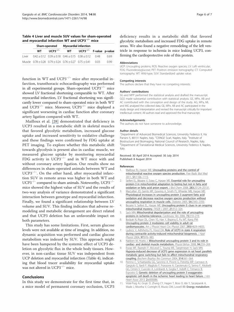
Table 4 Liver and muscle SUV values for sham-operatedand myocardial infarction WT and UCP3−/− mice
Sham-operated Myocardial infarction
WT UCP3−/− WT UCP3−/− F-value p-value
Liver 0.42 ± 0.12 0.39 ± 0.18 0.46 ± 0.15 0.38 ± 0.12 0.48 0.69
Muscle 0.78 ± 0.28 0.79 ± 0.24 0.76 ± 0.27 0.75 ± 0.41 0.03 0.99
Gargiulo et al. BMC Cardiovascular Disorders 2014, 14:98 Page 6 of 7http://www.biomedcentral.com/1471-2261/14/98
function in WT and UCP3−/− mice after myocardial in-farction, transthoracic echocardiography was performedin all experimental groups. Sham-operated UCP3−/− miceshowed LV fractional shortening comparable to WT. Aftermyocardial infarction, LV fractional shortening was signifi-cantly lower compared to sham-operated mice in both WTand UCP3−/− mice. Moreover, UCP3−/− mice displayed asignificant worsening in cardiac function after coronaryartery ligation compared with WT.Mailloux et al. [28] demonstrated that deficiency in
UCP3 resulted in a metabolic shift in skeletal musclesthat favored glycolytic metabolism, increased glucoseuptake and increased sensitivity to oxidative challengeand these findings were confirmed by FDG uptake atPET imaging. To explore whether this metabolic shifttowards glycolysis is present also in cardiac muscle, wemeasured glucose uptake by monitoring myocardialFDG activity in UCP3−/− and in WT mice with andwithout coronary artery ligation. Our results show nodifferences in sham-operated animals between WT andUCP3−/−. On the other hand, after myocardial infarc-tion SUV in remote areas was higher in both WT andUCP3−/− compared to sham animals. Noteworthy, UCP3−/−
mice showed the highest value of SUV and the results oftwo-way analysis of variance demonstrated a significantinteraction between genotype and myocardial infarction.Finally, we found a significant relationship between LVvolume and SUV. This finding indicates that adverse re-modeling and metabolic derangement are direct relatedand that UCP3 deletion has an unfavorable impact onboth parameters.This study has some limitations. First, serum glucose
levels were not available at time of imaging. In addition, nodynamic acquisition was performed and cardiac glucosemetabolism was indexed by SUV. This approach mighthave been hampered by the systemic effect of UCP3 de-letion on glycolytic flux in the whole body tissues. How-ever, in non-cardiac tissue SUV was independent fromUCP deletion and myocardial infarction (Table 4), indicat-ing that blood tracer availability for myocardial uptakewas not altered in UCP3−/− mice.
ConclusionsIn this study we demonstrate for the first time that, ina mice model of permanent coronary occlusion, UCP3
deficiency results in a metabolic shift that favoredglycolytic metabolism and increased FDG uptake in remoteareas. We also found a negative remodeling of the left ven-tricle in response to ischemia in mice leaking UCP3, con-firming the cardioprotective role of this protein.
AbbreviationsUCP: Uncoupling proteins; ROS: Reactive oxygen species; LV: Left ventricular;FDG: Fluorodeoxyglucose; PET: Positron emission tomography; CT: Computedtomography; WT: Wild-type; SUV: Standardized uptake value.
Competing interestsThe authors declare that they have no competing interests.
Authors’ contributionsSG and MPP performed the statistical analysis and drafted the manuscript.GGS made substantial contribution with statistical analysis. GE, MPe, AB andAC contributed with the conception and design of the study. AG, MPa, MLand MG analyzed the collected data. GE, MPe, AB and AC participated in thestudy design and interpretation and revised the manuscript critically for importantintellectual content. All authors read and approved the final manuscript.
AcknowledgementsThe authors do not have someone to acknowledge.
Author details1Department of Advanced Biomedical Sciences, University Federico II, ViaPansini 5, 80131 Naples, Italy. 2CEINGE Scarl, Naples, Italy. 3Institute ofBiostructure and Bioimaging, National Council of Research, Naples, Italy.4Department of Translational Medical Sciences, University Federico II, Naples,Italy.
Received: 30 April 2014 Accepted: 30 July 2014Published: 8 August 2014
References1. Mailloux RJ, Harper ME: Uncoupling proteins and the control of
mitochondrial reactive oxygen species production. Free Radic Biol Med2011, 51:1106–1115.
2. Seifert EL, Bézaire V, Estey C, Harper ME: Essential role for uncouplingprotein-3 in mitochondrial adaptation to fasting but not in fatty acidoxidation or fatty acid anion export. J Biol Chem 2008, 283:25124–25131.
3. MacLellan JD, Gerrits MF, Gowing A, Smith PJ, Wheeler MB, Harper ME:Physiological increases in uncoupling protein 3 augment fatty acidoxidation and decrease reactive oxygen species production withoutuncoupling respiration in muscle cells. Diabetes 2005, 54:2343–2350.
4. Bezaire V, Seifert EL, Harper ME: Uncoupling protein-3: clues in an ongoingmitochondrial mystery. FASEB J 2007, 21:312–324.
5. Sack MN: Mitochondrial depolarization and the role of uncouplingproteins in ischemia tolerance. Cardiovasc Res 2006, 72:210–219.
6. Bodyak N, Rigor DL, Chen YS, Han Y, Bisping E, Pu WT, Kang PM:Uncoupling protein 2 modulates cell viability in adult ratcardiomyocytes. Am J Physiol Heart Circ Physiol 2007, 293:H829–H835.
7. Ljubicic V, Adhihetty PJ, Hood DA: Role of UCP3 in state 4 respirationduring contractile activity-induced mitochondrial biogenesis. J ApplPhysiol 2004, 97:976–983.
8. Nabben M, Hoeks J: Mitochondrial uncoupling protein 3 and its role incardiac- and skeletal muscle metabolism. Physiol Behav 2008, 94:259–269.
9. Essop MF, Razeghi P, McLeod C, Young ME, Taegtmeyer H, Sack MN:Hypoxia-induced decrease of UCP3 gene expression in rat heart parallelsmetabolic gene switching but fails to affect mitochondrial respiratorycoupling. Biochem Biophys Res Commun 2004, 314:561–564.
10. Perrino C, Schiattarella GG, Sannino A, Pironti G, Petretta MP, Cannavo A,Gargiulo G, Ilardi F, Magliulo F, Franzone A, Carotenuto G, Serino F, AltobelliGG, Cimini V, Cuocolo A, Lombardi A, Goglia F, Indolfi C, Trimarco B,Esposito G: Genetic deletion of uncoupling protein 3 exaggeratesapoptotic cell death in the ischemic heart leading to heart failure. J AmHeart Assoc 2013, 2:e000086.
11. Vidal-Puig AJ, Grujic D, Zhang CY, Hagen T, Boss O, Ido Y, Szczepanik A,Wade J, Mootha V, Cortright R, Muoio DM, Lowell BB: Energy metabolism
Gargiulo et al. BMC Cardiovascular Disorders 2014, 14:98 Page 7 of 7http://www.biomedcentral.com/1471-2261/14/98
in uncoupling protein 3 gene knockout mice. J Biol Chem 2000,275:16258–16266.
12. Curcio A, Noma T, Naga Prasad SV, Wolf MJ, Lemaire A, Perrino C, Mao L,Rockman HA: Competitive displacement of phosphoinositide 3-kinase frombeta-adrenergic receptor kinase-1 improves postinfarction adverse myocardialremodeling. Am J Physiol Heart Circ Physiol 2006, 291:H1754–H1760.
13. Esposito G, Perrino C, Cannavo A, Schiattarella GG, Borgia F, Sannino A,Pironti G, Gargiulo G, Di Serafino L, Franzone A, Scudiero L, Grieco P, Indolfi C,Chiariello M: EGFR trans-activation by urotensin II receptor is mediated bybeta-arrestin recruitment and confers cardioprotection in pressure overloadinduced cardiac hypertrophy. Basic Res Cardiol 2011, 106:577–589.
14. Greco A, Petretta MP, Larobina M, Gargiulo S, Panico M, Nekolla SG, EspositoG, Petretta M, Brunetti A, Cuocolo A: Reproducibility and accuracy ofnoninvasive measurement of infarct size in mice with high-resolutionPET/CT. J Nucl Cardiol 2012, 19:492–499.
15. Higuchi T, Nekolla SG, Jankaukas A, Weber AW, Huisman MC, Reder S,Ziegler SI, Schwaiger M, Bengel FM: Characterization of normal andinfarcted rat myocardium using a combination of small-animal PET andclinical MRI. J Nucl Med 2007, 48:288–294.
16. Nekolla SG, Miethaner C, Nguyen N, Ziegler SI, Schwaiger M:Reproducibility of polar map generation and assessment of defectseverity and extent assessment in myocardial perfusion imaging usingpositron emission tomography. Eur J Nucl Med 1998, 25:1313–1321.
17. Himms-Hagen J, Harper ME: Physiological role of UCP3 may be export offatty acids from mitochondria when fatty acid oxidation predominates:an hypothesis. Exp Biol Med 2001, 226:78–84.
18. Argyropoulos G, Brown AM, Willi SM, Zhu J, He Y, Reitman M, Gevao SM,Spruill I, Garvey WT: Effects of mutations in the human uncouplingprotein 3 gene on the respiratory quotient and fat oxidation in severeobesity and type 2 diabetes. J Clin Invest 1998, 102:1345–1351.
19. Bezaire V, Hofmann W, Kramer JK, Kozak LP, Harper ME: Effects of fasting onmuscle mitochondrial energetics and fatty acid metabolism in Ucp3−/− andwild-type mice. Am J Physiol Endocrinol Metab 2001, 281:E975–E982.
20. Lu Z, Sack MN: ATF-1 is a hypoxia-responsive transcriptional activator ofskeletal muscle mitochondrial-uncoupling protein 3. J Biol Chem 2008,283:23410–23418.
21. Cadenas S, Aragonés J, Landàzuri MO: Mitochondrial reprogrammingthrough cardiac oxygen sensor in ischemic heart disease. Cardiovasc Res2010, 88:219–228.
22. Safari F, Anvari Z, Moshtaghioun S, Javan M, Bayat G, Forosh SS,Hekmatimoghaddam S: Differential expression of cardiac uncouplingproteins 2 and 3 in response to myocardial ischemia-reperfusion in rats.Life Sci 2014, 98:68–74.
23. Laskowski KR, Russell RR 3rd: Uncoupling proteins in heart failure.Curr Heart Fail Rep 2008, 5:75–79.
24. Anedda A, López-Bernardo E, Acosta-Iborra B, Saadeh Suleiman M, LandázuriMO, Cadenas S: The transcription factor Nrf2 promotes survival byenhancing the expression of uncoupling protein 3 under conditions ofoxidative stress. Free Radic Biol Med 2013, 61C:395–407.
25. Ozcan C, Palmeri M, Horvath TL, Russell KS, Russell RR 3rd: Role ofuncoupling protein 3 in ischemia-reperfusion injury, arrhythmias, andpreconditioning. Am J Physiol Heart Circ Physiol 2013, 304:H1192–H1200.
26. Lopaschuk G: The role of fatty acid oxidation in cardiac ischemia andreperfusion. Adv Stud Med 2004, 4:S803–S807.
27. Senese R, Valli V, Moreno M, Lombardi A, Busiello RA, Cioffi F, Silvestri E,Goglia F, Lanni A, de Lange P: Uncoupling protein 3 expression levelsinfluence insulin sensitivity, fatty acid oxidation, and related signalingpathways. Pflugers Arch 2011, 461:153–164.
28. Mailloux RJ, Dumouchel T, Aguer C, de Kemp R, Beanlands R, Harper ME:Hexokinase II acts through UCP3 to suppress mitochondrial reactiveoxygen species production and maintain aerobic respiration.Biochem J 2011, 437:301–311.
doi:10.1186/1471-2261-14-98Cite this article as: Gargiulo et al.: Genetic deletion in uncouplingprotein 3 augments 18F-fluorodeoxyglucose cardiac uptake in theischemic heart. BMC Cardiovascular Disorders 2014 14:98.
Submit your next manuscript to BioMed Centraland take full advantage of:
• Convenient online submission
• Thorough peer review
• No space constraints or color figure charges
• Immediate publication on acceptance
• Inclusion in PubMed, CAS, Scopus and Google Scholar
• Research which is freely available for redistribution
Submit your manuscript at www.biomedcentral.com/submit