detection of aortic graft infection by fluorodeoxyglucose positron emission tomography: comparison...
TRANSCRIPT
Detection of aortic graft infection byfluorodeoxyglucose positron emissiontomography: Comparison withcomputed tomographic findingsKazuki Fukuchi, MD,a,b Yoshio Ishida, MD,b Masahiro Higashi, MD,b Tomohiro Tsunekawa, MD,c
Hitoshi Ogino, MD,c Kenji Minatoya, MD,c Keisuke Kiso, MD,b and Hiroaki Naito, MD,b Osaka, Japan
Objective: Radionuclide imaging with fluorodeoxyglucose (FDG) and positron emission tomography (PET) has beenproposed for the identification of vascular graft infection; however, its accuracy has not been determined. We performedthis prospective study to compare the usefulness of FDG-PET in the assessment of vascular graft infection relative tocomputed tomography (CT).Methods: FDG-PET was performed for 33 consecutive patients with a suspected arterial prosthetic graft infection. ThePET images were then assessed visually in terms of the density of uptake. In cases with positive uptake, the pattern ofaccumulation was also defined, such as focal or diffuse uptake. We compared the diagnostic efficiency of PET withcontemporaneous CT in detection of infection of the arterial prosthetic graft.Results: On the basis of the surgical, microbiological, and clinical follow-up findings, the aortic grafts were consideredinfected in 11 patients and not infected in 22 patients. Although the sensitivity of PET (91%) was higher than that of CT(64%), its specificity (64%) was lower than that of CT (86%). When focal uptake was set as the positive criterion in FDG,the specificity and positive predictive value of PET for the diagnosis of aortic graft infection improved significantly to 95%(P < .05 for both).Conclusions: Although both techniques are useful in evaluation of patients with suspected aortic graft infection, using thecharacteristic FDG uptake pattern described previously as a diagnostic criterion made the efficacy of FDG superior to that
of CT in the diagnostic assessment of patients with suspected aortic graft infection. (J Vasc Surg 2005;42:919-25.)Aortic prosthetic graft infection is associated with highmorbidity and mortality in the absence of immediate, de-finitive antibiotic therapy and surgical intervention.1 Com-puted tomography (CT) has been used as a complementaryimaging approach for the assessment of graft infection,because the high spatial resolution of CT provides exquisitedetail of the vascular structure and perivascular spaces.However, hematomas and seromas in the vicinity of avascular graft appear anatomically similar to an abscess, thusmaking it sometimes difficult to distinguish between non-infected and infected prosthetic grafts on CT images.2
Thus, a reliable physiological approach is required to eval-uate the inflammatory activity, to detect infected prosthe-ses, and to determine the extent of infection, in addition toanatomic information.
Investigators have recently suggested that fluorodeoxy-glucose (FDG) positron emission tomography (PET) im-aging may be useful for detection of infection3,4 and eval-
From the Department of Nuclear Medicine, Osaka Medical Center forCancer and Cardiovascular Diseases,a and Departments of Radiologyb andCardiovascular Surgery,c National Cardiovascular Center.
Competition of interest: none.Reprint requests: Kazuki Fukuchi, MD, Department of Nuclear Medicine,
Osaka Medical Center for Cancer and Cardiovascular Diseases, 1-3-3Nakamichi, Higashinari-ku, Osaka 565-8511, Japan (e-mail: [email protected]).
0741-5214/$30.00Copyright © 2005 by The Society for Vascular Surgery.
doi:10.1016/j.jvs.2005.07.038uation of infected vascular grafts.5-7 However, because thevascular graft regions can exhibit a substantial inflammatoryreaction that results in some FDG accumulation,8 the valueof FDG-PET in the assessment of vascular graft infection isstill ambiguous. We hypothesized that PET, because of itsability to detect inflammation, would be a more sensitiveand specific test for aortic graft infection. To clarify thisissue, we conducted this preliminary study to examine thefeasibility of using FDG-PET for the diagnosis of aorticgraft infection in comparison with CT.
METHODS
Patient recruitment. This study was a prospective anal-ysis of consecutive patients undergoing combined FDG-PETand CT between September 2002 and November 2004 inNational Cardiovascular Center Hospital. Thirty-three pa-tients who underwent aortic reconstructive surgery with graft-ing for the treatment of aortic aneurysm, aortic dissection, oraortoiliac occlusive disease (mean age, 71 � 14 years [mean� SD]; age range, 22-83 years) were enrolled in the study.These patients were categorized into three groups accord-ing to the criteria of Fiorani et al.9 Nine patients hadsuspected advanced aortic graft infection with manifesta-tions of severe infection, 17 had suspected low-grade aorticgraft infection with nonspecific manifestations of infection,and 7 were asymptomatic control patients selected from areview of patients at our institution who had undergone
FDG whole-body PET mainly for oncologic purposes dur-919
ction;
JOURNAL OF VASCULAR SURGERYNovember 2005920 Fukuchi et al
ing the same period. The final diagnosis was based on thesurgical and microbiological findings. In cases in which nosurgical treatment or no microbiologic samples were avail-able, clinical follow-up for more than 4 months served asthe standard reference. Twenty-five patients had receivedsome antibiotic therapy before the FDG-PET and CTstudy, but the drugs were not changed until after bothimaging studies. Patients with diabetes mellitus were ex-cluded from the study because this condition can affect theFDG uptake.10 This study was conducted with the approvalof our institutional review board, and we obtained writteninformed consent from all the subjects before their partic-ipation in the study.
CT protocol. CT scanning was conducted within 1week before FDG-PET by using a helical or multidetectorCT system (Aquilion; Toshiba Medical Co, Tochigi, Ja-pan). Contiguous 1-cm sections were obtained at 1-cmintervals from the lung apices to the inguinal region afterbolus intravenous injection of contrast material, except in
Table I. Patient characteristics
PatientNo. Age/sex
Primarydisease
Type ofoperation
1 75/M AAA Y-graft2 74/M TAA TAR3 80/M AAA I-F bypass4 62/M TAA TAR5 78/M AAA Y-graft6 83/F IA TAR7 83/M AAA Y-graft8 74/M TAA TAR9 22/M AE Ascending graft
10 31/M AD TAR11 80/M TAA TAR12 75/M IA S-graft13 70/M AAA Y-graft14 77/F AAA Y-graft15 77/M AAA Y-graft16 75/M AAA A-F bypass17 64/M AAA Y-graft18 82/F AAA S-graft19 83/M TAA TAR20 73/M AAA Y-graft21 63/M AAA Y-graft22 66/M AAA Y-graft23 72/M AAA Y-graft24 76/M AAA Y-graft25 75/M AAA Y-graft26 68/F AAA A-F bypass27 80/M AAA Y-graft28 76/M AAA Y-graft29 83/M TAA Descending gra30 75/M AD Descending gra31 49/M TAA Descending gra32 82/M TAA TAR33 72/M TAA Descending gra
CT, Computed tomography; PET, positron emission tomography; AAA, abdTAA, thoracic aortic aneurysm; TAR, total arch replacement; MF, microbectasia; AD, aortic dissection; S-graft, straight graft; NEI, no evidence of infepositive.
four patients with acute or chronic renal failure.
PET protocol. Patients were instructed to fast for atleast 5 hours before FDG-PET studies. Transmissionscans were initially obtained by using a line source ofgermanium 68/gallium 68, and then 185 MBq of FDGwas administered intravenously. One hour later, emis-sion from the entire body was imaged for 25 minutes; atleast five bed positions were used. The emission dataobtained from the ECAT EXACT 47 (Siemens/CTI,Knoxville, Tenn) were consecutively reconstructed withmeasured attenuation correction based on the transmis-sion data.
Image interpretation. All the CT scans were re-viewed independently by two of the authors, who had noknowledge of the clinical or operative findings. In accor-dance with a previously described method,11 each CTscan was assessed for the presence of ectopic gas, peri-graft fluid (�20 Hounsfield Units), perigraft soft tissue(�20 Hounsfield Units), pseudoaneurysm formation,discontinuity of the aneurysmal wrap, and an increased
Finaldiagnosis
Proof ofinfection
CTfindings
PETfindings
Infection ST TP TPInfection MF FN TPInfection ST TP TPInfection ST FN TPInfection ST TP TPInfection ST FN TPInfection MF TP TPInfection MF FN TPInfection MF TP TPInfection CF FN TPInfection CF TP TPNEI CF TN TNNEI AsC TN FPNEI AsC TN TNNEI CF TN TNNEI CF TN TNNEI ST FP TNNEI AsC TN TNNEI ST FP TNNEI CF TN TNNEI AsC TN FPNEI CF TN TNNEI CF TN FPNEI AsC TN FPNEI CF FP TNNEI AsC TN FPNEI ST TN TNNEI CF TN FPNEI CF TN TNNEI CF TN TNNEI ST TN TNNEI AsC TN FPNEI CF TN FP
al aortic aneurysm; I-F, iliofemoral; ST, surgical treatment; TP, true positive;ical findings; FN, false negative; IA, infected aneurysm; AE, annuloaorticTN, true negative; CF, clinical feature; AsC, asymptomatic controls; FP, false
ftftft
ft
ominiolog
amount of soft tissue (�5 mm) between the graft and the

JOURNAL OF VASCULAR SURGERYVolume 42, Number 5 Fukuchi et al 921
surrounding aneurysmal wrap. If the interpretations ofthe observers disagreed, a consensus of the two observerswas obtained.
The FDG-PET images were analyzed by two indepen-dent experienced physicians who specialized in nuclearmedicine and who were blinded to the results of the otherimaging studies. They used the same image-evaluationcriteria as those used in a previous FDG-PET study fordetection of orthopedic infection.12 The intensity of FDGuptake was graded on a five-point scale, as follows: grade 0,FDG uptake similar to that in the background; grade 1, lowFDG uptake, comparable to that by inactive muscles and fat;grade 2, moderate FDG uptake, clearly visible and dis-tinctly higher than the uptake by inactive muscles and fat;grade 3, strong FDG uptake, but distinctly less than thephysiologic uptake by the bladder; and grade 4, very strongFDG uptake, comparable to the physiologic urinary uptakeby the bladder. In a previous investigation by Stumpeet al,12 the results of a receiver operating characteristicanalysis had shown that classifying lesions with grade 3 or 4uptake as infected lesions yielded the best discriminationbetween infected and noninfected lesions. Therefore, in-creased grade 3 or 4 FDG uptake by a prosthesis was used asthe diagnostic criterion for infection in our study. In addi-tion, in cases showing abnormal FDG uptake, the readersalso described the pattern of abnormal uptake, namely,whether it was focal or diffuse. An abnormality was inter-preted as diffuse if it was located along the prosthesisconsecutively. An abnormality was called focal if it waslocated in a region other than along the prosthesis and wasdotted in configuration.
Statistical analysis. Data are expressed as means �SD. The interobserver agreement of image interpretationwas estimated by using the � statistic.13 Concordance wasconsidered to be good for � values more than 0.6, moder-ate for values from 0.6 to 0.4, and poor for values less than0.4.14 Comparisons between groups were conducted byusing the unpaired t test for continuous variables. Thediagnostic performance was expressed in terms of the sensitiv-ity, specificity, accuracy, positive predictive value (PPV), and
Table II. Demographic data of patients withand without aortic graft infection
Variable
Group
P valueWith infection Without infection
Age (y) 68 � 21 73 � 8 NSSex (F/M) 1/10 3/19 NSSmoking 7/11 (64%) 15/22 (68%) NSCRP (mg/dL) 5.6 � 5.0 3.5 � 4.5 NSFBS (mg/dL) 100 � 10 97 � 14 NSDuration (mo) 11.8 � 9.4 12.5 � 9.2 NSLocation of graft
(thorax/abdomen) 7/4 6/16 .04
Data are n (%) or mean � SD.CRP, C-reactive protein; FBS, fasting blood sugar; NS, not significant.
negative predictive value (NPV), with 95% confidence in-
tervals. Differences in the diagnostic performance betweenthe two imaging modalities and criteria were consideredsignificant when the 95% confidence intervals did not over-lap.15
RESULTS
The characteristics of patients and the results of imag-ing are presented in Table I. Eleven of 33 patients weredefinitively categorized as having infected grafts on thebasis of the surgical procedure, including graft removal,aortic ligation, and extra-anatomic bypass grafting (n � 5),and microbiological findings, including blood culture andfollow-up imaging studies (n � 6). Another 22 patientswere definitively categorized in the noninfected-graftgroup. There were no significant differences between thetwo groups in the patients’ mean age, sex distribution,
Fig 1. Example of true-positive findings on computed tomogra-phy (CT) and fluorodeoxyglucose (FDG) positron emission to-mography in a patient with graft infection. The patient had under-gone total aortic arch replacement 4 months before the imagingexaminations. Focal FDG accumulation in the graft (black arrows)is accompanied by fluid collection and extraluminal air on theenhanced CT image (white arrows).
history of smoking, serum C-reactive protein, or blood
JOURNAL OF VASCULAR SURGERYNovember 2005922 Fukuchi et al
sugar at the time of the PET study or during follow-up aftersurgery. However, the incidence of infection in thoracicaortic grafts was significantly higher than that in abdominalgrafts (Table II).
Regarding the interpretation of CT images, the inter-observer agreement on positive observations was 0.85, andthe � value was 0.67. CT revealed true-positive findings in7 cases and false-positive findings in 3, and it showed atrue-negative result in 19 and a false-negative result in 4.
All FDG-PET images were considered appropriate forinterpretation. Interobserver agreement on positive obser-vations was 0.82, and the � value was 0.61. FDG-PETshowed true-positive findings in 10 cases and false-positivefindings in 8, and it showed a true-negative result in 14 anda false-negative result in one. All patients with true-positivefindings on CT also showed increased FDG uptake by thecorresponding lesions (Fig 1). Additionally, in all cases withfalse-negative CT findings, FDG-PET could depict the
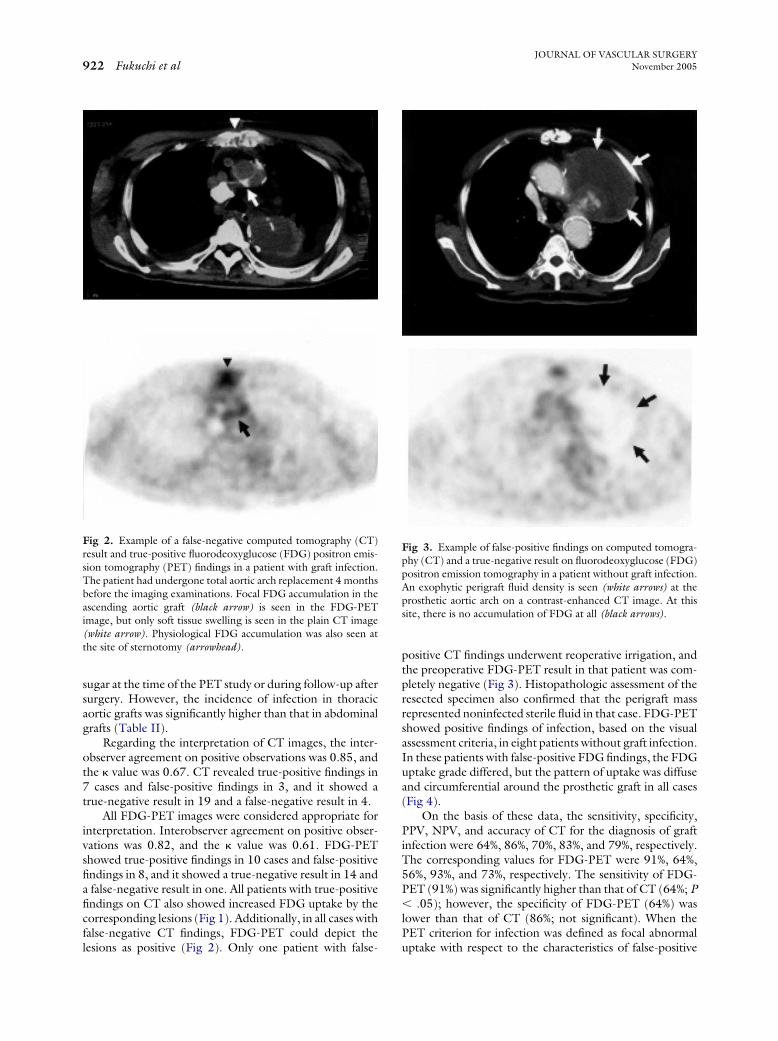
Fig 2. Example of a false-negative computed tomography (CT)result and true-positive fluorodeoxyglucose (FDG) positron emis-sion tomography (PET) findings in a patient with graft infection.The patient had undergone total aortic arch replacement 4 monthsbefore the imaging examinations. Focal FDG accumulation in theascending aortic graft (black arrow) is seen in the FDG-PETimage, but only soft tissue swelling is seen in the plain CT image(white arrow). Physiological FDG accumulation was also seen atthe site of sternotomy (arrowhead).
lesions as positive (Fig 2). Only one patient with false-
positive CT findings underwent reoperative irrigation, andthe preoperative FDG-PET result in that patient was com-pletely negative (Fig 3). Histopathologic assessment of theresected specimen also confirmed that the perigraft massrepresented noninfected sterile fluid in that case. FDG-PETshowed positive findings of infection, based on the visualassessment criteria, in eight patients without graft infection.In these patients with false-positive FDG findings, the FDGuptake grade differed, but the pattern of uptake was diffuseand circumferential around the prosthetic graft in all cases(Fig 4).
On the basis of these data, the sensitivity, specificity,PPV, NPV, and accuracy of CT for the diagnosis of graftinfection were 64%, 86%, 70%, 83%, and 79%, respectively.The corresponding values for FDG-PET were 91%, 64%,56%, 93%, and 73%, respectively. The sensitivity of FDG-PET (91%) was significantly higher than that of CT (64%; P� .05); however, the specificity of FDG-PET (64%) waslower than that of CT (86%; not significant). When thePET criterion for infection was defined as focal abnormal
Fig 3. Example of false-positive findings on computed tomogra-phy (CT) and a true-negative result on fluorodeoxyglucose (FDG)positron emission tomography in a patient without graft infection.An exophytic perigraft fluid density is seen (white arrows) at theprosthetic aortic arch on a contrast-enhanced CT image. At thissite, there is no accumulation of FDG at all (black arrows).
uptake with respect to the characteristics of false-positive

JOURNAL OF VASCULAR SURGERYVolume 42, Number 5 Fukuchi et al 923
FDG uptake, all the false-positive cases were categorized asnegative. With this alteration of the diagnostic criteria, thespecificity and PPV of FDG-PET improved significantlyfrom 64% to 95% and 56% from 91%, respectively (P � .05).An overview of the diagnostic performance of CT and ofFDG using the two methods of assessment, as well as the95% confidence intervals, is shown in Table III.
DISCUSSION
Because infection of aortic prosthetic grafts remains amajor surgical challenge, it is essential for aortic graftinfection to be diagnosed accurately and safely.1,2 CT andscintigraphic techniques are currently the most commonlyused modalities for diagnosis of aortic graft infection.2
Because of the rapidity with which it can be performed, CTshould be the first examination ordered in cases of sus-pected aortic graft infection. Studies of the early 1980s haveshowed specificity and sensitivity of CT of approximately100%.16 However, studies of the late 1980s, which in-
Fig 4. Example of a true-negative computed tomography (CT)result and false-positive fluorodeoxyglucose positron emission to-mography (FDG-PET) findings in a patient without graft infec-tion. The prosthetic aortic graft can be seen as a high-density rimaround the ascending aortic region on CT (white arrows). TheFDG-PET image depicts a relatively dense and diffuse uptakearound the graft (black arrows).
cluded cases with low-grade infection, indicated an overall
diagnostic sensitivity of 55.5% and an overall diagnosticspecificity of 100% of CT for graft infection.9 This imagingmodality was considered an accurate method for diagnosisof advanced graft infection (periprosthetic abscess, aor-toenteric fistula, and so on), but the risk of false-negativeresults is high in cases with low-grade graft infection. In thisstudy, CT failed to identify three cases with aortic graftinfection; consequently, the sensitivity of CT for the diag-nosis of aortic graft infection decreased to 64%. Thus, itwould be desirable to identify another imaging modality,besides CT, with a high sensitivity for detecting aortic graftinfection.
Whereas CT affords visualization of the structuralchanges secondary to infection, nuclear medicine tech-niques allow such infections to be diagnosed on the basis ofmolecular biological changes.2 Although several types ofscintigraphic techniques have been used for many years,FDG-PET has drawn much attention recently for diagnosisof infectious diseases. Previous articles have shown thatFDG-PET can be a very sensitive imaging modality fordiagnosis of infection,3,4 because increased FDG uptake isobserved in the activated inflammatory cells, such as leuko-cytes, granulocytes, and macrophages.17 Although somecase reports have been published,5-7 the usefulness of FDG-PET in the diagnosis of aortic graft infection has not yetbeen evaluated on sufficiently large numbers of patients.
This study confirmed the feasibility of FDG-PET fordetecting aortic graft infection. As compared with theconventional nuclear medicine techniques, PET is consid-ered to have the following advantages.4 First, the PETprocedure is much faster than the conventional modalities,and its results can usually be made available within 2 hours.Second, the better spatial resolution of the PET system ascompared with that of a gamma camera results in a higherdiagnostic sensitivity. Third, PET generally provides higher-quality images with superior contrast compared with singlephoton emission CT. Thus, FDG-PET can be used toassess the extent of infection more sensitively and canquantify the inflammatory activity more accurately than agamma camera. The superiority of FDG-PET comparedwith other gamma camera techniques has been confirmedin oncology, but not yet in infectious disease.18,19 Unfor-tunately, we could not compare FDG-PET with the con-ventional gamma camera imaging modalities in this study,but the above-mentioned advantages suggest that FDG-PET has the possibility to replace them as a diagnostic toolfor patients with suspected aortic graft infection.
Although the ability of FDG-PET to diagnose severaltypes of infection with a high sensitivity has been encour-aging, high sensitivity inevitably means a certain number offalse-positive results.4 Previous reports indicated that pros-thetic vascular graft replacement was sometimes associatedwith a false-positive FDG uptake in the graft or stentregions.8,20 Our visual FDG-PET analysis also showed afalse-positive accumulation in 8 (36%) of 22 patients withnoninfected grafts. This false-positive accumulation mightbe explained as FDG uptake during the process of normal
foreign body reaction or inflammation during the normal
JOURNAL OF VASCULAR SURGERYNovember 2005924 Fukuchi et al
postoperative course after reconstruction of the aorta.Other scintigraphic studies, including white blood cell orleukocyte scans, also showed a certain number of false-positive results.21,22 This inevitable reaction might confoundthe critical diagnosis of aortic graft infection. However, thesenormal inflammatory reactions might be distinguished frominfection by using the characteristic uptake patterns of FDGas diagnostic criteria, because nonspecific inflammation wasassociated with diffuse uptake along the prosthetic graft,whereas true infection was associated with focal or segmen-tal FDG uptake, mostly at sites of abnormal CT findings. Inthis study, eight cases showed various degrees of false-positive FDG uptake, but the pattern of uptake was diffusealong the prosthetic graft in all cases. This feature can be auseful marker for differentiating between infected and non-infected aortic grafts. In fact, using focal uptake as a diag-nostic criterion resulted in a statistically significant increasein the specificity and PPV of FDG-PET for the diagnosis ofgraft infection as compared with that of the conventionalvisual assessment. Using this focal sign may not always beversatile, because some grafts are entirely infected. In thistype of infection, the focal findings do not work and causemisdiagnosis. We emphasize that the use of a combinationof intensity and pattern of FDG uptake with reference tothe information of CT scans may allow one to clearlydistinguish between infected and noninfected aortic grafts.More extensive clinical evaluation is warranted to deter-mine the accuracy of this method.
Recently, a fusion technology between FDG-PET andCT, acquired in a single session, has been developed thatenables precise localization of any abnormal FDG up-take.23,24 This hybrid PET/CT method is expected tobecome increasingly popular in the field of nuclear medi-cine, because FDG-PET always requires anatomic informa-tion for accurate localization of any abnormal tracer distri-bution. We could not use PET/CT in our present study,but a few case reports have confirmed the feasibility ofPET/CT for the diagnosis of vascular prosthesis infec-tion.5,7 An incremental benefit of PET/CT over PET alonecan be expected, but further studies should be undertakento determine the role of FDG-PET/CT in the diagnosis of
Table III. Diagnostic performances of CT and FDG-PETvascular grafts
Variable CT findings
Sensitivity 0.64 (0.48-0.80)Specificity 0.86 (0.74-0.98)Accuracy 0.79 (0.65-0.93)PPV 0.70 (0.54-0.86)NPV 0.83 (0.70-0.96)
Numbers in parentheses are 95% confidence intervals.CT, Computed tomography; PET, positron emission tomography; PPV, po*P � .05, FDG-PET vs CT for sensitivity.†P � .05, FDG-PET (focal) vs FDG-PET (positive) for specificity.‡P � .05, FDG-PET (focal) vs FDG-PET (positive) for PPV.
vascular prosthesis infection.
In this preliminary study, there were some limitations.First, the inability to confirm infection in these patientsand, thus, have a gold standard is a fundamental problem inour attempt to test the diagnostic performance of FDG-PET. In cases in which no surgical treatment was available,follow-up CT and FDG-PET examinations were per-formed until the patient’s fever, C-reactive protein levels,and positive blood cultures returned to the average level.However, it remains unsolved whether true infection ex-isted in cases with FDG-PET–positive but CT-negativeresults in this study. Although vascular graft infection is rarecomplication, further studies including a larger populationof precisely diagnosed graft infections are required. Second,we excluded patients with diabetes mellitus because thiscondition can affect the distribution of FDG uptake.10
Although extensively assessed in patients with malignan-cies, the effect of hyperglycemia on FDG uptake by inflam-matory and infectious processes is not well documented,and the effect of increased glucose serum levels on PETsensitivity is a controversial issue.25-27 Accordingly, wetested the feasibility of FDG-PET in patients without dia-betes mellitus. Recently, Keidar et al28 have tested the roleof FDG-PET/CT in the diagnosis of diabetic foot osteo-myelitis. In their study, although increased serum glucosevalues were found in half of the study population, this didnot lead to false-negative results. Because the large num-bers of patients with vascular prosthesis have overt diabetesmellitus, further studies should be carried out to determinethe feasibility of FDG in the diagnosis of vascular prosthesisinfection in such patients. Third, approximately 75% of thepatients had received antibiotics before CT and PET in thisstudy. It was anticipated that antibiotic therapy before PETscanning would have some effect on the individual “false-negative” findings. In our study, both PET and CT scansmight be equally influenced by antibiotics because medica-tion started before and did not change throughout theimaging studies. Thus, the comparison between the twomodalities is thought to be reliable. Fourth, PET is sub-stantially more expensive than CT.29-31 We did not haveenough data to test the cost-effectiveness of FDG-PET forinfectious diseases because the use of PET in this clinical
differentiation between infected and noninfected
Visual assessment of FDG-PET
Positive/negative Focal/not
0.91 (0.81-1.00)* 0.91 (0.81-1.00)*0.64 (0.48-0.80) 0.95 (0.88-1.02)†
0.73 (0.58-0.88) 0.94 (0.86-1.02)0.56 (0.39-0.73) 0.91 (0.81-1.01)‡
0.93 (0.84-1.02) 0.95 (0.88-1.02)
predictive value; NPV, negative predictive value.
for
sitive
field has just started. It would be expected that PET can
JOURNAL OF VASCULAR SURGERYVolume 42, Number 5 Fukuchi et al 925
reduce unnecessary invasive procedures and save health carecosts when used appropriately in the management of pa-tients with infected aortic grafts. To prove this, furtherstudies are necessary.
In conclusion, FDG-PET seems to be a promisingmodality for the evaluation and management of infectedaortic grafts and may serve as a useful tool for noninvasivediagnosis of this clinical problem. Moreover, FDG-PETshows a diagnostic performance superior to that of CTwhen specific uptake patterns of FDG are included in thediagnostic criteria.
The authors thank Yoshinori Miyake, Hisashi Oka, andMasayoshi Sagoh for their skillful technical support inconnection with the FDG studies.
REFERENCES
1. Seeger JM. Management of patients with prosthetic vascular graftinfection. Am Surg 2000;66:166-77.
2. Orton DF, LeVeen RF, Saigh JA, Culp WC, Fidler JL, Lynch TJ, et al.Aortic prosthetic graft infections: radiologic manifestations and impli-cations for management. Radiographics 2000;20:977-93.
3. Sugawara Y, Braun DK, Kison PV, Russo JE, Zasadny KR, Wahl RL.Rapid detection of human infections with fluorine-18 fluorodeoxyglu-cose and positron emission tomography: preliminary results. Eur J NuclMed 1998;25:1238-43.
4. Stumpe KD, Dazzi H, Schaffner A, Von Schulthess GK. Infectionimaging using whole-body FDG-PET. Eur J Nucl Med 2000;27:822-32.
5. Keidar Z, Engel A, Nitecki S, Bar Shalom R, Hoffman A, Israel O.PET/CT using 2-deoxy-2-[18F]fluoro-D-glucose for the evaluation ofsuspected infected vascular graft. Mol Imaging Biol 2003;5:23-5.
6. Krupnick AS, Lombardi JV, Engels FH, Kreisel D, Zhuang H, Alavi A,et al. 18-Fluorodeoxyglucose positron emission tomography as a novelimaging tool for the diagnosis of aortoenteric fistula and aortic graftinfection: a case report. Vasc Endovasc Surg 2003;37:363-6.
7. Stadler P, Bilohlavek O, Spacek M, Michalek P. Diagnosis of vascularprosthesis infection with FDG-PET/CT. J Vasc Surg 2004;40:1246-7.
8. Von Schulthess GK, Stumpe KD. PET and PET/CT of soft-tissueinfections. In: Von Schulthess GK, editor. Clinical molecular ana-tomical imaging. Philadelphia: Lippincott Williams & Wilkins; 2003.p. 403-11.
9. Fiorani P, Speziale F, Rizzo L, De Santis F, Massimi GJ, Taurino M,et al. Detection of aortic graft infection with leukocytes labeled withtechnetium 99m-hexametazime. J Vasc Surg 1993;17:87-95.
10. Zhao S, Kuge Y, Tsukamoto E, Mochizuki T, Kato T, Hikosaka K, et al.Fluorodeoxyglucose uptake and glucose transporter expression in ex-perimental inflammatory lesions and malignant tumours: effects ofinsulin and glucose loading. Nucl Med Commun 2002;23:545-50.
11. Low RN, Wall SD, Jeffrey RB Jr, Sollitto RA, Reilly LM, Tierney LM Jr.Aortoenteric fistula and perigraft infection: evaluation with CT. Radi-ology 1990; 175: 157-162.
12. Stumpe KD, Notzli HP, Zanetti M, Kamel EM, Hany TF, Gorres GW,et al. FDG-PET for differentiation of infection and aseptic loosening intotal hip replacements: comparison with conventional radiography andthree-phase bone scintigraphy. Radiology 2004;231:333-41.
13. Gjorup T, Brahm M, Fogh J, Munck O, Jensen AM. Interobservervariation in the detection of metastases on liver scans. Gastroenterology
1986;90:166-72.14. Fukuchi K, Katafuchi T, Fukushima K, Shimotsu Y, Toba M, HayashidaK, et al. Estimation of myocardial perfusion and viability using simulta-neous 99mTc-tetrofosmin-FDG collimated SPECT. J Nucl Med 2000;41:1318-23.
15. Kjaer A, Lebech AM, Eigtved A, Højgaard L. Fever of unknown origin:prospective comparison of diagnostic value of 18F-FDG PET and 111In-granulocyte scintigraphy. Eur J Nucl Med Mol Imaging 2004;31:622-6.
16. Mark A, Moss AA, Lusby R, Kaiser JA. CT evaluation of complicationsof abdominal aortic surgery. Radiology 1982;145:409-14.
17. Kaim AH, Weber B, Kurrer MO, Gottschalk J, Von Schulthess GK,Buck A. Autoradiographic quantification of 18F-FDG uptake in exper-imental soft-tissue abscesses in rats. Radiology 2002;223:446-51.
18. Yamamoto F, Tsukamoto E, Nakada K, Takei T, Zhao S, Asaka M, et al.18F-FDG PET is superior to 67Ga SPECT in the staging of non-Hodgkin’s lymphoma. Ann Nucl Med 2004;18:519-26.
19. Friedberg JW, Fischman A, Neuberg D, Kim H, Takvorian T, Ng AK,et al. FDG-PET is superior to gallium scintigraphy in staging and moresensitive in the follow-up of patients with de novo Hodgkin lymphoma:a blinded comparison. Leuk Lymphoma 2004;45:85-92.
20. Zhuang H, Alavi A. 18-Fluorodeoxyglucose positron emission tomo-graphic imaging in the detection and monitoring of infection andinflammation. Semin Nucl Med 2002;32:47-59.
21. Rämö OJ, Vorne M, Lantto E, Lantto T, Soiva M, Lehtonen J, et al.Postoperative graft incorporation after aortic reconstruction—compar-ison between computerized tomography and Tc-99m-HMPAO la-belled leucocyte imaging. Eur J Vasc Surg 1993;7:122-8.
22. Chung CJ, Wilson AA, Melton JW, Hartley WS, Allen DM. Uptake ofIn-111 labeled leukocytes by lymphocele. A cause of false-positivevascular graft infection. Clin Nucl Med 1992;17:368-70.
23. Beyer T, Townsend DW, Brun T, Kinahan PE, Charron M, Roddy R,et al. A combined PET/CT scanner for clinical oncology. J Nucl Med2000;41:1369-79.
24. Townsend DW, Beyer T, Blodgett TM. PET/CT scanners: a hardwareapproach to image fusion. Semin Nucl Med 2003;33:193-204.
25. Gorenberg M, Hallett WA, O’Doherty MJ. Does diabetes affect[18F]FDG standardized uptake values in lung cancer? Eur J Nucl Med MolImaging 2002;29:1324-7.
26. Diederichs CG, Staib L, Glatting G, Beger HG, Reske SN. FDG-PET:elevated plasma glucose reduces both uptake and detection rate ofpancreatic malignancies. J Nucl Med 1998;39:1030-3.
27. Zhuang HM, Cortes-Blanco A, Pourdehnad M, Adam LE, YamamotoAJ, Martinez-Lazaro R, et al. Do high glucose levels have differentialeffect on FDG uptake in inflammatory and malignant disorders? NuclMed Commun 2001;22:1123-8.
28. Keidar Z, Militianu D, Melamed E, Bar-Shalom R, Israel O. Thediabetic foot: initial experience with 18F-FDG PET/CT. J Nucl Med2005;46:444-9.
29. Bietendorf J. FDG-PET reimbursement. J Nucl Med Technol 2004;32:33-8.
30. Kelly RF, Tran T, Holmstrom A, Murar J, Segurola RJ Jr. Accuracy andcost-effectiveness of [18F]-2-fluoro-deoxy-D-glucose-positron emis-sion tomography scan in potentially resectable non-small cell lungcancer. Chest 2004;125:1413-23.
31. Beinfeld MT, Wittenberg E, Gazelle GS. Cost-effectiveness of whole-body CT screening. Radiology 2005;234:415-22.
Submitted Mar 29, 2005; accepted Jul 24, 2005.