general anatomy - aiims rishikesh
TRANSCRIPT
1
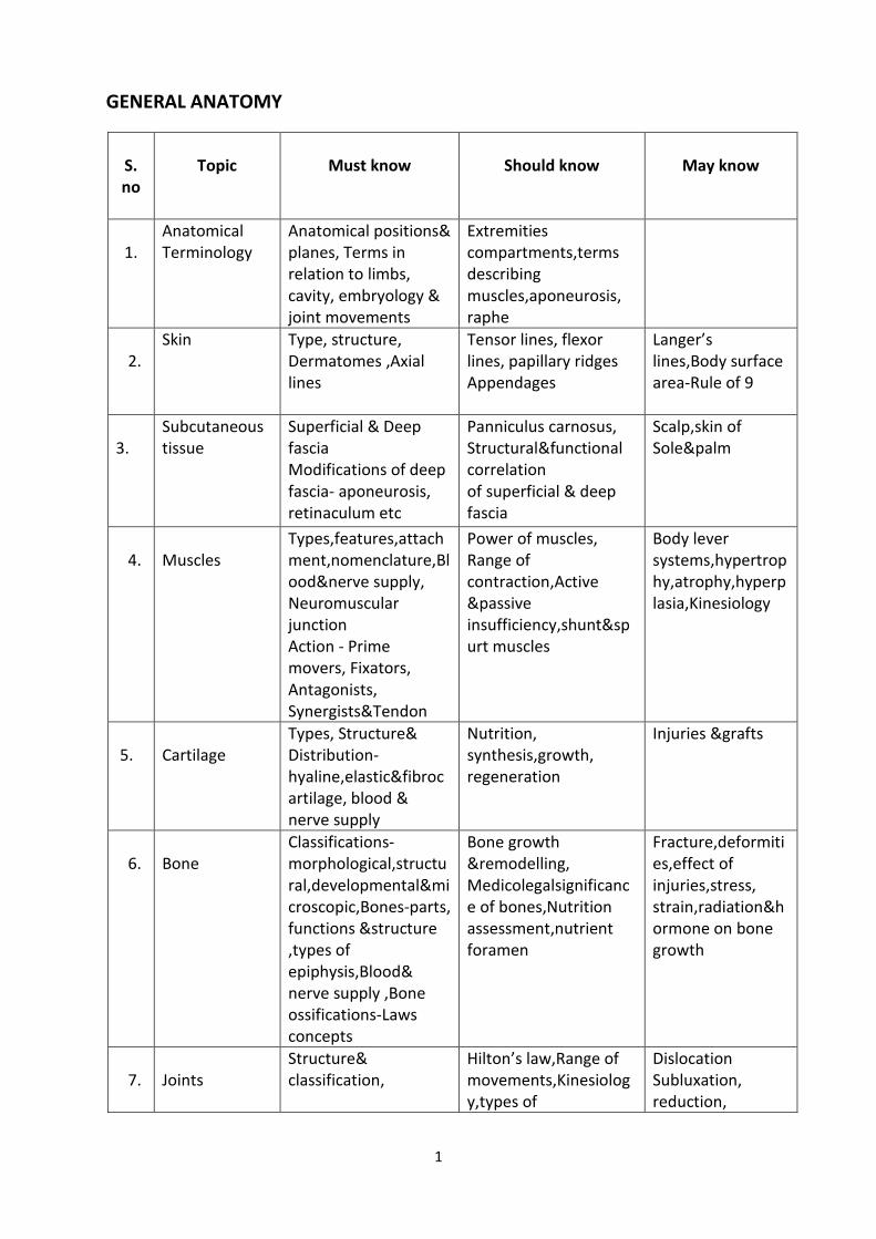
GENERAL ANATOMY
S. no
Topic
Must know
Should know
May know
1.
Anatomical Terminology
Anatomical positions& planes, Terms in relation to limbs, cavity, embryology & joint movements
Extremities compartments,terms describing muscles,aponeurosis, raphe
2.
Skin Type, structure, Dermatomes ,Axial lines
Tensor lines, flexor lines, papillary ridges Appendages
Langer’s lines,Body surface area-Rule of 9
3.
Subcutaneous tissue
Superficial & Deep fascia Modifications of deep fascia- aponeurosis, retinaculum etc
Panniculus carnosus, Structural&functional correlation of superficial & deep fascia
Scalp,skin of Sole&palm
4.
Muscles
Types,features,attachment,nomenclature,Blood&nerve supply, Neuromuscular junction Action - Prime movers, Fixators, Antagonists, Synergists&Tendon
Power of muscles, Range of contraction,Active &passive insufficiency,shunt&spurt muscles
Body lever systems,hypertrophy,atrophy,hyperplasia,Kinesiology
5.
Cartilage
Types, Structure& Distribution-hyaline,elastic&fibrocartilage, blood & nerve supply
Nutrition, synthesis,growth, regeneration
Injuries &grafts
6.
Bone
Classifications-morphological,structural,developmentalµscopic,Bones-parts, functions &structure ,types of epiphysis,Blood& nerve supply ,Bone ossifications-Laws concepts
Bone growth &remodelling, Medicolegalsignificance of bones,Nutrition assessment,nutrient foramen
Fracture,deformities,effect of injuries,stress, strain,radiation&hormone on bone growth
7.
Joints
Structure& classification,
Hilton’s law,Range of movements,Kinesiology,types of
Dislocation Subluxation, reduction,
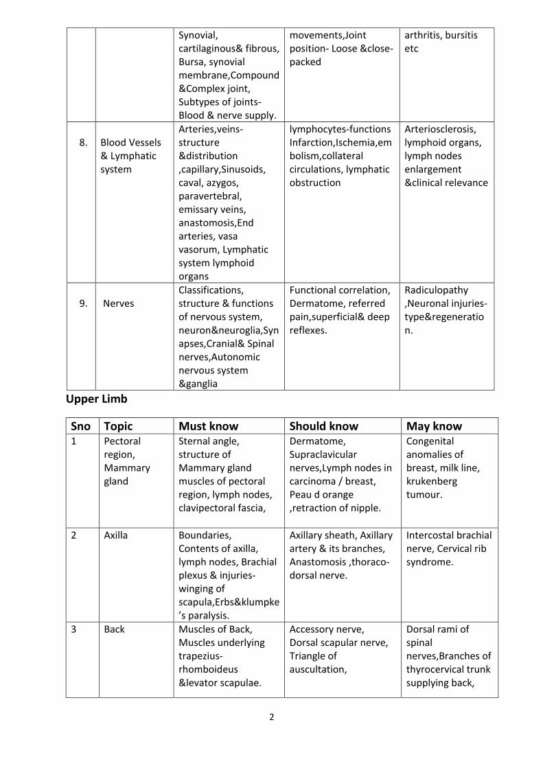
2
Synovial, cartilaginous& fibrous, Bursa, synovial membrane,Compound &Complex joint, Subtypes of joints- Blood & nerve supply.
movements,Joint position- Loose &close-packed
arthritis, bursitis etc
8.
Blood Vessels & Lymphatic system
Arteries,veins-structure &distribution ,capillary,Sinusoids, caval, azygos, paravertebral, emissary veins, anastomosis,End arteries, vasa vasorum, Lymphatic system lymphoid organs
lymphocytes-functions Infarction,Ischemia,embolism,collateral circulations, lymphatic obstruction
Arteriosclerosis, lymphoid organs, lymph nodes enlargement &clinical relevance
9.
Nerves
Classifications, structure & functions of nervous system, neuron&neuroglia,Synapses,Cranial& Spinal nerves,Autonomic nervous system &ganglia
Functional correlation, Dermatome, referred pain,superficial& deep reflexes.
Radiculopathy ,Neuronal injuries-type®eneration.
Upper Limb
Sno Topic Must know Should know May know 1 Pectoral
region, Mammary gland
Sternal angle, structure of Mammary gland muscles of pectoral region, lymph nodes, clavipectoral fascia,
Dermatome, Supraclavicular nerves,Lymph nodes in carcinoma / breast, Peau d orange ,retraction of nipple.
Congenital anomalies of breast, milk line, krukenberg tumour.
2 Axilla Boundaries, Contents of axilla, lymph nodes, Brachial plexus & injuries- winging of scapula,Erbs&klumpke’s paralysis.
Axillary sheath, Axillary artery & its branches, Anastomosis ,thoraco-dorsal nerve.
Intercostal brachial nerve, Cervical rib syndrome.
3 Back
Muscles of Back, Muscles underlying trapezius-rhomboideus &levator scapulae.
Accessory nerve, Dorsal scapular nerve, Triangle of auscultation,
Dorsal rami of spinal nerves,Branches of thyrocervical trunk supplying back,

3
anastomosis around scapula.
4 Scapular region
Musculo-tendinous cuff, Deltoid, Axillary nerve, Shoulder dislocation
Quadrangular , triangular spaces, their contents, Branches of third part of axillary artery.
Cutaneous supply of shoulder region, Intramuscular injection.
5 Shoulder joint Classification & structure, Intracapsular tendon of long head of biceps, ligament support, Axillary nerve damage,Shoulder dislocation.
Rotator cuff, Glenoid labrum, Relations of joint, Bursae around shoulder joint, frozen shoulder.
Impingement syndrome, Bursitis around shoulder joint,Bankart’slesion.
6 Front of arm&Cubital region
Muscles of flexor compartment ,boundaries of cubital region,Brachial artery,MusculocutaneousNerve,Median,radial& Ulnar nerve.
Anastomosis around elbow joint, Median cubital vein, superficial veins in cubital region, Intravenous injections
Median & ulnar nerve,entrapment , tourniquet application.
7 Back of arm
Triceps ,radial nerve in spiral groove, Saturday night palsy, crutch palsy, wrist drop.
Profunda brachii vessels.
Posterior cutaneous nerve of forearm, branches of profundabrachii artery.
8 Front of forearm & Hand
Muscles of forearm, lumbricals&interossei, radial & ulnar artery, Median radial & ulnar nerve in forearm, palmar Aponeurosis, thenar&hypothenar muscles.
Evolution of thumb & mechanism of grip flexor, retinaculum, palmarspaces,superficial& deep palmar arch,Synovial sheaths of flexor tendons, Carpal tunnel syndrome, claw hand, ape thumb deformity
Vincula longa,brevia,cutaneous veins & nerves of forearm, Surgical incisions of hand, Dupuytren’s contracture.
9 Back of forearm & dorsum of hand
Muscles of forearm,Dorsal digital expansion, posterior interosseus nerve, anatomical snuff box- boundaries & contents, wrist drop.
Superficial branch of radial nerve.
Extensor retinaculum, posterior interosseus artery.
4
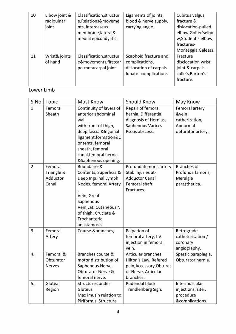
10 Elbow joint & radioulnar joint
Classification,structure,Relations&movements, interosseus membrane,lateral& medial epicondylitis.
Ligaments of joints, blood & nerve supply, carrying angle.
Cubitus valgus, fracture & dislocation-pulled elbow,Golfer’selbow,Student’s elbow, fractures-Monteggia,Galeazz
11 Wrist& joints of hand
Classification,structure&movements,firstcarpo-metacarpal joint
Scaphoid fracture and complications, dislocation of carpals- lunate- complications
Fracture disclocation wrist joint & carpals- colle’s,Barton’s fracture.
Lower Limb
S.No Topic Must Know Should Know May Know 1 Femoral
Sheath Continuity of layers of anterior abdominal wall with front of thigh, deep fascia &Inguinal ligament,formation&Contents, femoral sheath, femoral canal,femoral hernia &Saphenous opening.
Repair of femoral hernia, Differential diagnosis of Hernias, Saphenous Varices Psoas abscess.
Femoral artery &vein catherization, Abnormal obturator artery.
2 Femoral Triangle & Adductor Canal
Boundaries& Contents, Superficial& Deep Inguinal Lymph Nodes. femoral Artery , Vein, Great Saphenous Vein,Lat. Cutaneous N of thigh, Cruciate & Trochanteric anastamosis.
Profundafemoris artery Stab injuries at- Adductor Canal Femoral shaft Fractures.
Branches of Profunda famoris, Meralgia parasthetica.
3. Femoral Artery
Course &branches,
Palpation of femoral artery, I.V. injection in femoral vein.
Retrograde catheterisation / coronary angiography.
4. Femoral & Obturator Nerves
Branches course & motor distribution of Saphenous Nerve, Obturator Nerve & femoral nerve.
Articular branches Hilton's Law, Refered pain,Accessory,Obturator Nerve, Articular branches.
Spastic paraplegia, Obturator hernia.
5. Gluteal Region
Structures under Gluteus Max imusin relation to Piriformis, Structure
Pudendal block Trendlenberg Sign.
Intermuscular injections, site , procedure &complications.
5
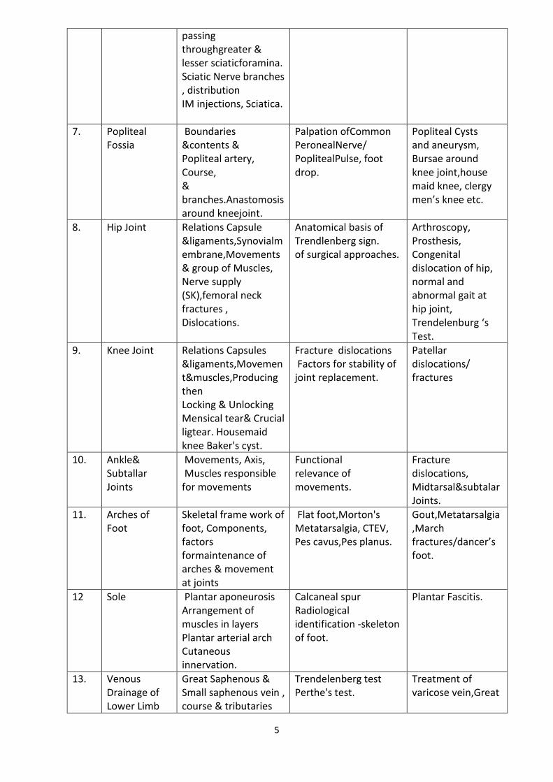
passing throughgreater & lesser sciaticforamina. Sciatic Nerve branches , distribution IM injections, Sciatica.
7. Popliteal Fossia
Boundaries &contents & Popliteal artery, Course, & branches.Anastomosis around kneejoint.
Palpation ofCommon PeronealNerve/ PoplitealPulse, foot drop.
Popliteal Cysts and aneurysm, Bursae around knee joint,house maid knee, clergy men’s knee etc.
8. Hip Joint Relations Capsule &ligaments,Synovialmembrane,Movements& group of Muscles, Nerve supply (SK),femoral neck fractures , Dislocations.
Anatomical basis of Trendlenberg sign. of surgical approaches.
Arthroscopy, Prosthesis, Congenital dislocation of hip, normal and abnormal gait at hip joint, Trendelenburg ‘s Test.
9. Knee Joint Relations Capsules &ligaments,Movement&muscles,Producing then Locking & Unlocking Mensical tear& Crucial ligtear. Housemaid knee Baker's cyst.
Fracture dislocations Factors for stability of joint replacement.
Patellar dislocations/ fractures
10. Ankle& Subtallar Joints
Movements, Axis, Muscles responsible for movements
Functional relevance of movements.
Fracture dislocations, Midtarsal&subtalar Joints.
11. Arches of Foot
Skeletal frame work of foot, Components, factors formaintenance of arches & movement at joints
Flat foot,Morton's Metatarsalgia, CTEV, Pes cavus,Pes planus.
Gout,Metatarsalgia,March fractures/dancer’s foot.
12 Sole Plantar aponeurosis Arrangement of muscles in layers Plantar arterial arch Cutaneous innervation.
Calcaneal spur Radiological identification -skeleton of foot.
Plantar Fascitis.
13. Venous Drainage of Lower Limb
Great Saphenous & Small saphenous vein , course & tributaries
Trendelenberg test Perthe's test.
Treatment of varicose vein,Great
6
Perforating vein, Positions, & communications Varicose veinVenesection, Coronary bypass.
Saphenous Veinous graft.
THORAX
S.No
Topics Must Know Should Know May Know
1. Intercostal Spaces
Muscles, Intercostal Nerve& vessels, Lymphatic’s, Intercostal Nerve Block, Pleural Tap.
Atypical Intercostal nerves & Vessels, Herpes Zoster.
Innermost intercostals Branches of intercostals Nerve & vessels Notching of ribs.
2. Pleura Parietal &Visceral pleura, Lines of pleural reflection, Pleural recessesBlood & Nerve Supply ,Pleural effusion, pleuralpleurisy& pleural tap.
Pulmonary ligament Lymphatic drainage.
Intercostal chest tube drainage, Pneumothorax .
3. Lungs Trachea & Bronchi
Anatomical position Fissures & lobes, Relations of mediastinal surface,Trachea,Interior , carina,Root of lung, Arrangement of structures within hilum ,bronchopulmonary segments& Importance.
Azygos lobe Palpation of trachea bronchopulmonary segments in Right & Left lungs, Foreign body in trachea/bronchoscopy, sites of auscultations, lung abscess, Postural drainage .
Mendelson’s syndrome Pan coast tumor Segmental pulmonary resection
4. Mediastinum Divisions, Boundaries& contents of each division.
Mediastinal syndrome Mediastinitis.
5. Pericardium & Heart
Pericardium division postion of heart, Dextrocardia, Chambers of heart orientation, Echocadiography, blood & Nerve supply, Refereed pain.
Pericarditis , paracentesis Pericardial tamponade.
Pericardial recesses.
7
6. Right Atrium External &Internal features interatrial septumASD .
SA Node AV Node
Triangle of Koch Torus aorticus.
7. Blood Supply of Heart
Rt. Coronary artery& Lt. Coronary artery, Origin, CourseTermination, Branches &Dominance. Coronary atherosclerosis Ischaemic Heart Disease (IHD),Angina pectoris Referred painHeart block.
Anastomosis & collateral circulation, Peculiarities of coronary arteries Veins of heart Coronary angiography Percutaneous transluminal coronary balloon angioplasty (PTCA).
Triple vessels disease of Heart.
8. Arch Of Aorta Course, Relations, Branches, Coarction of Aorta,PDA, Aneurysm.
Radiological appearance Aortic knucke, Anomalies of Aortic arch, Variation of branches, Aortic isthmus, Aortic spindle.
Aortic window.
9. Ventricles &Conducting System
Orifices &valvesArterialsupply,Conducting,system, VSD, Fallot’stetrology.
Cyanotic heart diseases
10. Azygos System of Veins
Azygos Vein Formation, Course Relations, Tributaries.
Hemi-azygos & Accessory azygos Vein.
SVC/IVC Obstruction.
11. Mechanism of Respiration
Joints of thorax: Costovertebral & costotransverse joints, Ligaments , Movements & axes,Pump handle Movement, Bucket handle & piston handle movement, Accessory muscles of respiration.
Emphysema Asthma Kyphosis & Scoliosis
12. Thoracic Duct Formation, Course Termination, Areas of drainage.
Chylothorax.
13. Oesophagus Course & relations Normal curvatures and constrictions, Blood supply.
Oesophagoscopy . Achalasia cardia &Sphincters.
8
Abdomen
S. No
Topic Must Know Should Know May Know
1. Anterior Abdominal Wall
Regions/Quadrants Landmarks, Joints,Superficial fascia &Muscles,Dermatomes, Blood Vessels, Porto-caval obstruction, Lymphatic Drainage, Superficial & deep inguinal rings, Renal angle, Murphy’s point.
Holden’s line Attachment of the muscles, Striae gravidarum & albicantes Extravasation of urine Abdominal incisions.
Langer’s lines Suspensory ligament of penis.
2. Rectus Sheath
Formation ,Arcuate line, Contents ,Linea alba & Linea semilunaris.
Functional aspects of rectus sheath,Divarication of recti, Meckel’s diverticulum.
Umbilical hernia Incisional hernia Faecal fistula Urinary fistula.
3. Inguinal Canal
Descent of testis &processes vaginalis Topographyboundaries, extent & contents of Indirect & direct inguinal hernia.
Mechanism of inguinal canal, Anatomical consideration of hernia repair.
Canal of Nuck.
4. Testis & Spermatic Cord
Coverings of spermatic cord/ testis, contents in males & females Spermatic cord vis-à-vis hernia sac in direct and indirect inguinal hernias Tunics of testis Blood &Nerve supply, lymphatic drainage, Hydrocele, Vasectomy,Cremasterreflex,Scrotum-nerve supply.
Varicocele Undescended testis ectopic testis.
Torsion of testis Appendix of testis Appendix of epididymis.
5. Peritoneum Vertical & horizontal disposition of peritoneum, Lesser &greater sacHepatorenal pouch&Pouch of Douglas .Nerve supply of peritoneum and referred pain.
Peritoneal recesses &bands,Functions of peritoneum, AscitisPeitoneal fossae-lesser sac, duodenal fossae, intersigmoidal recess.
Peritoneal recesses and bands.
9
6. Stomach Relations & Stomach bedBlood supply, Lymphaticdrainage,Nervesupply ,Gastric ulcer & vagotomy.
Peptic ulcers , Prepyloric vein of Mayo, gastric canal,Endoscopy ,Barium meal.
Ca stomach Trosier’s sign Traube’s space Congenital anomalies.
7. Duodenum Relations/Openings of bile&pancreatic duct, Blood supply,Duodenal ulcer &duodenal capparaduodenalfossadifference between small & large intestine.
Ligament of treitz Endoscopy &Endoscopic retrograde cholangiopancreaticography (ERCP).
Prepyloric vein of Mayo, Supraduodenal artery of Willkie, Retrodoudenal artery.
8. Caecum, Appendix &Colon
Relations.Blood supply, Interior Difference from large intestine, Gross features including relations Blood supply, Positions.
Shapes of caecum Clinical relevance of positons of appendix Burney’s point Appendicitis.
Recesses around caecum, Lump in right iliac fossa, Appendicitis vs salpingitis/ oophoritis Anatomical basis of surgical incisions.
9 Liver Relations, Blood supply Lobes of liver/vascular segments. Lymphatic drainage.
Surgical lobes. Hepatitis, Ca liver.
Liver resection & transplantation.
10. Extrahepatic Biliary Apparatus
Gross features,Blood supply of Gall Bladder , CBD-Parts & relations,gallstones,Callot’s triangle.
Sphincter of Oddi Cholecystography Endoscopic retrograde cholangiopancreatcography(ERCP)
Hartmann’s pouch Phrygian cap.
11. Pancreas Relations, Blood supply,lymphaticdrainage,Duct system, Sphincter of Oddi, Duodenal papillae.
Splenectomy vis-à-vis tail pancreas. Pancreatitis.
Pancreatic cysts &CA head pancreas.
12. Portal Vein Formation&tributaries, Porto-caval anastomosis, sites,haemorrhoids &esophageal varices caput medusae.
Porto-caval-shunt. Laminar blood flow.
13. Kidneys Relations,Coverings Blood supply,Vascularsegments,Coronal section with internal feature.
Morrison’ parallelogram, Horseshoe Kidney,Lt. Renal vein, Renal angle,Exposure of kidney from back, Pyelography.
Floating kidney Aberrant renal artery Renal transplant Lithotripsy.
10
14. Ureters Relations, extent, course constrictions, Blood supply&lymphatic drainageUrolithiasis &ureteric colic.
Congenital anomalies , relations in male/female.
Anomalies of the ureter.
15. Diaphragm Attachment,openings Nerve supply&Function.
Diaphragmatic hernia Trauma .
Foramen of Bochdalek Phrenic crush.
16.
Abdominal Aorta & Inferior Vena Cava
Extent, course , tributaries&termination of IVC Relations, branches & distribution of abdominal aorta.
Porto-caval, anastomosis Thoraco-epigastric Vein block of IVC.
Spread of carcinoma through systemic veins to vertebral venous plexus.
17.
18. Perineum Subdivisions Colle’s fascia & perineal membrane urogenital diaphragm Perineal pouches, Nerve supply.
Rupture of urethra & extravasation of urine Perineal tear.
Episiotomy.
19. Ischi-oanal Fossa
Location, boundaries & contents .Pudendal canal.
Inferior rectal vessels& pudendal nerve, Fistula in ano &Goodsall’s rule.
Hiatus of Schwalbe.
20. Urinary Bladder
relations in male/ female, Nerve &blood supply,Lymphatic drainage.
suprapubic cystostomy neurogenic bladder.
Ectopiavesicae patent urachus cystoscopy.
21. Prostate, Male Urethra Seminal Vesicle
Relation internal structure, blood supply& Age changes
Capsule vis-a- via prostatectomy benign prostatic hyperplasia, perrectal examination urethralcatheterization
TURP- transurethral resection of prostate Ca-prostate&spread.
22. Ovary,Uterus& Adnexa
Relation, flexures& Position, tubectomy Blood supply,Lymph drainage, Supports of uterus,Nerve supply and referred pain of ovary.
Rectouterine pouch&vesicouterine pouch prolapse of uterus hysterectomy Rectouterine fistula.
Uterine anomalies recurrent abortions in retroverted uterus.
23. Sigmoid Colon & Rectum
Relations/Internal feature, Blood supply & venous/Lymphatic drainage,Per rectal examinationFascia of Denonviller`sHemorrhoids.
Hirschsprung`s disease, prolapse of rectum,Imperforated anus.
Ca-rectum Proctoscopy.
24. Anal Canal Relations;anorectaljunctionInternal feature - white line&Pectinate line /anal columns.
Internal&externalhemorrhoids porto-caval anastomosis
Goodsall`s rule Embryological &surgical anal canal Imperforate anus.
11
Internal & external sphincters , nerve/Blood supply including venous, drainage Puborectalis-anorectal ring.
fissure –in –ano fistula-in-ano perianal abscesses vis-a vis ischio anal abscess.
25. Pelvic Diaphragm
Components/relationsActions,Nerve supply, sacral plexus and lumbosacral trunk Internal iliac artery.
Tear of levatorani in childbirth /episiotomy.Branches of external and internal iliac arteries
Role of levatorani in child birth Urinary stress in continence.
Head & Neck
S. No
Topic Must Know Should Know May Know
1. Scalp Layers,lymphatic drainage, sebaceouscysts, Closed&Open wounds Emissary veins-black eye safety valve haematoma.
Cephalohaematoma Caput succedanum.
Surgical scalp.
2. Cervical Fascia
investing layer ,Pretracheal&Preverteberal layer, prevertabral & carotid sheath.
axillary sheath facial spacesligament of berry.
Cold abscess other abscess.
3. Posterior Triangle
Boundaries, Subdivision-occipital , supraclavicular, content ,compression of subclavian artery to stop bleeding in upper limb wry neck.
air embolism Phrenic crush accessory phrenic nerve.
4. Orbit extrinsic muscles movement of eyeball
Squint Eye muscles testing
5. Lacrimal Apparatus
Lacrimal gland& Nasolacrimal duct.
Parts of lacrimal gland and relation to levatorpalpabraesuperioris, flow of lacrimalfluid.,nerve supply, spheno-palatine ganglia
Artificial tears.
6. Thyroid Gland
Border relation capsules,blood Supply, andvenous drainageGoiter&Thriodectomy.
pyramidal lobe levatorglandulaethyroidligament of berry, Venous plexus ,thyroid artery twigs from
Synthesis of throxine and its regulation.
12
oesophageal and tracheal branches ,vein of kocher congenital malformation.
7. Parotid Relation /Structure embedded in parotid gland Capsule parotid duct size lumen ,opening blood supply, nerve supply, applied anatomy-swelling parotidectomy, parotid abcess.
paraotid space-structure in depth of parotid space development parotidtumor.
Parotidectomy, frey’s syndrome
8. Temporal And Infratemporal Region
BoundariesContents,superfeicial-petrygoid venous plexues,sphenomndibularligament,maxillary artery deep mandibular nerve Chorda tympami , otic ganglion, Origin, insertion and actions of muscles,Mandibular nerve- course, relations and branches.
Branches of maxillary artery, Chorda tympani otic ganglion- mandibular injury laryngeal N block.
Spine of sphenoid.
9. Jugular Veins
FormationCourse,Tributaries.
Queckenstedt;s test Jugular venous pressure.
Anterior and oblique Jugular veins.
10. Temperomandibular Joint
Capsules and ligaments Articular disc Blood supply,Nerve supplyMovements,Dislocation o TMJ.
Sequence of movements for opening of mouthTreatment of dislocation.
Stability of TM joint.
11. Tongue Muscles a extrinsic,intrinsic,Blood supply-arterial /venous,Nerve supply lymphatic drainage, Applied anatomy,Tongue pulled anteriorly to prevent choking pulled anteriorly to prevent bleeding paralysis of XII nerve.
Falling back of tongue,causes& consequences
Carcinoma tongue operation Alternate taste pathway.
12. Submandibular Region
Submandibular &sublingual glands, RelationsWharton’s duct
Sialolithiasis Tumors.
Surgical approach.
13
Blood supply-lingual artery,Nerve supply lingual,9th,12th nerve Submandibular ganglion.
13. Pharynx Relations,Musculature Stylopharyngeus Palatophryngeus Salpingopharyngeus Constrictors Blood supply- Nerve supply-motor, sensory, Interior (Oropharynx,Nasopharynxand Laryngopharynx piriform fossa).
origin ,inseration and part of constrictorsstructure passing through gaps.
Anatomy of Space of morgagni Killian Jamison space Pharyngeal Diverticulum.
14. Palate intoduction and function Muscle of soft palate blood supply-nerve supply-motor &sensory anatomy:cleftplate.
Degluttiton Gag reflex . Passvant`s ridge.
15. Palatine Tonsil
Relations and surfaces Blood supply, nerve supply, tonsillitis , tonsillectomy.
Waldeyer’s ring Adenoids.
16. Larynx Cartilages ligament&Muscle,Vocal cords& Vestibular folds,Bloodsupply.lymphatic drainage nerve supply, functional consideration, Tracheostomy Damage to recurrent Laryngeal nerve
Age related changes and laryngeal carcinoma voice changes,Origin, insration,direction of muscle fibers and action of intrinsic muscle
Mechanism of sound production
17 Middle Ear Boundaries & contents.
Connection and opening .
otitis media infection of middle ear in children meningitis.
18 Hypoglossal Nerve
CourseIntraneural Extracranial&Branches- nerve testing.
location & function of nucleus.
thrombosis of anterior spinal artery care ofXII Nerve During Operation And Lingual Artery.
19 Glossopharyngeal Nerve
CourseIntraneural Intracranial, Extracranial&branches.
position of nuclei. eagles`s syndrome.
14
20 Facial Nerve functional component nuclei, course ,intracraninal extracranial&branches.
position of nuclei. Supranuclear lesion nuclear lesion infranuclearlesion.
21 Oculomotor Nerve
functional component NucleiCourse Intracranial,ExtracraninalandBrancheslightbreflex,accommodationreflex,Argyll Robertson `s supil.
positionand component of nucleus.
weber`s syndrome.
22. Abducent&Trochelear
functional Component Nuclei, course-intraneural ,intracranial&extracranial, muscle supplied by the nerves.
position of nuclei.
paralysis of muscle supplies –squint/diplopia.
23. Dural Venous Sinuses
diffrence between veins andsinusesclassificationflow of blood in sinuses cavernous sinuses- Situation,Formation Extent,SizeRelationTributariesdangerous area of face thrombosis of cavernous sinues.
Pulsating exophtalmos Queckenstedt`s test.
24 Anterior Triangle of The Neck
Boundaries and subdivisions Carotid triangleboundaries and content carotid arteries Trachea extant and relationOesophagusextent and relation.
sites of constrictions parathyroids.
Adam’s apple& relations
25 HypophysisCerebri
parts ,location ,relation blood supply, microscopic structure anddevelopment.
hormones secreted tumoures of the pituitary.
Hypophysectomy approaches.
26. Joints of The Head & Neck
atlanto occipital,atlanto axial, ligament .
Joints between the vertebral bodies.
Spondylosis.
Neuroanatomy
S no
Topics Must know Should know May know
1- Introduction to neuroanatomy
Overview of brain,brain stem & spinal cord,divisions of nervous system, cellular organization, neurons,
Nerve degeneration & regeneration, stretch reflex, muscles atrophy, hypertrophy, tendon jerks,reflex arc.
Peripheral neuropathy, entrapment syndromes overview,
15
cranial & spinal nerves, nerve plexus, autonomic & somatic ganglia, Dermatome.
radiculopathy, abnormalities of muscle tones, grading of muscle power.
2- spinal cord Spinal meninges,structure, nerve cell groups of gray column, ascending & descending tracts,blood supply.
Spinal segments,spinalenlargements,Lumbarpuncture,spinalreflex,brown-sequard syndrome, conusmedullaris syndrome, caudaequina syndrome.
Spastic & flaccid paralysis,lesion localization, syringomyelia,tabesdorsalis.
3 Brain stem Features of midbrain, pons & medulla, Internal structures- nerve cell groups of gray matter, ascending & descending tracts.
Blood supply of brain stem, Wallenberg syndrome, medial medullary syndrome, Millard-gublersyndrome,medial longitudinal fasciculus.
Cerebello-pontine angle syndrome,Mediallongitudinal fasciculus syndrome,weber’s,Benedikt’s syndrome, Argyll Robertson pupil.
4 Cranial nerve nuclei
Functional components & distributions, somatic & visceral afferent & efferent nuclei.
Nucleus ambiguous, motor,spinal,sensory& mesencephalic nuclei of trigeminal nerve, nuclei of vagus nerve,EW nucleus,nucleus of tractussolitarius, Bell’s Palsy.
Lesions of cranial nuclei & nerves, associated paralysis & syndromes, Trigeminal neuralgia, upper & lower motor type of palsy of 7th,12thnerves etc.
5 Cerebellum& fourth ventricle
features,division&subdivisions,internal structures- grey matter- nerve cell groups, white matter- connections of cerebellum.
Cerebellar peduncles, Fourth ventricle- features , boundaries & floor blood supply ,truncal ataxia, Romberg’s sign.
Cerebellar syndrome- tremors, intention tremors,dysarthria,dysmetria,dysdiadochokinesis,nystagamus.
6 Diencephalon & third ventricle
External features of thalamus, thalamic nuclei in different parts, connections &functions, hypothalamus- nuclei & connections & functions, third ventricle- communications.
Solitariothalmic tracts, connections of metathalmus(lateral & medial geniculate bodies),Hypothalmo-hypophyseal portal system, recess of ventricles, Choroid plexus.
Recent memory,exteroceptivesenses,thalmic syndrome, epithalmus,DiabetesInsipidus,hydrocephalus, hypothalamic syndrome.
7 Cerebrum External features- surfaces& borders,
Structure of cerebral cortex, sulci &gyri-
Hemiplegia & hemiparesis, motor
16
Sulci&Gyri-central ,lateral calcarine&parieto-occipital,collateralsulcus,lobes,functional areas-speech,sensory, motor,visual& auditory areas
Cingulate sulcus,paracentrallobule,cuneus, gustatory area, vestibular area, association areas,Brodmann classification
& sensory aphasia,agnosia,astereognosis,hemianopia, cerebral dominance, frontal eye field.
8 Basal Nuclei Corpus striatum- caudate nucleus &lentiform nucleus, internal capsule, connections &functions, functions of basal nucei, parkinsonism disease.
Amygdala,subthalmic nuclei, functions,Ballismus,Hemiballismus,chorea&athetosis,blood supply .
Claustrum,chorea- sydenham’s chorea & Huntington’s chorea.
9 White matter &lateral ventricle
Association,commissural& projection fibres, Corpus callosum-features,parts&functions, Internal capsule-parts,constituentfibres,blood supply of internal capsule, Lateral ventricle.
Corticofugal&corticopetal fibres, lateral ventricle- parts,boundaries, horns of lateral ventricle- choroid plexus & fissure.
Habenularcommissure,cerebralhaemorrhage,hemiplegia,ventricular system , ventriculography, CT & MRI imaging of ventricular system.
10 Blood supply of brain
Circle of Willis formation & its functional significance, vertebral & carotid system- branches
Arterial supply of cerebrum& occlusions of cerebral arteries, venous drainage of cerebrum, Blood brain barrier.
Blood –CSF barrier,Cerebralaneurism,subarachnoid haemorrhage, cerebral angiography, subdural haemorrhage.
12 Meninges & CSF
Meninges-dura mater, piamater& arachnoid mater, folds of duramater, Dural venous sinuses, extra dural&subarachnoid space- extensions& haemorrhage CSF production ,circulation& absorption.
Blood & nerve supply of duramater, cavernous sinus thrombosis, cisterna magna &cisternal puncture, functions of CSF, hydrocephalus.
Exophthalmos, arachnoid granulations,Virchow-Robin’s space,Froin’s syndrome, clinical features of hydrocephalus,Queckenstedt’s test.
13 Pathways Visual ,olfactory, auditory & gustatory Pathways,Pyramidal& extrapyramidal tracts,spinothalamictracts,dorsal column medial lemniscus pathway.
Lesions of motor & sensory tracts at various levels, spinocerebellar pathways.
Argyll Robertson pupil,hemianopia,deafness, Babinski’s sign,Examination of motor&sensory system.
17
Histology
S. No
Topics Must Know Should Know May Know
1. Introduction &Microscopy
Relevance of histology to MedicineLight microscope Magnification/resolution Setting up of a microscope Steps in paraffin block making,H& E staining.
Properties of light and electrons, Principles of microscopy, Factors influencing, magnification & resolution, Special stains for connective tissue, muscle and nerve tissue.
Wave theory of light Various kinds of microscopes Differences between light & electron microscopes Neuroanatomical stains Golgi staining methods.
2. Microtomy Parts of a rotary microtome,Steps in paraffin section cutting.
Freezing microtome. Cryotome and cryostat methods,Samplepreparations for biological specimens for transmission and electron microscopy
3. Animal Cell Cell organelle & Cell Division.
Euchromatin and hererochromatin Chemical nature of DNA &RNADistribution of the primary tissues in the body systems.
Lysosomal storage diseases,Lipid peroxidation and free radicals in biological tissues.
4. Epithelial Tissue I
Different types of epithelial tissues and their locations.
Cell junction Junctional complexes Nutrition of epithelial and connective tissues Surface modification of cell membranes (Cilia and microvilli) Cell types: FgGobet cells, APUD cells Renewal,Nerve supply.
Ultrastructure Metaplasia and hyperplasia.
5. Epithelial Tissue-Ii
Glandular epithelium: mucous, serous and mixed glands.
Ultra structure.
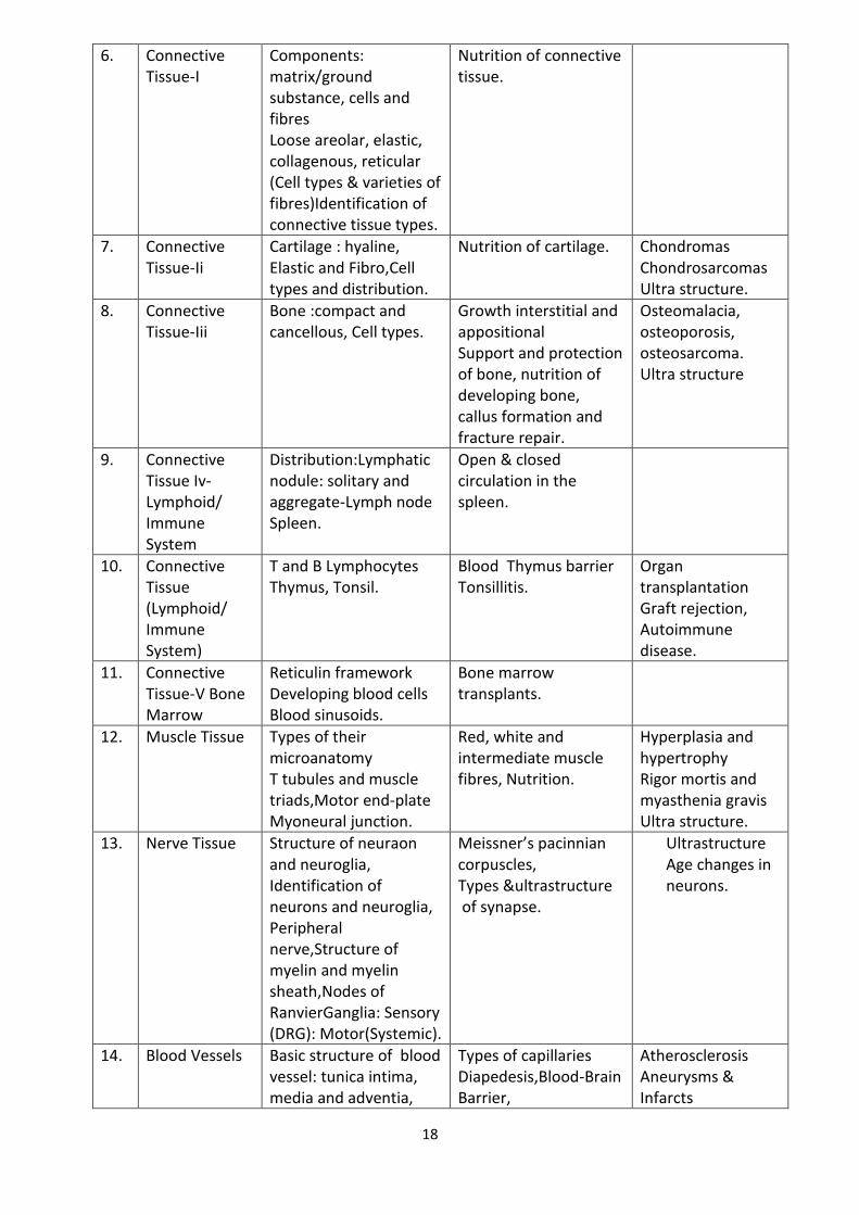
18
6. Connective Tissue-I
Components: matrix/ground substance, cells and fibres Loose areolar, elastic, collagenous, reticular (Cell types & varieties of fibres)Identification of connective tissue types.
Nutrition of connective tissue.
7. Connective Tissue-Ii
Cartilage : hyaline, Elastic and Fibro,Cell types and distribution.
Nutrition of cartilage. Chondromas Chondrosarcomas Ultra structure.
8. Connective Tissue-Iii
Bone :compact and cancellous, Cell types.
Growth interstitial and appositional Support and protection of bone, nutrition of developing bone, callus formation and fracture repair.
Osteomalacia, osteoporosis, osteosarcoma. Ultra structure
9. Connective Tissue Iv-Lymphoid/ Immune System
Distribution:Lymphatic nodule: solitary and aggregate-Lymph node Spleen.
Open & closed circulation in the spleen.
10. Connective Tissue (Lymphoid/ Immune System)
T and B Lymphocytes Thymus, Tonsil.
Blood Thymus barrier Tonsillitis.
Organ transplantation Graft rejection, Autoimmune disease.
11. Connective Tissue-V Bone Marrow
Reticulin framework Developing blood cells Blood sinusoids.
Bone marrow transplants.
12. Muscle Tissue Types of their microanatomy T tubules and muscle triads,Motor end-plate Myoneural junction.
Red, white and intermediate muscle fibres, Nutrition.
Hyperplasia and hypertrophy Rigor mortis and myasthenia gravis Ultra structure.
13. Nerve Tissue Structure of neuraon and neuroglia, Identification of neurons and neuroglia, Peripheral nerve,Structure of myelin and myelin sheath,Nodes of RanvierGanglia: Sensory (DRG): Motor(Systemic).
Meissner’s pacinnian corpuscles, Types &ultrastructure of synapse.
Ultrastructure Age changes in neurons.
14. Blood Vessels Basic structure of blood vessel: tunica intima, media and adventia,
Types of capillaries Diapedesis,Blood-Brain Barrier,
Atherosclerosis Aneurysms & Infarcts
19
Arteries(Large, Medium and Small)and Capillaries Veins & Sinusoids.
Thermoregulation Disorders of Clotting and Bleeding mechanisms.
15.
Skin Skin types & structure Cutaneous receptors Appendages of the skin(hair follicles, sebaceous and sweat glands, nails).
Renewal of the epidermis, keratinisation.
Psoriasis Vitiligo Albinism Malignant melanomaAcne,Lichen planus.
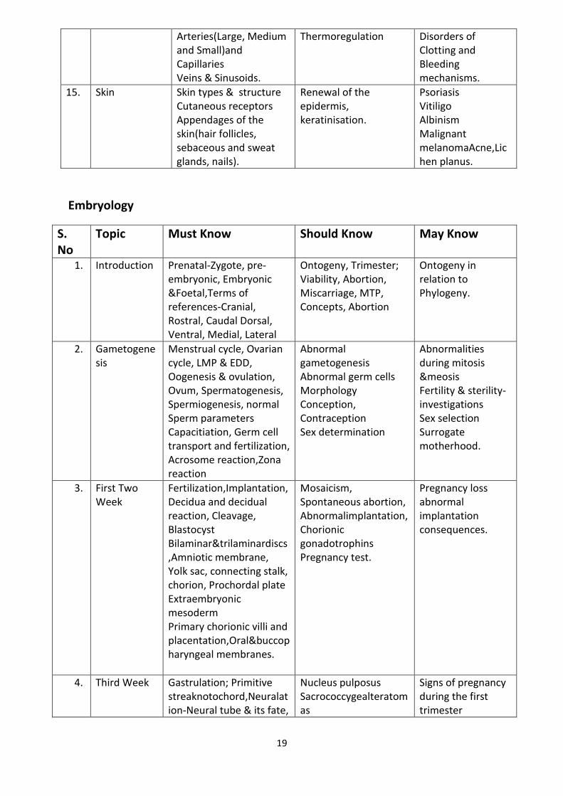
Embryology
S. No
Topic Must Know Should Know May Know
1. 1. Introduction Prenatal-Zygote, pre-
embryonic, Embryonic &Foetal,Terms of references-Cranial, Rostral, Caudal Dorsal, Ventral, Medial, Lateral
Ontogeny, Trimester; Viability, Abortion, Miscarriage, MTP, Concepts, Abortion
Ontogeny in relation to Phylogeny.
2. 2. Gametogenesis
Menstrual cycle, Ovarian cycle, LMP & EDD, Oogenesis & ovulation, Ovum, Spermatogenesis, Spermiogenesis, normal Sperm parameters Capacitiation, Germ cell transport and fertilization, Acrosome reaction,Zona reaction
Abnormal gametogenesis Abnormal germ cells Morphology Conception, Contraception Sex determination
Abnormalities during mitosis &meosis Fertility & sterility-investigations Sex selection Surrogate motherhood.
3. 3. First Two Week
Fertilization,Implantation, Decidua and decidual reaction, Cleavage, Blastocyst Bilaminar&trilaminardiscs,Amniotic membrane, Yolk sac, connecting stalk, chorion, Prochordal plate Extraembryonic mesoderm Primary chorionic villi and placentation,Oral&buccopharyngeal membranes.
Mosaicism, Spontaneous abortion, Abnormalimplantation,Chorionic gonadotrophins Pregnancy test.
Pregnancy loss abnormal implantation consequences.
4. 4 Third Week Gastrulation; Primitive streaknotochord,Neuralation-Neural tube & its fate,
Nucleus pulposus Sacrococcygealteratomas
Signs of pregnancy during the first trimester
20
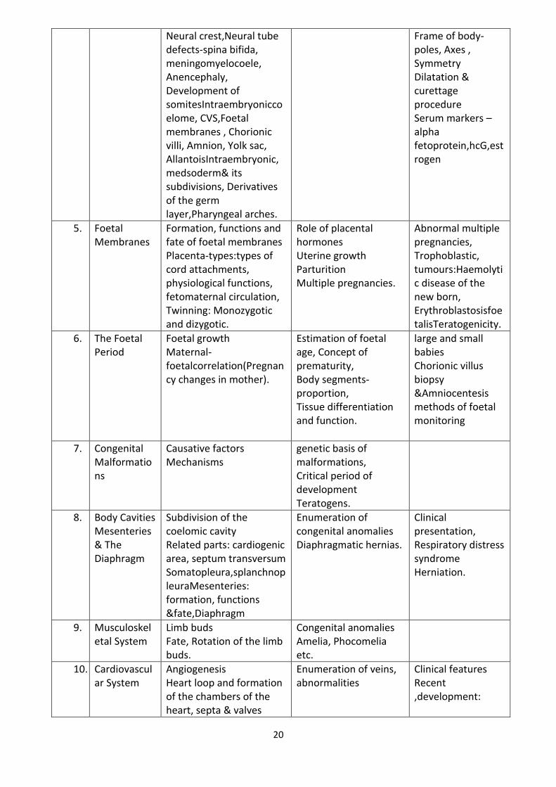
Neural crest,Neural tube defects-spina bifida, meningomyelocoele, Anencephaly, Development of somitesIntraembryoniccoelome, CVS,Foetal membranes , Chorionic villi, Amnion, Yolk sac, AllantoisIntraembryonic, medsoderm& its subdivisions, Derivatives of the germ layer,Pharyngeal arches.
Frame of body- poles, Axes , Symmetry Dilatation & curettage procedure Serum markers –alpha fetoprotein,hcG,estrogen
5. 5 Foetal Membranes
Formation, functions and fate of foetal membranes Placenta-types:types of cord attachments, physiological functions, fetomaternal circulation, Twinning: Monozygotic and dizygotic.
Role of placental hormones Uterine growth Parturition Multiple pregnancies.
Abnormal multiple pregnancies, Trophoblastic, tumours:Haemolytic disease of the new born, ErythroblastosisfoetalisTeratogenicity.
6. 6 The Foetal Period
Foetal growth Maternal-foetalcorrelation(Pregnancy changes in mother).
Estimation of foetal age, Concept of prematurity, Body segments-proportion, Tissue differentiation and function.
large and small babies Chorionic villus biopsy &Amniocentesis methods of foetal monitoring
7. 7 Congenital Malformations
Causative factors Mechanisms
genetic basis of malformations, Critical period of development Teratogens.
8. 6. Body Cavities Mesenteries & The Diaphragm
Subdivision of the coelomic cavity Related parts: cardiogenic area, septum transversum Somatopleura,splanchnopleuraMesenteries: formation, functions &fate,Diaphragm
Enumeration of congenital anomalies Diaphragmatic hernias.
Clinical presentation, Respiratory distress syndrome Herniation.
9. 7 Musculoskeletal System
Limb buds Fate, Rotation of the limb buds.
Congenital anomalies Amelia, Phocomelia etc.
10. 8. Cardiovascular System
Angiogenesis Heart loop and formation of the chambers of the heart, septa & valves
Enumeration of veins, abnormalities
Clinical features Recent ,development:
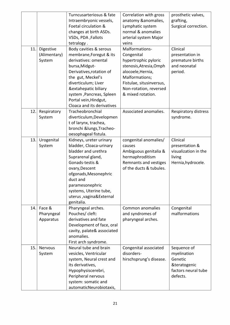
21
Turncusarteriosus & fate Intraembryonic vessels, Foetal circulation & changes at birth ASDs. VSDs, PDA ,Fallots tetralogy .
Correlation with gross anatomy &anomalies, Lymphatic system normal & anomalies arterial system Major veins
prosthetic valves, grafting, Surgical correction.
11. 9. Digestive (Alimentary) System
Body cavities & serous membrane,Foregut & its derivatives: omental bursa,Midgut-Derivatives,rotation of the gut, Meckel’s diverticulum; Liver &extahepatic biliary system ,Pancreas, Spleen Portal vein,Hindgut, Cloaca and its derivatives
Malformations- Congenital hypertrophic pyloric stenosis,Atresia,Omphalocoele,Hernia, Malformations; Fistulae, situsinversus, Non-rotation, reversed & mixed rotation.
Clinical presentation in premature births and neonatal period.
12. 10.
Respiratory System
Tracheobronchial diverticulum,Development of larynx, trachea, bronchi &lungs,Tracheo-oesophageal fistula.
Associated anomalies. Respiratory distress syndrome.
13. 11.
Urogenital System
Kidneys, ureter urinary bladder, Cloaca-urinary bladder and urethra Suprarenal gland, Gonads-testis & ovary,Descent ofgonads,Mesonephric duct and paramesonephric systems, Uterine tube, uterus ,vagina&External genitalia.
congenital anomalies/ causes Ambiguous genitalia & hermaphroditism Remnants and vestiges of the ducts & tubules.
Clinical presentation & visualization in the living Hernia,hydrocele.
14. 12.
Face & Pharyngeal Apparatus
Pharyngeal arches. Pouches/ cleft: derivatives and fate Development of face, oral cavity, palate& associated anomalies. First arch syndrome.
Common anomalies and syndromes of pharyngeal arches.
Congenital malformations
15. 13.
Nervous System
Neural tube and brain vesicles, Ventricular system, Neural crest and its derivatives, Hypophysiscerebri, Peripheral nervous system: somatic and automaticNeurobiotaxis,
Congenital associated disorders- hirschsprung’s disease.
Sequence of myelination Genetic &teratogenic factors neural tube defects.
22
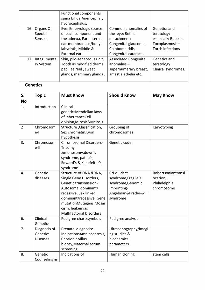
Functional components spina bifida,Anencephaly, hydrocephalus.
16. 14.
Organs Of Special Senses
Eye: Embryologic source of each component and the adnexa, Ear: Internal ear-membranous/bony labyrinth, Middle & External ear.
Common anomalies of the eye: Retinal detachment; Congenital glaucoma, Colobomairidis, Congenital cataract .
Genetics and teratology especially Rubella, Toxoplasmosis –Torch infections
17. 15.
Integumentary System
Skin, pilo-sebaceous unit, Tooth as modified dermal papillae,Nail , sweat glands, mammary glands .
Associated Congenital anomalies –supernumerary breast, amastia,athelia etc.
Genetics and teratology Clinical syndromes.
Genetics
S. No
Topic Must Know Should Know May Know
1. Introduction Clinical geneticsMendelian laws of inheritanceCell division,Mitosis&Meiosis.
2 Chromosome-I
Structure ,Classification, Sex chromatin,Lyon hypothesis
Grouping of chromosomes
Karyotyping
3. Chromosome-II
Chromosomal Disorders-Trisomy &monosomy,down’s syndrome, patau’s, Edward’s &,Klinefelter’s syndrome
Genetic code
4. Genetic diseases
Structure of DNA &RNA, Single Gene Disorders, Genetic transmission-Autosomal dominant/ recessive, Sex linked dominant/recessive, Gene mutationMutagens,Mosaicism, leukemias Multifactorial Disorders
Cri-du chat syndrome,Fragile X syndrome,Genomic Imprinting- Angelman&Prader-willi syndrome
Robertsoniantranslocation, Philadelphia chromosome
6. Clinical Genetics
Pedigree chart/symbols Pedigree analysis
7. Diagnosis of Genetics Diseases
Prenatal diagnosis:-IndicationsAmniocentesis, Chorionic villus biopsy,Maternal serum screening.
Ultrasonography/imaging studies & biochemical parameters
8. Genetic Counseling &
Indications of Human cloning, stem cells
23
Gene therapy,Human genome
Recommended Books for Undergraduate programme
S no Title Author Publisher Edition
1 Gray's Anatomy for students
Richard L.Drake , A Wayne Vogi, Adam W. Mitchel
Elsevier First South Asia Edition
2 Clinical Anatomy Richard S. Snell Wolters Kluwer 9th edition
3 Clinical Neuroanatomy Richard S. Snell Wolters Kluwer 7th edition
4 Cunningham's Manual of practical Anatomy Volume1,2,3
Rachel Koshi oxford 16th Edition
6 Di Fiore’s Atlas of Histology
Victor P. Eroschenko
Wolters Kluwer 13th Edition
7 Langman’sMedical Embryology
T.W. Sadler Wolters Kluwer 13th Edition
8 Surface and Radiological Anatomy
A. Halim CBS Publishers 3rd Edition
9 Human Osteology -A Clinical orientation
Nafis Ahmad Faruqi
CBS Publishers 3rd Edition
10 Gray's Anatomy-The Anatomical Basis of clinical practice
Susan Standring Elsevier 41th Edition
11- Synopsis of surgical anatomy
Lee McGregor’s Varghese publisher (Indian Edition)12th
12 A manual of clinical surgery
S Das S.S Das 12th
24
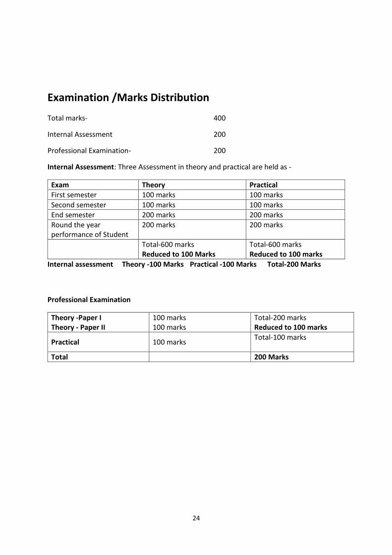
Examination /Marks Distribution
Total marks- 400
Internal Assessment 200
Professional Examination- 200
Internal Assessment: Three Assessment in theory and practical are held as -
Exam Theory Practical
First semester 100 marks 100 marks
Second semester 100 marks 100 marks
End semester 200 marks 200 marks
Round the year performance of Student
200 marks 200 marks
Total-600 marks Reduced to 100 Marks
Total-600 marks Reduced to 100 marks
Internal assessment Theory -100 Marks Practical -100 Marks Total-200 Marks
Professional Examination
Theory -Paper I Theory - Paper II
100 marks 100 marks
Total-200 marks Reduced to 100 marks
Practical 100 marks Total-100 marks
Total 200 Marks