gait parameters determination by 3d motion analyzer system for initial indonesian gait database
TRANSCRIPT
315
GAIT PARAMETERS DETERMINATION BY 3D
MOTION ANALYZER SYSTEM FOR INITIAL
INDONESIAN GAIT DATABASE
Nuha Desi Anggraeni1
, Ferryanto1, Suryo Tri Atmojo
1, Sandro Mihradi
1, Tatacipta
Dirgantara2, Andi Isra Mahyuddin
1
1Mechanical Design Research Group, Mechanical Engineering Department
2Lightweight Structures Research Group, Aeronautics & Astronautics Department
Faculty of Mechanical and Aerospace Engineering, Institut Teknologi Bandung
Jalan Ganesha 10, Bandung 40132, Indonesia,
Tel., +62 22 2504243, Email: [email protected]
Abstract
A 3D motion analyzer system to determine gait parameters as well as kinematics and kinetics of human
walking was developed previously. The system utilizes two 90 fps cameras, a personal computer and in-
house software developed to determine gait parameters of human walking motion as well as the
kinematics and kinetics of gait. The objectives of this research are two fold. First objective is to evaluate
the efficacy of the developed system and second objective is to obtain initial Indonesian normal gait
database. Spatio-temporal, kinematic, and kinetic parameters of 60 subjects (30 male, 30 female) obtained
by the 3D motion analyzer system are presented as preliminary results in an effort to establish Indonesia
gait database. Prior to measurement, each subject was evaluated to ensure normalcy of posture, and then
was instructed to walk normally in a specially-arranged walking area. The gait data obtained are
comparable to available normal gait data. This indicates that the initial development of the 3D normal gait
database is quiet successful. The data presented in this research could serve as the basis to establish
reference for 3D Indonesian normal gait database. The system developed could be further utilized in the
enrichment of the database as well as for clinical purpose by measuring and analyzing abnormal gait. The
resulting kinematic and kinetic parameters are useful in determining therapy protocol as well as keeping
track of the patient‟s progress. Hence, the system has the potential to be further developed into a medical
diagnostic tool.
Keywords: 3D Gait Parameters, 3D Motion Analyzer System, Gait Analysis, Gait Database
Introduction
Human movement analysis has long been studied and found applications in various fields, such
as medical rehabilitation, medical diagnostic, and sport science [1-5]. In medical rehabilitation,
information about position and orientation of various joints of a patient body is needed to
determine abnormalities. It is also reported that a knowledge of the expected changes in joint
angle allow therapists to better understand a patient gait pathology [6-7].
In general, human movement could be measured by direct measurement techniques and also
imaging (optical) measurement techniques. The main problem in direct measurement techniques
is the subject has to carry many cables or other components that could affect walking motion [5].
Most of the problems encountered by direct measurement techniques could be overcome by
imaging (optical) measurement techniques.
Unfortunately, human movement analyzer is not available in most Indonesian hospitals due to
its relatively high cost. Moreover, there is also no Indonesian reference data that could be used to
diagnose whether a patient has a normal or pathological gait.

316
Hence, an affordable motion analyzer system to obtain human gait parameters has been
developed. First, a system for 2D kinematics and kinetic analyses of human gait was developed
[8-9]. The system is then used to determine 2D gait parameters of Indonesian people as part of an
effort to develop the first Indonesian gait database [10-11].
Information obtained from 2D measurement is limited to the sagittal plane, while very useful
and in most cases are adequate, do not provide motion information in other planes. Although the
sagittal plane is probably the most important one, where much of the movement parameters could
be observed, there are certain gait pathologies where another plane (e.g. the frontal plane) would
yield useful information. Therefore, a 3D motion analyzer system is developed using similar
optical methodology [12-15]. In this work, 3D gait parameters based on optical measurement is
conducted to establish initial Indonesian gait reference data for medical diagnostic tool.
3D Motion Analyzer System
This work uses a previously developed 3D motion analyzer system consists of image processing
system, and kinematic and kinetic analyzer for human gait developed as reported in Mihradi et al.
[12-13]. As shown in Figure 1, the system consists of two 90 fps (frame per second) video
cameras, a personal computer, and computing software that was developed for tracking markers
attached to a human subject during motion and determining the real coordinates of markers along
their trajectories. Another software based on a multibody model of human, is also developed to
calculate gait parameters of human gait as well as the kinematics and kinetics of gait, based on
the markers real coordinates. Prior to recording the subject walking motion, a calibration
procedure is conducted to acquire camera parameters required for 3D reconstruction. The
calibration for this 3D motion analyzer system uses modified Zhang‟s technique proposed by
Ferryanto et al. [14].
Figure 16.Experimental setup.
317
The markers positioning and human body model employed in this study are described in more
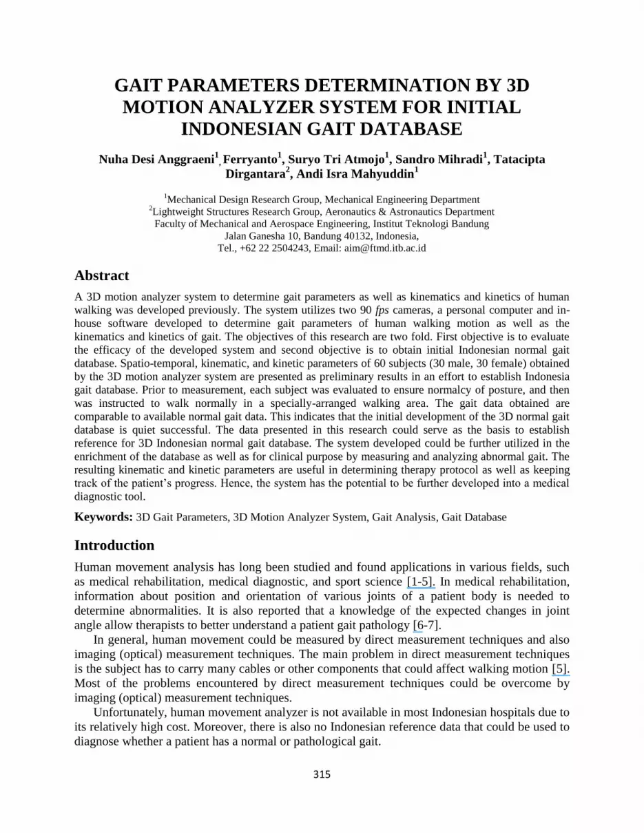
detail by Mihradi et al. [15]. Seven markers are attached on seven positions of right-side subject‟s
leg, i.e. pelvis, hip, mid-thigh, knee, tibia, malleolus, and lateral metatarsal. Marker locations on
the subject body are depicted in Figure 2. The image of the markers are processed to acquire their
real coordinate in 3D Cartesian, which are then employed as input for kinematic and kinetic
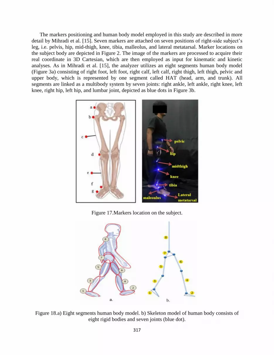
analyses. As in Mihradi et al. [15], the analyzer utilizes an eight segments human body model
(Figure 3a) consisting of right foot, left foot, right calf, left calf, right thigh, left thigh, pelvic and
upper body, which is represented by one segment called HAT (head, arm, and trunk). All
segments are linked as a multibody system by seven joints: right ankle, left ankle, right knee, left
knee, right hip, left hip, and lumbar joint, depicted as blue dots in Figure 3b.
Figure 17.Markers location on the subject.
Figure 18.a) Eight segments human body model. b) Skeleton model of human body consists of
eight rigid bodies and seven joints (blue dot).

318
Participating Subject and Data Collection
Spatio-temporal, kinematic, and kinetic parameters of 60 participating subjects (30 male, 30
female) obtained by the 3D motion analyzer system are presented as preliminary results in an
effort to establish Indonesia gait database. The subjects‟ weight, height, and anthropometric data
are measured and the results are summarized in Table 1.
Table 5. Age and Anthropometric Data of Participating Subjects
Variable Male Female
Mean (SD) Mean (SD)
Age (year) 21.87 (2.62) 22.47 (4.22)
Weight (Kg) 64.80 (9.14) 52.73 (6.46)
Height (m) 1.73 (0.09) 1.59 (0.06)
ASIS breadth (m) 0.32 (0.042) 0.31 (0.041)
Upper thigh diameter (m) 0.13 (0.012) 0.12 (0.013)
Midthigh circumference (m) 0.45 (0.087) 0.47(0.041)
Thigh length (m) 0.42 (0.038) 0.41 (0.049)
Knee diameter (m) 0.10 (0.008) 0.09 (0.008)
Calf circumference (m) 0.37 (0.031) 0.34 (0.024)
Calf length (m) 0.42 (0.032) 0.40 (0.025)
Malleolus width (m) 0.07 (0.009) 0.07 (0.009)
Foot width (m) 0.12 (0.007) 0.21 (0.028)
Malleolus height (m) 0.08 (0.004) 0.07 (0.006)
Foot length (m) 0.26 (0.047) 0.24 (0.012)
BMI 21.63 (2.77) 20.75 (1.93)
To check for normalcy, similar procedures as the ones conducted in Mahyuddin et al. [11]
were followed. Before the measurements, body posture and body mass index (BMI) of each
subject is evaluated to ensure normalcy. In order to assess posture normalcy, the shoulder and the
back of each subject are observed (see Figure 4). If it is unsymmetrical, it will be considered
abnormal that could be due to scoliosis or lordosis. The length of each leg is also measured to

319
check whether the two legs are having the same length (see Figure 4). A difference of more than
2 cm will be considered abnormal. Height and weight of each subject are measured to calculate
the BMI, which is considered normal if the value is between 18.5 and 24.9. Only subjects with
normal posture and BMI participated in the gait measurement.
Figure 19. Posture normalcy assessment and anthropometric measurement.
Results and Analysis
Following are several results obtained from the measurements of the subjects. The spatio-
temporal, kinematics, and kinetic parameters of the subjects are presented and then compared to
those obtained from other researches and literatures.
Spatio-Temporal Parameters
The spatio-temporal parameters, i.e. cadence, stride length, walking speed, and cycle time of the
subjects are summarized in Table 2.
Table 6. Spatio-Temporal Gait Parameters of Subjects
Variable Male Female
Mean (SD) Mean (SD)
Cadence (step/min) 84.13 (7.75) 84.97 (5.26)
Cycle period (s) 1.44 (0.13) 1.42 (0.09)
Stride length (m) 1.14 (0.12) 1.09 (0.11)
Walking speed (m/s) 0.8 (0.13) 0.75 (0.16)
From Table 2, it may be seen that the cadence of female subjects are slightly higher than male
subjects, while the stride length on male subjects are longer than female subjects. Differences in
male and female subjects may be attributed to the differences in anthropometric. Since male
subjects are taller than female, thus the stride length on male subjects are relatively longer than
female subjects. On the other hand, female tends to have a higher cadence than male. But, male
average walking speed is higher than female.
320
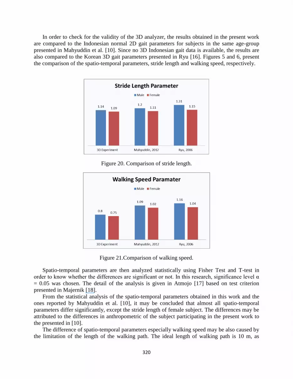
In order to check for the validity of the 3D analyzer, the results obtained in the present work
are compared to the Indonesian normal 2D gait parameters for subjects in the same age-group
presented in Mahyuddin et al. [10]. Since no 3D Indonesian gait data is available, the results are
also compared to the Korean 3D gait parameters presented in Ryu [16]. Figures 5 and 6, present
the comparison of the spatio-temporal parameters, stride length and walking speed, respectively.
Figure 20. Comparison of stride length.
Figure 21.Comparison of walking speed.
Spatio-temporal parameters are then analyzed statistically using Fisher Test and T-test in
order to know whether the differences are significant or not. In this research, significance level α
= 0.05 was chosen. The detail of the analysis is given in Atmojo [17] based on test criterion
presented in Majernik [18].
From the statistical analysis of the spatio-temporal parameters obtained in this work and the
ones reported by Mahyuddin et al. [10], it may be concluded that almost all spatio-temporal
parameters differ significantly, except the stride length of female subject. The differences may be
attributed to the differences in anthropometric of the subject participating in the present work to
the presented in [10].
The difference of spatio-temporal parameters especially walking speed may be also caused by
the limitation of the length of the walking path. The ideal length of walking path is 10 m, as
321
reported by Whittle [4]. The 10 m walking path should provide the first two steps for the
acceleration to steady walking speed and the last two steps for deceleration until the subject stop.
However, the length of walking speed of present work is only 3 m and could only provide
approximately 3 steps. As a result, subjects may have to decrease their speed while they are
accelerating their walking speed. Hence, the walking speed of the subject in this study may not
yet reach steady gait. Further measurement with longer walking path are ongoing.
Kinematic Parameters
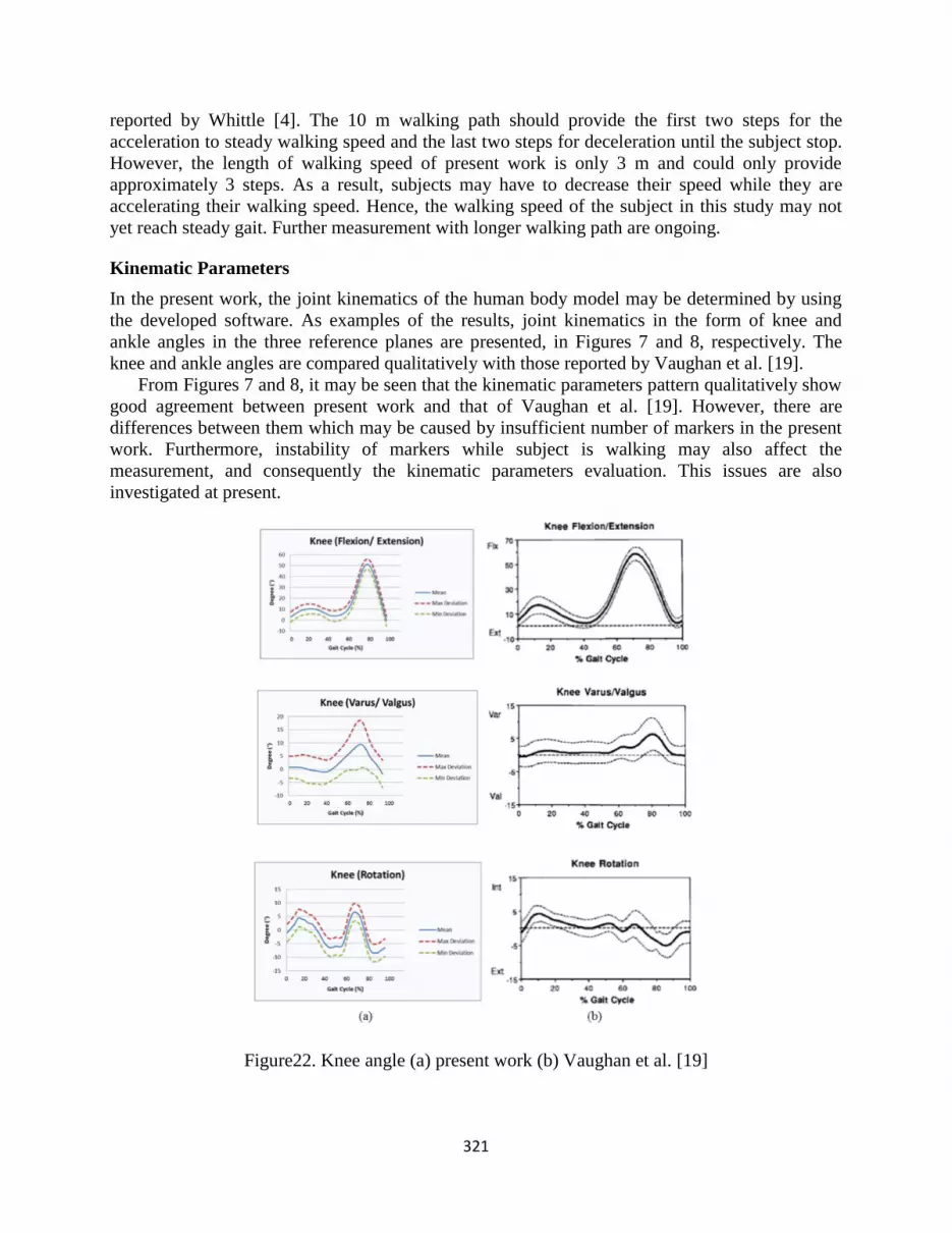
In the present work, the joint kinematics of the human body model may be determined by using
the developed software. As examples of the results, joint kinematics in the form of knee and
ankle angles in the three reference planes are presented, in Figures 7 and 8, respectively. The
knee and ankle angles are compared qualitatively with those reported by Vaughan et al. [19].
From Figures 7 and 8, it may be seen that the kinematic parameters pattern qualitatively show
good agreement between present work and that of Vaughan et al. [19]. However, there are
differences between them which may be caused by insufficient number of markers in the present
work. Furthermore, instability of markers while subject is walking may also affect the
measurement, and consequently the kinematic parameters evaluation. This issues are also
investigated at present.
Figure22. Knee angle (a) present work (b) Vaughan et al. [19]

322
Figure 23. Ankle angle (a) present work (b) Vaughan et al. [19]
Kinetic Parameters
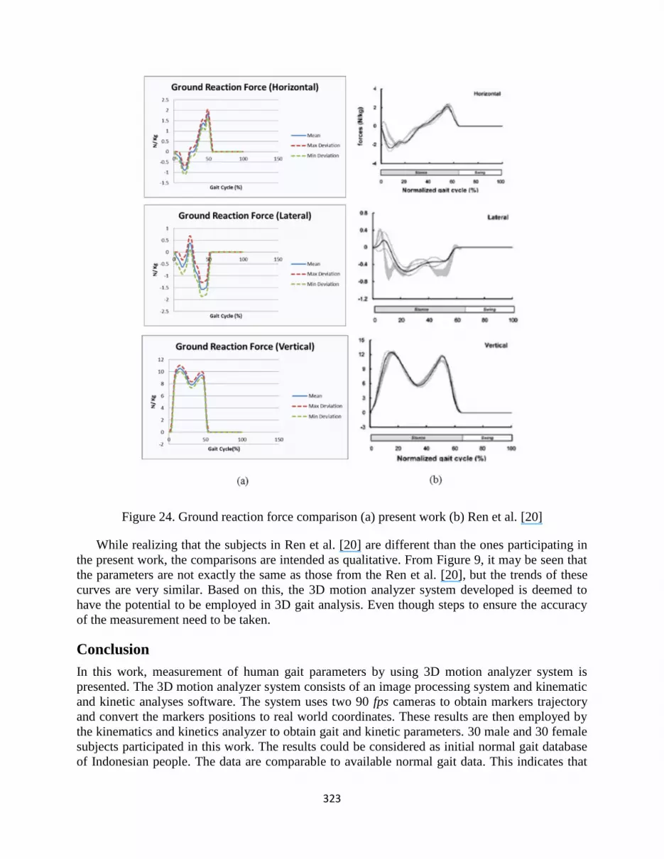
All kinetic parameters, i.e. ground reaction forces, joint forces, ground reaction moments, and
joint moments may be evaluated by employing the eight segments human body model and the
inertia and anthropometric data presented in Table 2. As example, ground reaction forces in the
three axes are presented in Figure 9, along with the ones obtained by Ren et al. [20].
323
Figure 24. Ground reaction force comparison (a) present work (b) Ren et al. [20]
While realizing that the subjects in Ren et al. [20] are different than the ones participating in
the present work, the comparisons are intended as qualitative. From Figure 9, it may be seen that
the parameters are not exactly the same as those from the Ren et al. [20], but the trends of these
curves are very similar. Based on this, the 3D motion analyzer system developed is deemed to
have the potential to be employed in 3D gait analysis. Even though steps to ensure the accuracy
of the measurement need to be taken.
Conclusion
In this work, measurement of human gait parameters by using 3D motion analyzer system is
presented. The 3D motion analyzer system consists of an image processing system and kinematic
and kinetic analyses software. The system uses two 90 fps cameras to obtain markers trajectory
and convert the markers positions to real world coordinates. These results are then employed by
the kinematics and kinetics analyzer to obtain gait and kinetic parameters. 30 male and 30 female
subjects participated in this work. The results could be considered as initial normal gait database
of Indonesian people. The data are comparable to available normal gait data. This indicates that

324
the initial development of the database is quite successful. The obtained data should serve as
initial database, while the system developed could be further utilized in the enrichment of the
database as well as for clinical purpose by measuring and analyzing abnormal gait. The resulting
kinematics and kinetic parameters are useful in determining therapy protocol as well as keeping
track of the patient‟s progress. Hence, the system has the potential to be further developed into a
medical diagnostic tool.
Acknowledgement
The authors gratefully acknowledge the support from Program Riset Desentralisasi DIKTI 2013
for the present work.
References
[1] R. Huston, Principle of Biomechanics, New York: CRC Press, Taylor & Francis Group,
2009.
[2] V. Medved, Measurement of Human Locomotion, New York: CRC Press, 2001.
[3] J. Perry, Gait Analysis: Normal and Pathological Function, Thorofare, NJ: Slack Inc., 1992.
[4] M.W. Whittle, Gait Analysis: An Introduction, USA: Butterworth-Heinemann, 2007.
[5] D.A. Winter, Biomechanics and Motor Control of Human Movement, New Jersey: John
Wiley and son Inc., 2009.
[6] T. Bajd, V. Valencic, M. Kljajic, R. Acimovic, and U. Stanic, “Standardization of kinematic
gait measurements and automatic pathological gait pattern diagnostics”, Scandinavian
Journal of Rehabilitation Medicine, Vol. 9, pp. 95-105, 1977.
[7] Rancho Los Amigos Research Centre, Observational Gait Analysis Handbook, Los Amigos
Research and Education Institute, Downey, CA, 1989.
[8] N. Juliyad, S. Mihradi, T. Dirgantara, and A. I. Mahyuddin, “2D experimental motion
analysis of human gait”, Regional Conference on Mechanical and Aerospace Technology,
Bali, Indonesia, 2010.
[9] I. Mahyuddin, S. Mihradi, T. Dirgantara, A.S. Jaya, N. Juliyad, and U.M. Purba,
“Development of an affordable system for 2D kinematics and dynamics analysis of human
gait”, Proc. SPIE, Singapore, 2010.
[10] I. Mahyuddin, S. Mihradi, T. Dirgantara, M. Moeliono, and T. Prabowo, "Development of
Indonesian gait database using 2D optical motion analyzer system," ASEAN Engineering
Journal, Vol. 2, No. 2, pp. 62-72, 2012.
[11] I. Mahyuddin, S. Mihradi, T. Dirgantara, and P.N. Maulido, “Gait parameters determination
by 2D optical motion analyzer system”, Applied Mechanics and Materials, Vol. 83, pp. 123-
129, 2011.
[12] S. Mihradi, Ferryanto, T. Dirgantara, and A.I. Mahyuddin, “Development of an optical
motion-capture system for 3D gait analysis”, International Conference on Instrumentation,
Communication, Information Technology and Biomedical Engineering, Bandung, Indonesia,
2011.
[13] S. Mihradi, A.I. Henda, T. Dirgantara, and A.I. Mahyuddin, “Development of 3D gait
analyzer software based on marker position data”, International Conference on
Instrumentation, Communication, Information Technology and Biomedical Engineering,
Bandung, Indonesia, 2011.

325
[14] Ferryanto, S. Mihradi, T. Dirgantara, and A.I. Mahyuddin, “Camera calibration technique
improvement for 3D optical gait analyzer”, Applied Mechanics and Materials, Vol. 393, pp.
976-981, 2013.
[15] S. Mihradi, A.I. Henda, T. Dirgantara, and A.I. Mahyuddin, “Development of 3D gait
analyzer software based on marker position data”, ASEAN Engineering Journal, Vol. 3, No.
2, pp. 4-14, 2013.
[16] T. Ryu, H. Choi, H. Choi, and M. Chung, “A comparison of gait characteristics between
korean and western people for establishing korean gait reference data”, International Journal
of Industrial Ergonomics, Vol. 36, 2006.
[17] S.T. Atmojo, “Initial Database Development of Indonesian Normal Gait Parameter Using 3
Dimension Motion System Analyzer (in Indonesia)," Faculty of Mechanical and Aerospace
Engineering, Institut Teknologi Bandung, Bandung, Indonesia, 2013.
[18] J. Majernik, “Normative human gait databases”, Statistics Research Letters, Vol. 2, No. 3,
pp. 69-74, 2013.
[19] L. Vaughan, B.L. Davis, and J.C. O'Connor, Dynamics of Human Gait, Cape Town: Human
Kinetics Publishers, 1999.
[20] L. Ren, R. Jones, and D. Howard, "Whole-body inverse dynamics over a complete gait cycle
based only on measured kinematics”, Journal of Biomechanics, Vol. 41, pp. 2750-2759,
2008.