gab2 alleles modify alzheimer's risk in apoe ɛ4 carriers
TRANSCRIPT
GAB2 Alleles Modify Alzheimer’s Risk in APOE ε4 Carriers
Eric M. Reiman1,2,3,17,*,18, Jennifer A. Webster1,17,18, Amanda J. Myers4,5,18, JohnHardy5,6, Travis Dunckley1,17, Victoria L. Zismann1,17, Keta D. Joshipura1,17, John V.Pearson1,17, Diane Hu-Lince1,17, Matthew J. Huentelman1,17, David W. Craig1,17, Keith D.Coon1,7,17, Winnie S. Liang1,17, RiLee H. Herbert1,17, Thomas Beach8,17, Kristen C.Rohrer5, Alice S. Zhao5, Doris Leung5, Leslie Bryden5, Lauren Marlowe5, Mona Kaleem5,Diego Mastroeni8, Andrew Grover8,17, Christopher B. Heward9, Rivka Ravid10, JosephRogers8,17, Michael L. Hutton11, Stacey Melquist11, Ron C. Petersen12, Gene E.Alexander13,17, Richard J. Caselli14,17, Walter Kukull16, Andreas Papassotiropoulos1,15,and Dietrich A. Stephan1,2,17,*
1 Neurogenomics Division, Translational Genomics Research Institute, Phoenix, AZ, 85004, USA 2 BannerAlzheimer’s Institute, Phoenix, AZ 85006, USA 3 Department of Psychiatry, University of Arizona, Tucson,AZ 85724, USA 4 Department of Psychiatry and Behavioral Sciences, University of Miami, Miller School ofMedicine, Miami, FL 33136, USA 5 Laboratory of Neurogenetics, National Institute on Aging, Bethesda, MD,20892, USA 6 Reta Lila Weston Laboratories, Department of Molecular Neuroscience, Institute of Neurology,Queen Square, London WC1N, 3BG, England 7 Division of Thoracic Oncology Research, St. Joseph’sHospital, Phoenix, AZ 85013, USA 8 Sun Health Research Institute, Sun City, AZ 85351, USA 9 KronosScience Laboratory, Phoenix, AZ 85016, USA 10 Netherlands Institute for Neurosciences, Dutch RoyalAcademy of Arts and Sciences, Meibergdreef 47 AB Amsterdam, The Netherlands 11 Department ofNeuroscience, Mayo Clinic, Jacksonville, FL 32224, USA 12 Department of Neurology, Mayo Clinic,Rochester, MN 55905, USA 13 Department of Psychology, Arizona State University, Tempe, AZ 85281, USA14 Department of Neurology, Mayo Clinic, Scottsdale, AZ 85259, USA 15 Division of Molecular Psychologyand Life Sciences Training Facility, Biozentrum, University of Basel, Switzerland 16 National Alzheimer’sCoordinating Center, Department of Epidemiology, School of Public Health and Community Medicine,University of Washington, Seattle, WA 98195, USA 17 Arizona Alzheimer’s Consortium, Phoenix AZ 85006,USA
SUMMARYThe apolipoprotein E (APOE) ε4 allele is the best established genetic risk factor for late-onsetAlzheimer’s disease (LOAD). We conducted genome-wide surveys of 502,627 single-nucleotidepolymorphisms (SNPs) to characterize and confirm other LOAD susceptibility genes. In ε4 carriersfrom neuropathologically verified discovery, neuropathologically verified replication, and clinicallycharacterized replication cohorts of 1411 cases and controls, LOAD was associated with six SNPsfrom the GRB-associated binding protein 2 (GAB2) gene and a common haplotype encompassingthe entire GAB2 gene. SNP rs2373115 (p = 9 × 10⁻¹¹) was associated with an odds ratio of 4.06(confidence interval 2.81–14.69), which interacts with APOE ε4 to further modify risk. GAB2 wasoverexpressed in pathologically vulnerable neurons; the Gab2 protein was detected in neurons,tangle-bearing neurons, and dystrophic neuritis; and interference with GAB2 gene expressionincreased tau phosphorylation. Our findings suggest that GAB2 modifies LOAD risk in APOE ε4carriers and influences Alzheimer’s neuropathology.
*Correspondence: [email protected] (E.M.R.), [email protected] (D.A.S.).18These authors contributed equally to this work.
NIH Public AccessAuthor ManuscriptNeuron. Author manuscript; available in PMC 2008 November 25.
Published in final edited form as:Neuron. 2007 June 7; 54(5): 713–720. doi:10.1016/j.neuron.2007.05.022.
NIH
-PA Author Manuscript
NIH
-PA Author Manuscript
NIH
-PA Author Manuscript
INTRODUCTIONAlzheimer’s disease (AD) afflicts about 10% of persons over 65 and almost half of those over85 (Evans et al., 1989) and the number of afflicted persons continues to grow. To date,researchers have firmly established associations between four genes and AD risk. Whereasmore than 150 mutations of the presenilin 1 (PS1), presenilin 2 (PS2), and amyloid precursorprotein (APP) genes account for many early-onset AD cases with autosomal dominantinheritance, the apolipoprotein E (APOE) ε4 allele accounts for many cases of late-onset AD(LOAD), with dementia onset after age 60 (Papassotiropoulos et al., 2006; Corder et al.,1993, 1994; Farrer et al., 1997). The APOE gene has three common variants, ε2, ε3, and ε4.The ε2 allele is associated with the lowest LOAD risk, while each copy of the ε4 allele in aperson’s APOE genotype is associated with a higher LOAD risk and a younger median age atdementia onset (Corder et al., 1994; Farrer et al., 1997). Although twin studies suggest thatthere are several susceptibility genes which, along with the APOE ε4 allele, contribute to upto 80% of LOAD cases (Gatz et al., 2006), discovery of other susceptibility genes has beenelusive (Bertram et al., 2007).
To identify susceptibility genes for common and genetically complex disorders like LOAD, ithas been proposed that it would help to conduct genome-wide surveys of at least 300,000single-nucleotide polymorphisms (SNPs) in unrelated cases and controls, compare the mosthomogeneous samples, and consider interactions between major and minor genes(Papassotiropoulos et al., 2006; Kruglyak, 1999; Coon et al., 2007). We individually genotyped502,627 SNPs to characterize and confirm LOAD susceptibility genes in three separate cohortsof LOAD cases and controls, including a discovery cohort of clinically and neuropathologicallycharacterized brain donors, a replication cohort of similarly characterized brain donors, and areplication cohort of clinically characterized living subjects. The brain donor cohorts wereselected to exclude clinically misdiagnosed cases and cognitively normal butneuropathologically affected elderly controls; the clinical cohort was selected to confirmgenetic associations independent of any brain donor selection bias. Within each cohort, LOADcases and controls were stratified into subgroups of APOE ε4 carriers and noncarriers,permitting us to investigate genes that modify LOAD risk in the ε4 carriers and genes thatmight otherwise be masked by disproportionately large ε4 effects.
RESULTS AND DISCUSSIONWe recently demonstrated the feasibility of high-density genome-wide association studies inour neuropathologically characterized cases and controls, providing empirical support for thesuggestion that the APOE locus is unparalleled in its contribution to LOAD risk (Coon et al.,2007). With the exception of an SNP only 14 kb pairs distal to and in linkage disequilibrium(LD) with the APOE ε4 variant on chromosome 19, no other SNP distinguished LOAD casesfrom controls after Bonferroni correction for multiple comparisons (Figure S1A athttp://www.tgen.org/neurogenomics/data). For the previously noted reasons, we divided eachcohort into two subgroups: allelic APOE ε4 carriers (Figure S1B) and APOE ε4 noncarriers(Figure S1C). We now report associations between a common gene and LOAD in APOE ε4carriers in our three cohorts; we show that the implicated gene is associated with ADneuropathology in neuronal microarray and immunohistochemical studies; and we consider apossible mechanism by which GAB2 modifies AD risk in a small-interfering RNA (siRNA)study. Finally, we deposit all of the data into the public domain for use by the community(http://www.tgen.org/neurogenomics/data).
High-Density Genome-Wide Association StudiesGenome-wide genotyping was performed on each individual sample from a “neuropathologicaldiscovery cohort” of 736 brain donors, a “neuropathological replication cohort” of 311 brain
Reiman et al. Page 2
Neuron. Author manuscript; available in PMC 2008 November 25.
NIH
-PA Author Manuscript
NIH
-PA Author Manuscript
NIH
-PA Author Manuscript
donors, and an additional “clinical replication cohort” of 364 living subjects who were at least65 years old at the time of their death or last clinical assessment and who were independentlyassessed for their APOE genotype. For the two neuropathological cohorts, brain tissue for DNAextraction, neuropathological diagnoses, and data were supplied by investigators from 20 ofthe National Institute on Aging (NIA)-sponsored Alzheimer’s Disease Centers (ADCs) (inaccordance with agreements with the NIA, the ADCs, and the National Alzheimer’sCoordinating Center) and from the Netherlands Brain Bank. For the hypothesis-testing clinicalreplication cohort, DNA extracted from blood, clinical diagnoses, and data from subjectsassessed in Rochester, MN were supplied by investigators from the Mayo Clinic.
The neuropathological discovery cohort included 446 LOAD cases (299 ε4 carriers and 147ε4 noncarriers) and 290 controls (61 ε4 carriers and 229 ε4 noncarriers); the neuropathologicalreplication cohort included 197 LOAD cases (113 ε4 carriers and 84 ε4 noncarriers) and 114controls (27 ε4 carriers and 87 ε4 noncarriers); and the clinical replication cohort included 218LOAD cases (115 ε4 carriers and 103 ε4 noncarriers) and 146 controls (29 ε4 carriers and 117ε4 noncarriers). Brain donor cases satisfied clinical and neuropathological criteria for LOAD,and were age 73.5 ± 6.2 at death. Brain donor controls did not have significant cognitiveimpairment or significant neuropathological features of AD, and were age 75.8 ± 7.5 at death.Clinical cases satisfied criteria for probable AD, and were age 78.9 ± 7.8 at last clinical visit.Clinical controls did not have clinically significant cognitive impairment and were age 81.7 ±6.6 at last clinical assessment.
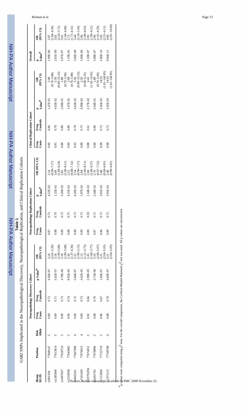
We initially surveyed SNPs in the neuropathological discovery cohort to explore LOADassociations in the ε4 carrier and noncarrier subgroups. Within the discovery subgroup ofAPOE ε4 carriers, 10 of the 25 SNPs with the most significant LOAD-association significancelevels (contingency test p = 9 × 10⁻⁵ to 1 × 10⁻⁷; uncorrected for multiple comparisons) werelocated in the GRB-associated binding protein 2 (GAB2) gene on chromosome 11q14.1 (Table1). LOAD associations in six of these SNPs were then confirmed in both the neuropathologicalreplication and clinical replication cohorts (Table 1). These ten SNPs were not significantlyassociated with LOAD in the APOE ε4 noncarrier group (contingency test p = 0.08 to 0.97)(Table S1A). Combining data from all 644 APOE ε4-carrying cases and controls, we foundhighly significant associations between LOAD and all ten GAB2 SNPs (contingency test p =1.19 × 10⁻⁵ to 9.66 × 10⁻¹¹), with five of the six consistently implicated alleles surviving thehighly conservative Bonferroni correction for 312,316 independent comparisons (p = 1.55 ×10⁻⁷) (Table 1). When data from the ε4 carriers and noncarriers were analyzed together, as inour previous report (Coon et al., 2007), the ten GAB2 SNPs were still associated with LOAD(contingency test p = 0.013 to 2.7 × 10⁻⁶, Table S1B), but these associations did not surviveBonferroni correction.
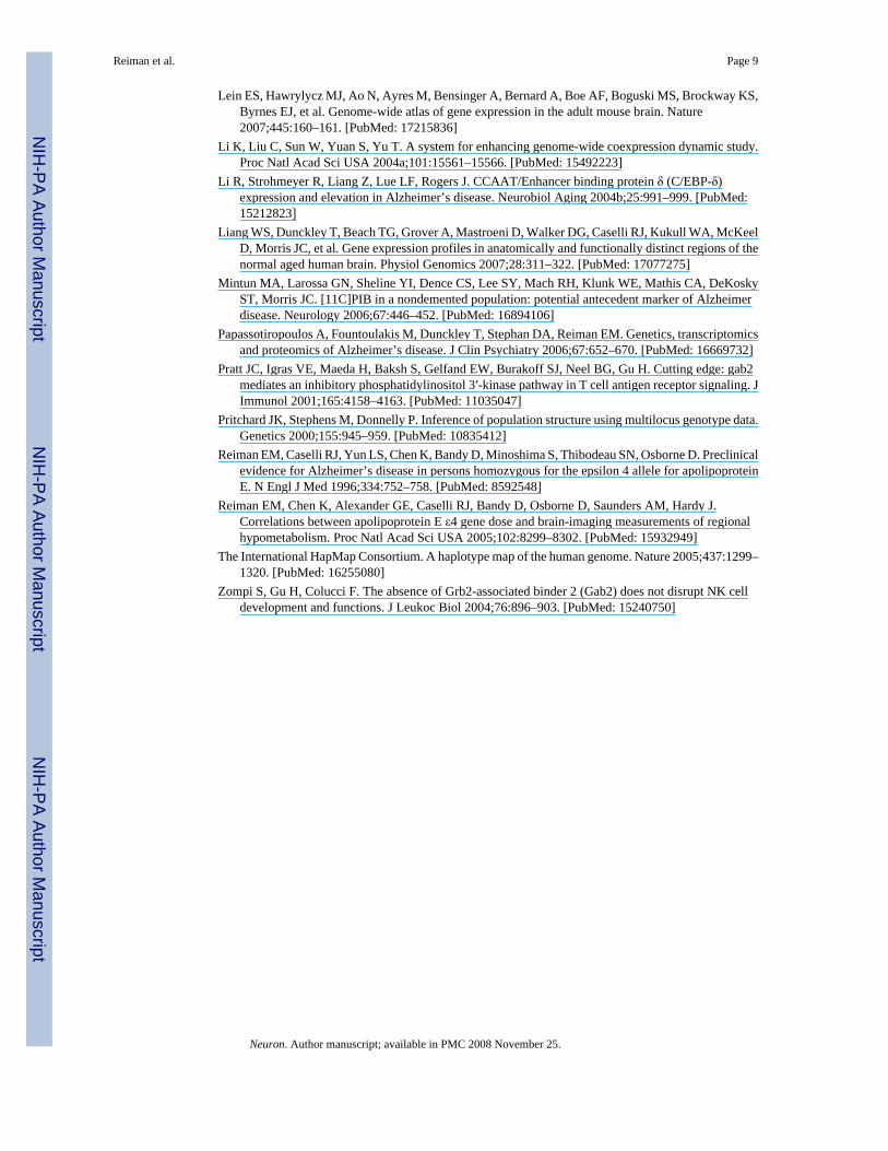
The PLINK analysis toolset (http://pngu.mgh.harvard.edu/~purcell/plink/index.shtml) wasused for whole-genome analysis. Haploview v3.32 was used to determine the LD structure ofthe chromosome 11q14.1 region surrounding GAB2 in each of the three APOE ε4-stratifiedcohorts (Figure 1). Three haplotype blocks are present in this region: one block upstream ofGAB2, roughly corresponding to the ALG8 locus; one 189 kb-pair block encompassing mostof the GAB2 locus; and one downstream block corresponding to the NARS2 locus. These blockswere consistent with the LD structure of the HapMap CEPH populations. The GAB2 gene iscompletely encompassed by a single haplotype block extending from rs901104 to rs2373115(SNPs 5–22 in Figure 1), which has three major haplotypes: an extremely common “GAB2risk haplotype,” a common “GAB2 protective haplotype,” and a relatively uncommon GAB2haplotype unrelated to LOAD risk in APOE ε4 carriers (Figure 1). In all three cohorts, theGAB2 CT-AAG-CAGATCAGACG haplotype was associated with higher LOAD risk, theGAB2 TC-GCA-TGAGGTGTCTT haplotype was associated with a lower LOAD risk, and the
Reiman et al. Page 3
Neuron. Author manuscript; available in PMC 2008 November 25.
NIH
-PA Author Manuscript
NIH
-PA Author Manuscript
NIH
-PA Author Manuscript
CT-AAG-CAGAGCA GCCG was unrelated to LOAD risk in the APOE ε4 carriers (Figure1).
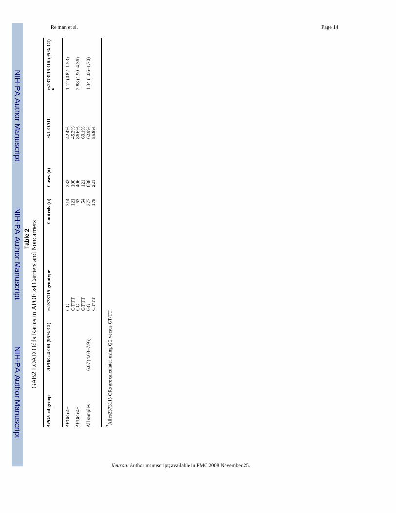
Data from the 1411 subjects (including 644 APOE ε4 carriers and 767 noncarriers) in all threecohorts were combined to characterize odds ratios (ORs) and 95% confidence intervals (CIs)for rs2373115, the most significant SNP in our screen (Table 2). In ε4 carriers, LOAD caseshad a risk genotype frequency of 0.88 and controls had a frequency of 0.71. In comparisonwith the other ε4 carriers, those with rs2373115 genotype GG had a significantly higher riskof LOAD (OR 2.36, 95% CI 1.55–3.58) than those with genotype GT and TT. In contrast,APOE ε4 noncarriers with rs2373115 genotype GG did not have a higher LOAD risk than theother ε4 noncarriers (OR 1.01, 95% CI 0.74–1.38).
Whereas we confirmed a younger age at dementia onset in the APOE ε4 carriers than innoncarriers (age 75.5 ± 7.2 versus 77.8 ± 7.9, p = 2.4 × 10⁻⁴, two-tailed unpaired t test; unequalvariance is statistical test used for all tests), there was no significant effect of rs2373115genotype on age at dementia onset in either the ε4 carriers (t test, p = 0.32) or noncarriers (ttest, p = 0.84).
Neuronal Microarray StudiesTo provide converging evidence that GAB2 is biologically relevant to AD neuropathology,expression profiling using the Affymetrix Human Genome U133 Plus 2.0 array was used tocharacterize and compare GAB2 expression in laser-capture microdissected non-tangle-bearingneurons of cases and controls in six brain regions differentially affected by AD. LOAD caseshad significantly greater neuronal GAB2 expression in the posterior cingulate cortex (9 cases,13 controls, 4.50-fold change, t test, p = 0.00039) and hippocampus (9 cases, 13 controls, 2.94-fold change, t test, p = 0.00085), and no significant expression differences in the entorhinalcortex (10 cases, 13 controls, 1.20-fold change, t test, p = 0.46), middle temporal gyrus (13cases, 12 controls, 1.44-fold change, t test, p = 0.14), superior frontal gyrus (22 cases, 11controls, 1.25-fold change, t test, p = 0.47), or primary visual cortex (17 cases, 12 controls,1.53-fold change, t test, p = 0.14).
The hippocampus is known to be especially vulnerable to AD-related neurofibrillary tangles(Braak and Braak, 1991), neuronal loss, and brain atrophy (Bobinski et al., 2000). It ispreferentially involved in AD-related memory impairment (Jack et al., 1999) and is associatedwith the highest cerebral Gab2 expression in the rodent brain (Lein et al., 2007). The posteriorcingulate cortex is known to be preferentially vulnerable to AD-related hypometabolicabnormalities and fibrillar amyloid deposition, and is also involved in AD-related memoryimpairment (Reiman et al., 1996, 2005; Johnson et al., 2006; Buckner et al., 2005; Mintun etal., 2006). While the entorhinal cortex and temporal and prefrontal regions are also affectedby AD neuropathology, the visual cortex is relatively spared. Using a repeated measuresanalysis of variance to analyze neuronal GAB2 gene expression data from the same eight LOADcases and ten controls, there was a significant group-by-region interaction (p = 0.011), withLOADrelated increases in neuronal GAB2 gene expression that were greater in the posteriorcingulate cortex and hippocampus than in the visual cortex.
Tau Phosphorylation siRNA StudyIn addition to its other properties, GAB2 is the principal activator of the phosphatidylinositol3-kinase (PI3K) signaling pathway (Pratt et al., 2001). PI3K activates Akt, which in turnpromotes glycogen synthase kinase-3 (Gsk3) phosphorylation/inactivation. This mechanismsuppresses Gsk3-dependent phosphorylation of tau at AD-related hyperphorylated tauresidues, the principal component of neurofibrillary tangles, and prevents apoptosis ofconfluent cells (Baki et al., 2004; Kang et al., 2005). Based on these findings, we hypothesized
Reiman et al. Page 4
Neuron. Author manuscript; available in PMC 2008 November 25.
NIH
-PA Author Manuscript
NIH
-PA Author Manuscript
NIH
-PA Author Manuscript
that Gab2 might function to protect cells from neuronal tangle formation and cell death andthat a loss-of-function GAB2 haplotype would diminish such protection. We thus postulatedthat interference with GAB2 expression using siRNA treatment would increase tauphosphorylation at the serine-262 residue known to be hyperphosphorylated in AD. As shownin Figure 2, GAB2 siRNA treatment was associated with a 1.70-fold increase in serine-262phosphorylated tau. This increase was not attributable to a concomitant increase in total taulevels. Additional siRNA and protein validation studies are now being performed to determinethe extent to which GAB2 affects tau phosphorylation at additional relevant epitopes.
Immunohistochemical ValidationGab2 immunohistochemistry was assessed in hippocampus and posterior cingulate cortex inLOAD cases. In hippocampus, Gab2 immunoreactivity was observed in structures with themorphology of dystrophic neurites or neuropil threads, neurons, and corpora amylacea. Theputative neurons were almost entirely dystrophic in appearance (Figure 3A) or had cytoplasmicinclusions resembling neurofibrillary tangles (Figure 3B). Dystrophic neurons and neurites(Figure 3C) and neurofibrillary tangle-bearing cells (Figure 3D) were also revealed by the Gab2antibody in posterior cingulate. Here, however, many relatively normal neurons were observedas well, with long stretches of immunoreactive apical dendrites ascending through the corticallayers (Figures 3C and 3D).
DiscussionIn order to characterize and confirm associations between the GAB2 gene and LOAD risk inAPOE ε4 carriers, our studies capitalized on the genome-wide survey of more than 300,000SNPs, two clinically characterized and neuropathologically verified cohorts of AD cases andcontrols, a third cohort of clinically well characterized subjects, and stratification of the sampleswith respect to carriers and noncarriers of a major LOAD susceptibility gene, APOE. Six SNPsthat are part of a common haplotype block encompassing the entire GAB2 gene were implicatedin three independent cohorts. Maximal significance of the association was at SNP rs2373115(p = 9 × 10⁻¹¹) with an odds ratio of 4.06 (CI 2.81–14.69). An odds ratio of 24.64 (CI 7.44–116.79) for overall genetic risk is achieved when both ε4 and the GAB2 rs2373115 risk allelesare present. Data from a microarray study of laser-capture microdissected neurons in LOADcases and controls, immunohistochemistry, and an siRNA study provided converging evidencefor the relevance of GAB2 to the neuropathology of LOAD, raising testable hypotheses aboutthe mechanisms by which GAB2 could modify LOAD risk in ε4 carriers and provide targetsat which to aim new treatments.
Although only one genotyping platform was used in all three cohorts, our findings are unlikelyto be attributable to any platform-related bias in genotyping calls because the observedassociation was not limited to a single SNP but was related to a large haplotype block inagreement with the LD structure of the HapMap CEPH population. Furthermore, all six of theimplicated SNPs had high-quality SNiPer-HD scores (greater than 0.45), indicating that thedata for each SNP clustered well (see Figure S2 for cluster diagrams).
Individual genotype data for all samples across >300,000 high-quality SNiPer-HD calls (seeExperimental Procedures for QC metrics) is made available to the community as a .ped file athttp://www.tgen.org/neurogenomics/data. This genome-wide scan data will enable replicationof putative common LOAD risk alleles, and also enable further discovery of both independentand combinatorial genetic associations.
The GAB2 haplotype block spans 189 kb and includes at least 614 known SNPs. Four of thesix hundred and fourteen known SNPs in this locus are nonsynonymous coding SNPs, whichare generally considered to be the best candidates for functional variation. However, all four
Reiman et al. Page 5
Neuron. Author manuscript; available in PMC 2008 November 25.
NIH
-PA Author Manuscript
NIH
-PA Author Manuscript
NIH
-PA Author Manuscript
of these SNPs are reported to have minor allele frequencies of 0.0% in the CEPH population(The International HapMap Consortium, 2005), and therefore are not candidates for thecommon functional variant on the GAB2 risk haplotype.
GAB2 is a scaffolding protein involved in multiple signaling pathways, which could affect AD-related tau, amyloid, metabolic, or other aspects of AD pathology and cell survival in differentways (Koncz et al., 2002; Gu et al., 2001; Zompi, Gu and Colucci, 2004), and it has been foundto be coexpressed with other putative AD-related genes (Li et al., 2004a). Discovery of thisLOAD susceptibility gene, if further replicated, provides new opportunities to investigateLOAD pathogenesis, predisposition, treatment, and prevention. Genome-wide studies usingeven higher density platforms and compound genetic analyses in sufficiently large samples ofwell-characterized cases and controls promise to play increasingly important roles in thescientific understanding, evaluation, treatment, and prevention of AD and other common andgenetically complex disorders. In the interim, public access to the raw genotyping data fromour series will provide valuable information to assess the contribution of other putative riskloci to this devastating disease.
EXPERIMENTAL PROCEDURESHigh-Density Genome-Wide Association Study
The 500K GeneChip (Affymetrix, Santa Clara, CA) was used to survey 502,267 SNPs in eachsubject as recently described (Coon et al., 2007). Genotypes were extracted using both SNiPer-HD (Hua et al., 2007) and BRLMM (Affymetrix) software. 312,316 SNPs were analyzed afterexcluding those that were monomorphic, did not cluster into three distinct Gaussiandistributions, clustered poorly, had Hardy Weinberg equilibrium p values less than 0.01, hadminor allele frequencies less than 2%, or exhibited less than 98% concordance between theSNiPer-HD and BRLMM calls. The software program STRUCTURE (Pritchard et al., 2000)was employed to test for underlying genetic stratification using 5000 randomly selected SNPsand including at least 100 SNPs per chromosome. The initial analysis yielded empiricalevidence of three populations. Since 14 subjects belonged to a population far removed fromthe rest of the study population, they were eliminated from further analyses. STRUCTUREthen was used to demonstrate a comparable admixture of the two populations in the cases andcontrols. After stratifying the LOAD cases and controls for presence or absence of the APOEε4 allele, allelic χ2 statistics were computed for each SNP. APOE genotypes were obtained ineach subject by either pyrosequencing (Ahmadian et al., 2000) or restriction fragment lengthpolymorphism (RFLP) analysis (Lai et al., 1998).
LD mapping was performed by importing genotypes into the Haplo-View program v3.32.Pairwise LD values (as measured by D′) reflect the likelihood that two genetic markers areinherited together.
Neuronal Microarray StudiesBrain samples (mean post-mortem interval of 2.5 hr) from six brain regions that are eitherhistopathologically or metabolically relevant to LOAD and aging were collected at the SunHealth Research Institute. Expression profiling was performed as described previously (Lianget al., 2007). Direct case-to-control comparisons were performed to analyze expressiondifferences in each region.
Immunohistochemical ValidationGab2 immunoreactivity in LOAD hippocampus and posterior cingulate cortex was examinedusing an affinity-purified goat polyclonal antibody directed against a C-terminal epitope ofGab2 (Santa Cruz Biotechnology, Santa Cruz, CA). Blocks were obtained from rapid autopsy
Reiman et al. Page 6
Neuron. Author manuscript; available in PMC 2008 November 25.
NIH
-PA Author Manuscript
NIH
-PA Author Manuscript
NIH
-PA Author Manuscript
LOAD cases (<3 hr postmortem) (n = 5). Hippocampus sections were derived from blocks thatwere fixed for 24 hr in 4% paraformaldehyde and sectioned at 40 μm on a freezing microtome.Posterior cingulate sections were derived from snap-frozen blocks that were sectioned at 6μm on a cryostat. Immunohistochemical protocols were as previously described (Li et al.,2004b). Immunoreactivity was visualized with nickel-intensified diaminobenzidine.
Tau Phosphorylation siRNA StudyNeuroglioma cells overexpressing wild-type tau protein were grown in 96-well plates andtransfected with siRNA directed at GAB2 mRNA. Following 4 days of transfection, cells werefixed, permeablized, and immunostained with antibodies against total tau protein and tauprotein phosphorylated on serine-262. A FITC- and Cy5-conjugated secondary antibodycocktail was then applied. After incubation and washing, images were captured and quantitatedusing the InCell imager 3000 (General Electric). The fold increase in serine-262phosphorylated tau levels was calculated against control samples that had been transfected witha scrambled siRNA sequence.
Supplementary MaterialRefer to Web version on PubMed Central for supplementary material.
AcknowledgementsThese studies were supported by Kronos Life Sciences Laboratories, the National Institute on Aging (ArizonaAlzheimer’s Disease Center P30 AG19610, RO1 AG023193, Mayo Clinic Alzheimer’s Disease Center P50 AG16574,and Intramural Research Program), the National Alzheimer’s Coordinating Center (U01 AG016976), and the state ofArizona. We thank our research volunteers and their families for their generous participation; Drs. Creighton Phelps,Marcelle Morrison-Bogorad, Marilyn Miller, and Walter Kukull for their assistance in the acquisition of tissue samplesand data; and directors, pathologists, and technologists from the following ADCs and brain banks: Lucia Sue (SunHealth Research Institute and Arizona Alzheimer’s Disease Center); Ruth Seemann and Dan Brady (National Instituteon Aging); Juan C. Troncoso and Olga Pletnikova (John Hopkins, P50 AG05146); Harry Vinters and Justine Pomakian(University of California, Los Angeles, P50 AG16570); Christine Hulette (The Kathleen Price Bryan Brain Bank,Duke University Medical Center, P50 AG05128, RO1 NS39764, RO1 MH60451, and GlaxoSmithKline); DikranHoroupian and Ahmad Salehi (Stanford University, P30 AG17824); Jean Paul Vonsattel (New York Brain Bank, TaubInstitute, Columbia University, P50 AG08702); E. Tessa Hedley-Whyte and Karlotta Fitch (Massachusetts GeneralHospital, P50 AG05134); Roger Albin, Lisa Bain, and Eszter Gombosi (University of Michigan, P50 AG08671);William Markesbery, Sonya Anderson (University of Kentucky, P50 AG05144); Dennis W. Dickson and NatalieThomas (Mayo Clinic, Jacksonville, P50 AG16574 and P50 AG25711); Carol A. Miller, Jenny Tang, and DimitriDiaz (University of Southern California, P50 AG05142); Dan McKeel, John C. Morris, Eugene Johnson, Jr., VirginiaBuckles, and Deborah Carter (Washington University, St. Louis, P50 AG 05681); Thomas Montine and Aimee Schantz(University of Washington, P50 AG05136); John Q. Trojanowski, Virginia M. Lee, Vivianna Van Deerlin, and TerrySchuck (University of Pennsylvania); Ann C. McKee and Carol Kubilus (Boston University, P30 AG13846); BruceH. Wainer and Marla Gearing (Emory University, AG025688); Charles L. White III, Roger Rosenberg, MarilynHowell, and Joan Reisch (University of Texas, Southwestern Medical School, P30-AG12300); William Ellis and MaryAnn Jarvis (University of California, Davis, P30 AG AG01542); David A. Bennett, Julie A. Schneider, Karen Skish,and Wayne T. Longman (Rush University Medical Center, P30 AG10161); and Deborah C. Mash, Margaret J. Basile,and Mitsuko Tanaka University of Miami/NPF Brain Endowment Bank). Additional support was provided by theJohnnie B. Byrd Sr. Alzheimer’s Disease and Research Institute, the Swiss National Science Foundation(PP00B-68859), the Verum foundation, the Bisgrove charitable donation, the NIH Neuroscience Blueprint(U24NS051872), the ENDGAME Consortium (UO1HL084744), and the National Institute on Aging(K01AG024079).
ReferencesAhmadian A, Gharizadeh B, Gustafsson AC, Sterky F, Nyren P, Uhlen M, Lundeberg J. Single-nucleotide
polymorphism analysis by pyrosequencing. Anal Biochem 2000;280:103–110. [PubMed: 10805527]Baki L, Shioi J, Wen P, Shao Z, Schwarzman A, Gama-Sosa M, Neve R, Robakis NK. PS1 activates
PI3K thus inhibiting GSK-3 activity and tau overphosphorylation: effects of FAD mutations. EMBOJ 2004;23:2586–2596. [PubMed: 15192701]
Reiman et al. Page 7
Neuron. Author manuscript; available in PMC 2008 November 25.
NIH
-PA Author Manuscript
NIH
-PA Author Manuscript
NIH
-PA Author Manuscript
Bertram L, McQueen MB, Mullin K, Blacker D, Tanzi RE. Systematic meta-analyses of Alzheimerdisease genetic association studies: the AlzGene database. Nat Genet 2007;39:17–23. [PubMed:17192785]
Bobinski M, de Leon MJ, Wegiel J, Desanti S, Convit A, Saint Louis LA, Rusinek H, Wisniewski HM.The histological validation of post mortem magnetic resonance imaging-determined hippocampalvolume in Alzheimer’s disease. Neuroscience 2000;95:721–725. [PubMed: 10670438]
Braak H, Braak E. Neuropathological staging of Alzheimer’s-related changes. Acta Neuropathol (Berl)1991;82:239–259. [PubMed: 1759558]
Buckner R, Snyder AZ, Shannon BJ, LaRossa G, Sachs R, Fotenos AF, Sheline YI, Klunk WE, MathisCA, Morris JC, Mintun MA. Molecular, structural and functional characterization of Alzheimer’sdisease: evidence for a relationship between default activity, amyloid and memory. J Neurosci2005;25:7709–7717. [PubMed: 16120771]
Coon KD, Myers AJ, Craig DW, Webster JA, Pearson JV, Hu-Lince D, Zismann VL, Beach T, LeungD, Bryden L, et al. A high density whole-genome association study reveals that APOE is the majorsusceptibility gene for sporadic late-onset Alzheimer’s disease. J Clin Psychiatry 2007;68:613–618.[PubMed: 17474819]
Corder EH, Saunders AM, Strittmatter WJ, Schmechel DE, Gaskell PC, Small GW, Roses AD, HainesJL, Pericak-Vance MA. Gene dose of apolipoprotein E type 4 allele and the risk of Alzheimer’s diseasein late onset families. Science 1993;261:921–923. [PubMed: 8346443]
Corder EH, Saunders AM, Risch NJ, Strittmatter WJ, Schemchel DE, Gaskell PC Jr, Rimmler JB, LockePA, Conneally PM, Schmader KE, et al. Protective effect of apolipoprotein E type 2 allele for lateonset Alzheimer disease. Nat Genet 1994;7:180–184. [PubMed: 7920638]
Evans DA, Funkenstein HH, Albert MS, Scherr PA, Cook NR, Chown MJ, Hebert LE, Hennekens CH,Taylor JO. Prevalence of Alzheimer’s disease in a community population of older persons: higherthan previously reported. JAMA 1989;262:2551–2556. [PubMed: 2810583]
Farrer LA, Cupples LA, Haines JL, Hyman B, Kukull WA, Mayeux R, Myers RH, Pericak-Vance MA,Risch N, van Duijn CM. Effects of age, sex and ethnicity on the association between apolipoproteinE genotype and Alzheimer disease. A metaanalysis. APOE and Alzheimer Disease Meta AnalysisConsortium. JAMA 1997;278:1349–1356. [PubMed: 9343467]
Gatz M, Reynolds CA, Fratiglioni L, Johansson B, Mortimer JA, Berg S, Fiske A, Pedersen NL. Role ofgenes and environments for explaining Alzheimer disease. Arch Gen Psychiatry 2006;63:168–174.[PubMed: 16461860]
Gu H, Saito K, Klaman LD, Shen J, Fleming T, Wang Y, Pratt JC, Lin G, Lim B, Kinet JP, Neel BG.Essential role for Gab2 in the allergic response. Nature 2001;412:186–190. [PubMed: 11449275]
Hua J, Craig DW, Brun M, Webster J, Zismann V, Tembe W, Joshipura K, Huentelman MJ, DoughertyER, Stephan DA. SNiPer-HD: Improved genotype calling accuracy by an expectation-maximizationalgorithm for high-density SNP arrays. Bioinformatics 2007;23:57–63. [PubMed: 17062589]
Jack CR Jr, Petersen RC, Xu YC, O’Brien PC, Smith GE, Ivnik RJ, Boeve BF, Waring SC, TangalosEG, Kokmen E. Prediction of AD with MRI-based hippocampal volume in mild cognitiveimpairment. Neurology 1999;52:1397–1403. [PubMed: 10227624]
Johnson SC, Schmitz TW, Moritz CH, Meyerand ME, Rowley HA, Alexander AL, Hansen KW, GleasonCE, Carlsson CM, Ries ML, et al. Activation of brain regions vulnerable to Alzheimer’s disease: Theeffect of mild cognitive impairment. Neurobiol Aging 2006;27:1604–1612. [PubMed: 16226349]
Kang DE, Yoon IS, Repetto E, Busse T, Yermian N, Ie L, Koo EH. Presenilins mediatephosphatidylinositol 3-kinase/AKT and ERK activation via select signaling receptors. Selectivity ofPS2 in platelet-derived growth factor signaling. J Biol Chem 2005;208:31537–31547. [PubMed:16014629]
Koncz G, Bodor C, Kovesdi D, Gati R, Sarmay G. BCR mediated signal transduction in immature andmature B cells. Immunol Lett 2002;82:41–49. [PubMed: 12008033]
Kruglyak L. Prospects for whole-genome linkage disequilibrium mapping of common disease genes. NatGenet 1999;22:139–144. [PubMed: 10369254]
Lai E, Riley J, Purvis I, Roses A. A 4-Mb high-density single nucleotide polymorphism-based map aroundhuman APOE. Genomics 1998;54:31–38. [PubMed: 9806827]
Reiman et al. Page 8
Neuron. Author manuscript; available in PMC 2008 November 25.
NIH
-PA Author Manuscript
NIH
-PA Author Manuscript
NIH
-PA Author Manuscript
Lein ES, Hawrylycz MJ, Ao N, Ayres M, Bensinger A, Bernard A, Boe AF, Boguski MS, Brockway KS,Byrnes EJ, et al. Genome-wide atlas of gene expression in the adult mouse brain. Nature2007;445:160–161. [PubMed: 17215836]
Li K, Liu C, Sun W, Yuan S, Yu T. A system for enhancing genome-wide coexpression dynamic study.Proc Natl Acad Sci USA 2004a;101:15561–15566. [PubMed: 15492223]
Li R, Strohmeyer R, Liang Z, Lue LF, Rogers J. CCAAT/Enhancer binding protein δ (C/EBP-δ)expression and elevation in Alzheimer’s disease. Neurobiol Aging 2004b;25:991–999. [PubMed:15212823]
Liang WS, Dunckley T, Beach TG, Grover A, Mastroeni D, Walker DG, Caselli RJ, Kukull WA, McKeelD, Morris JC, et al. Gene expression profiles in anatomically and functionally distinct regions of thenormal aged human brain. Physiol Genomics 2007;28:311–322. [PubMed: 17077275]
Mintun MA, Larossa GN, Sheline YI, Dence CS, Lee SY, Mach RH, Klunk WE, Mathis CA, DeKoskyST, Morris JC. [11C]PIB in a nondemented population: potential antecedent marker of Alzheimerdisease. Neurology 2006;67:446–452. [PubMed: 16894106]
Papassotiropoulos A, Fountoulakis M, Dunckley T, Stephan DA, Reiman EM. Genetics, transcriptomicsand proteomics of Alzheimer’s disease. J Clin Psychiatry 2006;67:652–670. [PubMed: 16669732]
Pratt JC, Igras VE, Maeda H, Baksh S, Gelfand EW, Burakoff SJ, Neel BG, Gu H. Cutting edge: gab2mediates an inhibitory phosphatidylinositol 3′-kinase pathway in T cell antigen receptor signaling. JImmunol 2001;165:4158–4163. [PubMed: 11035047]
Pritchard JK, Stephens M, Donnelly P. Inference of population structure using multilocus genotype data.Genetics 2000;155:945–959. [PubMed: 10835412]
Reiman EM, Caselli RJ, Yun LS, Chen K, Bandy D, Minoshima S, Thibodeau SN, Osborne D. Preclinicalevidence for Alzheimer’s disease in persons homozygous for the epsilon 4 allele for apolipoproteinE. N Engl J Med 1996;334:752–758. [PubMed: 8592548]
Reiman EM, Chen K, Alexander GE, Caselli RJ, Bandy D, Osborne D, Saunders AM, Hardy J.Correlations between apolipoprotein E ε4 gene dose and brain-imaging measurements of regionalhypometabolism. Proc Natl Acad Sci USA 2005;102:8299–8302. [PubMed: 15932949]
The International HapMap Consortium. A haplotype map of the human genome. Nature 2005;437:1299–1320. [PubMed: 16255080]
Zompi S, Gu H, Colucci F. The absence of Grb2-associated binder 2 (Gab2) does not disrupt NK celldevelopment and functions. J Leukoc Biol 2004;76:896–903. [PubMed: 15240750]
Reiman et al. Page 9
Neuron. Author manuscript; available in PMC 2008 November 25.
NIH
-PA Author Manuscript
NIH
-PA Author Manuscript
NIH
-PA Author Manuscript
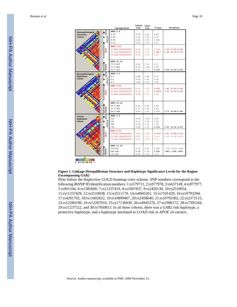
Figure 1. Linkage Disequilibrium Structure and Haplotype Significance Levels for the RegionEncompassing GAB2Plots follow the Haploview GOLD heatmap color scheme. SNP numbers correspond to thefollowing dbSNP ID identification numbers: 1:rs579711, 2:rs977978, 3:rs637149, 4:rs977977,5:rs901104, 6:rs1385600, 7:rs11237419, 8:rs1007837, 9:rs2450130, 10:rs2510054,11:rs11237429, 12:rs2510038, 13:rs2511170, 14:rs4945261, 15:rs7101429, 16:rs10793294,17:rs4291702, 18:rs11602622, 19:rs10899467, 20:rs2458640, 21:rs10793302, 22:rs2373115,23:rs12280198, 24:rs12287010, 25:rs17136630, 26:rs4945276, 27:rs1996172, 28:rs7395344,29:rs11237522, and 30:rs7950813. In all three cohorts, there was a GAB2 risk haplotype, aprotective haplotype, and a haplotype unrelated to LOAD risk in APOE ε4 carriers.
Reiman et al. Page 10
Neuron. Author manuscript; available in PMC 2008 November 25.
NIH
-PA Author Manuscript
NIH
-PA Author Manuscript
NIH
-PA Author Manuscript
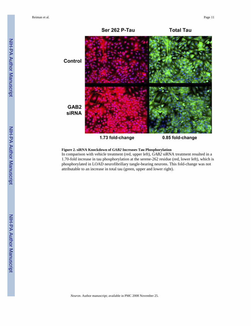
Figure 2. siRNA Knockdown of GAB2 Increases Tau PhosphorylationIn comparison with vehicle treatment (red, upper left), GAB2 siRNA treatment resulted in a1.70-fold increase in tau phosphorylation at the serene-262 residue (red, lower left), which isphosphorylated in LOAD neurofibrillary tangle-bearing neurons. This fold-change was notattributable to an increase in total tau (green, upper and lower right).
Reiman et al. Page 11
Neuron. Author manuscript; available in PMC 2008 November 25.
NIH
-PA Author Manuscript
NIH
-PA Author Manuscript
NIH
-PA Author Manuscript
Figure 3. Gab2 Colocalizes with Dystrophic Neurons in the AD Brain(A) LOAD hippocampus (neutral red counterstain) (40× objective). The arrow indicates ahighly dystrophic cell with the size and morphology of a cortical pyramidal neuron.Arrowheads point to one of many structures in the sections that resemble dystrophic neuritesor neuropil threads. (B) LOAD hippocampus (neutral red counterstain) (40×). The arrowdenotes a putative neurofibrillary tangle-containing neuron. Arrowheads again indicate adystrophic neurite. (C) LOAD posterior cingulate gyrus (40×). Filled arrows point to normal-appearing putative neurons. The open arrow points to a cell with the features of a neurofibrillarytangle-bearing neuron. Immunoreactive structures clearly resembling pyramidal cell apicaldendrites were also observed ascending through the cortical layers (arrowheads). (D) LOADposterior cingulate cortex (100× objective). Shown is a Gab2 immunoreactive cell with theflame-shaped cytoplasmic inclusion typical of the neurofibrillary tangle.
Reiman et al. Page 12
Neuron. Author manuscript; available in PMC 2008 November 25.
NIH
-PA Author Manuscript
NIH
-PA Author Manuscript
NIH
-PA Author Manuscript
NIH
-PA Author Manuscript
NIH
-PA Author Manuscript
NIH
-PA Author Manuscript
Reiman et al. Page 13Ta
ble
1G
AB2
SNPs
Impl
icat
ed in
the
Neu
ropa
thol
ogic
al D
isco
very
, Neu
ropa
thol
ogic
al R
eplic
atio
n, a
nd C
linic
al R
eplic
atio
n C
ohor
ts
Neu
ropa
thol
ogy
Dis
cove
ry C
ohor
tN
euro
path
olog
y R
eplic
atio
n C
ohor
tC
linic
al R
eplic
atio
n C
ohor
tO
vera
ll
dbsn
pR
S ID
Posi
tion
Ris
kA
llele
Freq
.C
ases
Freq
.C
ontr
ols
p V
alue
aO
R(9
5% C
I)Fr
eq.
Cas
esFr
eq.
Con
trol
sp V
alue
aO
R (9
5% C
I)Fr
eq.
Cas
esFr
eq.
Con
trol
sp V
alue
aO
R(9
5% C
I)p V
alue
aO
R(9
5% C
I)
rs90
1104
7760
8147
C0.
890.
724.
56E-
073.
24(2
.01–
5.20
)0.
870.
734.
52E-
022.
54(0
.90–
7.17
)0.
890.
801.
47E-
012.
08(0
.76–
5.88
)1.
99E-
062.
87(1
.84–
4.50
)rs
1385
600
7761
3814
T0.
890.
715.
02E-
073.
18(1
.99–
5.08
)0.
880.
701.
32E-
023.
00(1
.09–
8.24
)0.
950.
784.
63E-
025.
56(0
.89–
33.3
3)2.
81E-
093.
65(2
.34–
5.71
)rs
1007
837
7761
8724
C0.
890.
734.
78E-
063.
18(1
.99–
5.08
)0.
890.
733.
20E-
022.
85(1
.00–
8.11
)0.
880.
802.
04E-
011.
89(0
.70–
5.00
)3.
97E-
073.
01(1
.94–
4.68
)rs
2510
038
7764
3682
C0.
900.
748.
92E-
052.
44(1
.37–
4.36
)0.
880.
754.
35E-
022.
53(0
.88–
7.32
)0.
890.
801.
47E-
012.
08(0
.76–
5.88
)1.
19E-
052.
72(1
.72–
4.31
)rs
4945
261
7766
7908
G0.
890.
725.
66E-
073.
21(1
.99–
5.15
)0.
880.
734.
20E-
022.
54(0
.90–
7.17
)0.
950.
784.
63E-
025.
56(0
.89–
33.3
3)3.
06E-
083.
44(2
.18–
5.43
)rs
7101
429
7767
0615
A0.
890.
734.
62E-
052.
13(1
.21–
3.75
)0.
890.
731.
87E-
022.
84(1
.00–
8.11
)0.
880.
754.
99E-
022.
50(0
.98–
6.25
)1.
06E-
062.
96(1
.89–
4.63
)rs
1079
3294
7767
4051
C0.
820.
662.
98E-
052.
45(1
.60–
3.77
)0.
800.
591.
34E-
022.
66(1
.06–
6.57
)0.
880.
612.
17E-
024.
45(1
.18–
16.9
5)1.
59E-
072.
83(1
.90–
4.21
)rs
4291
702
7767
8896
C0.
880.
703.
73E-
063.
19(2
.00–
5.07
)0.
870.
722.
44E-
022.
70(0
.97–
7.52
)0.
880.
802.
04E-
011.
89(0
.70–
5.00
)5.
88E-
072.
96(1
.91–
4.59
)rs
7115
850
7772
2719
C0.
860.
671.
60E-
073.
21(2
.04–
5.05
)0.
860.
723.
91E-
022.
48(0
.90–
6.81
)0.
980.
723.
45E-
0314
.93
(1.6
1–14
0.85
)2.
80E-
103.
92(2
.51–
6.11
)rs
2373
115
7776
8798
G0.
880.
704.
60E-
073.
21(2
.04–
5.05
)0.
860.
723.
91E-
022.
48(0
.90–
6.81
)0.
980.
723.
45E-
0314
.93
(1.6
1–14
0.85
)9.
66E-
114.
06(2
.81–
14.6
9)
a p va
lues
wer
e co
mpu
ted
usin
g χ2
test
s. Fo
r the
ove
rall
com
paris
on, t
he C
ochr
an-M
ante
l-Hae
nsze
l χ2
test
was
use
d. A
ll p
valu
es a
re u
ncor
rect
ed.
Neuron. Author manuscript; available in PMC 2008 November 25.
NIH
-PA Author Manuscript
NIH
-PA Author Manuscript
NIH
-PA Author Manuscript
Reiman et al. Page 14Ta
ble
2G
AB
2 LO
AD
Odd
s Rat
ios i
n A
POE ε4
Car
riers
and
Non
carr
iers
APO
E ε4
gro
upAP
OE ε4
OR
(95%
CI)
rs23
7311
5 ge
noty
peC
ontr
ols (
n)C
ases
(n)
% L
OA
Drs
2373
115
OR
(95%
CI)
a
APO
E ε4−
GG
314
232
42.4
%1.
12 (0
.82–
1.53
)G
T/TT
121
100
45.2
%AP
OE ε4
+G
G63
406
86.6
%2.
88 (1
.90–
4.36
)G
T/TT
5412
169
.1%
All
sam
ples
6.07
(4.6
3–7.
95)
GG
377
638
62.9
%1.
34 (1
.06–
1.70
)G
T/TT
175
221
55.8
%
a All
rs23
7311
5 O
Rs a
re c
alcu
late
d us
ing
GG
ver
sus G
T/TT
.
Neuron. Author manuscript; available in PMC 2008 November 25.