fungal planet description sheets: 128–153
TRANSCRIPT

© 2012 Nationaal Herbarium Nederland & Centraalbureau voor Schimmelcultures
You are free to share - to copy, distribute and transmit the work, under the following conditions:Attribution: You must attribute the work in the manner specified by the author or licensor (but not in any way that suggests that they endorse you or your use of the work).Non-commercial: You may not use this work for commercial purposes.No derivative works: You may not alter, transform, or build upon this work.For any reuse or distribution, you must make clear to others the license terms of this work, which can be found at http://creativecommons.org/licenses/by-nc-nd/3.0/legalcode. Any of the above conditions can be waived if you get permission from the copyright holder. Nothing in this license impairs or restricts the author’s moral rights.
Persoonia 29, 2012: 146–201www.ingentaconnect.com/content/nhn/pimj http://dx.doi.org/10.3767/003158512X661589RESEARCH ARTICLE
Acknowledgements We thank the technical staff, A. van Iperen (cultures), M. Vermaas (photographic plates), and M. Starink-Willemse (DNA isolation, amplification and sequencing) for their invaluable assistance. Sincere thanks to Dr Barry Sneddon and Dr Patrick Brownsey for their help in confirming the host substrate (FP 130), and to Kerie McCombe and Andrew Millar for some of the photographs used. Kathie Hodge, Rebecca Bennett and D.H. DeFoe are thanked for collecting some of the specimens studied here (FP
150). The South African National Antarctic Programme is thanked for logistic support and Tristan da Cunha’s Conservation Department for permission to collect samples (FP 147). Fundação para a Ciência e a Tecnologia, Portugal is thanked for grant SFRH/BD/46038/2008 (M. Couthinho) and PEst-OE/BIA/UI0457/2011 (A.J.L. Phillips). The contribution of L. Marvanová is part of the project MSM 0021622416 of the Ministry of Education, Youth and Sports, Czech Republic.
Fungal Planet description sheets: 128–153P.W. Crous1, R.G. Shivas2, M.J. Wingfield3, B.A. Summerell4, A.Y. Rossman5, J.L. Alves6, G.C. Adams7, R.W. Barreto6, A. Bell8, M.L. Coutinho9, S.L. Flory10, G. Gates11, K.R. Grice12, G.E.St.J. Hardy13, N.M. Kleczewski14, L. Lombard1, C.M.O. Longa15, G. Louis-Seize16, F. Macedo9, D.P. Mahoney8, G. Maresi17, P.M. Martin-Sanchez18, L. Marvanová19, A.M. Minnis20, L.N. Morgado21, M.E. Noordeloos21, A.J.L. Phillips22, W. Quaedvlieg1, P.G. Ryan23, C. Saiz-Jimenez18, K.A. Seifert16, W.J. Swart24, Y.P. Tan2, J.B. Tanney16, P.Q. Thu25, S.I.R. Videira1, D.M. Walker26, J.Z. Groenewald1
Key words
ITS DNA barcodesLSUnovel fungal speciessystematics
Abstract Novel species of microfungi described in the present study include the following from Australia: Catenulostroma corymbiae from Corymbia, Devriesia stirlingiae from Stirlingia, Penidiella carpentariae from Carpentaria, Phaeococcomyces eucalypti from Eucalyptus, Phialophora livistonae from Livistona, Phyllosticta aristolochiicola from Aristolochia, Clitopilus austroprunulus on sclerophyll forest litter of Eucalyptus regnans and Toxicocladosporium posoqueriae from Posoqueria. Several species are also described from South Africa, namely: Ceramothyrium podocarpi from Podocarpus, Cercospora chrysanthemoides from Chrysanthemoides, Devriesia shakazului from Aloe, Penidiella drakensbergensis from Protea, Strelitziana cliviae from Clivia and Zasmidium syzygii from Syzygium. Other species include Bipolaris microstegii from Microstegium and Synchaetomella acerina from Acer (USA), Brunneiapiospora austropalmicola from Rhopalostylis (New Zealand), Calonectria pentaseptata from Eucalyptus and Macadamia (Vietnam), Ceramothyrium melastoma from Melastoma (Indonesia), Collembolispora aristata from stream foam (Czech Republic), Devriesia imbrexigena from glazed decorative tiles (Portugal), Microcyclospora rhoicola from Rhus (Canada), Seiridium phylicae from Phylica (Tristan de Cunha, Inaccessible Island), Passalora lobeliaefistulosis from Lobelia (Brazil) and Zymoseptoria verkleyi from Poa (The Netherlands). Valsalnicola represents a new ascomycete genus from Alnus (Austria) and Parapenidiella a new hyphomycete genus from Eucalyptus (Australia). Morphological and culture characteristics along with ITS DNA barcodes are also provided.
Article info Received: 1 October 2012; Accepted: 26 October 2012; Published: 20 December 2012.
1 CBS-KNAW Fungal Biodiversity Centre, Uppsalalaan 8, 3584 CT Utrecht, The Netherlands; corresponding author e-mail: [email protected].
2 Biosecurity Queensland, Ecosciences Precinct, Level 2C East, GPO Box 267, Brisbane 4001, Queensland, Australia.
3 Forestry and Agricultural Biotechnology Institute, University of Pretoria, South Africa.
4 Royal Botanic Gardens and Domain Trust, Mrs. Macquaries Road, Syd-ney, NSW 2000, Australia.
5 Systematic Mycology & Microbiology Laboratory, USDA-ARS, Rm. 246, B010A, 10300 Baltimore Ave., Beltsville, MD 20705, USA.
6 Universidade Federal de Viçosa UFV, Campus Universitário, 36570-00, Viçosa, Brazil.
7 Department of Plant Pathology, University of Nebraska, Lincoln, NE 68583, USA.
8 Gurney Road 45, Lower Hutt, New Zealand. 9 REQUIMTE – CQFB and Departamento de Conservação e Restauro,
Faculdade de Ciências e Tecnologia, Universidade Nova de Lisboa, Monte de Caparica, 2829-516 Caparica, Portugal.
10 Agronomy Department, University of Florida, Gainesville, FL 32611, USA.11 School of Plant Science, University of Tasmania, Hobart, Australia.12 Agri-Science Queensland, PO Box 1054, Mareeba 4880, Queensland,
Australia.13 School of Biological Sciences and Biotechnology, Murdoch University,
Murdoch, Western Australia, 6150.14 Department of Botany and Plant Pathology, Purdue University, South-
west Purdue Agricultural Program, 4369 North Purdue Rd., Vincennes, IN 47591, USA.
15 FEM-IASMA – Research and Innovation Centre, Sustainable Agro-Ecosystems and Bioresources Department. Via E. Mach 1, 38010 San Michele all’Adige (TN), Italy.
16 Biodiversity (Mycology & Botany), Agriculture & Agri-Food Canada, 960 Carling Ave., Ottawa, Ontario K1A 0C6, Canada.
17 IASMA - Centre for Technology Transfer, Via E. Mach 1, 38010 San Michele all’Adige (TN), Italy.
18 Instituto de Recursos Naturales y Agrobiologia, IRNAS-CSIC, Av. Reina Mercedes 10, 41012 Sevilla, Spain.
19 Czech Collection of Microorganisms, Institute of Experimental Bio logy, Faculty of Science, Masaryk University, Tvrdého 14, 602 00 Brno, Czech Republic.
20 Center for Forest Mycology Research, Northern Research Station, USDA- Forest Service, One Gifford Pinochet Dr., Madison, WI 53726, USA.
21 National Herbarium of the Netherlands, Naturalis Biodiversity Center, Leiden University, P.O. Box 9514, 2300 RA Leiden, The Netherlands.
22 CREM, Departamento de Ciências da Vida, Faculdade de Ciências e Tecnologia, Universidade Nova de Lisboa, Monte de Caparica, 2829-516 Caparica, Portugal.
23 Percy FitzPatrick Institute of African Ornithology, University of Cape Town, Rondebosch 7701, South Africa.
24 Department of Plant Pathology, University of the Free State, P.O. Box 339, Bloemfontein 9300, South Africa.
25 Forest Science Institute of Vietnam, Dong Ngac, Tu Liem, Hanoi, Viet-nam.
26 Department of Natural Sciences, The University of Findlay, Findlay, OH 45840, USA.

147Fungal Planet description sheets
© 2012 Nationaal Herbarium Nederland & Centraalbureau voor Schimmelcultures
Neighbour-joining tree obtained using a distance analysis with a general time reversible (GTR) substitution model on the partial 28S nrRNA gene alignment (817 nucleotides including alignment gaps) as implemented in PAUP v. 4.0b10 (Swofford 2003). Novel species are indicated in a bold font and the orders are indicated on the right-hand side of the figure. The scale bar indicates the number of substitutions per site and the bootstrap support values (based on 1 000 replicates) are shown by colour-coded dots for values > 79 % (see legend on figure). The tree was rooted to a sequence of Saccharomyces cerevisiae (GenBank Z73326.)
Incertae sedis
Botryosphaeriales
Capnodiales
Xylariales
Hypocreales
Diaporthales
Pleosporales
Helotiales
Chaetothyriales
Bootstrap supportvalues: = 100 % = 95 % to 99 % = 90 % to 94 % = 80 % to 89 %
0.10
Saccharomyces cerevisiae Z73326 Dothideomycetes sp. GU323986
Fungal Planet 131 - Phyllosticta aristolochiicola JX486128
Phaeococcomyces nigricans AF050278
Phyllosticta abietis EU754193 Phyllosticta minima EU754194
Fungal Planet 133 - Phaeococcomyces eucalypti CPC 17606 Dothideomycetes sp. GU323985
Toxicocladosporium irritans EU040243 Toxicocladosporium strelitziae JX069858 Toxicocladosporium pseudoveloxum JF499868
Seiridium unicorne DQ414532
Fungal Planet 146 - Penidiella carpentariae CPC 19439
Zasmidium citri GQ852733
Fungal Planet 144 - Toxicocladosporium posoqueriae CPC 19305
Setosphaeria monoceras AY016368
Fungal Planet 132 - Calonectria pentaseptata CBS 133349
Fungal Planet 147 - Seiridium phylicae CPC 19964 Seiridium eucalypti DQ414533
Fungal Planet 134 - Penidiella drakensbergensis CPC 19778
Ramichloridium cerophilum GU214485
Zymoseptoria passerinii JQ739843
Cladosporium ramotenellum JF499859 Cladosporium grevilleae JF770462
Dendryphiella salina EU848587
Exophiala placitae EU040215 Fungal Planet 135 - Ceramothyrium melastoma CPC 19837 Strelitziana australiensis GQ303326 Fungal Planet 136 - Strelitziana cliviae CPC 19822
Calonectria queenslandica GQ280741 Calonectria terrae-reginae GQ280779
Fungal Planet 128 - Valsalnicola oxystoma JX519563 Melanconis marginalis AF408373 Gnomonia petiolorum AY818963 Amphiporthe hranicensis DQ323521
Fungal Planet 148 - Microcyclospora rhoicola
Parapenidiella pseudotasmaniensis GQ852625 Parapenidiella tasmaniensis GU214452
Fungal Planet 145 - Catenulostroma corymbiae CPC 19437
Fungal Planet 139 - Devriesia shakazului CPC 19782
Fungal Planet 141 - Devriesia stirlingiae CPC 19948
Zasmidium nocoxi GQ852735 Fungal Planet 140 - Zasmidium syzygii CPC 19792
Fungal Planet 142 - Cercospora chrysanthemoides CPC 20529 Cercospora sojina GU253861 Mycosphaerella valgourgensis JF951175
Fungal Planet 143 - Zymoseptoria verkleyi CBS 133618
Fungal Planet 129 - Bipolaris microstegii AR 4840
Fungal Planet 150 - Synchaetomella acerina DAOM 242271
Pilidium acerinum AY487089 Pilidium concavum AY487098
Collembolispora barbata CBS 115944 Fungal Planet 149 - Collembolispora aristata CPC 21145 Cadophora luteo-olivacea HM116760 Cadophora fastigiata JN938877
Vonarxia vagans FJ839673
Ceramothyrium carniolicum FJ358232
Cyphellophora laciniata FJ358239 Phialophora reptans EU514699 Fungal Planet 138 - Phialophora livistonae CPC 19433 Phialophora sessilis EU514700
Microcyclospora malicola GU570550
Catenulostroma chromoblastomycosum EU019251 Teratosphaeria encephalarti FJ372417
Devriesia hilliana GU214414 Fungal Planet 151 - Devriesia imbrexigena CAP1373
Devriesia lagerstroemiae GU214415 Teratosphaeria knoxdaviesii EU707865
Fungal Planet 152 - Passalora lobelia-fistulosae VIC 31840
Zymoseptoria tritici EU019298 Zymoseptoria brevis JQ739832
Cochliobolus heterostrophus AY544645 Cochliobolus sativus DQ678045
Chaetomella acutiseta AY544679 Chaetomella oblonga AY487083
Cyphellophora eugeniae FJ839652
Fungal Planet 137 - Ceramothyrium podocarpi CPC 19826 Ceramothyrium thailandicum HQ895835
Microcyclospora pomicola GU570551 Microcyclospora tardicrescens GU570552
Passalora brachycarpa GU214664 Pantospora guazumae JN190954
Exophiala eucalyptorum EU035417 Cyphellophora hylomeconis EU035415

148 Persoonia – Volume 29, 2012
Valsalnicola oxystoma
c
b
d
a
e

149Fungal Planet description sheets
© 2012 Nationaal Herbarium Nederland & Centraalbureau voor Schimmelcultures
Basionym. Valsa oxystoma Rehm, Ber. Naturhist. Vereins Augsburg 26: 70. 1881.
≡ Cryptodiaporthe oxystoma (Rehm) Z. Urb., Preslia 29: 395. 1957.
Twig lesions in surface view (511–)591–890(–893) µm diam (mean = 654, S.D. 122, n = 13). Ectostroma well-developed, brown to black, thick disc from which perithecial necks emerge. Ascomatal cavity (690–)765–909(–950) µm high × (1610–)1710–2346(–3947) µm diam (mean = 816 × 2198, S.D. 109, 703, n1 = 5, n2 = 9). Ascomata perithecial, immersed beneath ectostroma, causing host tissue to swell and rupture, perithe-cia converging into 5–20 necks, emerging at surface through ecto stromatic disc, perithecia grouped 13–23. Ascomata glossy black, subglobose to globose (240–)266–298(–320) µm high × (253–)260–335(–337) µm diam (mean = 282 × 294, S.D. 25, 36, n1 = 7, n2 = 13); necks central, straight to curved, length (426–)428–550(–563) µm (mean = 476, S.D. 54, n = 9). Asci fusiform, (38–)39–48(–49) × (8–)9–12(–13) µm (mean = 44×11, S.D. 4, 1.2, n1 = 17, n2 = 18), apex broadly rounded, with indistinct apical ring, stipe acute, rounded, or ta-pering to a point, ascospores arranged irregularly multiseriate. Ascospores allantoid with rounded ends, mostly curved, rarely straight, (9–)10–11(–12) × 2–3 µm (mean = 11 × 2, S.D. 0.9, 0.5, n = 30), 1-septate, median, slightly constricted or not at septum, each cell with several small guttules, hyaline. Cultures slow-growing, 3–6 mm in 10 d on potato-dextrose agar, my-celium low, pale brown to greyish brown, reverse dark brown. In culture on synthetic nutrient-poor agar — Dimorphic, forming a synanamorph. Conidiomata pycnidial, exuding masses of brown conidia. Conidiophores reduced to conidio-genous cells, or one supporting cell, proliferating percurrently. Conidia cylindrical, brown, finely verruculose, apex obtuse, base truncate, 3–5-euseptate, 15–23 × 4–5 µm. Conidia of synanamorph intermingled in same conidioma, but conidio-genous cells proliferating percurrently or sympodially; conidia hyaline to subhyaline, narrowly obclavate, apex subobtuse, base truncate, straight to curved, 25–80 × 2.5–3 µm, up to 11-septate. Synanamorph also developing in aerial mycelium (on PNA); conidiophores subcylindrical, straight to curved, 0–2-septate, hyaline to subhyaline, 8–15 × 2–3 µm, prolifer-ating sympodially at apex. Conidiophores solitary or fascicu-late or on a reduced stroma.
Fungal Planet 128 – 20 December 2012
Valsalnicola D.M. Walker & Rossman, gen. nov. Etymology. Named for its valsa-like appearance and occurrence on species of Alnus.
Causing linear cankers and lesions. Ectostromata well-devel-oped, brown to black, thick disc from which perithecial necks emerge. Ascomata perithecial, immersed beneath ectostroma,
aggregated in groups of 13–23, converging into 5–20 necks. Asci fusiform, with indistinct apical ring. Ascospores allantoid with rounded ends, 1-septate, hyaline.
Type species. Valsalnicola oxystoma. MycoBank MB801277.
Valsalnicola oxystoma (Rehm) D.M. Walker & Rossman, comb. nov. Typus. AustriA, Tyrol, Längenfeld, on dead branch of Alnus viridis, c. 3 500 ft., Aug. 1874, coll. Rehm. This type specimen was issued as Rehm, Ascomyceten no. 280. Of the two specimens at BPI, the more plenti-ful one is in the bound set of Rehm, Ascomyceten, and is herein designated as Lectotype BPI 884138. Isolectotypes examined BPI 738235 and NY, MycoBank MB801277.
Additional specimens examined. Belgium, Brussels, Soignes, on branch of Alnus glutinosa, Oct. 1899, P. Nypels, comm. H. Rehm, Vestergren, Micro mycetes rariores selecti 409 as Valsa oxystoma (BPI 574854). – CAn-AdA, British Columbia, Yoho National Park, Chancellor Mountain Camp, on Alnus sp., 11 Aug. 1962, R.F. Cain, TRTC 40116 (NY); Ontario, Kenora District, Tustin Township, Gordon Lake, Rd., on Alnus sp., 26 Sept. 1959, coll. D. Bowen, det. J. Reid as Valsa oxystoma (BPI 574855). – itAly, Tren-to, Monte Bondone Trento, E11°03'51" N46°02'20", on Alnus viridis, Apr. 2011, G. Maresi, isol. A. Rossman AR 4833 = CBS 133337, ITS sequence JX519559, and LSU sequence JX519563 (BPI 884137); Trento, val Sadole (E11.60, N46.15), 2009, G. Maresi & C.M.O. Longa (BPI 884136). – swe-den, Umea, on dead branch of Alnus ‘borealis’, Sept. 1910, Vlengel, det. F. Bubak (BPI 574856). – USA, Alaska, near Fairbanks, Moose Creek, En-vironmental Monitoring Plot 316 MC UM11 MRC, N64.72, W147.23, elev. 150 m, on Alnus incana var. tenuifolia, May 2010, G.C. Adams, culture AR 5137 = CBS 133329, ITS sequence JX519561 (BPI 884135).
Habitat — Alnus viridis ssp. viridis, causing a twig coloniza-tion and canker disease involved in green alder decline (Pi-setta et al. 2012); also known from Alnus glutinosa, A. incana, A. incana var. tenuifolia, A. rubra, A. viridis ssp. fruticosa and A. viridis ssp. maximowiczii. Distribution — Asia: Japan (Kobayashi 2007); Europe: Aus-tria, Belgium, Italy, Sweden, also United Kingdom (Cannon et al. 1985); North America: Canada (Ontario); USA: Alaska.
Notes — Valsalnicola is based on a species that was de-scribed in the genus Valsa. Although it resembles Valsa in having allantoid ascospores, the ascospores of Valsalnicola are 1-septate while the majority of species of Valsa and closely related Leucostoma and Valsella have aseptate ascospores. However, one species of Valsa, V. melanodiscus, also has 1-septate ascospores, occurs on Alnus spp., and produces lin-ear cankers on the host. A distinguishing feature of Valsalnicola is the lack of a black line surrounding stromata in the asco-matal cavity, which is characteristic of Valsa melanodiscus. In addition, the growth rate of cultures of Valsalnicola oxystoma is considerably slower than species of Valsa. Molecular se-quence data place this new genus within the GnomoniaceaeMelanconidaceae complex. Allantoid, 1-septate ascospores have not previously been reported in the Gnomoniaceae or Melanconidiaceae. ITS sequences of specimens from Alaska and Italy are identical. The basionym has been cited as Rehm: Ascomyceten 270 (1875) in ‘Index Fungorum’ reflecting an er-ror in Saccardo (1882) but the correct number is Rehm: Asco-myceten 280, which does not include a description.
Donald M. Walker, Department of Natural Sciences, The University of Findlay, Findlay, OH 45840, USA; e-mail: [email protected] Y. Rossman, Systematic Mycology & Microbiology Laboratory, USDA-ARS, Beltsville, MD 20705, USA; e-mail: [email protected]
Gerard C. Adams, Department of Plant Pathology, University of Nebraska, Lincoln, NE 68583, USA; e-mail: [email protected] Maria Oliveira Longa, FEM-IASMA - Research and Innovation Centre, Sustainable Agro-Ecosystems and Bioresources Department. Via E. Mach 1,
38010 San Michele all’Adige (TN), Italy; e-mail: [email protected] Maresi, IASMA - Centre for Technology Transfer, Via E. Mach 1, 38010 San Michele all’Adige (TN), Italy; e-mail: [email protected]
Colour illustrations. Italy, Trentino, Val Sadole, showing trees of Alnus viridis with green alder decline (Giorgio Maresi). a. Rehm: Ascomyceten 280, scale bar = 500 µm. b–d. BPI 884137, scale bars of perithecia = 100 µm, scale bar of ascus = 10 µm. e. Rehm: Ascomyceten 280, scale bar = 10 µm.

150 Persoonia – Volume 29, 2012
Bipolaris microstegii

151Fungal Planet description sheets
© 2012 Nationaal Herbarium Nederland & Centraalbureau voor Schimmelcultures
Etymology. Named after the host, Microstegium vimineum (Poaceae), from which the species was isolated originally.
Leaf spots on Microstegium, up to 2 × 0.5 cm, ellipsoid to irregular, brown with a darker, near black border. Conidiophores macronematous, mononematous, erect, more or less straight to slightly flexuous, simple or with a single dichoto-mous branch, cylindrical, geniculate at apex, pale to medium brown, often darker towards apices, smooth walled, septate, up to at least 750 µm long × 5–8 µm diam. Conidiogenous cells integrated, terminal or intercalary, with sympodial prolif-eration, monotretic or polytretic with darkened, circular scars. Conidia solitary, curved, cylindrical to obclavate, apex obtuse, base obtuse with inconspicuous hilum, pale brown, becoming medium to dark brown, end cells usually paler, walls smooth or faintly granulose, 5–10 distoseptate, with septa becoming accentuated at maturity, 40–97.5(–105) × 12.5–15(–17.5) µm, Q = 3.2–7.8 (Lm = 69.6 µm, Wm = 13.5 µm, Qm = 5.2). Germination via a germ tube at each end cell of conidium. Culture characteristics — Colonies 10–44(–70) mm diam on potato-dextrose agar (Difco) after 7 d at 24 °C with a 12 h light/dark diurnal cycle; surface near dull green (30D4, 30E3), dark green (28F3, 30F3), to greenish grey (28F2), velutinous to tomentose with sparse, white, aerial hyphae and dark co-nidiophores; margin uneven and lobed, whitish; reverse near greenish grey (30F2), dark green (30F5), to almost black.
Typus. USA, West Virginia, near Arnoldsburg, Crummies Creek Tree Farm, on living leaves of Microstegium vimineum, Aug. 2009, coll. R. Richardson, Bipolaris 4 isolated by N.M. Kleczewski, holotype BPI 883727 (dried culture on PDA); culture ex-type CBS 132550; ITS sequence Gen-Bank JX089579, gpd sequence GenBank JX089575, LSU sequence Gen-Bank JX100808, MycoBank MB801569.
Additional specimens examined. USA, West Virginia, near Arnoldsburg, cove near Crummies Creek Tree Farm, on living leaves of Microstegium vimineum, Aug. 2009, coll. R. Richardson, Bipolaris 2 isolated by N.M. Kleczewski, BPI 883728 (dried culture on PDA); culture CBS 132549; ITS sequence GenBank JX089577 and gpd sequence GenBank JX089573; sa-vannah near Crummies Creek Tree Farm, on living leaves of Micro stegium vimineum, Aug. 2009, coll. R. Richardson, Bipolaris 3 isolated by N.M. Kleczewski, BPI 883729 (dried culture on PDA); culture CBS 132548; ITS sequence GenBank JX089578 and gpd sequence GenBank JX089574.
Phylogenetic analysis — The gpd and ITS sequences for all three isolates, Bipolaris 2–4 (Flory et al. 2011), were iden-tical. A concatenated alignment of both loci was made using sequence data from the ex-type and Group 1 species (Berbee
et al. 1999). A maximum likelihood search was then performed using the RAxML BlackBox (http://phylobench.vital-it.ch/rax-ml-bb/) with gamma, partitioned model, and per gene branch length optimization; 100 bootstrap replicates were included.
Notes — The microscopic description is based on PDA cul-tures and colony colour is based on Kornerup & Wanscher (1978). Many species of Bipolaris are important pathogens of grasses. This new species was isolated from Microstegium vimineum, an invasive plant in the USA. The fungus causes disease on Microstegium, but it also infects a wider range of hosts (Kleczewski & Flory 2010, Flory et al. 2011, Kleczewski et al. 2012). Comparison of ITS and gpd sequence data to sequences in GenBank and subsequent phylogenetic analy-ses based on Group 1 species (Berbee et al. 1999), referred to herein as Bipolaris (sensu Manamgoda et al. 2012), sug-gest that the present species is distinct and closely related to B. victoriae and B. zeicola. These species of Bipolaris consist of a highly pathogenic species complex that shows large dif-ferences in virulence and host ranges in spite of few genetic differences in the sequenced loci. Using Sivanesan (1987), B. microstegii is morphologically similar to B. miyakei and B. zeicola. A probable original culture of B. miyakei (CBS 197.29) is not closely related to B. microstegii based on ITS (JX089580) and gpd (JX089576) sequences. Bipolaris microstegii differs from B. zeicola by its longer and sometimes branched conidio phores.Microstegium vimineum is native to Asia. Several isolates of Bipolaris are known from Microstegium in Asia (Shimizu et al. 1998), but the origin of B. microstegii is unknown. Species of Bipolaris in Group 1 (Berbee et al. 1999) are highly pathogen-ic on a wide range of native and non-native hosts and these include major pathogens of corn and oats.
The best scoring tree from the maximum likelihood analysis. Bootstrap values ≥ 70 % are indicated. GenBank numbers of included sequences for each species are given as gpd/ITS. An asterisk denotes that gpd and ITS sequences were from different isolates.
Fungal Planet 129 – 20 December 2012
Bipolaris microstegii Minnis, Rossman, Kleczewski & S.L. Flory, sp. nov.
Colour illustrations. Landscape invaded by Microstegium vimineum; leaf spots on M. vimineum; surface view of culture on PDA; conidiophore; co-nidia. Scale bars = 30 µm.
Andrew M. Minnis, Center for Forest Mycology Research, Northern Research Station, USDA-Forest Service, One Gifford Pinochet Dr., Madison, WI 53726, USA; e-mail: [email protected]
Amy Y. Rossman, Systematic Mycology & Microbiology Laboratory, USDA-ARS, Rm. 246, B010A, 10300 Baltimore Ave., Beltsville, MD 20705, USA; e-mail: [email protected]
Nathan M. Kleczewski, Department of Botany and Plant Pathology, Purdue University, Southwest Purdue Agricultural Program, 4369 North Purdue Rd., Vincennes, IN 47591, USA; e-mail: [email protected]
S. Luke Flory, Agronomy Department, University of Florida, Gainesville, FL 32611, USA; e-mail: [email protected]

152 Persoonia – Volume 29, 2012
Brunneiapiospora austropalmicola

153Fungal Planet description sheets
© 2012 Nationaal Herbarium Nederland & Centraalbureau voor Schimmelcultures
Etymology. austropalmicola, meaning Southern palm referring to the Nikau palm (Rhopalostylis sapida) upon which the fungus was found.
Ascomata perithecial, in small clusters developing on black-ened stroma bursting through the plant tissue. Individual as-comata black, c. 1 mm diam, densely covered with brown, septate hairs mixed with host tissue, each with small papillate ostiole. Outer peridium black, brittle and structure less, inner peridium composed of areolate tissue. Copious centrum con-tents embedded in sticky material. Paraphyses hyaline, free-ended, longer than asci with densely granular contents c. 3–4 µm wide. Asci cylindrical, c. 250 × 7 µm, (tapering stipe con-stituting approx. a quarter of the length), ascus with prominent apical J+ ring, each ascus containing 8 uniseriate to overlap-ping ascospores. Ascospores 2-celled, septate in the lower part, upper cell pale brown, fusiform and symmetrical in one view, but flattened on one side sometimes strongly so, rather variable in size ranging from 19–30 × 3–5 μm (n = 50), lower hyaline cell 3–4 μm long.
Typus. new ZeAlAnd, on dead water-soaked fibrous Rhopalostylis sapida, Rimutaka Forest Park, 9 Nov. 2011, Bell & Mahoney Herb. no. 1172 (holotype PDD 102614), MycoBank MB800261.
Notes — During a recent foray into Rimutaka Forest Park near Wellington a new species of Brunneiapiospora was found on dead portions of Rhopalostylis sapida (Nikau palm). For a full description of the former placement of fungi with apiospo-rous ascospores the reader is referred to the paper by Hyde et al. (1998). In it the genus Brunneiapiospora was established to accommodate apiosporous species with cylindrical asci and whose ascospores consist of a larger brown cell and a smaller basal hyaline cell. It differs from the apiosporous ge-nus Anthostomella which have broadly cylindrical asci and as-cospores usually provided with a prominent longitudinal germ slit in the darker ascospore cell. Hyde et al. (1998) provide a key to the six known species of Brunneiapiospora all of which are pan-tropical in origin found on decaying material of palms in Ecuador, tropical Australia, Sierra Leone, Tanzania and Indonesia. They placed the genus (together with other gen-era), in a new family the Apiosporaceae. Kang et al. (1999) redefine the family Clypeosphaeriaceae, and indicate that the genus Brunneiapiospora might be placed therein, although their earlier molecular studies on the Amphisphaeriales (Kang et al. 1998) did not include any Brunneiapiospora samples. Our species B. austropalmicola differs in the ascospore di-mensions from those previously described. They are approxi-mately the length of B. deightoniella but much narrower (3–5 µm wide vs 7.5–10 µm for B. deightoniella). It is also the first described species of the genus from the cooler climates typi-cal of the temperate rain forests of New Zealand.The substrate upon which this species was found was quite unlike the woody substrate, which we normally collect on for-ays. It was quite friable and light in both colour and weight. At first we considered it could be from a tree fern trunk but this was proved not to be the case when we consulted those with a good knowledge of fern anatomy. Since all other species of Brunneiapiospora have been found on palms, we set about making several slides of the substrate together with portions of freshly collected Rhopalostylis sapida. By examination of these and conferring with the article by Tomlinson (2006), we are confident that the abraded material upon which B. austropalmicola was growing is a stem portion of the palm Rhopalostylis sapida. This palm is common in the Rimutaka Forest Park.
Fungal Planet 130 – 20 December 2012
Brunneiapiospora austropalmicola A.E. Bell & Mahoney, sp. nov.
Ann Bell & Daniel P. Mahoney, 45, Gurney Road, Lower Hutt, New Zealand; e-mail: [email protected]
Colour illustrations. Forest of Rhopalostylis sapida in Nikau Reserve, Paraparaumu, New Zealand (www.wikimedia.org). Photo plate: A–A¹. Paraphyses, asci and ascospores; B. ascus and ascospores; C, D. as-cospores; E. areolate peridial fragment; F. ascus apical ring complex in Melzer’s reagent. All except F in Shear’s mounting fluid. A–A¹ phase mi-croscopy, others brightfield. Scale bars: A–A¹, B, E = 25 µm, C, D = 10 µm, F = 5 µm. Water colour: A. Perithecia on substrate; B. excised perithecium showing vestiture; C. aerolate inner perithecial tissue; D. paraphyses and asci; E. mature ascospores; F. scus apical rings showing J+ reaction in Melzer’s reagent.

154 Persoonia – Volume 29, 2012
Phyllosticta aristolochiicola

155Fungal Planet description sheets
© 2012 Nationaal Herbarium Nederland & Centraalbureau voor Schimmelcultures
Etymology. Name derived from the host plant genus, Aristolochia (Aristolochiaceae).
Leaf spots amphigenous, circular, up to 1 cm diam, grey to pale brown, solitary, surrounded by a slightly raised black border about 1 mm wide; centres of lesions often tear or fall out producing symptoms of shot-hole. Conidiomata pycnidial, mostly epiphyllous, black, solitary, unilocular, globose, 40–70 μm diam, erumpent; wall composed of layers of textura angularis, outer layer dark reddish brown. Conidiophores reduced to conidiogenous cells or with a supporting branched cell. Conidiogenous cells terminal, hyaline, smooth, subcylindrical to ampulliform, 10–20 × 2–4 μm. Conidia globose, subglobose, broadly ellipsoidal or obovoid, with a truncate base and round-ed apex, hyaline, 7–16 × 6.5–11 μm, aseptate; wall uniformly 0.5–1 μm thick, enclosed in a mucilaginous sheath, with a minute basal frill and an apical hyaline tapered appendage 3–7 μm long. Teleomorph not observed. Culture characteristics — (after 1 wk in the dark and a further 2 wk under 12 h ultraviolet light / 12 h dark cycle, at 23 °C): Colonies on potato-dextrose agar 4 cm diam, flat with no aerial mycelium, olivaceous black (Rayner 1970) with a white-grey, 2 mm entire margin, narrowly zonate towards the margin.
Typus. AustrAliA, Queensland, Kuranda, Kennedy Highway, on leaves of Aristolochia acuminata, 1 Apr. 2010, K.R.E. Grice & P. Wright (holotype BRIP 53316a; includes ex-type culture), ITS sequence GenBank JX486129, LSU sequence GenBank JX486128; Queensland, Emmagen Creek, Cape Tribulation National Park, 1 Aug. 1993, R.G. Shivas, paratype BRIP 21785, MycoBank MB801322.
Notes — Species of Phyllosticta have Guignardia sexual morphs, and are common endophytes or pathogens, occur-ring on a wide range of plant hosts (Glienke et al. 2011). Two species of Phyllosticta, P. aristolochiae on A. clematitis and P. aristolochiae (replacement name P. tassiana) on A. sempervirens, have been described from Aristolochia. Neither species was considered a Phyllosticta in a more recent revi-sion of the genus (van der Aa & Vanev 2002). Furthermore, the latter name and its replacement name (P. tassiana) were both homonyms and thus both are illegitimate (van der Aa & Vanev 2002). Phyllosticta aristolochiicola was first collected in north Queensland in 1993 in association with leaf spot and shot-hole of Aristolochia (Shivas & Alcorn 1996). Based on a megablast search of NCBIs GenBank nucleotide database, the closest hit using the ITS sequence is Phyllosticta cordylinophili (GenBank AB454357; Identities = 591/612 (97 %), Gaps = 5/612 (1 %)), followed by Phyllostica ardisiicola (GenBank AB454274; Identities = 584/614 (95 %), Gaps = 10/614 (2 %)), and Guignardia vaccinii (GenBank JQ936158; Identities = 583/614 (95 %), Gaps = 6/614 (1 %)). Using the LSU se-quence, the closest hits are to Phyllosticta abietis (GenBank EU754193; Identities = 1311/1328 (99 %), Gaps 0/1328 (0 %)), followed by Phyllosticta bidwellii (GenBank DQ678085; Iden-ti ties = 1299/1313 (99 %), Gaps = 0/1313 (0 %)), and Phyl losticta minima (GenBank EU754194; Identities = 1291/1303 (99 %), Gaps = 0/1303 (0 %)).
Fungal Planet 131 – 20 December 2012
Phyllosticta aristolochiicola R.G. Shivas, Y.P. Tan & Grice, sp. nov.
Colour illustrations. Aristolochia acuminata with leaf spots associated with P. aristolochiicola at Kuranda, northern Queensland; leaf spot with pyc-nidia; 3 wk old culture on potato-dextrose agar; conidiophores and conidia; conidia with appendages apparent. Scale bars (from top left to bottom right) = 1 mm, 1 cm, 10 µm, 10 µm.
Roger G. Shivas & Yu Pei Tan, Biosecurity Queensland, Ecosciences Precinct, Level 2C East, GPO Box 267, Brisbane 4001, Queensland, Australia; e-mail: [email protected] & [email protected]
Kathy R. Grice, Agri-Science Queensland, PO Box 1054, Mareeba 4880, Queensland, Australia; e-mail: [email protected]

156 Persoonia – Volume 29, 2012
Calonectria pentaseptata

157Fungal Planet description sheets
© 2012 Nationaal Herbarium Nederland & Centraalbureau voor Schimmelcultures
Etymology. Name refers to the 5-septate macroconidia produced by this fungus.
Sexual morph unknown. Conidiophores consisting of a stipe bearing a suit of penicillate fertile branches, a stipe extension, and terminal vesicle; stipe septate, hyaline, smooth 47–133 × 6–10 µm; stipe extension septate, straight to flexuous, 168–350 µm long, 3–6 µm wide at the apical septum, terminating in a narrowly clavate vesicle, 2–6 µm diam. Conidiogenous apparatus 70–99 µm long, 23–90 µm wide; primary branches 0–1-septate, 19–31 × 4–7 µm; secondary branches aseptate, 16–34 × 4–7 µm; tertiary branches aseptate, 14–22 × 4–6 µm, each terminal branch producing 1–3 phialides; phialides cylindrical to allantoid, obpyriform when carried singly, hya-line, aseptate, 15–24 × 4–6 µm; apex with minute periclinal thickening and inconspicuous collarette. Macroconidia cylin-drical, rounded at both ends, straight, (75–)87–109(–115) × (5–)6–8(–10) µm (av. = 98 × 7 µm), 5(–8)-septate, lacking a visible abscission scar, held in parallel cylindrical clusters by colourless slime. Megaconidia and microconidia not seen. Culture characteristics — (in the dark, 24 °C after 1 wk): Colonies fast growing, with optimum growth at 24 °C on MEA; surface sienna to dark brick, reverse sepia-brown; abundant aerial mycelium and sporulation; chlamydospores extensive throughout the medium, forming microsclerotia.
Typus. VietnAm, Bavi, Hanoi, Eucalyptus hybrid, Sept. 2011, P.Q. Thu, holo- type CBS H-21062, culture ex-type CBS 133349, β-tubulin (TUB) sequence GenBank JX855942, Histone H3 (HIS3) sequence GenBank JX855946, ITS sequence GenBank JX855950, LSU sequence GenBank JX855954 and translations elongation factor 1-alpha (TEF1-α) sequence GenBank JX855958, MycoBank MB801468.
Other specimens examined. VietnAm, Bavi, Hanoi, Macadamia sp., Sept. 2011, P.Q. Thu, CBS 133351, TUB sequence GenBank JX855944, HIS3 sequence GenBank JX855948, ITS sequence GenBank JX855952, LSU sequence GenBank JX855956 and TEF1-α sequence GenBank JX855960; ibid., E. urophylla, Sept. 2011, P.Q. Thu, CBS 133350, TUB sequence GenBank JX855943, HIS3 sequence GenBank JX855947, ITS sequence GenBank JX855951 and TEF1-α sequence GenBank JX855959; ibid., Eucalyptus hybrid, Sept. 2011, P.Q. Thu, CBS 133352, TUB sequence GenBank JX855945, HIS3 sequence GenBank JX855949, ITS sequence GenBank JX855953 and TEF1-α sequence GenBank JX855961.
Notes — Calonectria pentaseptata resides in the C. reteaudii species complex (Kang et al. 2001, Lombard et al. 2010a, b, c) based on morphological characteristics supported by phylogenetic inference. The macroconidia of C. pentaseptata (av. = 98 × 7 µm) are smaller than those of C. pseudoreteaudii (av. = 104 × 8 µm), and larger than those of C. queenslandica (av. = 69 × 6 µm), C. reteaudii (av. = 84 × 6.5 µm) and C. terrae reginae (av. = 76 × 6 µm) (Lombard et al. 2010c). As with C. queenslandica and C. terraereginae, C. pentaseptata failed to produce microconidiophores and microconidia, distinguish-ing this fungus from C. pseudoreteaudii and C. reteaudii, which readily form these structures in culture (Lombard et al. 2010a, b, c).
One of two equally most parsimonious trees (Tl = 380, CI = 0.942, RI = 0.921, RC = 0.868) obtained from a heuristic search with 1 000 random taxon additions of the combined sequences of TUB, HIS3 and TEF1-α sequence alignments of the C. reteaudii complex using PAUP v. 4.0b10. The bootstrap support values from 1 000 replicates are shown at the nodes. The tree was rooted to C. chinensis (CBS 112744) and C. colombiensis (CBS 112221). The ex-type strains are printed in bold.
Fungal Planet 132 – 20 December 2012
Calonectria pentaseptata L. Lombard, M.J. Wingf., P.Q. Thu & Crous, sp. nov.
Lorenzo Lombard & Pedro W. Crous, CBS-KNAW Fungal Biodiversity Centre, P.O. Box 85167, 3508 AD Utrecht, The Netherlands; e-mail: [email protected] & [email protected]
Pham Q. Thu, Forest Science Institute of Vietnam, Dong Ngac, Tu Liem, Hanoi, Vietnam; e-mail: [email protected] J. Wingfield, Forestry and Agricultural Biotechnology Institute, University of Pretoria, South Africa;
e-mail: [email protected]
Colour illustrations. Eucalyptus plantation in Vietnam; conidiophore; cla-vate vesicles; conidiogenous apparatus; conidia. Scale bars = 10 µm.
C. chinensis CBS 112744
C. colombiensis CBS 112221
C. queenslandica CBS 112146
C. queenslandica CBS 112155
C. terrae-reginae CBS 112151
C. terrae-reginae CBS 112634
C. reteaudii CBS 112143
C. reteaudii CBS 112144
C. pseudoreteaudii CBS 123694
C. pseudoreteaudii CBS 123696
CBS 133349
CBS 133351
CBS 133350
CBS 13335210 changes
100
80
97
75
97
97
71
99
62
C. pentaseptata sp. nov.

158 Persoonia – Volume 29, 2012
Phaeococcomyces eucalypti

159Fungal Planet description sheets
© 2012 Nationaal Herbarium Nederland & Centraalbureau voor Schimmelcultures
Etymology. Named after the host genus from which it was isolated, Eucalyptus.
Colonies lacking mycelium but consisting of a globular mass of chlamydospore-like cells; cells aseptate, brown (hyaline when young), 4–8 µm diam, verruculose, covered in mucus, globose, thick-walled, remaining attached to one another through younger end cells at colony margin, which detach during slide preparation; ellipsoid to globose, hyaline, thick-walled, covered in mucus, finely verruculose, 3–5 × 2.5–5 µm. Colonies dense, with cells remaining attached on malt extract agar (MEA), potato-dextrose agar (PDA) and synthetic nutrient-poor agar (SNA), but on oatmeal agar (OA) colonies form profuse amounts of mucous and appear looser with cells forming smaller clusters, and many conidia separate from one another; conidia also darker brown, and have a thicker wall and are more verruculose than on other media. Culture characteristics — (in the dark, 25 °C after 3 wk): Colonies erumpent, spreading, surface folded, lacking aerial mycelium, and margins with lobate, irregular margins, reach-ing 25 mm diam. On MEA, PDA and OA, iron-grey, slimy.
Typus. AustrAliA, Queensland, Anderson Park Botanic Garden, Towns-ville, S19°17'28.5" E146°47'13.5", on leaf litter of Eucalyptus sp., together with ascomata of Thyriopsis sphaerospora, 5 Aug. 2009, P.W. Crous, holo -type CBS H-21091, cultures ex-type CPC 17606 = CBS 132526, ITS se-quence GenBank KC005769, LSU sequence GenBank KC005791, Myco-Bank MB801769.
Notes — Phaeococcomyces eucalypti was isolated while trying to culture Thyriopsis sphaerospora, a foliar leaf patho-gen of eucalypts that is known from South Africa, South America (Brazil, Chile) (Park et al. 2000) and Australia. Asco-spores of T. sphaerospora germinate (on MEA and PDA), but die soon afterwards, which is probably due to its biotrophic growth habit. Colonies of Phaeococcomyces eucalypti started growing from an ascoma with a portion of host tissue that was plated onto malt extract agar. The logical inference that P. eu calypti represents the yeast phase of T. sphaerospora, is highly unlikely, as T. sphaerospora appears to be an obligate patho-gen, with ascomata occurring on green, healthy leaf tissue. Phaeococcomyces eucalypti clusters among unidentified spe-cies of Dothideomycetes (rock fungi), and is allied to P. nigri cans, although it has smaller conidia (de Hoog 1977).
Based on a megablast search of NCBIs GenBank nucleotide database, only more distant hits were obtained using the ITS sequence, e.g. with Umbilicaria rigida (GenBank AF096212; Identities = 457/533 (86 %), Gaps = 35/533 (7 %)), Endoconidioma populi (GenBank AY604526; Identities = 454/537 (85 %), Gaps = 33/537 (6 %)) and Phaeococcomyces nigricans (GenBank AY843154; Identities = 432/509 (85 %), Gaps = 18/509 (4 %)). Closest hits using the LSU sequence had highest similarity to ‘Dothideomycetes sp. TRN 452’ (Gen-Bank GU323985; Identities = 805/812 (99 %), Gaps = 0/812 (0 %)), ‘Dothideomycetes sp. TRN 456’ (GenBank GU323986; Identities = 788/812 (97 %), Gaps = 0/812 (0 %)) and Phaeococcomyces nigricans (GenBank AF050278; Identities = 830/ 860 (97 %), Gaps = 2/860 (0 %)).
Fungal Planet 133 – 20 December 2012
Phaeococcomyces eucalypti Crous & R.G. Shivas, sp. nov.
Colour illustrations. Giant Eucalyptus tree in Anderson Park Botanic Garden, Townsville; colonies on PDA; colony sporulating in culture, forming brown melanised cells, and small, ellipsoid, hyaline conidia. Scale bars = 10 µm.
Pedro W. Crous & Johannes Z. Groenewald, CBS-KNAW Fungal Biodiversity Centre, P.O. Box 85167, 3508 AD Utrecht, The Netherlands; e-mail: [email protected] & [email protected]
Roger G. Shivas, Biosecurity Queensland, Ecosciences Precinct, Level 2C East, GPO Box 267, Brisbane 4001, Queensland, Australia; e-mail: [email protected]

160 Persoonia – Volume 29, 2012
Penidiella drakensbergensis
161Fungal Planet description sheets
© 2012 Nationaal Herbarium Nederland & Centraalbureau voor Schimmelcultures
Etymology. Named after the Drakensberg Mountains, where this fungus was collected.
Colonies on synthetic nutrient-poor agar. Mycelium consist-ing of smooth, pale brown, septate, branched, 3–4 µm diam hyphae. Conidiophores solitary, erect, subcylindrical, pale brown, smooth, straight or geniculate-sinuous, unbranched to branched, 3–5-septate, up to 70 µm tall, 4–6 µm wide at base. Conidiogenous cells terminal, integrated, subcylindri-cal, smooth, medium brown, proliferating sympodially, 8–15 × 4–5 µm; scars flattened, unthickened, aggregated, somewhat darkened, not refractive, 2–3 µm diam. Primary ramoconidia subcylindrical, brown, smooth, 0–1-septate, 10–15 × 4–5 µm. Secondary ramoconidia ellipsoid to obclavate or obovoid, with 1–3 apical hila, 9–13 × 3–4 µm. Intermediate and terminal conidia subcylindrical to ellipsoidal, brown, smooth, in branched chains, with up to six conidia, (6–)7–8(–10) × 2.5–3(–3.5) µm, aseptate; hila flattened, truncate, unthickened, somewhat darkened, 0.5–1 µm diam. Culture characteristics — (in the dark, 25 °C after 2 wk): Colonies on malt extract agar, potato-dextrose agar and oat-meal agar spreading, erumpent, with smooth, lobate margin, and sparse aerial mycelium. Surface and reverse olivaceous-grey; reaching 7 mm diam.
Typus. south AfriCA, KwaZulu-Natal, Drakensberg Mountains, Giant’s Castle, close to Bushman’s Pass, on leaves of Protea sp. (Proteaceae), 18 July 2011, P.W. Crous, holotype CBS H-21076, cultures ex-type CPC 19778 = CBS 133575, ITS sequence GenBank KC005770, LSU sequence GenBank KC005792, MycoBank MB801770.
Notes — A blast search of NCBIs GenBank nucleotide data base using the LSU sequence placed this species in Teratosphaeriaceae with closest hits being Penidiella aggre gata (GenBank JF499862; Identities = 849/858 (99 %), Gaps = 0/858 (0 %)), Readeriella brunneotingens (GenBank EU019286; Identities = 839/860 (98 %), Gaps = 2/860 (0 %)) and Teratosphaeria profusa (GenBank FJ493220; Identities = 838/860 (97 %), Gaps = 2/860 (0 %)). Closest hits using the ITS se-quence had highest similarity to Penidiella aggregata (Gen-Bank JF499862; Identities = 502/551 (91 %), Gaps = 18/551 (3 %)), Catenulostroma hermanusense (GenBank JF499833; Identities = 496/560 (89 %), Gaps = 24/560 (4 %)) and Tera tosphaeria jonkershoekensis (GenBank EU707864; Identities = 486/547 (89 %), Gaps = 20/547 (4 %)). Although phyloge-netically allied to P. aggregata (conidia (5–)6–8 × (2–)2.5(–3) μm), P. drakensbergensis has larger intermediate and termi-nal conidia (Crous & Groenewald 2011).
Fungal Planet 134 – 20 December 2012
Penidiella drakensbergensis Crous, sp. nov.
Colour illustrations. Protea sp. growing at Giant’s Castle, Drakensberg Mountains; colony sporulating on synthetic nutrient-poor agar; conidio-phores, conidiogenous cells and conidia. Scale bars = 10 µm.
Pedro W. Crous & Johannes Z. Groenewald, CBS-KNAW Fungal Biodiversity Centre, P.O. Box 85167, 3508 AD Utrecht, The Netherlands; e-mail: [email protected] & [email protected]

162 Persoonia – Volume 29, 2012
Ceramothyrium melastoma

163Fungal Planet description sheets
© 2012 Nationaal Herbarium Nederland & Centraalbureau voor Schimmelcultures
Etymology. Named reflects the host genus, Melastoma.
Description of colonies sporulating on synthetic nutrient-poor agar (SNA). Mycelium consisting of pale brown, septate, branched, finely verruculose, 2–3 µm diam hyphae. Conidiophores reduced to conidiogenous cells. Conidiogenous cells integrated, lateral on hyphae, phialidic with small collarette (flaring or not), 2 µm wide, 1–1.5 µm high. Conidia pale brown to subhyaline, subcylindrical to obclavate, apex subobtuse, base tapering, truncate, 1–12-septate, but commonly forming lateral branches as in Stanhughesia morphs of Ceratothyrium (especially on potato-dextrose agar (PDA) and malt extract agar (MEA), but less so on SNA), conidial body (25–)40–60(–90) × (2.5–)3 µm, lateral branches 7–25 × 2.5–3 µm. Triposporium morph on PDA and MEA: central conidial body 15–30 µm long, 3–4 µm wide at clavate apex, giving rise to two apical, lateral branches that angle upwards, of unequal length, lateral arms 15–35 × 2.5–3 µm; constricted at septa where lateral arms join the conidial body. Culture characteristics — (in the dark, 25 °C after 2 wk): Colonies on MEA, PDA and oatmeal agar erumpent, spread-ing, with smooth, even margin and sparse aerial mycelium. Surface pale olivaceous-grey, reverse olivaceous-grey, reach-ing 5 mm diam.
Typus. indonesiA, North Sumatra, Lake Toba, on leaves of Melastoma sp. (Melastomataceae), 20 Aug. 2011, M.J. Wingfield, holotype CBS H-21077, culture ex-type CPC 19837 = CBS 133576, ITS sequence GenBank KC005771, LSU sequence GenBank KC005793, MycoBank MB801771.
Notes — Based on a megablast search of NCBIs GenBank nucleotide database, the closest hits using the LSU sequence are Phaeococcomyces catenatus (GenBank AF050277; Identities = 847/875 (97 %), Gaps = 0/875 (0 %)), Exophiala placitae (GenBank EU040215; Identities = 841/871 (97 %), Gaps = 0/871 (0 %)), and Sarcinomyces petricola (Gen-Bank FJ358249; Identities = 835/865 (97 %), Gaps = 0/865 (0 %)). Closest hits using the ITS sequence had highest simi-larity to Trichomerium deniqulatum (GenBank JX313654; Identities = 559/664 (84 %), Gaps = 38/664 (6 %)), Phaeococcomyces chersonesos (GenBank AJ507323; Identities = 534/641 (83 %), Gaps = 43/641 (7 %)), and Trichomerium gleosporum (GenBank JX313656; Identities = 417/480 (87 %), Gaps = 18/480 (4 %)). Ceramothyrium melastoma clusters in a basal lineage to the Chaetothyriales, and renders Ceramothyrium paraphyletic. For a discussion on Ceramothyrium, see Fungal Planet 137.
Fungal Planet 135 – 20 December 2012
Ceramothyrium melastoma Crous & M.J. Wingf., sp. nov.
Colour illustrations. Flower and leaves of Melastoma sp.; colonies grow-ing on synthetic nutrient-poor agar; conidiogenous cells giving rise to co-nidia, which become star-shaped with age. Scale bars = 10 µm.
Pedro W. Crous & Johannes Z. Groenewald, CBS-KNAW Fungal Biodiversity Centre, P.O. Box 85167, 3508 AD Utrecht, The Netherlands; e-mail: [email protected] & [email protected]
Michael J. Wingfield, Forestry and Agricultural Biotechnology Institute, University of Pretoria, South Africa; e-mail: [email protected]

164 Persoonia – Volume 29, 2012
Strelitziana cliviae

165Fungal Planet description sheets
© 2012 Nationaal Herbarium Nederland & Centraalbureau voor Schimmelcultures
Etymology. Named after the host genus from which it was collected, Clivia.
Description of colonies sporulating on synthetic nutrient-poor agar. Mycelium consisting of pale brown, septate, branched, smooth, 3–4 µm diam hyphae, frequently constricted at sep-ta, forming sterile, brown, globose, sclerotium-like bodies, 20–40 µm diam. Conidiophores reduced to conidiogenous cells. Conidiogenous cells integrated, lateral or terminal on hyphae, phialidic with small collarette (flaring or not), solitary or aggregated (–3), 2–3 µm high, 2 µm wide. Conidia pale brown, smooth, obclavate, apex subobtuse, base obconically truncate, 3–7-septate, (35–)42–55(–70) × (3–)3.5(–4) µm, apex and base frequently with mucoid caps, and conidia form-ing lateral branches in older cultures (onset of microcyclic co-nidiation). Culture characteristics — (in the dark, 25 °C after 2 wk): Colonies on potato-dextrose agar, malt extract agar and oatmeal agar erumpent, spreading, with lobate margins and moderate aerial mycelium; surface folded, pale olivaceous-grey; reverse iron-grey, reaching 15 mm diam.
Typus. south AfriCA, Mpumalanga, Nelspruit, Lowveld Botanical Gar-den, on leaves of Clivia miniata (Amaryllidaceae), 16 July 2011, P.W. Crous, holotype CBS H-21078, culture ex-type CPC 19822 = CBS 133577, ITS se-quence GenBank KC005772, LSU sequence GenBank KC005794, Myco-Bank MB801772.
Notes — Four species are presently known from the ge-nus Strelitziana (Table 1), which is characterised by having polyphialides, rhexolytic conidiation, pigmented structures, and unthickened conidial scars (Arzanlou & Crous 2006). Al-though S. africana lacks mucoid conidial appendages, these have since been observed in S. eucalypti, S. australiensis, and now also in S. cliviae (Cheewangkoon et al. 2009). Co-nidia of S. eucalypti (40–130 × 3–4 µm; Crous et al. 2010b) are larger than those of S. cliviae, while those of S. australiensis are again narrower (30–73 × 2.8–3.2 µm; Cheewangkoon et al. 2009). Based on a megablast search of NCBIs Gen-Bank nucleotide database, the closest hits using the LSU se-quence are Strelitziana australiensis (GenBank GQ303326; Identities = 870/903 (96 %), Gaps = 0/903 (0 %)), Capronia peltigerae (GenBank HQ613813; Identities = 870/904 (96 %), Gaps = 2/904 (0 %)), and Glyphium elatum (GenBank AF346420; Identities = 870/905 (96 %), Gaps = 2/905 (0 %)). Closest hits using the ITS sequence had highest similar-ity to Strelitziana albiziae (GenBank HQ599584; Identities = 546/646 (85 %), Gaps = 33/646 (5 %)), Strelitziana africana (GenBank DQ885895; Identities = 550/653 (84 %), Gaps = 42/653 (6 %)) and Strelitziana eucalypti (GenBank HQ599596; Identities = 548/653 (84 %), Gaps = 45/653 (7 %)).
Fungal Planet 136 – 20 December 2012
Strelitziana cliviae Crous, sp. nov.
Colour illustrations. Clivia miniata growing in the Lowveld Botanical Gar-den; colony on synthetic nutrient-poor agar; microsclerotia or sterile fruiting bodies; conidiogenous cells giving rise to conidia that can undergo micro-cyclic conidiation. Scale bar = 10 µm.
Pedro W. Crous & Johannes Z. Groenewald, CBS-KNAW Fungal Biodiversity Centre, P.O. Box 85167, 3508 AD Utrecht, The Netherlands; e-mail: [email protected] & [email protected]
Table 1 Comparison of hosts, distribution and micromorphology of currently described Strelitziana species.
Species Host Origin Morphology Reference
Conidial dimensions (μm) Conidial septation
S. africana Strelitzia South Africa (18–)50–70(–95)×3(–3.5) 3–5(–10) Arzanlou & Crous 2006S. australiensis Eucalyptus Australia (30–)50–60(–73)×2.8–3.2 4–8 Cheewangkoon et al. 2009S. cliviae Clivia South Africa (35–)42–55(–70)×(3–)3.5(–4) 3–7 Present studyS. eucalypti Rumex Iran (40–)60–80(–130)×(3–)3.5(–4) 6–10 Crous et al. (2010b)S. mali Malus China (12–)35–60(–100)×7(–35) (2–)5–10 Zhang et al. (2009)

166 Persoonia – Volume 29, 2012
Ceramothyrium podocarpi

167Fungal Planet description sheets
© 2012 Nationaal Herbarium Nederland & Centraalbureau voor Schimmelcultures
Etymology. Named after the host genus from which it was collected, Podocarpus.
Description of colonies sporulating on synthetic nutrient-poor agar. Mycelium consisting of pale brown, septate, branched, smooth, 3–4 µm diam hyphae, frequently constricted at septa. Conidiophores reduced to conidiogenous cells. Conidiogenous cells integrated, lateral on hyphae, 20–30×3–5 µm, phialidic with small collarette, solitary, 1 µm high, 1–2 µm wide, rather inconspicuous. Conidia highly variable regard-ing morphology, hyaline to subhyaline, smooth, obclavate, but quickly constricting at septa, and developing lateral branches, which again branch further, forming a star-shaped conidium with numerous branches; conidial cells 4–6 µm wide, arms 25–90 µm long, 1–9-septate, apices obtuse, base truncate, with hilum 1.5–2 µm diam, at times with marginal frill. Culture characteristics — (in the dark, 25 °C after 2 wk): Colonies on potato-dextrose agar, malt extract agar and oat-meal agar erumpent, spreading, with uneven, feathery mar-gins and sparse aerial mycelium. Surface folded, pale oliva-ceous-grey; reverse olivaceous-grey, reaching 10 mm diam.
Typus. south AfriCA, Mpumalanga, Drakensberg escarpment, God’s Window, on leaves of Podocarpus falcatus (Podocarpaceae), 14 July 2011, P.W. Crous, holotype CBS H-21079, culture ex-type CPC 19826 = CBS 133578, ITS sequence GenBank KC005773, LSU sequence GenBank KC005795, MycoBank MB801773.
Notes — Based on a megablast search of NCBIs GenBank nucleotide database, the closest hits using the LSU sequence are Ceramothyrium thailandicum (GenBank HQ895835; Iden-tities = 817/843 (97 %), Gaps = 2/843 (0 %)), Ceramothyrium carniolicum (GenBank FJ358232; Identities = 811/845 (96 %), Gaps = 3/845 (0 %)) and Cyphellophora hylomeconis (Gen-Bank EU035415; Identities = 809/844 (96 %), Gaps = 2/844 (0 %)). Closest hits using the ITS sequence had highest simi-larity to Cyphellophora hylomeconis (GenBank EU035415; Identities = 510/593 (86 %), Gaps = 39/593 (7 %)), Exophiala eucalyptorum (GenBank EU035417; Identi ties = 500/587 (85 %), Gaps = 25/587 (4 %)), and Cyphello phora eugeniae (GenBank FJ839617; Identities = 514/606 (85 %), Gaps = 36/ 606 (6 %)). The genus Ceramothyrium has Stanhughesia asexual morphs (Constantinescu et al. 1989) and represents a genus of epi-phyllous ascomycetes in the Chaetothyriales for which DNA data has been lacking until the recent study of Chomnunti et al. (2012). Although only the asexual morph of Ceramothyrium podocarpi was observed in the present study, we choose to name it in the older sexual genus, Ceramothyrium (1955; with 34 taxa), accepting Stanhughesia (1989; with only four taxa, three having existing names in Ceramothyrium) as later synonym.
Fungal Planet 137 – 20 December 2012
Ceramothyrium podocarpi Crous, sp. nov.
Colour illustrations. View from God’s Window, Mpumalanga; colony growing on synthetic nutrient-poor agar; conidiophores giving rise to star-shaped conidia. Scale bars = 10 µm.
Pedro W. Crous & Johannes Z. Groenewald, CBS-KNAW Fungal Biodiversity Centre, P.O. Box 85167, 3508 AD Utrecht, The Netherlands; e-mail: [email protected] & [email protected]

168 Persoonia – Volume 29, 2012
Phialophora livistonae

169Fungal Planet description sheets
© 2012 Nationaal Herbarium Nederland & Centraalbureau voor Schimmelcultures
Etymology. Named after the host genus from which it was collected, Livistona.
Colonies on synthetic nutrient-poor agar. Mycelium consisting of spreading, septate, branched hyphae, smooth, pale brown, 2–3 µm diam. Conidiophores reduced to conidiogenous cells. Conidiogenous cells intercalary and integrated on hyphae, pale brown, subcylindrical to narrowly ellipsoid, at times erect on hyphae, ampulliform to doliiform, monophialidic, 4–10 ×3–4 µm; collarette flaring, 1–2×1–1.5 µm. Conidia solitary, hyaline to pale brown, smooth, clavate to fusoid-ellipsoid, apex obtuse, tapering to a truncate base, 0.5–1 µm diam, (4–)7–8(–10)×(2–)3(–3.5) µm; at times becoming 1-septate with age. Chlamydospores intercalary, pale brown to brown, smooth, globose to narrowly ellipsoid, 0–1-septate, 8–10 ×3–5 µm. Culture characteristics — (in the dark, 25 °C after 2 wk): Colonies on potato-dextrose agar, malt extract agar and oat-meal agar erumpent, spreading, with even, smooth margin and sparse aerial mycelium; surface olivaceous-grey to iron-grey; reverse iron-grey; reaching 8 mm diam.
Typus. AustrAliA, Northern Territory, Litchfield National Park, S13°01.226' E130°56.349', on leaves of Livistona humilis (Arecaceae), 25 Apr. 2011, P.W. Crous & B.A. Summerell, holotype CBS H-21080, cultures ex-type CPC 19433 = CBS 133589, ITS sequence GenBank KC005774, LSU sequence GenBank KC005796, MycoBank MB801774.
Notes — Based on a megablast search of NCBIs Gen-Bank nucleotide database, the closest hits using the LSU se-quence are Phialophora sessilis (GenBank FJ147173; Identi-ties = 728/733 (99 %), Gaps = 1/733 (0 %)), Cyphellophora eucalypti (GenBank GQ303305; Identities = 859/882 (97 %), Gaps = 4/882 (0 %)) and Cyphellophora fusarioides (Gen-Bank JQ766486; Identities = 745/766 (97 %), Gaps = 4/766 (1 %)). Closest hits using the ITS sequence had highest simi-larity to Phialophora sessilis (GenBank AB190381; Identities = 570/630 (90 %), Gaps = 27/630 (4 %)), Cyphellophora eucalypti (GenBank GQ303274; Identities = 536/622 (86 %), Gaps = 33/622 (5 %)) and Phialophora olivacea (GenBank AB190379; Identities = 544/633 (86 %), Gaps = 41/633 (6 %)). Although phylogenetically allied to P. sessilis (conidia 3 × 1.8 µm; de Hoog et al. 1999), conidia of P. livistonae are larger and easily distinguishable.
Fungal Planet 138 – 20 December 2012
Phialophora livistonae Crous & Summerell, sp. nov.
Colour illustrations. Livistona humilis growing in Litchfield National Park, Northern Territory; colony on synthetic nutrient-poor agar; hyphae, conidi-ogenous cells and conidia. Scale bars = 10 µm.
Pedro W. Crous & Johannes Z. Groenewald, CBS-KNAW Fungal Biodiversity Centre, P.O. Box 85167, 3508 AD Utrecht, The Netherlands; e-mail: [email protected] & [email protected]
Brett A. Summerell, Royal Botanic Gardens and Domain Trust, Mrs. Macquaries Road, Sydney, NSW 2000, Australia; e-mail: [email protected]

170 Persoonia – Volume 29, 2012
Devriesia shakazului

171Fungal Planet description sheets
© 2012 Nationaal Herbarium Nederland & Centraalbureau voor Schimmelcultures
Etymology. Named after Shaka kaSenzangakhona (also known as Shaka Zulu), a former king of the Zulu Nation, who used to send his hand-maidens to collect dried salt off the rocks (Salt Rock) at low tide.
Colonies on synthetic nutrient-poor agar. Mycelium consisting of smooth, pale brown, septate, branched, 1.5–2 µm diam hy-phae. Conidiophores erect, subcylindrical, pale brown, smooth, straight or flexuous, branched or not, reduced to conidioge-nous cells or up to 2-septate, 5–25×3–4 µm. Conidiogenous cells terminal, integrated, subcylindrical, smooth, pale brown, proliferating sympodially, 5–15×2.5–4 µm; scars flattened, thickened, somewhat darkened, 0.5–1.5 µm diam. Ramoconidia 0(–1)-septate, guttulate, subcylindrical, smooth, pale brown, 10–15×2–3 µm; hila somewhat thickened and dark-ened, 1–1.5 µm diam, giving rise to conidia in long branched or unbranched chains (–15). Intercalary conidia subcylindrical to somewhat fusoid-ellipsoidal, pale brown, smooth, guttulate, 1-septate, 10–15×2–2.5 µm. Terminal conidia subcylindrical to fusoid-ellipsoidal, apex obtuse, pale brown, smooth, guttu-late, (6–)8–9(–11)×2(–2.5) µm, (0–)1-septate; hila flattened, truncate, somewhat thickened and darkened, 0.5–1 µm diam. Chlamydospores not observed. Culture characteristics — (in the dark, 25 °C after 2 wk): Colonies on potato-dextrose agar (PDA), malt extract agar (MEA) and oatmeal agar (OA) erumpent, spreading, with smooth, even margin and moderate aerial mycelium. Surface grey-olivaceous (OA and PDA) to hazel (MEA), reverse iron-grey, reaching 16 mm diam.
Typus. south AfriCA, KwaZulu-Natal, Durban, Salt Rock, on leaves of Aloe sp. (Xanthorrhoeaceae), 24 July 2011, P.W. Crous, holotype CBS H-21081, cultures ex-type CPC 19784, CPC 19782 = CBS 133579, ITS sequence GenBank KC005775–KC005776, LSU sequence GenBank KC005797, MycoBank MB801775.
Notes — Based on a megablast search of NCBIs Gen-Bank nucleotide database, the closest hits using the LSU sequence are Devriesia queenslandica (GenBank JF951168; Identities = 880/887 (99 %), Gaps = 0/887 (0 %)), Devriesia hilliana (GenBank GU214414; Identities = 875/885 (99 %), Gaps = 0/885 (0 %)) and Devriesia xanthorrhoeae (Gen-Bank HQ599606; Identities = 867/879 (99 %), Gaps = 0/879 (0 %)). Closest hits using the ITS sequence had highest similarity to Devriesia queenslandica (GenBank JF951148; Identities = 554/575 (96 %), Gaps = 8/575 (1 %)), Devriesia lagerstroemiae (GenBank GU214634; Identities = 526/577 (91 %), Gaps = 24/577 (4 %)) and Devriesia hilliana (Gen-Bank GU214633; Identities = 530/587 (90 %), Gaps = 27/587 (5 %)). Although phylogenetically closely related to D. queenslandica (conidiophores 5–45×3–4 μm, ramoconidia 10–20×2–3 μm, terminal conidia (5–)7–9(–11)×2–2.5 μm; Crous et al. 2011), structures of D. shakazului are slightly shorter.
Fungal Planet 139 – 20 December 2012
Devriesia shakazului Crous, sp. nov.
Colour illustrations. Salt Rock, KwaZulu-Natal; colony sporulating on oatmeal agar; conidiophores, conidiogenous cells and conidia. Scale bars = 10 µm.
Pedro W. Crous & Johannes Z. Groenewald, CBS-KNAW Fungal Biodiversity Centre, P.O. Box 85167, 3508 AD Utrecht, The Netherlands; e-mail: [email protected] & [email protected]

172 Persoonia – Volume 29, 2012
Zasmidium syzygii

173Fungal Planet description sheets
© 2012 Nationaal Herbarium Nederland & Centraalbureau voor Schimmelcultures
Etymology. Named after the host genus from which it was collected, Syzygium.
Occurring as secondary invader on leaf spots of Pseudocercospora punctata, sporulating sparsely between prominent, dense fascicles (sporodochia) of P. punctata. Description based on colonies on synthetic nutrient-poor agar (SNA). Mycelium consisting of septate, branched, verruculose, brown, 2–3 µm diam hyphae. Conidiophores solitary on superficial mycelium, erect, unbranched, straight to somewhat flexuous, subcylindrical, brown, finely verruculose, 1–5-septate, 30–70 × 2–3 µm. Conidiogenous cells integrated, terminal, subcylin-drical, finely verruculose, brown, 10–20 × 2–3 µm, with apical taper towards rounded or flattened apex, with one to several conidiogenous loci; scars thickened, darkened, somewhat re-fractive, 0.5 µm diam. Conidia brown, verruculose, narrowly obclavate or subcylindrical, apex obtusely rounded, base long obconically truncate, hilum thickened, darkened, somewhat refractive, 1 µm diam, 1–2(–5)-septate, (10–)22–25(–50) × (2–)3(–3.5) µm; conidia occurring in branched chains. Culture characteristics — (in the dark, 25 °C after 2 wk): Colonies spreading, flat with even, lobate margin and mod-erate aerial mycelium. On malt extract agar olivaceous-grey (surface), iron-grey (reverse). On oatmeal agar iron-grey in centre, surrounded by broad orange outer zone. On potato-dextrose agar olivaceous-grey and iron-grey in reverse. On SNA sienna, reaching 25 mm diam.
Typus. south AfriCA, Mpumalanga, Nelspruit, Lowveld Botanical Gar-den, on leaves of Syzygium cordatum (Myrtaceae), 16 July 2011, P.W. Crous, M.K. Crous, M. Crous & K.L. Crous, holotype CBS H-21082, cultures ex-type CPC 19792 = CBS 133580, ITS sequence GenBank KC005777, LSU sequence GenBank KC005798, MycoBank MB801776.
Notes — Based on a megablast search of NCBIs Gen-Bank nucleotide database, the closest hits using the LSU se-quence are Zasmidium angulare (GenBank JQ622096; Identi-ties = 785/798 (98 %), Gaps = 1/798 (0 %)), Mycosphaerella aleuritidis (GenBank EU167594; Identities = 825/839 (98 %), Gaps = 0/839 (0 %)) and Ramichloridium cerophilum (Gen-Bank GU214485; Identities = 873/888 (98 %), Gaps = 0/888 (0 %)). Closest hits using the ITS sequence had highest simi-larity to ‘Ramichloridium sp. CATASR1’ (GenBank JQ768795; Identities = 531/535 (99 %), Gaps = 1/535 (0 %)), Zasmidium nocoxi (GenBank GQ852842; Identities = 518/544 (95 %), Gaps = 9/544 (2 %)) and Mycosphaerella aleuritidis (Gen-Bank EU167594; Identities = 515/543 (95 %), Gaps = 8/543 (1 %)). Morphologically Z. syzygii is distinguishable from other species of Zasmidium occurring on Syzygium based on its smaller conidia (Crous 1999).
Fungal Planet 140 – 20 December 2012
Zasmidium syzygii Crous, sp. nov.
Colour illustrations. Lowveld Botanical Garden, Nelspruit; verruculose hy-phae giving rise to conidiophores and conidia in chains. Scale bars = 10 µm.
Pedro W. Crous & Johannes Z. Groenewald, CBS-KNAW Fungal Biodiversity Centre, P.O. Box 85167, 3508 AD Utrecht, The Netherlands; e-mail: [email protected] & [email protected]
174 Persoonia – Volume 29, 2012
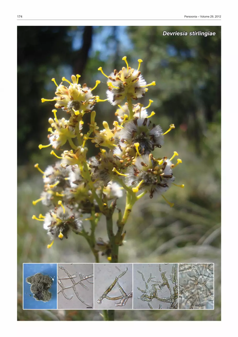
Devriesia stirlingiae

175Fungal Planet description sheets
© 2012 Nationaal Herbarium Nederland & Centraalbureau voor Schimmelcultures
Etymology. Named after the host genus from which it was isolated, Stirlingia.
Colonies on synthetic nutrient-poor agar. Mycelium consist-ing of smooth, pale brown, septate, branched, 2–3 µm diam hyphae. Conidiophores erect, subcylindrical, pale brown, smooth, straight or flexuous, branched or not, reduced to conidiogenous cells or 1–8-septate, 10–50 ×4–5 µm. Conidiogenous cells terminal, integrated, subcylindrical, smooth, pale brown, proliferating sympodially, 8–15×3–4 µm; scars flattened, thickened, somewhat darkened, 1–2 µm diam. Ramoconidia 1–3-septate, granular to guttulate, subcylindrical, smooth, pale brown, 15–30×4–5 µm, frequently with lateral branch at apex, up to 10 µm long, hila somewhat thickened and darkened, 1.5–2(–3) µm diam. Conidia subcylindrical to fusoid-ellipsoidal, apex obtuse, pale brown, smooth, guttulate, (7–)12–16(–20)×(3–)4(–5) µm, 0–3-septate; hila flattened, truncate, somewhat thickened and darkened, 1–2 µm diam. Chlamydospores thick-walled, brown, globose, in intercalary chains, up to 10 µm diam. Culture characteristics — (in the dark, 25 °C after 2 wk): Colonies erumpent with even, smooth margins and sparse aerial mycelium. On potato-dextrose agar, malt extract agar and oatmeal agar surface olivaceous-grey, reverse iron-grey, reaching 7 mm diam.
Typus. western AustrAliA, Perth, Wandoo National Park, on leaves of Stirlingia latifolia (Proteaceae), 13 July 2011, W. Gams, holotype CBS H-21083, cultures ex-type CPC 19948 = CBS 133581, ITS sequence GenBank KC005778, LSU sequence GenBank KC005799, MycoBank MB801777.
Notes — Based on a megablast search of NCBIs Gen-Bank nucleotide database, the closest hits using the LSU se-quence are Devriesia hilliana (GenBank GU214414; Identities = 843/856 (98 %), Gaps = 2/856 (0 %)), Devriesia xanthorrhoeae (GenBank HQ599606; Identities = 841/856 (98 %), Gaps = 2/856 (0 %)), and Teratosphaeria knoxdaviesii (Gen-Bank EU707865; Identities = 839/853 (98 %), Gaps = 0/853 (0 %)). Closest hits using the ITS sequence had highest simi-larity to Devriesia fraseriae (GenBank HQ599602; Identities = 491/501 (98 %), Gaps = 2/501 (0 %)), Devriesia lagerstroemiae (GenBank GU214634; Identities = 478/508 (94 %), Gaps = 13/508 (3 %)), and Teratosphaeria knoxdaviesii (Gen-Bank EU707866; Identities = 473/507 (93 %), Gaps = 11/507 (2 %)). Although phylogenetically closely related to D. fraseriae (intercalary and terminal conidia (6–)8–10(–11)×3(–4) µm; Crous et al. 2010a), D. stirlingiae is easily distinguishable by having larger conidia.
Fungal Planet 141 – 20 December 2012
Devriesia stirlingiae Crous, sp. nov.
Colour illustrations. Flowers of Stirlingia latifolia; colony sporulating on potato-dextrose agar; conidiophores, conidiogenous cells and conidia. Scale bar = 10 µm.
Pedro W. Crous & Johannes Z. Groenewald, CBS-KNAW Fungal Biodiversity Centre, P.O. Box 85167, 3508 AD Utrecht, The Netherlands; e-mail: [email protected] & [email protected]
Giles E.St.J. Hardy, School of Biological Sciences and Biotechnology, Murdoch University, Murdoch, Western Australia, 6150; e-mail: [email protected]

176 Persoonia – Volume 29, 2012
Cercospora chrysanthemoides

177Fungal Planet description sheets
© 2012 Nationaal Herbarium Nederland & Centraalbureau voor Schimmelcultures
Etymology. Named after the host genus on which it occurs, Chrysanthemoides.
Description based on host material, incubated in moist cham-bers. Leaf spots amphigenous, subcircular, 2–10 mm diam, with concentric darker circles, margin dark brown, raised. Sporulation amphigenous, but more prominently hypophyl-lous. Mycelium internal, consisting of branched, septate, smooth, pale brown, 2–3 µm diam hyphae. Stromata substo-matal, globose, consisting of brown, pseudoparenchymatal cells, becoming erumpent, up to 60 µm diam, giving rise to co-nidiophores. Conidiophores fasciculate, containing numerous conidiophores in dense clusters, subcylindrical, straight, rare-ly once-geniculate, brown, finely verruculose, 1–3-septate, 30–70 × 6–7 µm. Conidiogenous cells terminal, integrated, 25–55 × 5–7 µm, brown, finely verruculose, subcylindrical; loci terminal, single, rarely with lateral locus, scars flattened, darkened, thickened, 3–4 µm diam. Conidia solitary, hyaline, obclavate to subcylindrical, straight to slightly curved, apex subobtuse, widest at or below basal septum, (38–)42–55 (–70) × (4–)5(–6) µm, 3–5-septate; hila thickened, darkened and refractive, 3–4 µm diam. Culture characteristics — (in the dark, 25 °C after 2 wk): Colonies spreading, with moderate aerial mycelium and even, lobate margin. On potato-dextrose agar surface dirty white, surrounded by broad red-purple zone of diffuse pigment in agar, dark red in reverse. On oatmeal agar centre dirty white, outer region olivaceous-grey. On malt extract agar surface dirty white with patches of olivaceous-grey, reverse iron-grey, reaching 30 mm diam.
Typus. south AfriCA, Free State Province, Bloemfontein, Free State National Botanical Garden, on leaves of Chrysanthemoides monilifera (Asteraceae), 7 May 2012, P.W. Crous & W.J. Swart, holotype CBS H-21084, cultures ex-type CPC 20605, CPC 20529 = CBS 133582, ITS sequences GenBank KC005779–KC005780, ACT sequences GenBank KC005764–KC005765, TEF sequences GenBank KC005813–KC005814, CAL se-quences GenBank KC005767–KC005768, LSU sequences GenBank KC005800–KC005801, MycoBank MB801778.
Notes — Based on a megablast search of NCBIs GenBank nucleotide database, the closest hits using the LSU sequence are Cercospora cf. apii (GenBank JN941176; Identi ties = 900/902 (99 %), Gaps = 1/902 (0 %)), Cercospora acaciae mangii (GenBank JN941175; Identities = 900/902 (99 %), Gaps = 1/902 (0 %)) and Cercospora sp. (GenBank JN941174; Identities = 900/902 (99 %), Gaps = 1/902 (0 %)). Closest hits using the ITS sequence had highest similarity to Cercospora zebrina (GenBank JX390615; Identities = 529/530 (99 %), Gaps = 0/530 (0 %)), Cercospora piaropi (Gen-Bank HQ902254; Identities = 529/530 (99 %), Gaps = 0/530 (0 %)) and Cercospora capsici (GenBank GU214654; Iden-tities = 529/530 (99 %), Gaps = 0/530 (0 %)). Closest hits using the ACT sequence had highest similarity to Cercospora althaeina (GenBank JX143036; Identities = 192/194 (99 %), Gaps = 0/194 (0 %)), Cercospora zebrina (GenBank JX143260; Identities = 211/214 (99 %), Gaps = 0/214 (0 %)) and Cercospora armoraciae (GenBank JX143058; Identi-ties = 190/194 (98 %), Gaps = 0/194 (0 %)). Closest hits using the TEF sequence had highest similarity to Cercospora delaireae (GenBank JX143346; Identities = 288/292 (99 %), Gaps = 0/292 (0 %)), Cercospora ricinella (Gen-Bank JX143406; Identities = 287/291 (99 %), Gaps = 0/291 (0 %)) and Cercospora cf. zinniae CPC 15075 (GenBank JX143519; Identities = 287/292 (98 %), Gaps = 0/292 (0 %)). Closest hits using the CAL sequence had highest si milarity to Cercospora cf. chenopodii (GenBank JX142839; Identi-ties = 388/398 (97 %), Gaps = 0/398 (0 %)), Cercospora ricinella (GenBank JX142913; Identities = 287/297 (97 %), Gaps = 0/297 (0 %)) and Cercospora cf. coreopsidis (Gen-Bank JX142851; Identities = 285/296 (96 %), Gaps = 0/296 (0 %)) (see Groenewald et al. (In press) for morphological de-tails pertaining to the species cited above).
Fungal Planet 142 – 20 December 2012
Cercospora chrysanthemoides Crous & W.J. Swart, sp. nov.
Colour illustrations. Chrysanthemoides monilifera in the Free State National Botanical Garden; leaf spots; lesion; conidiophores and conidia. Scale bars = 10 µm.
Pedro W. Crous & Johannes Z. Groenewald, CBS-KNAW Fungal Biodiversity Centre, P.O. Box 85167, 3508 AD Utrecht, The Netherlands; e-mail: [email protected] & [email protected]
Wijnand J. Swart, Department of Plant Pathology, University of the Free State, P.O. Box 339, Bloemfontein 9300, South Africa; e-mail: [email protected]

178 Persoonia – Volume 29, 2012
Zymoseptoria verkleyi

179Fungal Planet description sheets
© 2012 Nationaal Herbarium Nederland & Centraalbureau voor Schimmelcultures
Etymology. Named after Gerard J.M. Verkley, for the contribution that he has made to further our understanding of the genus Septoria.
On sterile barley leaves on water agar: Conidiomata pycnidial, substomatal, immersed to erumpent, globose, dark brown, up to 200 µm diam, with central ostiole, 10–15 µm diam; wall of 3–4 layers of brown textura angularis. Conidiophores re-duced to conidiogenous cells, or with one supporting cell, lin-ing the inner cavity. Conidiogenous cells hyaline, smooth (in older cultures on malt extract agar becoming brownish, ver-ruculose), tightly aggregated, subcylindrical to ampulliform, straight to curved, 7–15×3–4.5 µm, with inconspicuous, per-current proliferations at apex, but also proliferating sympodial-ly. Conidia of all three types present. Type I conidia (pycnidial conidia) solitary, hyaline, smooth, granular, acicular to narrow-ly obclavate, tapering towards subacutely rounded apex, with truncate or obconically truncate base, straight to flexuous, 1–6(–12)-septate, (30–)40–65(–80) × (2–)2.5(–3) µm; hila not thickened nor darkened, 1–2 µm. On synthetic nutrient-poor agar, yeast-like growth and microcyclic conidiation (Type III conidia) present, as well as aerial hyphae and older conidia disarticulating into phragmoconidia (Type II conidia). Culture characteristics — (in the dark, 25 °C after 2 wk): Colonies erumpent, with even to feathery margins and sparse aerial mycelium. On potato-dextrose agar and malt extract agar surface pale olivaceous-grey to olivaceous-grey; reverse iron-grey, colonies reaching 12 mm diam.
Typus. netherlAnds, Utrecht, Houten, on leaves of Poa annua (Poaceae), 2012, S. Videira, holotype CBS H-21085, cultures ex-type S657 = CBS 133618, ITS sequence GenBank KC005781 and LSU sequence Gen-Bank KC005802, MycoBank MB801779.
Notes — Based on a megablast search of NCBIs Gen-Bank nucleotide database, the closest hits using the LSU sequence are Zymoseptoria brevis (GenBank JQ739832; Identities = 862/865 (99 %), Gaps = 2/865 (0 %)), Zymoseptoria tritici (GenBank GU214436; Identities = 862/865 (99 %), Gaps = 2/865 (0 %)) and Zymoseptoria passerinii (GenBank JQ739843; Identities = 855/863 (99 %), Gaps = 0/863 (0 %)). Closest hits using the ITS sequence had highest simi larity to Zymoseptoria passerinii (GenBank AF181699; Iden tities = 494/508 (97 %), Gaps = 5/508 (1 %)), Zymoseptoria tritici (GenBank FN428877; Identities = 473/479 (99 %), Gaps = 3/479 (1 %)) and Zymoseptoria halophila (GenBank JF700876; Identities = 461/475 (97 %), Gaps = 5/475 (1 %)). Although phylogenetically closely related to Z. passerinii (conidia 1–3- septate, 21–52 × 1.5–2.2 µm; Quaedvlieg et al. 2011, Stuken-brock et al. 2012), conidia of Z. verkleyi are much larger.
Fungal Planet 143 – 20 December 2012
Zymoseptoria verkleyi Crous, Videira & Quaedvlieg, sp. nov.
Species Host Origin Morphology Reference
Conidial dimensions (µm) Conidial septation
Z. ardabiliae Lolium Iran (15–)20–25(–30) × 2(–3) (0–)1 Stukenbrock et al. (2012)Z. brevis Phalaris Iran (12–)13–16(–17) × 2(–2.5) 0–1 Quaedvlieg et al. (2011)Z. halophila Hordeum Iran (30–)33–38(–50) × 2(–3) 1(–3) Quaedvlieg et al. (2011)Z. passerinii Hordeum Italy 21–52 × 1.5–2.2 1–3 Quaedvlieg et al. (2011)Z. pseudotritici Dactylis Iran (7–)10–12(–22) × 2.5(–3) 0(–1) Stukenbrock et al. (2012)Z. tritici Triticum France 28–85 × 1.5–2 (0–)3 Quaedvlieg et al. (2011)Z. verkleyi Poa Netherlands (30–)40–65(–80) × (2–)2.5(–3) 1–6(–12) Present study
Table 1 Comparison of hosts, distribution and micromorphology of currently described Zymoseptoria species.
Pedro W. Crous, Sandra I.R. Videira & William Quaedvlieg, CBS-KNAW Fungal Biodiversity Centre, P.O. Box 85167, 3508 AD Utrecht, The Netherlands;
e-mail: [email protected], [email protected] & [email protected]
Colour illustrations. Poa annua growing next to the roadside in Houten; colony sporulating on synthetic-nutrient poor agar; conidiogenous cells and conidia with microcyclic conidiation and phragmoconidia. Scale bars = 10 µm.

180 Persoonia – Volume 29, 2012
Toxicocladosporium posoqueriae

181Fungal Planet description sheets
© 2012 Nationaal Herbarium Nederland & Centraalbureau voor Schimmelcultures
Etymology. Named after the host genus from which it was collected, Posoqueria.
Description based on colonies sporulating on synthetic nutri-ent poor agar. Mycelium internal, pale brown, smooth, 2–3 µm diam (in culture brown, thick-walled, constricted at septa, smooth, 3–7 µm diam); giving rise to conidiophores that arise from stomata (hypophyllous, on brown leaf spots with con-centric brown rings, associated with a Colletotrichum sp., the presumed primary pathogen), erect, solitary, straight, subcy-lindrical, main axis unbranched on host (frequently branched in culture), 50–200 × 4–7 µm; apex branched with lateral branches, 15–50×3–5 µm, 1–3-septate, becoming clavate towards apex, thick-walled, smooth to finely verruculose. Conidiogenous cells integrated, terminal and lateral, in whorls of 3–4, clavate or broadly cylindrical to doliiform, 10–20×4–7 µm, aseptate, medium to dark brown, concolorous with co-nidiophores, polyblastic with numerous loci at conidiogenous tip; loci truncate, circular, thickened, slightly darkened and refractive, 0.5–1(–1.5) µm. Ramoconidia dark brown, clavate to subcylindrical, finely verruculose, thick-walled, aseptate, 5–15×4–5 µm, with numerous apical loci, resembling those on conidiogenous cells. Conidia in branched, short chains, subglobose, ellipsoid to fusoid, (4–)6–7 × (3–)4 µm, pale brown, smooth, thin-walled; hila with circular, thickened, dark-ened and refractive loci, 1–1.5 µm diam. Culture characteristics — (in the dark, 25 °C after 2 wk): Colonies erumpent, spreading, with moderate aerial myce-lium and even, lobate margins. On malt extract agar surface folded, grey-olivaceous, reverse olivaceous-grey. On oatmeal agar surface grey-olivaceous in centre, sienna in outer region. On potato-dextrose agar grey-olivaceous in centre, oliva-ceous-grey in outer region, iron-grey in reverse, reaching 30 mm diam.
Typus. AustrAliA, Northern Territory, Darwin, on leaves of Posoqueria latifolia (Rubiaceae), 12 Apr. 2011, R.G. Shivas, holotype CBS H-21086, cultures ex-type CPC 19305 = CBS 133583, ITS sequence GenBank KC005782, LSU sequence GenBank KC005803, MycoBank MB801780.
Notes — The genus Toxicocladosporium, based on T. irritans, presently accommodates eight species (Crous et al. 2007, 2009d, Crous & Groenewald 2011). Toxicocladosporium posoqueriae differs from other members of the genus in that it has whorls of conidiogenous cells, resembling Parapericoniella asterinae, the type species of the genus Parapericoniella (Bensch et al. 2012). However, P. asterinae is mycophylic, growing on Asterina contigua, thus it differs ecologically from T. posoqueriae, which appears to be plant pathogenic, colo-nising lesions of a Colletotrichum sp. Nevertheless, if these genera are eventually found to be synonymous, Parapericoniella (2005) would represent an older name than Toxicocladosporium (2007). Based on a megablast search of NCBIs GenBank nucleotide database, the closest hits using the LSU sequence are Toxicocladosporium pseudoveloxum (Gen-Bank JF499868; Identities = 924/938 (99 %), Gaps = 0/938 (0 %)), Toxicocladosporium strelitziae (GenBank JX069858; Identities = 922/938 (98 %), Gaps = 0/938 (0 %)) and Toxicocladosporium irritans (GenBank EU040243; Identities = 922/938 (98 %), Gaps = 0/938 (0 %)). Closest hits using the ITS sequence had highest similarity to Toxicocladosporium strelitziae (GenBank JX069874; Identities = 532/562 (95 %), Gaps = 10/562 (2 %)), Toxicocladosporium pseudoveloxum (GenBank JF499847; Identities = 660/698 (95 %), Gaps = 11/698 (2 %)) and Toxicocladosporium rubrigenum (GenBank FJ790285; Identities = 638/675 (95 %), Gaps = 7/ 675 (1 %)).
Fungal Planet 144 – 20 December 2012
Toxicocladosporium posoqueriae Crous & R.G. Shivas, sp. nov.
Colour illustrations. Rocky outcrop in Northern Territories, Darwin; col-ony on synthetic nutrient-poor agar; conidiophores with whorls of conidio-genous cells and conidia. Scale bars = 10 µm.
Pedro W. Crous & Johannes Z. Groenewald, CBS-KNAW Fungal Biodiversity Centre, P.O. Box 85167, 3508 AD Utrecht, The Netherlands; e-mail: [email protected] & [email protected]
Roger G. Shivas, Biosecurity Queensland, Ecosciences Precinct, Level 2C East, GPO Box 267, Brisbane 4001, Queensland, Australia; e-mail: [email protected]

182 Persoonia – Volume 29, 2012
Catenulostroma corymbiae

183Fungal Planet description sheets
© 2012 Nationaal Herbarium Nederland & Centraalbureau voor Schimmelcultures
Etymology. Named after the host genus from which it was collected, Corymbia.
Leaf spots amphigenous, subcircular, 2–3 mm diam, grey-brown with a dark brown, raised border. Ascomata pseudothe-cial, amphigenous, black, subepidermal, erumpent, globose, up to 120 µm diam; central ostiole 10–20 µm diam; wall con-sisting of 2–3 layers of medium brown textura angularis. Asci aparaphysate, fasciculate, bitunicate, subsessile, obovoid to subcylindrical, straight to curved, 8-spored, 20–30×7–9 µm. Ascospores multiseriate, overlapping, hyaline, guttulate, thin-walled, straight, obovoid with obtuse ends, widest in middle of apical cell, medianly 1-septate, not or slightly constricted at septum, tapering towards both ends, but more prominently towards lower end, 7–8(–10)×(2–)3(–3.5) µm. Ascospores become distorted upon germination, brown, verruculose, 7–10 µm diam; initial germ tubes parallel to the long axis, but additional tubes at various angles to the long axis. Colonies on SNA. Mycelium consisting of septate, branched, smooth, pale brown, 2–3 µm diam hyphae, that give rise to globose or elongated sclerotial-like bodies of brown, multiseptate, thick-walled cells (variously shaped and branched, up to 25 µm diam). Conidiophores developing mostly on terminal hyphal ends, subcylindrical, brown, straight or variously curved, with multiple septa, up to 80 µm tall, and 4–6 µm wide. Conidiogenous cells subcylindrical, brown, smooth, terminal and lateral, 5–15×4–6 µm, with flattened, truncate locus, 2–3 µm diam, mono- to polyblastic. Conidia brown, smooth, subcylindrical to fusoid-ellipsoidal, straight to variously curved, at times with lateral branches, 0–3 transversely septate, apex obtuse or truncate, base truncate, 2 µm diam, somewhat darkened, not thickened, 8–20×3.5–4 µm; commonly arranged in branched chains that branch irregularly below or near apex of conidial chain. Culture characteristics — (in the dark, 25 °C after 2 wk): Colonies erumpent, spreading, with moderate aerial mycelium and even, lobate margins. On malt extract agar (MEA) centre olivaceous-grey, outer region iron-grey, reverse iron-grey. On oatmeal agar surface olivaceous-grey. On potato-dextrose-agar same as MEA, reaching 13 mm diam.
Typus. AustrAliA, Northern Territory, Darwin, just off Arnhem Highway, S12°44.839' E131°31.558', on leaves of Corymbia sp. (Myrtaceae), 9 May 2011, P.W. Crous & B.A. Summerell, holotype CBS H-21087, cultures ex-type CPC 19435, CPC 19437 = CBS 133584, ITS sequence GenBank KC005783, LSU sequences GenBank KC005805–KC005805, MycoBank MB801781.
Notes — Based on a megablast search of NCBIs GenBank nucleotide database, the closest hits using the LSU sequence are Teratosphaeria encephalarti (GenBank FJ372417; Identi- ties = 836/850 (98 %), Gaps = 0/850 (0 %)), Catenulostroma chromoblastomycosum (GenBank EU019251; Identities = 877/892 (98 %), Gaps = 0/892 (0 %)) and Penidiella rigidophora (GenBank EU019276; Identities = 833/853 (98 %), Gaps = 0/853 (0 %)). Closest hits using the ITS sequence yielded highest similarity to Catenulostroma protearum (Gen-Bank GU214628; Identities = 600/668 (90 %), Gaps = 29/668 (4 %)), Catenulostroma hermanusense (GenBank JF499833; Identities = 599/668 (90 %), Gaps = 29/668 (4 %)) and Teratosphaeria encephalarti (GenBank FJ372400; Identities = 592/661 (90 %), Gaps = 21/661 (3 %)). Although phylogeneti-cally closely related to C. protearum (conidia 12–45×7–25 μm; Crous & Groenewald 2011), C. corymbiae has much smaller conidia.
Fungal Planet 145 – 20 December 2012
Catenulostroma corymbiae Crous & Summerell, sp. nov.
Pedro W. Crous & Johannes Z. Groenewald, CBS-KNAW Fungal Biodiversity Centre, P.O. Box 85167, 3508 AD Utrecht, The Netherlands; e-mail: [email protected] & [email protected]
Brett A. Summerell, Royal Botanic Gardens and Domain Trust, Mrs. Macquaries Road, Sydney, NSW 2000, Australia; e-mail: [email protected]
Colour illustrations. Corymbia and Eucalyptus spp. in Northern Terri-tory, Darwin; symptomatic Corymbia leaf; germinating ascospores; asci and asco spores. Scale bars = 10 µm.

184 Persoonia – Volume 29, 2012
Penidiella carpentariae

185Fungal Planet description sheets
© 2012 Nationaal Herbarium Nederland & Centraalbureau voor Schimmelcultures
Etymology. Named after the host genus from which it was isolated, Carpentaria.
Colonies on synthetic nutrient-poor agar. Mycelium con-sisting of smooth, pale brown, septate, branched, 2–3 µm diam hyphae. Conidiophores erect, subcylindrical, pale brown, smooth to finely verruculose, straight or flexuous, un-branched, 1–3(–7)-septate, 20–90×2–4 µm. Conidiogenous cells terminal, integrated, subcylindrical, smooth, pale brown, proliferating sympodially, 15–25 × 2–3 µm; numerous scars aggregated at apex, flattened, thickened, somewhat dark-ened, 0.5–1.5 µm diam. Ramoconidia 0(–1)-septate, granu-lar to guttulate, subcylindrical to fusoid-ellipsoidal, smooth, pale brown, 10–18×2–3 µm, with one to numerous loci at apex (especially on OA); hila somewhat thickened and dark-ened, 0.5–1 µm diam. Conidia fusoid-ellipsoidal, pale brown, smooth, guttulate, (6–)7–8(–10) × (1.5–)2(–2.5) µm, asep-tate; hila flattened, truncate, somewhat thickened and dark-ened, 0.5–1 µm diam. Chlamydospores not observed. Culture characteristics — (in the dark, 25 °C after 2 wk): Colonies erumpent, spreading, with smooth, lobate margins and sparse to moderate aerial mycelium. On potato-dextrose
agar, malt extract agar and oatmeal agar surface and reverse iron-grey, colonies reaching 20 mm diam.
Typus. AustrAliA, Northern Territory, Litchfield National Park, Wangi Falls, on leaves of Carpentaria acuminata (Arecaceae), 24 Apr. 2011, P.W. Crous & B.A. Summerell, holotype CBS H-21088, cultures ex-type CPC 19439 = CBS 133586, ITS sequence GenBank KC005784, LSU sequence GenBank KC005806, MycoBank MB801782.
Notes — Based on a megablast search of NCBIs GenBank nucleotide database, the closest hits using the LSU sequence are Parapenidiella tasmaniensis (GenBank GU214452; Iden-tities = 865/895 (97 %), Gaps = 2/895 (0 %)), Parapenidiella pseudotasmaniensis (GenBank GQ852625; Identities = 859/889 (97 %), Gaps = 2/889 (0 %)) and Phacellium pas pali (GenBank GU214669; Identities = 863/895 (96 %), Gaps = 3/895 (0 %)). Closest hits using the ITS sequence had high-est similarity to Devriesia tardicrescens (GenBank JF499840; Identities = 453/527 (86 %), Gaps = 21/527 (4 %)), Teratosphaeria associata (GenBank EU707857; Identities = 360/ 391 (92 %), Gaps = 9/391 (2 %)) and Teratosphaeria parva (GenBank AY626980; Identities = 458/534 (86 %), Gaps = 32/534 (6 %)). Penidiella carpentariae clusters basal to a clade that contains Parapenidiella tasmaniensis and P. pseudotasmaniensis.
Fungal Planet 146 – 20 December 2012
Penidiella carpentariae Crous & Summerell, sp. nov.
Etymology. Para (= close to) + its morphological similarity to Penidiella.
Mycelium consisting of branched, septate, smooth subhya-line to pale brown hyphae. Conidiophores macronematous, occasionally micronematous; macronematous conidiophores arising from superficial mycelium, solitary, erect, pale brown, thin-walled, smooth to finely verruculose; terminally penicil-late, unbranched in terminal part; conidiogenous apparatus composed of a series of conidiogenous cells and/or ramoco-nidia. Conidiogenous cells integrated, terminal or intercalary, unbranched, pale brown, smooth, tapering to a flattened or rounded apical region, mono- or polyblastic, sympodial, giving
Parapenidiella Crous & Summerell, gen. nov.rise to a single or several sets of ramoconidia on different lev-els; with relatively few conidiogenous loci, slightly thickened, slightly darkened. Conidia in branched acropetal chains. Ramoconidia 0–1-septate, pale brown, smooth, thin-walled, fusoid-ellipsoidal to subcylindrical. Conidia subcylindrical, fusoid to ellipsoid-ovoid, aseptate, pale olivaceous to pale brown, smooth, thin-walled, catenate; hila truncate, slightly thickened, somewhat darkened.
Type species. Parapenidiella tasmaniensis (Crous & M.J. Wingf.) Crous. MycoBank MB801783.
Pedro W. Crous & Johannes Z. Groenewald, CBS-KNAW Fungal Biodiversity Centre, P.O. Box 85167, 3508 AD Utrecht, The Netherlands; e-mail: [email protected] & [email protected]
Brett A. Summerell, Royal Botanic Gardens and Domain Trust, Mrs Macquaries Rd, Sydney, NSW 2000, Australia; e-mail: [email protected]
Colour illustrations. Wangi Falls, Litchfield National Park, Northern Ter-ritory; colony sporulating on synthetic nutrient-poor agar; conidiophores, conidiogenous cells and conidia. Scale bars = 10 µm.
Basionym. Mycovellosiella tasmaniensis Crous & M.J. Wingf., Mycol. Res. 102: 527. 1998. ≡ Passalora tasmaniensis (Crous & M.J. Wingf.) Crous & U. Braun, in Mycosphaerella and its anamorphs. 1. Names published in Cercospora and Passalora: 472. 2003. = Mycosphaerella tasmaniensis Crous & M.J. Wingf., Mycol. Res. 102: 527. 1998.
Notes — Parapenidiella represents a genus between Devriesia (Seifert et al. 2004) and Penidiella (Crous et al. 2007),
which are known to be paraphyletic (Crous et al. 2009a, b). All three genera have Teratosphaeria-like teleomorphs (Crous et al. 2008, 2012). Parapenidiella is distinguished from Penidiella and Devriesia by having pale brown, unbranched, penicil-late conidiophores, with olivaceous to pale brown, branched conidial chains. Penidiella carpentariae strongly resembles Parapenidiella in morphology, yet appears to represent a dif-ferent lineage in this generic complex.
MycoBank MB801785.
Parapenidiella tasmaniensis (Crous & M.J. Wingf.) Crous, comb. nov.
Parapenidiella pseudotasmaniensis (Crous) Crous, comb. nov. Penidiella pseudotasmaniensis Crous, Persoonia 23: 126. 2009. MycoBank MB801784.

186 Persoonia – Volume 29, 2012
Seiridium phylicae

187Fungal Planet description sheets
© 2012 Nationaal Herbarium Nederland & Centraalbureau voor Schimmelcultures
Etymology. Name refers to the host genus, Phylica.
Caulicolous. Conidiomata stromatic, pycnidia, scattered to aggregated, erumpent, conical, up to 350 µm diam, unilocu-late, dark brown to black, opening by irregular rupture; basal stroma of dark brown textura angularis. Conidiophores lining cavity, filamentous, creating impression of paraphyses, sep-tate, branched, hyaline, smooth, up to 80 µm long, and 3.5 µm wide. Conidiogenous cells subcylindrical, terminal and lateral, integrated, smooth, hyaline, 10–20 × 1.5–3 µm; proliferating percurrently. Conidia fusoid to ellipsoid, dark to golden brown, granular, 5-septate, not constricted at septa, with visible cen-tral septal pore, (23–)28–30(–35) × (9–)10(–11) µm; basal cell conical with truncate hilum, pale brown to hyaline, 3–5 µm long; 4 median cells doliiform to subcylindrical, brown, with wall and septa being darker, cells together 17–23 µm long; apical cell broadly conical, apex rounded, hyaline, 2–4 µm long. Api-cal appendages tubular, unbranched, eccentric, 6–8 µm long; basal appendages unbranched, centric, 2–5 µm long. Culture characteristics — (in the dark, 24 °C after 2 wk): Colonies erumpent, spreading, with moderate aerial myce-lium and even, lobate margins. On malt extract agar surface pale olivaceous-grey, with patches of dirty white; reverse cinnamon. On potato-dextrose agar surface dirty white with patches of black sporulation; reverse dirty white. On oatmeal agar surface pale grey-olivaceous with patches of dirty white, reaching 30 mm diam.
Typus. UK, British Overseas Territory of Saint Helena, Ascension and Tristan da Cunha, Inaccessible Island, Blenden Hall, S37°17'41" W12°42'08", stems of Phylica arborea (Rhamnaceae), Sept. 2011, P.G. Ryan, holotype CBS H-21089, cultures ex-type CPC 19962–19965 (CPC 19964 = CBS 133587), β-tubulin (TUB) sequence GenBank KC005819–KC005821, TEF1-α sequences GenBank KC005815–KC005817, ITS sequences GenBank KC005785–KC005785, LSU sequences GenBank KC005807–KC005810, MycoBank MB801788.
Notes — Conidia of Seiridium cardinale are 21–30 × 8–10 µm, with basal appendage being 1 µm long when present, and apical appendage 0.5–1.5 µm (Sutton 1980), which clearly dis-tinguishes it from Seiridium phylicae. Based on a megablast search of NCBIs GenBank nucleotide database, the closest hits using the LSU sequence are Seiridium eucalypti (Gen-Bank DQ414533; Identities = 833/833 (100 %), Gaps = 0/833 (0 %)), Seiridium unicorne (GenBank DQ414532; Identities = 833/833 (100 %), Gaps = 0/833 (0 %)) and Lepteutypa cupressi (GenBank AF382379; Identities = 872/875 (99 %), Gaps = 3/875 (0 %)). Closest hits using the ITS sequence had highest similarity to Seiridium cardinale (GenBank AF409995; Identities = 552/558 (99 %), Gaps = 2/558 (0 %)), Seiridium cupressi (GenBank FJ430600; Identities = 558/567 (98 %), Gaps = 4/567 (1 %)) and Seiridium unicorne (Gen-Bank AF377299; Identities = 567/578 (98 %), Gaps = 2/578 (0 %)). Closest hits using the TUB sequence had highest similarity to Seiridium cardinale (GenBank DQ926973; Iden-tities = 353/366 (96 %), Gaps = 3/366 (1 %)) and Seiridium cupressi (GenBank AF320495; Identities = 385/401 (96 %), Gaps = 2/401 (0 %)). Only distant hits (e.g. Identities = 218/249 (88 %), Gaps = 12/249 (5 %)) with Pestalotiopsis spp. were obtained when the TEF sequences were used in a megablast search.
Fungal Planet 147 – 20 December 2012
Seiridium phylicae Crous & M.J. Wingf., sp. nov.
Pedro W. Crous & Johannes Z. Groenewald, CBS-KNAW Fungal Biodiversity Centre, P.O. Box 85167, 3508 AD Utrecht, The Netherlands; e-mail: [email protected] & [email protected]
Peter G. Ryan, Percy FitzPatrick Institute of African Ornithology, University of Cape Town, Rondebosch 7701, South Africa; e-mail: [email protected]
Michael J. Wingfield, Forestry and Agricultural Biotechnology Institute, University of Pretoria, South Africa; e-mail: [email protected]
Colour illustrations. Phylica arborea growing on Inaccessible Island; colony on synthetic nutrient-poor agar; conidiophores, conidiogenous cells and conidia. Scale bars = 10 µm.

188 Persoonia – Volume 29, 2012
Microcyclospora rhoicola

189Fungal Planet description sheets
© 2012 Nationaal Herbarium Nederland & Centraalbureau voor Schimmelcultures
Etymology. Named after the host from which it was collected, Rhus typhina.
Colonies on Rhus typhina forming dark, sometimes slimy, crust on petiole and twig surfaces, forming a stroma-like sheath on trichomes, textura prismatica in surface view. Mycelium con-sisting of pale brown, branched, thick-walled (c. 1 μm), septate hyphae, (2–)4–6.5(–7.5) μm diam, smooth. Micromorphology identical to that in culture, described below.Colonies on malt extract agar (MEA). Mycelium consist-ing of pale brown, branched, septate hyphae, 1.5–3.5 μm diam, smooth. In older cultures, hyphae becoming darker, thick-walled (c. 1 μm), ossiform, and fragmenting to form a yeast-like colony. Conidiophores reduced to conidiogenous cells. Conidiogenous cells integrated, lateral on hyphae, soli-tary, subdenticulate, 3–5 μm tall, 2–3 μm wide, pale brown, smooth. Conidia (0–)1–3(–6)-septate, 3-septate conidia most frequent, 5–6-septate conidia rarely observed, aseptate co-nidia (8–)9–17.5(–29.5)× (2–)2.5–3 μm, 1-septate conidia (10.5–)11.5–22.5(–36)×(2–)2.5–3(–3.5) μm, 2-septate co-nidia (17–)19–28(–32)×(2.5–)3–3.5(–4), 3-septate conidia (19–)26–35.5(–40)×(2–)2.5–3(–3.5) μm, 4-septate conidia (36–)37.5–44(–47.5) × (2.5–)2.5–3(–3.5) μm, 5-septate conidia (47–)48.5–56(–57) × 2.5–3 μm, 6-septate conidia 48.5×3 μm, hyaline, smooth, cylindrical, straight to variously curved, apex obtuse, base truncate, older conidia somewhat constricted at septa, guttulate, aggregated in mucoid masses; hila neither thickened nor darkened; anastomosis among co-nidia sometimes observed; microcyclic conidiation commonly observed. Culture characteristics — (in the dark, 25 °C after 2 wk on MEA): Colonies convex, with moderate to woolly aerial my-celium; surface irregular, slimy, dark grey to olive (1F1–1F3)
(Kornerup & Wanscher 1978), aerial mycelium greyish off-white to pastel grey (1B2–1B3), margin diffuse; reverse dark grey (1F1); diam up to 4 mm. In older colonies (> 6 wk), aerial
mycelium becoming yellowish brown to tobacco brown (5E8–5F6), collapsing, centre carbonaceous, slimy and yeast-like, margin lobate.
Typus. CAnAdA, Ontario, Ottawa, Dominion Arboretum, on twigs of Rhus typhina var. laciniata (Anacardiaceae), 20 Oct. 2011, J.B. Tanney, holotype DAOM 242272, dried culture ex-type DAOM 242276, ITS sequence Gen-Bank KC012605, LSU sequence GenBank KC012606, TEF1 sequence GenBank KC012604, MycoBank MB801439.
Notes — Microcyclospora was first described in 2010, with three species causing sooty blotch on Malus domestica fruit (Frank et al. 2010). The genus is characterised by 1–multi-septate, smooth, pale brown, scolecosporous to cylindrical conidia borne from reduced and integrated mono- to polyblas-tic conidiogenous cells. Conidia occur in mucoid masses and microcyclic conidiation is common (Frank et al. 2010). Mor-phologically, M. rhoicola conforms with the generic concept of Microcyclospora and can be differentiated from other species by its shorter conidia with fewer septa (Table 1). The discovery of M. rhoicola represents the first record of Microcyclospora in North America and on its host, Rhus typhina.The phylogenetic analysis below is based on internal tran-scribed spacer (ITS) sequences derived from two M. rhoicola isolates (specimens collected c. 250 km apart) and previously published data (Frank et al. 2010, Crous et al. In press). Microcyclospora rhoicola has distinct ITS sequences from those sequenced to date and appears to be rather distant from the currently described species.Several cercosporoid fungi are described from Rhus spp. in North America (Farr et al. 1989), including Cercosporella toxicodendri and Pseudocercospora rhoina. Both species occur as leaf spots and have more complex conidiophores compared to the reduced and integrated conidiogenous cells characterising M. rhoicola.
Fungal Planet 148 – 20 December 2012
Microcyclospora rhoicola Tanney, sp. nov.
Colour illustrations. Rhus typhina var. laciniata at the Dominion Arbo-retum, Ottawa, Ontario, Canada (type host, photo K. Seifert); mycelium on individual trichomes (scale bar = 100 μm); conidiogenous cells and conidia exhibiting microcyclic conidiation. Scale bars = 10 μm.
Joey B. Tanney, Biodiversity (Mycology & Botany), Agriculture & Agri-Food Canada, 960 Carling Ave., Ottawa, Ontario K1A 0C6, Canada; e-mail: [email protected]
Species Host Origin Morphology Reference
Conidial dimensions (µm) Conidial septation
M. malicola Malus Germany, Slovenia 45–75 × 2.5 (1–)5–7(–13) Frank et al. (2010) M. pomicola Malus Germany 50–75 × 2.5–3 1–13 Frank et al. (2010)M. quercina Quercus Netherlands 30–45 × 2.5–3 (1–)3–4(–11) Crous et al. (In press)M. rhoicola Rhus Canada 26–36 × 2.5–3 (0–)1–3(–6) Present studyM. rumicis Rumex Iran 37–54 × 2.5 1–10 Arzanlou & Bakhshi (2011)M. tardicrescens Malus Slovenia 35–55 × 2 1–9 Frank et al. (2010)
Table 1 Comparison of hosts, distribution and micromorphology of currently described Microcyclospora species.
Fig. 1 Consensus phylogram (50 % majority rule) of 15 002 trees result-ing from a Bayesian inference analysis of an ITS sequence alignment us-ing MrBayes v. 3.1.2. Posterior probabilities indicated with colour-coded branches (see legend).

190 Persoonia – Volume 29, 2012
Collembolispora aristata

191Fungal Planet description sheets
© 2012 Nationaal Herbarium Nederland & Centraalbureau voor Schimmelcultures
Etymology. aristatus (L.) = with bristles.
Conidia isolated from foam according to the methodology of Marvanová et al. (2003). Colonies on malt agar medium fast growing, reaching 25 mm after 20 d at 12 °C, dark grey, re-verse black. Aerial mycelium abundant, lanose to funiculose. Hyphae glabrous, hyaline, thin-walled, 1–3 µm wide or dark brown, thicker-walled, up to 5 µm wide. Sporulation initially directly on agar, in subcultures after submergence in standing distilled water at 15 °C within a few days. Conidiophores inter-calary, lateral or terminal, simple to profusely branched; stipes, if present, cylindrical or distally slightly widening up to 32 × 1.5–3.5 µm, with branches on various levels along the stipes or in a penicillate head; often concurrent with conidiogenous cells, sometimes verticillate, cylindrical or subclavate, 4–8 × 2–3 µm. Conidiogenous cells subclavate to narrow-doliiform, usually 1–3 per conidiophore branch, polyblastic sympodial, 5–11 × 1.5–3 µm, with one to few denticles at the apex, scars flat. Conidia in slimy masses when formed outside water, ap-pearing in close sequence. Axis 31–46 × 2–3.5 µm, proximal part obclavate and unequilateral, mildly curved or straight, 3(–5)-septate, basal scar truncate, sometimes eccentric, apex with an integrated, setose extension sometimes slightly curved away; branch single (exceptionally two), typically ven-tral, rarely dorsal, usually arising from the second suprabasal cell of the axis, often strongly retrorse, but also perpendicular to the axis, straight or often slightly curved abaxially, rarely adaxially, proximal part obclavate, base often slightly sinuous, 16–30 × 1.5–2.7 µm, insertion unequally constricted, distally protracted into setose extension. Some considerably swollen conidia are usually present in submerged cultures after sev-eral weeks. Hyphopodia-like outgrowths may appear in aged, submerged cultures on hyphae, and also on conidia.
Typus. CZeCh repuBliC, South Moravian region, between the villages Ochoz uTišnova and Lomnice, c. 440 m alt., isolated from foam in an un-named left tributary of the Křeptovský potok stream (the streamlet is shal-low, 80–100 cm wide, slow-flowing, with grasses on the banks and Typha latifolia and Glyceria maxima in the littoral zone), Mar. 1984, L. Marvanová, holotype CBS H-21090, culture ex-type CPC 21145 = CCM F-01585 = CBS 115662; ITS sequence GenBank KC005789, LSU sequence GenBank KC005811, MycoBank MB491201.
Notes — The hyphomycete genus Collembolispora is based on C. barbata, isolated from Alnus glutinosa leaf baits submerged in a slow-flowing, oligotrophic softwater stream in North Portugal (Marvanová et al. 2003). Collembolispora aristata has similar colonies, conidiogenesis as well as conidia like C. barbata, but the conidia of the latter differ from those of C. aristata by having a branched, terminal, setose extension on the conidial axis and on the conidial branch, and by the presence of a hyphomycetous, phialidic (?spermatial) morph.
As far as we know, there is thus far only one other report on conidia of C. aristata (Roldán & Puig 1992, f. 3C, as Gyoerffyella sp.). These authors collected detached conidia in a stream in the river Esva basin in the Asturias Province of Northern Spain, 285 m alt., in a site where the riparian vegeta-tion consists predominantly of grasses.Conidia of C. aristata resemble those of Ramulispora bromi, which is a grass parasite causing spots on Bromus spp. In fact the conidia illustrated by Sprague (1950, f. 76), resemble un-derdeveloped conidia of C. aristata, without long extensions. According to Braun (1995), R. bromi is an insufficiently known species, with conidia resembling those of Mycocentrospora or Spermospora. Ramulispora is based on R. andropogonis, which according to Braun (1995) is a facultative (taxonomic) synonym of R. sorghi, based on Septorella sorghi. Conidia of R. sorghi are filiform to narrow obclavate, sometimes with 1–2 lateral branches. Ramulispora sorghi is anamorphic Mycosphaerellaceae, Dothideomycetes (Crous et al. 2009a, c). Collembolispora aristata conidia are superficially also simi-lar to those of two Gyoerffyella species with single-branched non-coiled conidia (G. entomobryoides and G. tricapillata). However, members of this holoanamorphic genus have pale colonies and polyblastic, clavate conidiogenous cells, which do not proliferate.Phylogenetically Collembolispora clusters in the Helotiales, with the nearest group formed by strains of Leptodontidium orchidicola. Leptodontidium was established for dematia-ceous endophytes in roots of various plants growing in cool soils rich in humus (Fernando & Currah 1995). Based on a megablast search of NCBIs GenBank nucleotide database, the closest hits using the LSU sequence are Cadophora luteoolivacea (GenBank HM116760; Identities = 907/913 (99 %), Gaps = 0/913 (0 %)) and Mollisia dextrinospora (GenBank HM116757; Identities = 906/913 (99 %), Gaps = 0/913 (0 %)). Closest hits using the ITS sequence had highest similarity to Collembolispora barbata (GenBank GQ411302; Identities = 559/576 (97 %), Gaps = 7/576 (1 %)) and Leptodontidium orchidicola (GenBank GU586841; Identities = 555/580 (96 %), Gaps = 10/580 (2 %)).There is little information on the ecology of species of Collembolispora. It is not known whether they should be considered indwellers, residents or transients (in the sense of Park 1972) in the water environment. In both localities of C. aristata, Poaceae were present on the stream banks or in the littoral zone. Although the occurrence of Poaceae at the type locality may support the hypothesis about relationships between R. bromi and C. aristata, the phylogenetic affinity suggests this not to be the case.
Fungal Planet 149 – 20 December 2012
Collembolispora aristata Marvanová & J.Z. Groenew., sp. nov.
Colour illustrations. Left tributary of the stream Křeptovský potok be-tween the villages Ochoz u Tišnova and Lomnice; conidiophores, conidio-genous cells and appendaged conidia. Scale bars = 10 µm.
Ludmilla Marvanová, Czech Collection of Microorganisms, Institute of Experimental Biology, Faculty of Science, Masaryk University, Tvrdého 14, 602 00 Brno, Czech Republic; e-mail: [email protected]
Johannes Z. Groenewald, CBS-KNAW Fungal Biodiversity Centre, P.O. Box 85167, 3508 AD Utrecht, The Netherlands; e-mail: [email protected]

192 Persoonia – Volume 29, 2012
Synchaetomella acerina

193Fungal Planet description sheets
© 2012 Nationaal Herbarium Nederland & Centraalbureau voor Schimmelcultures
= Stilbella acerina Overh., Mycologia 35: 253. 1943. nom. inval. Art. 36.
Etymology. Named after the genus of its host, duplicating the epithet proposed by the original discoverer of this species.
Synnemata 250–825 µm tall, subulate, capitate, slender, 30–50 µm wide at the base, narrowing to 20–40 µm wide, black or dark brown, fading below the capitulum, usually unbranched, solitary, scattered or gregarious, sometimes associated with necrotic leaf spots of a Phyllosticta sp. Hyphae of stipe in two zones: marginal hyphae 2–3 µm wide, golden brown, un-branched, with walls up to 1 µm thick, sometimes seta-like and projecting into the capitulum and supporting the conidial mass; core hyphae 2–2.5 µm wide, hyaline, branching in the capitu-lum to give rise to the conidiophores. Conidiophores biverticil-late or terverticillate, with terminal branches comprising 2–3 co-nidiogenous cells in an acropleurogenous chain; metulae 7–11 × 1.5–2 µm. Conidiogenous cells phialidic; terminal phialides 7–13 × 1–2 µm, subulate or acerose; intercalary phialides 6–9 × 1–2.5 µm, cylindrical with a lateral, apically directed, termi-nal conidiogenous extension, 1–11.5 µm long, usually longest near the base of the chain; periclinal thickening and collarettes not seen. Conidial mass globose, at first hyaline, becoming white, usually orange when dry, sometimes white, yellow, or red, about 75–125 µm diam when dry. Conidia 5–7.5(–9.5) × 1.5–2.5(–3) µm (6.53 ± 0.06 × 2.37 ± 0.02, n = 25), aseptate, allantoid to ellipsoidal, hyaline. Culture characteristics — Typical synnemata develop on oatmeal agar, as do sessile conidiomata with identical conidio-phores and conidia. Mononematous conidiophores, similar to those in conidiomata, are produced on the agar surface. There is more variation in conidial size than is seen on the natural substrate. On 2 % malt extract agar, parts of the colony are yeast-like, with irregularly shaped yeast cells. Other parts of the colony have a mycelial, micronematous anamorph, perhaps a degenerated version of conidiophores seen in conidiomata. Synnemata that develop in a damp chamber are often apically branched 1–4 times.
Typus. USA, Tennessee, Gatlinburg, Great Smokey Mountains National Park, Cataloochee Campground, on leaves of Acer rubrum, 15 July 2004, R. Bennett (holotype DAOM 242271, culture ex-type CCFC 242271); ITS sequence GenBank JX989830, LSU sequence GenBank JX989831, SSU sequence GenBank JX989832, Cox1 sequence GenBank JX989833, Myco Bank MB801762.
Additional material examined. Same location and host, Park Headquar-ters Building, L.O. & F.M. Overholts, 18 Aug. 1939 (PAC 22851, holotype of Stilbella acerina nom. inval.); Park Headquarters Building, D.H. DeFoe, 27 June 1984 (culture CBS 543.84).
Notes — The hyphomycete genus Synchaetomella is based on S. lunatospora, a species with falcate, 1-septate co-nidia, which was originally isolated from leaf-litter collected in Singapore (Decock et al. 2005). Synchaetomella acerina has similar conidiomata and conidiophore branching and is phylo-genetically closely related, but differs by having aseptate, al-lantoid conidia, and occurs on living leaves of Acer rubrum. The lignicolous Exophiala calicioides also has acropleurogenous conidiogenous cells terminating in dark synnemata, but they have distinct annellations, and other characters are dissimilar to those of S. acerina (Ellis 1971, as Graphium). Stilbella acerina was invalidly described by Overholts (1943) without a Latin diagnosis, and excluded from Stilbella by Seif-ert (1985) without redisposition. In 1984, park rangers at Great Smokey Mountains National Park sent us living leaves of Acer rubrum from near the park headquarters, where Overholts re-ported finding the fungus. To our surprise, the synnematous fungus emerged from the leaves when damp chambered, and was easily cultured. Twenty years after that, K. Hodge and her student R. Bennett of Cornell University recollected the fungus again during a foray of the Mycological Society of America in the same park, providing the specimen used as the holotype of the species here.Synchaetomella belongs to the complex of anamorph genera including Chaetomella, Hainesia, Pilidium and Sphaerographium (Rossman et al. 2004). These genera have species with similar conidia and phialidic conidiogenous cells that develop in acropleurogenous chains. They differ primarily in the nature of their conidiomata, which in the other genera are coelomy-cetous. The addition of S. acerina to the complex calls into question the monophyly of these genera as presently circum-scribed, but the species clearly belongs to Synchaetomella morphologically, based on its synnematous conidiomata. Supplementary material in MycoBank includes line drawings of S. acerina and its yeast-like form in vitro.
Fungal Planet 150 – 20 December 2012
Synchaetomella acerina Seifert, sp. nov.
Keith A. Seifert & Gerry Louis-Seize, Biodiversity (Mycology & Botany), Agriculture & Agri-Food Canada, 960 Carling Ave., Ottawa, Ontario K1A 0C6, Canada;
e-mail: [email protected] & [email protected]
Colour illustrations. Acer rubrum near the Park Headquarters of the Great Smokey Mountains National Park (Andrew Miller), with synnemata, conidiophores and conidia from the type of Synchaetomella acerina.

194 Persoonia – Volume 29, 2012
Devriesia imbrexigena

195Fungal Planet description sheets
© 2012 Nationaal Herbarium Nederland & Centraalbureau voor Schimmelcultures
Etymology. Name derived from the Latin word for tile (imbrex) relating to the habitat where it was found.
On half-strength potato-dextrose agar. Mycelium immersed or superficial, consisting of dark brown, branched, septate, 4–5 μm diam hyphae. Arthroconidia brown, smooth, barrel-shaped or globose, thick-walled, irregular, (5–)7–9(–10) ×(4.5–)5–6(–6.5) μm, occurring in branched chains, buds aris-ing at intervals along the chain. Chlamydospores intercalary or terminal, thick-walled, brown, 5–6.5×4.5–5 μm. Culture characteristics — (In the dark, 25 °C after 4 wk): Colonies spreading with mostly appressed mycelium and lo-bate margins, reaching 35 mm diam, olivaceous to brown.
Typus. portugAl, Sintra, Palácio da Pena, on glazed decorative tiles in association with Trebouxia sp. (Chlorophyta), 29 Oct. 2011, M.L. Coutinho (holotype LISE 96109, culture ex-type CAP1373), ITS sequence GenBank JX915746, LSU sequence GenBank JX915750, MycoBank MB801761.
Additional material examined. Same collection: CAP1371, ITS sequence GenBank JX915745, LSU sequence GenBank JX915749; CAP1374, ITS sequence GenBank JX915747, LSU sequence GenBank JX915751; CAP1375, ITS sequence GenBank JX91574, LSU sequence GenBank JX915752.
Notes — Devriesia is paraphyletic (Frank et al. 2010), com-prising at least four lineages, three of which are distantly re-lated to D. staurophora, the type species of the genus. In the LSU phylogeny Devriesia imbrexigena clusters in one of these lineages, but not with the typical cluster of soil-inhabiting, heat resistant strains. In the nutrient-poor habitat where D. imbrexigena was found it seems to derive nutrition by parasitizing algae that colonise tiles (Coutinho et al. 2012).A megablast search of NCBIs GenBank nucleotide sequence database with the LSU sequence revealed highest similarities to Devriesia hilliana (GenBank GU214414; Identities = 913/920 (99 %), Gaps = 0/920 (0 %)), Passalora sp. (Gen-Bank GQ852622; Identities = 912/920 (99 %), Gaps = 0/920 (0 %)) and Devriesia queenslandica (GenBank JF951168; Identities = 911/920 (99 %), Gaps = 0/920 (0 %)). Closest hit with the ITS sequence is Teratosphaeria capensis (GenBank JN712501; Identities = 436/471 (93 %), Gaps = 12/471 (3 %)), followed by Devriesia sp. (GenBank HQ914861; Identi ties = 434/476 (91 %), Gaps = 14/476 (3 %)) and Devriesia lagerstroemiae (GenBank GU214634; Identities = 427/471 (91 %), Gaps = 15/471 (3 %)).
Fungal Planet 151 – 20 December 2012
Devriesia imbrexigena A.J.L. Phillips & M.L. Coutinho, sp. nov.
Alan J.L. Phillips, CREM, Departamento de Ciências da Vida, Faculdade de Ciências e Tecnologia, Universidade Nova de Lisboa, Monte de Caparica, 2829-516 Caparica, Portugal;
e-mail: [email protected] L. Coutinho & Filomena Macedo, REQUIMTE – CQFB and Departamento de Conservação e Restauro, Faculdade de Ciências e Tecnologia,
Universidade Nova de Lisboa, Monte de Caparica, 2829-516 Caparica, Portugal;e-mail: [email protected]
Cesareo Saiz-Jimenez & Pedro M. Martin-Sanchez, Instituto de Recursos Naturales y Agrobiologia, IRNAS-CSIC, Av. Reina Mercedes 10, 41012 Sevilla, Spain;
e-mail: [email protected] & [email protected]
Colour illustrations. Pena National Palace, Sintra, Portugal. Branched chain of arthroconidia, buds developing on the chain of arthroconidia, mul-tiple buds, detail of a budding cell. Scale bars = 5 μm.

196 Persoonia – Volume 29, 2012
Passalora lobeliae-fistulosis

197Fungal Planet description sheets
© 2012 Nationaal Herbarium Nederland & Centraalbureau voor Schimmelcultures
Etymology. Named after the host species from which it was collected, Lobelia fistulosa.
Leaf spots circular, centrally white greyish, with distinct dark brown margins, becoming subcircular to irregular, tissue col-lapsing in the necrotic areas and often torn, 2–16 ×3–20 mm. Internal mycelium, inter- and intracellular, 1.5–2 µm wide, branched, septate, pale brown, smooth. External mycelium absent. Stromata subepidermal, globose to subglobose, 25–85.5 × 28–63.5 µm, composed of dark brown textura angularis. Conidiophores amphigenous, aggregated in dense erect synnemata, subcylindrical, straight to slightly sinuous at apex, up to 330 µm long, 2–4 µm wide, multiseptate, unbranched, chestnut-brown at base becoming yellowish brown at apex, geniculate, smooth. Conidiogenous cells integrated, terminal, slightly sinuous, tapering to flat-tipped apical loci, subcylindri-cal 6–25×1.5–4 µm, pale brown. Conidiogenous loci con-spicuous, 1–3 per cell, truncate to slightly convex, 1.5–2 µm diam, thickened and darkened. Conidia brown, smooth, dry, guttulate, solitary or catenulate, forming branched chains, cy-lindrical to subcylindrical, 10–59×1.9–5.5 µm, apex round-ed, base obconically truncate, (1–)2–3(–4)-septate; hilum thickened and darkened. Culture characteristics — Colonies slow growing (reaching 10 mm diam after 15 d) on vegetable broth agar (VBA; Pereira et al. 2003) at 25 °C; circular or irregular, raised centrally of dense cottony aerial mycelium, olivaceous-black, with black-grey uneven margins. Reverse on VBA iron-grey, alternate with olivaceous-black; sporulation abundant.
Typus. BrAZil, Rio de Janeiro, Nova Friburgo, Riograndina, Alto dos Micheis, on leaves of Lobelia fistulosa, 9 July 2011, R.W. Barreto (holo-type VIC 31840, culture ex-type COAD 1116), LSU sequence GenBank JX171142, ITS sequence GenBank JX494388, MycoBank MB800217.
Notes — Passalora lobeliae-fistulosis was found associ-ated with distinct leaf spotting of Lobelia fistulosa (Campanulaceae). There is only one species of Passalora described on a member of the genus Lobelia, namely P. lobeliaecardinalis (http://nt.ars-grin.gov/fungaldatabases/index.cfm). The pres-ence of long synnemata, conidial chains and also conidial size allows for an easy distinction of P. lobeliae-fistulosis from P. lobeliaecardinalis and the other species of Passalora described hitherto on members of the Campanulaceae, namely: P. codonopsis, P. effusa, P. ferruginea, P. isotomae and P. lobeliaecardinalis. These species were originally treated as members of Cercospora or Mycovellosiella but have been transferred to Passalora (Braun 1995, Braun & Crous 2003). The closest species of Passalora as compared by LSU sequences avail-able in public databases is P. brachycarpa. Nevertheless, this is a pathogen of Solanum spp. (Solanaceae) which is both phylogenetically and morphologically distinct from P. lobeliae fistulosis. No sexual morph was observed on the leaves, nor were we able to induce any in culture. BLASTn results of the LSU sequence of P. lobeliae-fistulosis (VIC 31840, VIC 31841) had an E-value of 0.0 with the LSU sequence of P. brachycarpa (GenBank GU214664, 100 % query cover-age).
Fungal Planet 152 – 20 December 2012
Passalora lobeliae-fistulosis J.L. Alves & R.W. Barreto, sp. nov.
Janaina L. Alves & Robert W. Barreto, Universidade Federal de Viçosa UFV, Campus Universitário, 36570-00, Viçosa, Brazil; e-mail: [email protected]
Colour illustrations. Lobelia fistulosa growing on a humid slope at type locality in the Atlantic rainforest at Nova Friburgo, state of Rio de Janeiro, Brazil. Close-up of leaf spot; dense and erect synnema; conidia with thick-ened and darkened hila. Scale bars = 10 um

198 Persoonia – Volume 29, 2012
Clitopilus austroprunulus
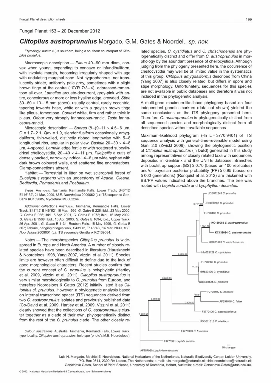
199Fungal Planet description sheets
© 2012 Nationaal Herbarium Nederland & Centraalbureau voor Schimmelcultures
Etymology. austro (L) = southern, being a southern counterpart of Clitopilus prunulus.
Macroscopic description — Pileus 40–90 mm diam, con-vex when young, expanding to concave or infundibuliform, with involute margin, becoming irregularly shaped with age with undulating marginal zone. Not hygrophanous, not trans-lucently striate, uniformly pale grey, sometimes with a slight brown tinge at the centre (10YR 7/3–4), adpressed-tomen-tose all over. Lamellae arcuate-decurrent, grey-pink with en-tire, concolorous or more or less hyaline edge, crowded. Stipe 30–60×10–15 mm (apex), usually central, rarely eccentric, tapering towards base, white or with a greyish brown tinge like pileus, tomentose. Context white, firm and rather thick in pileus. Odour very strongly farinaceous-rancid. Taste farina-ceous-rancid. Microscopic description — Spores (8–)9–11 × 4.5–6 µm, Q = 1.7–2.1, Qav = 1.9, slender fusiform occasionally amyg-daliform, thin-walled, distinctly ribbed lengthwise with 5–8 longitudinal ribs, angular in polar view. Basidia 20–30 × 4–8 µm, 4-spored. Lamella edge fertile or with scattered subcylin-drical cheilocystidia, 20–40 × 4–11 µm. Pileipellis a cutis of densely packed, narrow cylindrical, 4–8 µm wide hyphae with dark brown coloured walls, and scattered fine encrustations. Clamp-connections absent. Habitat — Terrestrial in litter on wet sclerophyll forest of Eucalyptus regnans with an understorey of Acacia, Olearia, Bedfordia, Pomaderris and Phebalium.
Typus. AustrAliA, Tasmania, Kermandie Falls, Lower Track, S43°12' E146°52', 24 Mar. 2009, M.E. Noordeloos 2009062 (L); ITS sequence Gen-Bank KC139085, MycoBank MB802264.
Additional collections AustrAliA, Tasmania, Kermandie Falls, Lower Track, S43°12' E146°52', 16 Mar. 1999, G. Gates E 226; ibid., 23 May 2000, G. Gates E 936; ibid., 5 Apr. 2001, G. Gates E 1072; ibid., 16 May 2002, G. Gates E 1508; ibid., 10 Apr. 2003, G. Gates E 1694; ibid., Upper Track, 26 Apr. 2001, G. Gates E 1131; Reuben Falls, 15 May 1999, G. Gates E 507; Tahune, hanging bridges walk, S43°06', E146°43', 14 Mar. 2009, M.E. Noordeloos 2009001 (L), ITS sequence GenBank KC139084.
Notes — The morphospecies Clitopilus prunulus is wide-spread in Europe and North America. A number of closely re-lated species have been described in literature (Hausknecht & Noordeloos 1998, Yang 2007, Vizzini et al. 2011). Species limits are however often difficult to define due to the lack of good morphological characters. Recent studies confirm that the current concept of C. prunulus is polyphyletic (Hartley et al. 2009, Vizzini et al. 2011). Clitopilus austroprunulus is very similar morphologically to C. prunulus from Europe, and therefore Noordeloos & Gates (2012) initially listed it as Clitopilus cf. prunulus. However, a phylogenetic analysis based on internal transcribed spacer (ITS) sequences derived from two C. austroprunulus isolates and previously published data (Co-David et al. 2009, Hartley et al. 2009, Vizzini et al. 2011) clearly showed that the collections of C. austroprunulus clus-ter together as a clade of their own, phylogenetically distinct from the rest of the C. prunulus clade. The other closely re-
lated species, C. cystidiatus and C. chrischonensis are phy-logenetically distinct and differ from C. austroprunulus in mor-phology by the abundant presence of cheilocystidia. Although judging from the phylogeny presented here, the occurrence of cheilocystidia may well be of limited value in the systematics of this group. Clitopilus amygdaliformis described from China (Yang 2007) is also closely related, but differs in spore and stipe morphology. Unfortunately, sequences for this species are not available in public databases and therefore it was not included in the phylogenetic analysis. A multi-gene maximum-likelihood phylogeny based on four independent genetic markers (data not shown) yielded the same conclusions as the ITS phylogeny presented here. Therefore C. austroprunulus is phylogenetically distinct from all sequenced species and morphologically distinct from all described species without available sequences.
Maximum-likelihood phylogram (-ln L = 3770.9401) of ITS sequence analysis with general-time-reversible model using Garli 2.0 (Zwickl 2006), showing the phylogenetic position of Clitopilus austroprunulus (in bold) generated in this study among representatives of closely related taxa with sequences deposited in GenBank and the UNITE database. Branches with bootstrap support (BS) ≥ 0.70 (based on 100 replicates) and/or bayesian posterior probability (PP) ≥ 0.95 (based on 5 000 generations) (Ronquist et al. 2012) are thickened with BS/PP values indicated above the branches. The tree was rooted with Lepista sordida and Lyophyllum decastes.
Fungal Planet 153 – 20 December 2012
Clitopilus austroprunulus Morgado, G.M. Gates & Noordel., sp. nov.
Luis N. Morgado, Machiel E. Noordeloos, National Herbarium of the Netherlands, Naturalis Biodiversity Center, Leiden University, P.O. Box 9514, 2300 RA Leiden, The Netherlands; e-mail: [email protected]; [email protected];
Genevieve Gates, School of Plant Science, University of Tasmania, Hobart, Australia; e-mail: [email protected].
Colour illustrations. Australia, Tasmania, Kermandi Falls, Lower Track, type-locality. Clitopilus austroprunulus, holotype (photo’s M.E. Noordeloos).

200 Persoonia – Volume 29, 2012
REFEREnCES
Aa HA van der, Vanev S. 2002. A revision of the species described in Phyl-losticta. Centraalbureau voor Schimmelcultures, Utrecht, The Nether-lands.
Arzanlou M, Bakhshi M. 2011. Microcyclospora rumicis, a new species on Rumex crispus from Iran. Mycotaxon 118: 181–186.
Arzanlou M, Crous PW. 2006. Strelitziana africana. Fungal Planet No. 8. Centraalbureau voor Schimmelcultures, Utrecht, The Netherlands.
Bensch K, Braun U, Groenewald JZ, Crous PW. 2012. The genus Clado-sporium. Studies in Mycology 72: 1–401.
Berbee ML, Pirseyedi M, Hubbard S. 1999. Cochliobolus phylogenetics and the origin of known, highly virulent pathogens, inferred from ITS and glyceraldehyde-3-phosphate dehydrogenase sequences. Mycologia 91: 964–977.
Braun U. 1995. A monograph of Cercosporella, Ramularia and allied gen-era (Phytopathogenic Hyphomycetes). IHW Verlag, München, Germany.
Cannon PF, Hawksworth DL, Sherwood-Pike MA. 1985. The British Asco-mycotina. An annotated checklist. Commonwealth Mycological Institute, Kew, Surrey, England.
Cheewangkoon R, Groenewald JZ, Summerell BA, Hyde KD, To-anun C, Crous PW. 2009. Myrtaceae, a cache of fungal biodiversity. Persoonia 23: 55–85.
Chomnunti P, Ko Ko TW, Chukeatirote E, Hyde KD, Cai L, et al. 2012. Phy-logeny of the Chaetothyriaceae in northern Thailand including three new species. Mycologia 103: 382–395.
Co-David D, Langeveld D, Noordeloos M. 2009. Molecular phylogeny and spore evolution of Entolomataceae. Persoonia 23: 147–176.
Constantinescu O, Holm K, Holm L. 1989. Teleomorph-anamorph connec-tions in Ascomycetes. 1–3. Stanhughesia (Hyphomycetes) new genus, the anamorph of Ceramothyrium. Studies in Mycology 31: 69–84.
Coutinho ML, Miller AZ, Gutierrez-Patricio S, Hernandez-Marine M, Gomez- Bolea A, et al. 2012. Microbial communities on deteriorated artistic tiles from Pena National Palace (Sintra, Portugal). International Biodeteriora-tion & Biodegradation. doi.org/10.1016/j.ibiod.2012.05.028.
Crous PW. 1999. Species of Mycosphaerella and related anamorphs oc-curring on Myrtaceae (excluding Eucalyptus). Mycological Research 103: 607–621.
Crous PW, Braun U. 2003. Mycosphaerella and its anamorphs. 1. Names published in Cercospora and Passalora. CBS Biodiversity Series 1: 1– 571. Utrecht, The Netherlands.
Crous PW, Braun U, Groenewald JZ. 2007. Mycosphaerella is polyphyletic. Studies in Mycology 58: 1–32.
Crous PW, Braun U, Hunter GC, Wingfield MJ, Verkley GJM, et al. In press. Phylogenetic lineages in Pseudocercospora. Studies in Mycology 75: 37–114.
Crous PW, Groenewald JZ. 2011. Why everlastings don’t last. Persoonia 26: 70–84.
Crous PW, Groenewald JZ, Shivas RG. 2010a. Devriesia fraseriae. Fungal Planet 65. Persoonia 25: 150–151.
Crous PW, Groenewald JZ, Shivas RG. 2010b. Strelitziana eucalypti. Fun-gal Planet 62. Persoonia 25: 144–145.
Crous PW, Groenewald JZ, Shivas RG, Edwards J, Seifert KA, et al. 2011. Fungal Planet description sheets: 69–91. Persoonia 26: 108–156.
Crous PW, Schoch CL, Hyde KD, Wood AR, Gueidan C, et al. 2009a. Phy-logenetic lineages in the Capnodiales. Studies in Mycology 64: 17–47.
Crous PW, Summerell BA, Carnegie AJ, Wingfield MJ, Groenewald JZ. 2009b. Novel species of Mycosphaerellaceae and Teratosphaeriaceae. Persoonia 23: 119–146.
Crous PW, Summerell BA, Carnegie AJ, Wingfield MJ, Hunter GC, et al. 2009c. Unravelling Mycosphaerella: do you believe in genera? Persoonia 23: 99–118.
Crous PW, Summerell BA, Shivas RG, Burgess TI, Decock CA, et al. 2012. Fungal Planet description sheets: 107–127. Persoonia 28: 138–182.
Crous PW, Wingfield MJ, Groenewald JZ. 2009d. Niche sharing reflects a poorly understood biodiversity phenomenon. Persoonia 22: 83–94.
Crous PW, Wood AR, Okada G, Groenewald JZ. 2008. Foliicolous micro-fungi occurring on Encephalartos. Persoonia 21: 135–146.
Decock C, Delgado Rodriguez G, Seifert KA. 2005. Phylogeny of Synchae-tomella lunatospora, a new genus and species of synnematous fungi from Southeast Asia. Antonie van Leeuwenhoek 88: 231–240.
Ellis MB. 1971. Dematiaceous hyphomycetes. CAB International, Kew, UK.Farr DF, Bills GF, Chamuris GP, Rossman AY. 1989. Fungi on plants and
plant products in the United States. APS Press, USA. Fernando AA, Currah RS. 1995. Leptodontidium orchidicola (Mycelium
radicis atrovirens complex): Aspects of its conidiogenesis and ecology. Mycotaxon 54: 287–294.
Flory SL, Kleczewski N, Clay K. 2011. Ecological consequences of patho-gen accumulation on an invasive grass. Ecosphere 2, 120: 1–12.
Frank J, Crous PW, Groenewald JZ, Oertel B, Hyde KD, et al. 2010. Micro-cyclospora and Microcyclosporella: novel genera accommodating epi-phytic fungi causing sooty blotch on apple. Persoonia 24: 93–105.
Glienke C, Pereira OL, Stringari D, Fabris J, Kava-Cordeiro V, et al. 2011. Endophytic and pathogenic Phyllosticta species, with reference to those associated with Citrus Black Spot. Persoonia 26: 47–56.
Groenewald JZ, Nakashima C, Nishikawa J, Shin H-D, Park JH, et al. In press. Species concepts in Cercospora: spotting the weeds among the roses. Studies in Mycology 75: 115–170.
Hartley AJ, Mattos-Shipley K, Collins CM, Kilaru S, Foster GD, Bailey AM. 2009. Investigating pleuromutilin-producing Clitopilus species and related basidiomycetes. FEMS Microbiology Letters 297: 24–30.
Hausknecht A, Noordeloos ME. 1998. Neue oder seltene arten der En-tolomataceae (Agaricales) aus Mittel- und Südeuropa. Österreichische Zeitschrift für Pilzkunde 8: 199–221.
Hoog GS de. 1977. Rhinocladiella and allied genera. Studies in Mycology 15: 1–140.
Hoog GS de, Weenink XO, Gerrits van den Ende AHG. 1999. Taxonomy of the Phialophora verrucosa complex with the description of two new spe-cies. Studies in Mycology 43: 107–121.
Hyde KD, Frölich J, Taylor JE. 1998. Fungi from palms XXXVI. Sydowia 50: 21–79.
Kang JC, Crous PW, Old KM, Dudzinski MJ. 2001. Non-conspecificity of Cylindrocladium quinqueseptatum and Calonectria quinqueseptata based on beta-tubulin gene phylogeny and morphology. Canadian Journal of Botany 79: 1241–1247.
Kang JC, Hyde KD, Kong RYC. 1999. Studies on the Amphisphaeriales 1. The Clypeosphaeriaceae. Mycoscience 40: 151–164.
Kang JC, Kong RYC, Hyde KD. 1998. Studies on the Amphisphaeriales 1. Amphisphaeriaceae (sensu stricto) and its phylogenetic relationships in-ferred from 5.8SrDNA and ITS2 sequences. Fungal Diversity 1: 147–157.
Kleczewski NM, Flory SL. 2010. Leaf blight disease of the invasive grass Microstegium vimineum caused by a Bipolaris sp. Plant Disease 94: 807–811.
Kleczewski NM, Flory SL, Clay K. 2012. Variation in pathogenicity and host range of Bipolaris sp. causing leaf blight disease on the invasive grass Microstegium vimineum. Weed Science 60: 486–493.
Kobayashi T. 2007. Index of fungi inhabiting woody plants in Japan. Host, distribution and literature. Zenkoku-Noson-Kyoiku Kyokai Publishing Co., Ltd., Japan.
Kornerup A, Wanscher JH. 1978. Methuen handbook of colour. Eyre Methuen Ltd., UK.
Lombard L, Crous PW, Wingfield BD, Wingfield MJ. 2010a. Species con-cepts in Calonectria (Cylindrocladium). Studies in Mycology 66: 1–14.
Lombard L, Crous PW, Wingfield BD, Wingfield MJ. 2010b. Phylogeny and systematics of the genus Calonectria. Studies in Mycology 66: 31–69.
Lombard L, Zhou XD, Crous PW, Wingfield BD, Wingfield MJ. 2010c. Calo-nectria species associated with cutting rot of Eucalyptus. Persoonia 24: 1–11.
Manamgoda DS, Cai L, McKenzie EHC, Crous PW, Madrid H, et al. 2012. A phylogenetic and taxonomic re-evaluation of the Bipolaris – Cochliobolus – Curvularia complex. Fungal Diversity 56: 131–144.
Marvanová L, Pascoal C, Cássio F. 2003. New and rare hyphomycetes from streams of Northwest Portugal. Part I. Cryptogamie Mycologie 24: 339–358.
Noordeloos ME, Gates GM. 2012. The Entolomataceae of Tasmania. Fun-gal Diversity Research Series, vol. 22.
Overholts L. 1943. Mycological notes for 1939–40. Mycologia 35: 243–254.Park D. 1972. On the ecology of heterotrophic micro-organisms in fresh-
water. Transactions of the British Mycological Society 58: 291–299.Park RF, Keane PJ, Wingfield MJ, Crous PW. 2000. Fungal diseases of euca-
lypt foliage. In: Keane PJ, Kile GA, Podger FD, Brown BN (eds), Diseases and pathogens of eucalypts: 153–239. CSIRO publishing, Australia.
Pereira JM, Barreto RW, Ellison AC, Maffia LA. 2003. Corynespora casi-icola f. sp. lantanae: a potential biocontrol agent from Brazil for Lantana camara. Biological Control 26: 21–31.
Pisetta M, Montecchio L, Longa CMO, Salvadori C, Zottele F, Maresi G. 2012. Green alder decline in the Italian Alps. Forest Ecology and Manage-ment 281: 75–83.
Quaedvlieg W, Kema GHJ, Groenewald JZ, Verkley GJM, Seifbarghi S, et al. 2011. Zymoseptoria gen. nov.: a new genus to accommodate Septoria-like species occurring on graminicolous hosts. Persoonia 26: 57–69.
Rayner RW. 1970. A mycological colour chart. Commonwealth Mycological Institute, Kew, Surrey, England.

201Fungal Planet description sheets
© 2012 Nationaal Herbarium Nederland & Centraalbureau voor Schimmelcultures
Roldán A, Puig MA. 1992. Hifomycetos acuáticos en la cuenca del río Esva (Asturias, norte de Espana). Anales del Real Jardín Botánico de Madrid 17: 3–11.
Ronquist F, Teslenko M, Mark P, Ayres D, Darling A, Hohna S, Larget B, Liu L, Suchard M, Huelsenbeck J. 2012. MrBayes 3.2: Efficient Bayesian phylogenetic inference and model choice across a large model space. Systematic Biology 61: 539–542.
Rossman AY, Aime MC, Farr DF, Castlebury LA, Peterson KR, Leahy R. 2004. The coelomycetous genera Chaetomella and Pilidium represent a newly discovered lineage of inoperculate discomycetes. Mycological Progress 4: 275–290.
Saccardo PA. 1882. Sylloge fungorum omnium hucusque cognitorum I. Patavii, Italy.
Seifert KA. 1985. A monograph of Stilbella and some allied hyphomycetes. Studies in Mycology 27: 1–235.
Seifert KA, Nickerson NL, Corlett M, Jackson ED, Lois-Seize G, Davies RJ. 2004. Devriesia, a new hyphomycete genus to accommodate heat-resis-tant, cladosporium-like fungi. Canadian Journal of Botany 82: 914–926.
Shimizu K, Tanaka C, Peng Y-L, Tsuda M. 1998. Phylogeny of Bipolaris inferred from nucleotide sequences of Brn1, a reductase gene involved in melanin biosynthesis. Journal of General and Applied Microbiology 44: 251–258.
Shivas RG, Alcorn JL. 1996. A checklist of plant pathogenic and other mi-crofungi in the rainforests of the wet tropics of northern Queensland. Aus-tralasian Plant Pathology 25: 158–173.
Sivanesan A. 1987. Graminicolous species of Bipolaris, Curvularia, Drechs-lera, Exserohilum and their teleomorphs. Mycological Papers 158: 1–261.
Sprague R. 1950. Diseases of cereals and grasses in North America. Ronald Press Co., New York, USA.
Stukenbrock EH, Quaedvlieg W, Javan-Nikhah M, Zala M, Crous PW, Mc-Donald BA. 2012. Zymoseptoria ardabiliae and Z. pseudotritici, two pro-genitor species of the septoria tritici leaf blotch fungus Z. tritici (synonym: Mycosphaerella graminicola). Mycologia 104: 1397–1407.
Sutton BC. 1980. The Coelomycetes. Fungi imperfecti with pycnidia, acer-vuli and stromata. Commonwealth Mycological Institute, Kew, Surrey, England.
Swofford DL. 2003. PAUP* 4.0b10. Phylogenetic Analysis Using Parsimony (*and other methods). Version 4. Sinauer Associates, Sunderland, MA, USA.
Tomlinson PB. 2006. The uniqueness of palms. Botanical Journal of the Lin- nean Society 151: 5–14.
Vizzini A, Musumeci E, Ercole E, Contu M. 2011. Clitopilus chrischonensis sp. nov. (Agaricales, Entolomataceae), a striking new fungal species from Switzerland. Nova Hedwigia 92: 425–434.
Yang ZL. 2007. Clitopilus amygdaliformis, a new species from tropical China. Mycotaxon 100: 241–246.
Zhang R, Yang HL, Sun GY, Li HY, Zhuang JL, Zhai XR, Gleason ML. 2009. Strelitziana mali, a new species causing sooty blotch on apple fruit. Myco-taxon 110: 477–485.
Zwickl D. 2006. Genetic algorithm approaches for the phylogenetic analysis of large biological sequence datasets under the maximum likelihood crite-rion. PhD thesis, The University of Texas at Austin.