functional characterization of d9, a novel deazaneplanocin a (dznep) analog, in targeting acute...
TRANSCRIPT
RESEARCH ARTICLE
Functional Characterization of D9, a NovelDeazaneplanocin A (DZNep) Analog, inTargeting Acute Myeloid Leukemia (AML)Xia Jiang1, Cheryl Zi Hui Lim1, Zhimei Li1, Puay Leng Lee1, Siti Maryam J. M. Yatim1,Peiyong Guan2, Juntao Li2, Jianbiao Zhou3, Jingxuan Pan4, Wee-Joo Chng3,5,6, ChristinaL. L. Chai7,8, Qiang Yu1,9,10*
1 Cancer Therapeutics and Stratified Oncology, Genome Institute of Singapore, A*STAR (Agency forScience, Technology and Research), Biopolis, 138672, Singapore, 2 Computational and Systems Biology,Genome Institute of Singapore, A*STAR (Agency for Science, Technology and Research), Biopolis, 138672,Singapore, 3 Cancer Science Institute of Singapore, National University of Singapore, 14 Medical Drive,Singapore, 117599, Singapore, 4 Department of Pathophysiology, School of Medicine, Sun Yat-SenUniversity, 74 Zhongshan Road II, Guangzhou, 510089, China, 5 Department of Haematology-Oncology,National University Cancer Institute of Singapore, National University Health System, Singapore, Singapore,6 Department of Medicine, Yong Loo Lin School of Medicine, National University of Singapore, Singapore,Singapore, 7 Institute of Chemical and Engineering Sciences, 8 Biomedical Grove, Neuros #07–01,Singapore, 138665, Singapore, 8 Department of Pharmacy, 18 Science Drive 4, National University ofSingapore, Singapore 117543, 9 Cancer Institute, Jinan University, Guangzhou, 510632, China,10 Department of Physiology, Yong Loo Lin School of Medicine, National University of Singapore,Singapore, 117597, Singapore
AbstractAberrant epigenetic events contribute to tumorigenesis of all human cancers. Significant ef-
forts are underway in developing new generation of epigenetic cancer therapeutics. Al-
though clinical trials for agents targeting DNA hypermethylation and histone deacetylation
have yielded promising results, developing agents that target histone methylation remains
to be in the early stage. We and others have previously reported that 3-Deazaneplanocin A
(DZNep) is a histone methylation inhibitor that has a wide range of anticancer effects in vari-
ous human cancers. Here, focusing on acute myeloid leukemia (AML) as a model, we re-
ported a less toxic analog of DZNep, named D9, which is shown to be efficacious in AML
cell lines and patient-derived samples in vitro, as well as AML tumorigenesis in vivo. Gene
expression analysis in a panel of AML cell lines treated with D9 identified a set of genes that
is associated with D9 sensitivity and implicated in multiple oncogenic signaling pathways.
Moreover, we show that D9 is able to deplete the leukemia stem cells (LSC) and abolish
chemotherapy-induced LSC enrichment, leading to dramatic elimination of AML cell surviv-
al. Thus, D9 appears to be a robust epigenetic compound that may constitute a potential for
AML therapy.
PLOS ONE | DOI:10.1371/journal.pone.0122983 April 30, 2015 1 / 20
OPEN ACCESS
Citation: Jiang X, Lim CZH, Li Z, Lee PL, YatimSMJM, Guan P, et al. (2015) FunctionalCharacterization of D9, a Novel Deazaneplanocin A(DZNep) Analog, in Targeting Acute MyeloidLeukemia (AML). PLoS ONE 10(4): e0122983.doi:10.1371/journal.pone.0122983
Academic Editor: Javier S Castresana, University ofNavarra, SPAIN
Received: December 9, 2014
Accepted: February 26, 2015
Published: April 30, 2015
Copyright: © 2015 Jiang et al. This is an openaccess article distributed under the terms of theCreative Commons Attribution License, which permitsunrestricted use, distribution, and reproduction in anymedium, provided the original author and source arecredited.
Data Availability Statement: All relevant data arewithin the paper.
Funding: This work was supported by the JointCouncil Office and Exploit Technology of Agency forScience, Technology, and Research (A�STAR) to Q.Y. and C.C.; by the Singapore National ResearchFoundation; and by the Ministry of Education underthe Research Centre of Excellence Program to W.-J.C. W.-J.C. was supported by the National MedicalResearch Council Clinician Scientist InvestigatorAward. X.J. was supported by A�STAR graduatefellowship. The funders had no role in study design,
IntroductionAcute myeloid leukemia (AML) is an aggressive hematological disorder in which the haemato-poietic progenitor cells lose their ability to differentiate normally and continue to proliferate.AML is an extremely heterogeneous disease with variable long term survival rate ranging from20%-90% [1]). Although a number of targeted therapeutics have been proposed for treatingAML, chemotherapy, such as cytarabine (Ara-C), adriamycin (ADR) or their combination, re-mains to be the first-line treatment option for most of the AML patients [2, 3]. In spite of aninitial complete remission (CR) in nearly 70% of AML patients following the chemotherapy, alarge portion of these patients subsequently relapse and eventually die of the disease progres-sion [4]. It is generally thought that the disease recurrence stems from a rare subset of leukemiastem cells (LSCs) that are resistant to standard chemotherapy [5–8], which therefore raises astrong need to develop therapeutics to target LSCs.
Although there are growing interests in developing epigenetic therapy for hematologicalmalignancies, the success for clinical advancement of histone deacetylase (HDAC) inhibitorsand DNA methylation inhibitors remains to be limited for AML. Additionally, aberrant his-tone methylations, such as those induced by Polycomb protein Enhancer of Zeste homolog 2(EZH2) [9, 10], Mixed-Lineage Leukemia (MLL) [11–13] and G9a [14–16] have been alsoshown to be attractive therapeutic targets. To date, efforts for developing histone methylationinhibitors are still in their infancy and no drugs have ever been approved by FDA or in latestages of clinical trials in AML and other malignancies.
We have previously reported that S-adenosylhomocysteine hydrolase inhibitor 3-deazane-planocin A (DZNep) is a potent histone methylation inhibitor that is able to deplete the onco-genic PRC2 and associated histone H3 Lysine 27 trimethylation (H3K27me3) together withother histone methylations and induce robust apoptosis in cancer cells but not in normal cells[17]. Hereafter, there have been increasing numbers of reports showing the impressive antican-cer effects of DZNep as a new epigenetic compound in a variety of cancer models both in vitroand in vivo [18–23]. In particular, DZNep alone, or in combination with HDAC inhibitor, hasbeen shown to be effective in inducing growth arrest and apoptosis in AML cells [24, 25].Therefore, there are interests in exploring DZNep and its analogs as a potential new class ofepigenetic cancer therapeutics for AML.
In this study, we reported a comprehensive study of a novel analog of DZNep, named D9, inAML. We show that D9 induces strong growth inhibition and apoptosis in AML cells and itssensitivity in AML cells is associated with its ability to modulate gene expression associatedwith multiple oncogenic signaling pathways. Importantly, D9 is able to deplete both the basaland chemotherapy-enriched LSCs, and abrogate chemotherapy-induced gene expression asso-ciated with chemoresistance. Our results demonstrated a unique utility of D9 as a novel histonemethylation inhibitor in targeting both bulk leukemic cells and leukemia stem cells (LSCs),thus providing a novel potential for eradication of AML.
Materials and Methods
Cell culture and drugsA panel of acute myeloid leukemia (AML) cell lines, that is, MV4-11, Kasumi-1, KG-1, KG-1a,TF-1, TF-1a, MOLM-14, THP-1, HL-60 and Mono-Mac-1 was obtained as described previous-ly [24]. D9 (MW: 308.16) was synthesized at Institute of Chemical and Engineering Sciences,Singapore. Suberoylanilide hydroxamic acid (SAHA), Adriamycin, Ara-C and Decitabine werepurchased from Sigma-Aldrich.
Therapeutic Effects of DZNep Analog D9 on AML
PLOSONE | DOI:10.1371/journal.pone.0122983 April 30, 2015 2 / 20
data collection and analysis, decision to publish, orpreparation of the manuscript.
Competing Interests: The authors have declaredthat no competing interests exist.

Patient samplesFrozen bone marrow (BM) blasts from newly diagnosed AML patients were obtained at the Na-tional University Hospital in Singapore with informed consent and approved by InstitutionalReview Board of National University Hospital. Written consents were obtained from the partici-pants. Immediately after recovery, primary AML cells were cultured in IMDM supplementedwith 10% FBS (Invitrogen), FLT3 ligand (20 ng/ml), SCF (20 ng/ml), IL-3 (20 ng/ml), G-CSF(50 ng/ml) and thrombopoietin (TPO; 50 ng/ml) (R&D Systems).
Cell viability assayCells were seeded at a density of 1 × 10³ cells per well of 96-well optical bottom plate (Corning)24 hours before D9 treatment. A range of concentrations of D9 diluted with basal RPMI orIMDMmedium (Invitrogen) were added into the wells of cell suspension and incubated for 96hours before detection. Six replicas were conducted for each sample. CellTiter-Glo LuminescentCell Viability Assay (Promega) was used to evaluate the viable cell numbers. The luminescencesignal was measured by MicroLumat Plus LB96V system (BERTHOLD TECHNOLOGIES). Tocalculate EC50, nonlinear regression sigmoidal dose response curves were generated using Graph-Pad PRISM3.
Transwell migration and invasion assayFor invasion assay, 24-well Falcon FluoroBlok Transwell inserts (BD Biosciences) with a poresize of 8 μmwere precoated with growth factor-reduced Matrigel (BD Biosciences) for 3–4hours at 37°C before seeding the cells. For migration assay, the coating step was omitted.5 × 104 pretreated cells in 200 μl RPMI containing 0.25% FBS were seeded into the upperchamber of each insert. 750 μl RPMI containing 10% FBS were added outside the chamber aschemoattractant. Inserts were fixed after 24 hours of incubation at 37°C by using 3.7% formal-dehyde for 20 minutes at room temperature. After several washes, the inserts were stained with25 μg/ml propidium iodid for 30 minutes at room temperature in the dark. The migrated andinvaded cells in 10 individual fields beneath the inserts were scanned and counted with Cello-mics ArrayScan VTI (Thermo Scientific.). Triplicates were conducted for each sample, and atleast three independent experiments were performed.
Cell adhesion assay96-well optical bottom plates were coated with 10 μg/ml Fibronectin (Sigma-Aldrich) for over-night at 4°C. The plates were washed three times using PBS prior to seeding the cells. 1 × 10³
TF-1a cells were seeded into each well and treated with D9 (100 nM) with or without Ara-C(50 nM). After 72 hours, the wells were gently washed by PBS three times to remove the unat-tached cells. CellTiter-Glo Luminescent Cell Viability Assay (Promega) was used for the quan-tification of the remaining adherent cells.
AntibodiesThe following primary antibodies were used. Cleaved PARP, EZH2, p-ERK (T202/204), p-AKT (S473), total ERK and total AKT (Cell Signaling Technology); Bim and Survivin (BD Bio-sciences); H3K27me3, H3K9me3, H3K4me3, H3K79me2, H4K20me3 and total H3 (UpstateBiotechnology); and actin (Sigma-Alrich) were used as primary antibodies. Anti-rabbit andanti-mouse horseradish peroxidise linked whole antibodies (GE Healthcare) were used assecondary antibodies.
Therapeutic Effects of DZNep Analog D9 on AML
PLOSONE | DOI:10.1371/journal.pone.0122983 April 30, 2015 3 / 20

PI staining, active caspase3 staining and flow cytometric analysisTreated cells were washed twice with cold PBS before fixing by 70% ethanol for at least 1 hourat 4°C. The fixed cells were washed twice with PBS, treated by 100 μl RNase (100 μg/ml) for 5minutes, and then stained with 400 μl propidium iodide (PI) (50 μg/ml) for 30 minutes in thedark. The DNA contents were measured by FACSCalibur (Becton Dickson Instrument), and1 × 104 cells from each sample were analyzed by using Cell Quest software (Becton DickinsonInstrument). For active caspase3 assay, cytofix/cytoperm fixation/permeabilization kit (BDBiosciences) coupled with FITC rabbit anti active Caspase-3 antibody (BD Biosciences) wereused to stain the cells for flow cytometric analysis.
Immunofluorescent staining and flow cytometric analysisTF-1a Cells were stained with CD38-FITC antibody and CD34-APC antibody (Miltenyi Bio-tec) sequentially. After staining, the cell pellets were suspended with 0.5 ml of 1 μg/ml propi-dium iodide (PI) just before harvested for analysis by FACSCalibur (BD Bioscience). Celldebris and dead cells were excluded from the analysis based on scatter signals and PI fluores-cence. The stained cells were analyzed by using CellQuest software (BD bioscience).
Microarray gene expression analyses and RT-PCRTotal RNA was isolated using the RNeasy Mini Kit (Qiagen). Microarray hybridization wasperformed using Human HT-12 V4.0 expression beadchip (Illumina), and data analysis wasperformed using GeneSpring GX (Agilent Technologies). The finalized genesets were importedinto Ingenuity Pathway Analysis (IPA) (www.ingenuity.com) for gene ontology analysis. Themicroarray data reported herein have been deposited at the National Centre for BiotechnologyInformation’s Gene Expression Omnibus (accession no. GSE59624 and GSE59625). Reverse-transcription and quantitative PCR (qPCR) assays were performed using the High-CapacitycDNA Archive Kit and Kapa SyBr Fast qPCR Kit (Kapa Biosystems), respectively. Primer se-quences are listed in S1 Table.
Mouse xenografts and drug treatmentFor subcutaneous implantation, 5 x 10⁶ MOLM-14 cells were subcutaneously injected into femaleathymic BALB/c nude mice (5–8 week-old). D9 at three different doses (30, 60 and 90 mg/kg)was administered though intraperitoneal (i.p.) injection as qd x 5d/w for 3 weeks in these recipi-ents. Tumor diameters were measured every 3–4 days with calliper and the body weight of micewas monitored overtime. For bone marrow engraftment, 1 x 10⁶ MOLM-14 cells were trans-planted into the sublethally irradiated NOD/SCIDmice (5–8 week-old) mice through lateral tailvein injection. D9 (60 mg/kg) was administered through intraperitoneal (i.p.) injection two daysafter the tumor engraftment as q2d/w for 3 weeks. All animal studies were conducted in compli-ance with animal protocols approved by the A�STAR-Biopolis Institutional Animal Care andUse Committee (IACUC) of Singapore.
Statistical analysisStatistical analysis of this study was performed using GraphPad PRISM3. All values are ex-pressed as mean ± SEM (standard error of mean) of at least three independent experiments.P values were calculated with the two-tailed Student’s t test. A p value< 0.05 is consideredstatistically significant.
Therapeutic Effects of DZNep Analog D9 on AML
PLOSONE | DOI:10.1371/journal.pone.0122983 April 30, 2015 4 / 20

Results
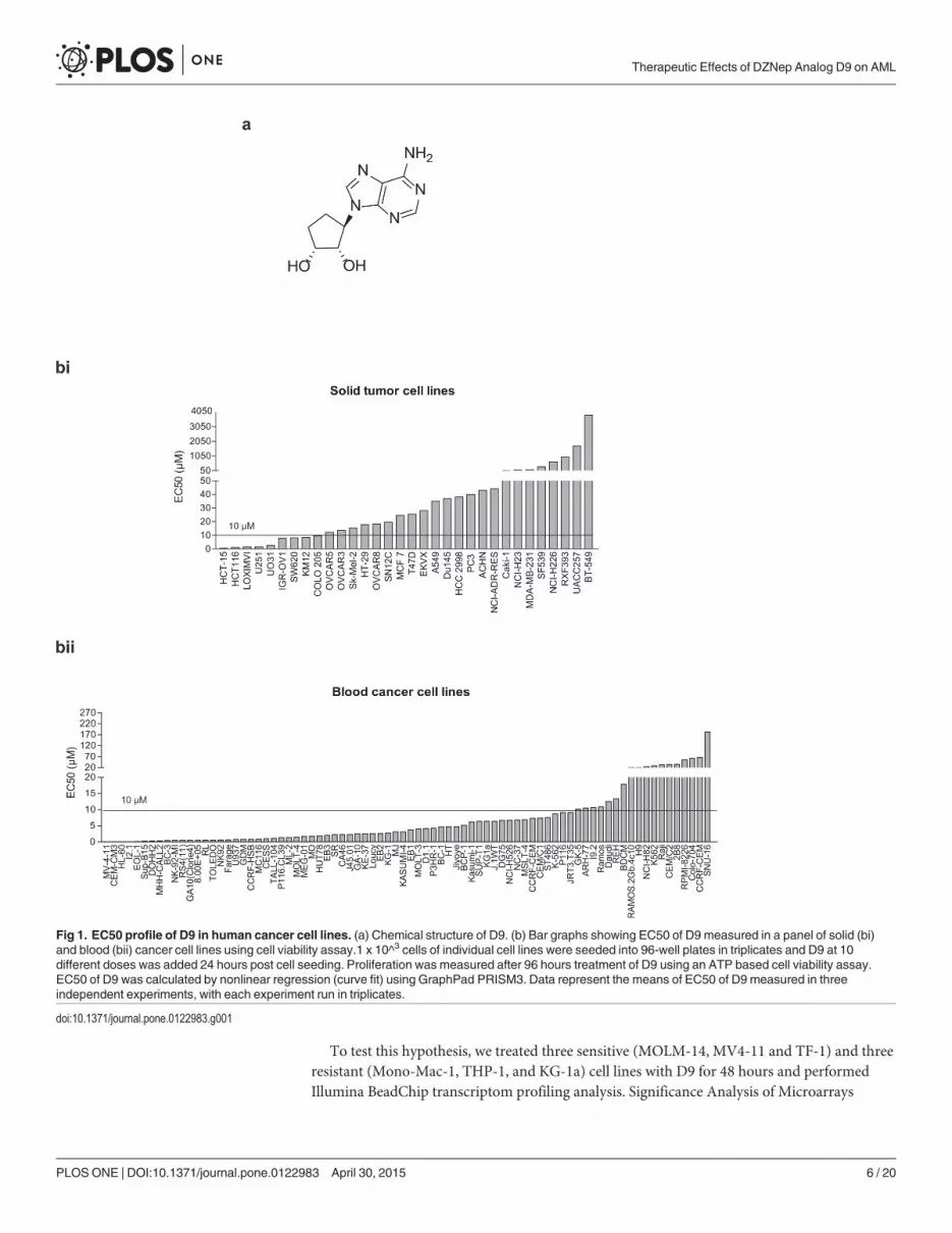
EC50 profiling of D9 in human cancer cell linesIn an effort to identify DZNep analogs with better potency and safety profile, we have synthe-sized a series of DZNep-like compounds and identified several lead compounds throughstructure and cellular activity relationship (SAR) analysis as well as correlation of chemicaland physical properties. One such compound, D9 (Fig 1A), showed a comparable cellular ac-tivity with DZNep but a 20-fold less toxicity in mice [26] and was thus chosen for furtherinvestigations.
We first set out to determine which type of cancer cells will be most sensitive to D9. To dothis, we screened a large panel of human cancer cell lines, including 32 solid tumor cell lines and80 blood cancer cell lines, and determined their EC50 responses to D9. Overall, these cancer celllines showed heterogeneous responses to D9; and blood cancer cell lines in general appeared tobe more sensitive to D9 compared to solid tumor cell lines (median EC50 of 3.14 μM in bloodcancer cell lines versus 25.1 μM in solid tumor cell lines) (Fig 1Bi and 1Bii; S2 and S3 Tables).Among the blood cancer cell lines, a significant number of AML cell lines such as MV4-11, HL-60, EOL1, and GDM-1 were highly sensitive to D9, with EC50s of< 1μM (Fig 1Bii). We thus de-cided to focus on AML for further investigation of the anti-cancer effect of D9.
In vitro activity of D9 in AML cell linesTo investigate the effect of D9 on AML, we further validated the effect of D9 on the short-termviability of a panel of 10 well-established AML cell lines. For comparison, we also included his-tone deacetylase (HDAC) inhibitor SAHA and DNAmethylation inhibitor Decitabine, both ofwhich are FDA approved for the treatment of hematological malignancies [27, 28]. The resultsshowed that in general AML cell lines were remarkably more sensitive to D9 compared to ei-ther SAHA or DAC, with a median EC50 of 0.31 μM for D9, 1.08 μM for SAHA and 0.94 μMfor DAC, respectively (Fig 2B and 2C), indicating that D9 is potentially a more potent com-pound in AML cell lines compared to SAHA and DAC. Moreover, in a 14-day long soft agarcolony formation assay, D9 was also effective in reducing the colony growth of MOLM-14,KG-1 and HL-60 cells in a dose-dependent manner, with similar EC50s as determined by theshort-term cell viability assay (Fig 2D). Finally, we show that D9 was able to induce a sub-G1cell death in MV4-11, MOLM-14, TF-1, KG-1, Kasumi-1 and TF-1a cells (45–75%), but to less-er extents in THP-1, HL-60, Mono-Mac-1and KG-1a cells (< 20%) (Fig 2E). The D9-inducedcell death was further confirmed to be apoptosis by measuring Caspase 3 activation in MOLM-14 and MV4-11 cells (Fig 2F).
Effects of D9 on histone methylation and transcriptome changesassociated with D9-induced apoptosis in AML cellsWe next evaluated the overall effects of D9 on histone lysine methylations in sensitive MOLM-14 and resistant KG-1a to determine if D9 would have differential effects on the histone meth-ylations in the two cell lines. In both cell lines we showed that D9 induced similar levels of sup-pression of H3K27me3 and H4K20me3 and to lesser extents on H3K4me3 and H3K79me2,while it had little effects on H3K9me2 and H3K9me3 (Fig 3A and 3B). This observation is simi-lar to the parental compound DZNep that has been shown to act as a selective but non-specifichistone methylation inhibitor in cancer cells, including AML [17, 24]. Thus, the differentialsensitivities of AML cells to D9 do not seem to be associated with its ability to inhibit the bulkhistone methylation per se, but perhaps more likely to be associated with the differential geneexpression response to D9.
Therapeutic Effects of DZNep Analog D9 on AML
PLOSONE | DOI:10.1371/journal.pone.0122983 April 30, 2015 5 / 20
To test this hypothesis, we treated three sensitive (MOLM-14, MV4-11 and TF-1) and threeresistant (Mono-Mac-1, THP-1, and KG-1a) cell lines with D9 for 48 hours and performedIllumina BeadChip transcriptom profiling analysis. Significance Analysis of Microarrays
Fig 1. EC50 profile of D9 in human cancer cell lines. (a) Chemical structure of D9. (b) Bar graphs showing EC50 of D9 measured in a panel of solid (bi)and blood (bii) cancer cell lines using cell viability assay.1 x 10^3 cells of individual cell lines were seeded into 96-well plates in triplicates and D9 at 10different doses was added 24 hours post cell seeding. Proliferation was measured after 96 hours treatment of D9 using an ATP based cell viability assay.EC50 of D9 was calculated by nonlinear regression (curve fit) using GraphPad PRISM3. Data represent the means of EC50 of D9 measured in threeindependent experiments, with each experiment run in triplicates.
doi:10.1371/journal.pone.0122983.g001
Therapeutic Effects of DZNep Analog D9 on AML
PLOSONE | DOI:10.1371/journal.pone.0122983 April 30, 2015 6 / 20

Fig 2. In vitro anti-leukemia activity of D9 in AML cell lines. (a) Bar graph showing EC50 of D9 measured in a panel of AML cell lines using cell viabilityassay. (b) Dot plot showing EC50 of D9, SAHA and DACmeasured in the same panel of AML cell lines. The short lines represent the median EC50 of eachcompound in the AML cell line panel. (c) Table showing EC50 of D9, SAHA and DACmeasured in the AML cell line panel. (d) Representative images of softagar assay conducted in MOLM-14, KG-1 and HL-60 cells. 1 x 10^3 cells were mixed with D9 at various doses in semi-solid media and incubated for 2 weeks.Colony growth was assessed and EC50 of D9 measured by soft agar assay was indicated in the brackets. Data represent the means of EC50 of D9measured in three independent experiments, with each experiment run in triplicates. (e) Bar graphs showing sub-G1 percentage of 10 AML cell lines treatedwith indicated concentrations of D9 for 72 hours, followed by propidium iodide (PI) staining and fluorescence-activated cell sorting (FACS) analysis. (f) Bargraphs showing the percentage of cells positive for active Caspase 3 in MOLM-14 and MV4-11 cells treated with indicated concentrations of D9 for 72 hours,
Therapeutic Effects of DZNep Analog D9 on AML
PLOSONE | DOI:10.1371/journal.pone.0122983 April 30, 2015 7 / 20
(SAM) analysis shows that D9 treatment resulted in transcriptional changes of 547 genes, in-cluding 327 upregulated genes and 220 downregulated genes in sensitive cell lines but not in re-sistant cell lines, as shown in a heatmap of supervised gene clustering (Fig 3C; S4 and S5Tables). Ingenuity pathway analysis (IPA) of both gene sets revealed gene networks linked tothe PI3K/AKT and MEK/ERK pathways (Fig 3Di and 3Dii), whose aberrant activation is fre-quently observed in AML [29, 30]. This analysis raised the possibility that D9 treatment in-duced a transcriptional change that may result in modulations of PI3K/AKT and MAPKsignaling cascades in D9-sensitive cell lines but not resistant cell lines.
D9 treatment suppresses PI3K and MAPK signaling in both AML celllines and primary patient samplesTo validate the above hypothesis, we investigated the effects of D9 on the PI3K and MAPKpathways by western blot analysis in two most sensitive cell lines MOLM-14 and MV4-11 andthree resistant cell lines Mono-Mac-1 (MM), THP-1 and KG-1a. Consistent with what we havepredicted from the gene expression analysis, D9 treatment resulted in significant suppressionof MAPK and PI3K signaling as indicated by reduced phosphorylation of ERK (T202/204) orAKT (S473) in sensitive MOLM-14 and MV4-11 cells at both 24 and 48 hours without affect-ing the total levels of either ERK or AKT (Fig 4A). In contrast, the three resistant cells did notshow such changes on p-ERK (T202/204) or p-AKT (S473) (Fig 4A).
It is well known that ERK inhibition results in upregulation of pro-apoptotic Bim induction[31, 32]. Accordingly, we found that all the three Bim isoforms, in particular BimS, the mostpotent isoform for apoptosis [33], were upregulated by D9 in sensitive lines but not in resistantlines. On the other hand, expression of the anti-apoptotic molecule, Survivin, which is a down-stream effector of AKT [34, 35], was suppressed by D9 only in the sensitive cell lines (Fig 4B).Thus, the combined changes in Bim and Survivin induced by D9 are consistent with the strongapoptosis observed in the sensitive cell lines. Based on these observations, we proposed that themolecular mechanisms underlying the D9 sensitivity in AML cells is associated with a tran-scriptomic response resulting in downregulation of oncogenic PI3K-AKT and MAPK signalingpathways, leading to apoptosis.
To validate the clinical relevance of these findings, we investigated whether D9 has a similareffect on patient-derived primary AML cells. To this end, we managed to test the effect of D9on 4 sets of primary AML blasts isolated from four newly diagnosed AML patients. The resultshows that the primary blasts, like AML cell lines, exhibited heterogeneous responses to D9,with EC50 of D9 ranging from 0.96 to 13.43 μM (Fig 4Cii and S6 Table). In the sensitive patientsample AD353, we detected a robust PARP cleavage and strong reductions of phosphorylatedAKT and ERK, which was similar to MOLM-14 line included here as a positive control. In con-trast, we did not see such changes in resistant AD409 sample. In fact, we even observed a slightactivation of AKT and ERK signaling in AD409 (Fig 4Ciii), probably a result of a feedback acti-vation. Thus, D9 is able to target multiple oncogenic signaling pathways and this effect seemsto be associated with the therapeutic effect in sensitive AML cells sourced from both the celllines and patient-derived primary cells.
followed by active Caspase 3 antibody staining and FACS analysis. Data are mean ± SEM; N = 3; *P < 0.05, **P < 0.01, ***P < 0.001, unpaired two tailedt test.
doi:10.1371/journal.pone.0122983.g002
Therapeutic Effects of DZNep Analog D9 on AML
PLOSONE | DOI:10.1371/journal.pone.0122983 April 30, 2015 8 / 20

Fig 3. Effects of D9 on histonemethylation and transcriptome in AML.Western blot analysis showingthe level of cleaved PARP, EZh2 and a series of histone lysine methylation marks in MOLM-14 (a) and KG-1acells (b) treated by D9 at indicated concentrations for 48 and 72 hours. (c) Heat map of differential genesetsbetween sensitive and resistant cell lines with D9 treatment. Three sensitive (MOLM-14, MV4-11 and TF-1)and three resistant cell lines (Mono-Mac-1, KG-1a and THP-1) were treated with D9 at 1 or 5 μM for 48 hours.
Therapeutic Effects of DZNep Analog D9 on AML
PLOSONE | DOI:10.1371/journal.pone.0122983 April 30, 2015 9 / 20
D9 inhibits AML tumorigenesis in vivoTo evaluate the therapeutic potential of D9 in vivo, we used both the xenograft tumor growthmodel and bone marrow engraftment model. First, in a model of MOLM-14-derived subcuta-neous transplantation in nude mice, D9 administered daily at 30, 60 and 90 mg/kg thoughintraperitoneal (i.p.) injection resulted in marked inhibition of tumor growth in a dose-depen-dent manner (Fig 5Ai). The treatment did not cause overt signs of toxicity as judged by insig-nificant body weight loss of mice before and after the D9 treatment (Fig 5Aii), suggesting thatthe effective doses of D9 were well tolerated in the recipient mice.
We next determined the anticancer efficacy of D9 in a more therapeutically relevant modelof AML, in which MOLM-14 cells were injected through the tail vein of sublethally irradiatedNOD/SCID mice to induce leukemic dissemination and mortality. We found that D9 given at60 mg/kg through i.p. injection caused dramatic reduction in leukemic burden (data notshown) and significant prolonged life span of recipient mice (median survivals of 42 days inD9 group vs. 36 days in control group, p< 0.001) (Fig 5Bi). Again, D9 in this model was alsowell tolerated and no overt toxicity was observed in recipient mice (Fig 5Bii). Collectively, ourresults showed that D9 possessed potent anti-AML effects both in vitro and in vivo.
D9 treatment depletes both basal and chemotherapy-induced LSCBecause tumor initiating cells or cancer stem cells have been thought to have undergone epige-netic reprogramming [36, 37], we sought to determine if D9 as a histone methylation inhibitorwould be effective in targeting AML leukemia stem cells (LSCs). To test the effect of D9 onLSCs, we used TF-la, an AML primitive myeloid progenitor cell line, which has a high fractionof stem-like CD34+CD38- cells and thus considered as an excellent model for studying LSC[38]. Treatment of TF-1a cells with increasing low nanomolar concentration of D9 for 72hours resulted in efficient depletion of CD34+CD38- population (Fig 6A). For a comparison,we also included HDAC inhibitor SAHA and DNAmethylation inhibitor DAC in this experi-ment. Interestingly, we did not observe such an effect for SAHA and DAC (Fig 6B).
Chemotherapy is well known to induce enrichment of cancer stem cell, which is believed tocause chemoresistance and disease recurrence. We next tested if D9 is able to reduce chemo-therapy-induced LSC enrichment. As expected, both Ara-C and ADR treatment of TF-1a cellsfor 72 hours resulted in dramatic increases of CD34+CD38- populations, while co-treatmentwith D9 (100 nM) nearly completely abrogated such increases (Fig 6B and 6C). Moreover, in a14 days long-term experiment, D9 used even at 20 nM was also able to sufficiently deplete theCD34+CD38- enrichment by Ara-C (Fig 6D). Again, SAHA did not show such an effect to an-tagonize the chemotherapy-induced LSC enrichment, while DAC showed a much weaker effectthan D9 in targeting LSC (Fig 6E).
To further evaluate the effects of D9 in the context of LSC survival associated with chemore-sistance, we used a colony-forming-unit (CFU) assay, a gold standard in vitro assay to evaluatea long term survival of haematopoietic or leukemia stem/progenitor cells. In this experiment,TF-1a cells were treated with Ara-C or ADR, and then the dead cells were removed by usingDead Cell Removal Kit and the remaining live cells were seeded with equal numbers for CFUassay. Consistent with the induction of LSC by chemotherapy, we observed more coloniesformed in Ara-C- or ADR-treated cells compared to untreated control (Fig 6F). Strikingly, co-
Total RNA was isolated for microarray and SAM analysis. 327 genes were up-regulated and 220 genes weredown-regulated upon D9 treatment in sensitive cells relative to resistant cells using 10% false discovery rate(FDR) cut-off. (d) Ingenuity Pathway Analysis (IPA) of differentially up-regulated geneset and down-regulatedgeneset (dii) showing their strong connections to PI3K/AKT and MEK/ERK signaling pathways.
doi:10.1371/journal.pone.0122983.g003
Therapeutic Effects of DZNep Analog D9 on AML
PLOSONE | DOI:10.1371/journal.pone.0122983 April 30, 2015 10 / 20

Therapeutic Effects of DZNep Analog D9 on AML
PLOSONE | DOI:10.1371/journal.pone.0122983 April 30, 2015 11 / 20

treatment with D9, but not SAHA, nearly completely abolished the colony formation inducedby Ara-C or ADR (Fig 6F). Under the same condition, DAC exerted comparable inhibitory ef-fect on the colony formation of TF-1a cells (Fig 6F). These findings further suggest that D9 as ahistone methylation inhibitor is equipped with a special ability to remove LSC and thus thechemoresistance in AML.
Fig 4. Effects of D9 on AKT and ERK phosphorylation in AML. (a)Western blot analysis of p-ERK (T202/204), total ERK, p-AKT (S473), total AKT and ACTIN in indicated AML cell lines treated with D9 for 24 hoursand 48 hours. (b) Western blot analysis showing the effects of D9 on Bim and Survivin in AML cell lines as in(a). (c) The frozen primary AML patient blasts were recovered for 24 hours and the dead cells were removedusing Dead Cell Removal Kit just before D9 treatment. The blasts were treated for 96 hours for cell viabilityassay and 48 hours for western blot analysis. The diagrams showed the drug response curves of AML patientblasts towards D9 (ci), measurement of EC50 of D9 using cell viability assay (cii) and western blot analysis ofPARP, p-ERK (T202/204), total ERK, p-AKT (S473), total AKT and ACTIN in AML patient blasts as well asMOLM-14 with and without D9 treatment (ciii). Each data point in the plots of drug response curves of D9represents the mean ± SEM of six replicates at each specified concentration of D9, N = 3.
doi:10.1371/journal.pone.0122983.g004
Fig 5. In vivo effects of D9 in AMLmousemodels. (a) Subcutaneous xenograft tumor growth of MOLM-14cells in Balb/c mice treated with D9. 5 x 10^6 of MOLM-14 cells were injected into mice subcutaneously (s.c.).D9 at three different doses (30, 60 and 90mg/kg) was administered through intraperitoneal (i.p.) injection asqd x 5d/w for 3 weeks in these recipients from day 8 to day 23 after transplantation, with vehicle treatment ascontrol. The graphs show the tumor sizes (ai) and body weight change (aii). Data are mean ± SEM,***P < 0.001, unpaired two tailed t test. (b) Kaplan–Meier curves of NOD/SCIDmice treated with D9. 1 x 10^6of MOLM-14 cells were injected into sublethally irradiated NOD/SCIDmice through tail vein. D9 at 60mg/kgwas administered through i.p. injections as q2d/w for 3 weeks in these recipients from day 2 aftertransplantation, with vehicle treatment as control. The graphs showed the survival rate (bi) and body weightchange (bii) during D9 treatment. Data are mean ± SEM, ***P <0.001, log-rank (Mantel-Cox) test conducted.
doi:10.1371/journal.pone.0122983.g005
Therapeutic Effects of DZNep Analog D9 on AML
PLOSONE | DOI:10.1371/journal.pone.0122983 April 30, 2015 12 / 20

Fig 6. Effective anti-leukemia stem cells (LSC) activity of D9 in AML. (a) Bar graphs showing FACS analysis of the proportion of CD34+CD38- populationin TF-1a after the single treatment with D9, SAHA or DAC for 72 hours. (b) Representative FACS histogram profiles of CD34+CD38- cell population in TF-1acells treated with D9 (100 nM) alone or in combination with either Ara-C (20 nM) or ADR (50 nM). (c) Bar graphs showing the proportion of CD34+CD38-population in TF-1a cells treated as in B. (d) Bar graphs showing the proportion of CD34+CD38- population in TF-1a treated with D9 (20 nM) with or withoutAra-C (20 nM) for 14 days. (e). Bar graphs showing the percentage of CD34+CD38- cell population in TF-1a cells treated as indicated. (f) Colony FormationUnit (CFU) assay showing the effects of D9, SAHA or DAC on basal or Ara-C or ADR-induced colony formation capacity of TF-1a cells. The medium anddrugs were replenished every 3 to 4 days and the dead cells were removed by Dead Cell Removal Kit. 1 x 10^3 live cells were seeded with semi-solid colonyformation medium and incubated for 2 weeks before enumeration. Representative images of the colony formation of TF-1a were shown on (fi) and bar graphswere shown the colony numbers on (fii). Data are mean ± SEM; N = 3; *P < 0.05, **P < 0.01, ***P < 0.001, ns represents no significance, unpaired twotailed t test.
doi:10.1371/journal.pone.0122983.g006
Therapeutic Effects of DZNep Analog D9 on AML
PLOSONE | DOI:10.1371/journal.pone.0122983 April 30, 2015 13 / 20

D9 abolishes chemotherapy-induced gene expression associated withdrug resistance in AMLWe next sought to characterize the molecular changes induced by the chemotherapy that is an-tagonized by D9. To do this, TF-1a cells were treated with Ara-C or ADR or co-treated withD9 were harvested. Total RNA was isolated and subjected to gene expression analysis. A super-vised gene cluster analysis revealed a common set of 720 genes that are induced by both Ara-Cand ADR but repressed by D9 (Fig 7A and S7 Table). Ingenuity Pathway Analysis (IPA) indi-cates that the gene categories including “Cellular Movement” and “Cell-To-Cell Signaling andInteraction” appeared among the top five Bio Functions (Fig 7B). In addition, among the topten canonical pathways which were significantly associated with chemotherapeutic gene re-sponse, “Leukocytes Extravasation Signaling”, “IL-8 Signaling” and “Integrin Signaling” wereof particular interests as they have been previously linked to AML tumorigenesis (Fig 7C). Inparticular, the network analysis indicated the involvements of integrins-AKT signalling in che-motherapy induced gene changes (Fig 7D). Our microarray data showed that multiple mem-bers of integrins were upregulated by Arac, which were abolished by co-treatment with D9.The same was also observed for multiple ECM ligands of integrins, particularly the lamininfamily members, and numerous cytokines and their respective receptors, all well known to beassociated with cancer progression, self-renewal and drug resistance (Fig 7E, S8–S11 Tables).The microarray results were further validated by quantitative RT-PCR analysis of 15 represen-tative genes of the implicated gene categories (Fig 7F).
Furthermore, in consonance with the above molecular changes, we noticed that Ara-C treat-ment induced an emergence of a small proportion of TF-1a cells that were attached to the tis-sue culture plate and exhibited spread and mesenchymal-like morphology, which wasreversible upon D9 treatment (Fig 7G). We also found that Ara-C triggered a significant num-ber of migratory, invasive and adherent cells, which were effectively abolished by D9 but not bySAHA. While DAC exerted much weaker effects compared with D9 (Fig 7H, 7I and 7J). Theseresults showed that D9 is able to inhibit the enhanced migration, invasion and adhesion capaci-ties of AML cells induced by chemotherapy. Collectively, these findings demonstrated the po-tential ability of D9 to target chemoresistance of AML through modulating a wide range ofgene sets functionally important in LSC self-renewal, invasion and tumor dissemination.
DiscussionAML is the most common leukemia of adults, and its incidence increases with age. Althoughchemotherapy is capable of producing complete remission in almost 70% of patients, many ofthem will ultimately relapse and succumb to their disease. Thus, there is a great need of newtreatment strategies for AML.
In this study, we provide a comprehensive preclinical investigation of D9, an analog of his-tone methylation inhibitor DZNep in AML. DZNep is a SAM hydrolase inhibitor that inhibitsmultiple histone methylations and is able to effectively deplete oncogenic PRC2 protein com-plex and associated H3K27me3. DZNep has been shown to be effective in inducing apoptosisin a variety of cancer cells both in vitro and in vivo and has been used as a useful tool for study-ing EZH2-mediated gene silencing. In contrast, the specific catalytic inhibitors of EZH2, suchas GSK126 or GSK343, though potent in killing some hematological malignant cells such as thediffuse large B-cell lymphoma (DLBCL) that carry EZH2 activating mutations [10, 39], do notseem to be effective in cancer cells overexpressing a wild type EZH2 and only have a modest ef-fect, if any, on EZH2 target gene expression [9, 40].
Like DZNep, D9 inhibits multiple histone methylation marks such as H3K27me3 andH4K20me3 and also depletes EZH2 protein expression in AML cell lines, which was
Therapeutic Effects of DZNep Analog D9 on AML
PLOSONE | DOI:10.1371/journal.pone.0122983 April 30, 2015 14 / 20

Fig 7. D9 targets cell adhesion-mediated drug resistance (CAM-DR) in AML. (a) Heat map of 720genesets induced by both Ara-C and ADR which were suppressed by D9. (b) The diagram showing the topfive Bio Functions of commonly upregulated 720 genes by Ara-C and ADR suggested by IPA. (c) Thediagram showing the top ten canonical pathways of commonly upregulated 720 genes by Ara-C and ADR
Therapeutic Effects of DZNep Analog D9 on AML
PLOSONE | DOI:10.1371/journal.pone.0122983 April 30, 2015 15 / 20
accompanied by effective growth inhibition and apoptosis in both AML cell lines and primaryAML cells. It also has a potent anti-tumor effect in vivo with a low level of toxicity. However,the mechanisms governing the pharmacologic response are certainly more complex as theabove mentioned changes were seen in both sensitive and resistant AML cell lines. Rather, webelieve that the intrinsic differences of AML cells in transcriptional response to the chromatinchanges induced by D9 may be responsible for the differential sensitivity to D9. Indeed, weidentified gene expression changes that are associated with D9 sensitivity/resistance in AMLand these changes indicate the downregulation of a number of key oncogenic pathways and in-duction of apoptosis, which was validated in both cell lines and primary samples of AML.Thus, the histone methylation inhibitor D9 has produced epigenetic changes that blunted mul-tiple oncogenic signaling pathways in AML simultaneously. Despite the complexity, this actu-ally may provide a much desired advantage in treating cancer cells. Moreover, the molecularchanges in p-AKT (S473) and p-ERK (T202/204) as well as the upregulation of Bim and thedownregulation of Survivin in response to D9 in an ex vivo test might serve as surrogate mark-ers for evaluating patient’s response to D9 or similar compounds in the future.
A key finding in our study is the observed effect of D9 on leukemia stem cells. This involvesthe stem-like CD34+CD38- cells in AML which can be induced by chemotherapy. The effect ofD9 on chemotherapy-induced CD34+CD38- LSC population correlated well with its ability todeplete the colony forming capacity of LSCs when combined with chemotherapy. It is impor-tant to note that in these experiments D9 given at a low nanomolar concentration is sufficientto deplete the LSCs and overcome the chemoresistance, suggesting a potential therapeutic ben-efit of D9 when combined with chemotherapy in treating AML patients.
Mechanistically, we show that the transcriptional gene expression response in which a largenumber of genes implicated in cancer stemness, EMT, inflammation, migration and drug resis-tance are induced by chemotherapy but abolished by D9. In particular, a group of adhesionmolecules and their corresponding ligands, multiple cytokines, chemokines, as well as EMT re-lated genes were markedly induced by Ara-C but prevented by D9 co-treatment, which is con-sistent with the phenotypic changes showing that D9 significantly abrogated the EMT-likeconversion as well as increased cell adhesion, migration and invasion following the chemother-apeutic treatment. Numerous studies reported that epithelial-mesenchymal transition [41]cross-talks with metastasis, cancer stem cell and drug resistance [41–44]. Moreover, accumu-lated evidence suggests that leukemic stem cells (LSCs) in contact with bone marrow (BM)stromal cells and extracellular matrix (ECM) components may acquire resistance to chemo-therapy by a process known as cell adhesion-mediated drug resistance (CAM-DR) [45, 46].Thus, we propose that AML treated with chemotherapeutic agent like Ara-C may utilize thesimilar mechanism for disease dissemination. The successful mitigation of CAM-DR by D9suggested the therapeutic potential of D9 to target chemoresistance and AML recurrence.
We also compared D9 with two other FDA approved epigenetic drugs, HDAC inhibitorSAHA and DNAmethylation inhibitor DAC in various anti-cancer aspects in AML cell line
suggested by IPA. (d) The diagrams showing Integrins and AKT are highly associated networks of commonlyupregulated 720 genes by Ara-C and ADR suggested by IPA. (e) Bar graphs showing the averaged values of46 probes of Integrin members, 24 probes of Laminins, 8 probes of cytokines and 8 probes of receptors ofcytokines extracted from normalized microarray data of CD34+CD38- double-selected TF-1a cells treated asin A. (f) Bar graphs showing qRT-PCR validation of microarray expression data of 15 representative genes asindicated. (g) Representative phase-contrast images of TF-1a cells treated with D9 (100 nM), Ara-C (50 nM)alone or combination. Bar graphs showing transwell migration assay (h), invasion assays (i) and adhesionassay (j) conducted on TF-1a cells treated with D9 (100 nM), SAHA (100 nM) or DAC (100 nM) with or withoutAra-C (100 nM). Data are mean ± SEM; N = 3; *P < 0.05, **P < 0.01, ***P < 0.001, ns represents nosignificance, unpaired two tailed t test.
doi:10.1371/journal.pone.0122983.g007
Therapeutic Effects of DZNep Analog D9 on AML
PLOSONE | DOI:10.1371/journal.pone.0122983 April 30, 2015 16 / 20

model. Of significant notice is that our results demonstrated D9 was more potent to suppressAML than SAHA and DAC. Thus, these findings demonstrated a special utility of this class ofhistone methylation inhibitors for treating AML.
In conclusion, we show that D9 may represent a new generation epigenetic compound thatis capable of exerting multifactorial anti-cancer activities including inhibiting multiple onco-genic signaling pathways, targeting LSC and chemoresistance. Regardless of the exact mecha-nism of D9, its intriguing anti-cancer effects make it a promising cancer drug candidate forfurther investigation.
Supporting InformationS1 Table. qRT-PCR primers. Table showing the primer sequences from 5’to 3’.(DOCX)
S2 Table. EC50 of D9 in solid cancer cell lines.(DOCX)
S3 Table. EC50 of D9 in blood cancer cell lines. S2–S3 Tables showing EC50 of D9 measuredin 32 solid tumor cell lines and 80 blood cancer cell lines. Data are mean ± SEM; N = 3.(DOCX)
S4 Table. 220 downregulated genes in response to D9 in sensitive cell lines but not in resis-tant cell lines.(DOCX)
S5 Table. 327 upregulated genes in response to D9 in sensitive cell lines but not in resistantcell lines. Three sensitive (MOLM-14, MV4-11 and TF-1) and three resistant cell lines (Mono-Mac-1, KG-1a and THP-1) were treated with D9 at 1 or 5 μM for 48 hours. Total RNA was iso-lated for microarray and SAM analysis. S4–S5 Tables showing 220 genes were down-regulatedand 327 genes were up-regulated upon D9 treatment in sensitive cells relative to resistant cellsusing 10% false discovery rate (FDR) cut-off.(DOCX)
S6 Table. Information of primary cells from AML patients. Table showing EC50 of D9 in 4AML patients. Data are mean ± SEM; N = 3.(DOCX)
S7 Table. 720 chemotherapy induced genes. Table showing 720 genesets induced by bothAra-C and ADR which were suppressed by D9.(DOCX)
S8 Table. Normalized microarray data of Integrins. Table showing the averaged values of 46probes of Integrin members.(DOCX)
S9 Table. Normalized microarray data of Laminins. Table showing the averaged values of 24probes of Laminins.(DOCX)
S10 Table. Normalized microarray data of cytokines. Table showing the averaged values of 8probes of cytokines.(DOCX)
S11 Table. Normalized microarray data of the receptors of cytokines. Table showing 8probes of receptors of cytokines extracted from normalized microarray data of CD34+CD38-
Therapeutic Effects of DZNep Analog D9 on AML
PLOSONE | DOI:10.1371/journal.pone.0122983 April 30, 2015 17 / 20
double-selected TF-1a cells treated as indicated.(DOCX)
AcknowledgmentsThis work is supported by Joint Council Office and Exploit Technology of Agency for Science,Technology, and Research (A�STAR) to Q.Y and C.C and Singapore National Research Foun-dation and the Ministry of Education under the Research Centre of Excellence Program to W.-J.C. We thank Mei Yee Aau for performing microarray analysis. W.-J.C. was supported by theNational Medical Research Council Clinician Scientist Investigator Award. X.J was supportedby A�STAR graduate fellowship.
Author ContributionsConceived and designed the experiments: XJ QY. Performed the experiments: XJ CZHL ZLPLL SMJMY. Analyzed the data: XJ PG JL. Contributed reagents/materials/analysis tools: JZ JPWJC CLLC. Wrote the paper: XJ QY.
References1. Tallman MS, Gilliland DG, Rowe JM. Drug therapy for acute myeloid leukemia. Blood. 2005 Aug 15;
106(4):1154–63. PMID: 15870183
2. Stone RM. Targeted agents in AML: much more to do. Best Pract Res Clin Haematol. 2007 Mar; 20(1):39–48. PMID: 17336253
3. Yates JW,Wallace HJ Jr, Ellison RR, Holland JF. Cytosine arabinoside (NSC-63878) and daunorubicin(NSC-83142) therapy in acute nonlymphocytic leukemia. Cancer Chemother Rep. 1973 Nov-Dec; 57(4):485–8. PMID: 4586956
4. Roboz GJ. Novel approaches to the treatment of acute myeloid leukemia. Hematology Am Soc Hema-tol Educ Program. 2011; 2011:43–50. doi: 10.1182/asheducation-2011.1.43 PMID: 22160011
5. Ninomiya M, Abe A, Katsumi A, Xu J, Ito M, Arai F, et al. Homing, proliferation and survival sites ofhuman leukemia cells in vivo in immunodeficient mice. Leukemia. 2007 Jan; 21(1):136–42. PMID:17039228
6. Ishikawa F, Yoshida S, Saito Y, Hijikata A, Kitamura H, Tanaka S, et al. Chemotherapy-resistanthuman AML stem cells home to and engraft within the bone-marrow endosteal region. Nat Biotechnol.2007 Nov; 25(11):1315–21. PMID: 17952057
7. Roboz GJ, Guzman M. Acute myeloid leukemia stem cells: seek and destroy. Expert Rev Hematol.2009 Dec; 2(6):663–72. doi: 10.1586/ehm.09.53 PMID: 21082958
8. Goardon N, Marchi E, Atzberger A, Quek L, Schuh A, Soneji S, et al. Coexistence of LMPP-like andGMP-like leukemia stem cells in acute myeloid leukemia. Cancer Cell. 2011 Jan 18; 19(1):138–52. doi:10.1016/j.ccr.2010.12.012 PMID: 21251617
9. Knutson SK, Wigle TJ, Warholic NM, Sneeringer CJ, Allain CJ, Klaus CR, et al. A selective inhibitor ofEZH2 blocks H3K27 methylation and kills mutant lymphoma cells. Nat Chem Biol. 2012 Nov; 8(11):890–6. doi: 10.1038/nchembio.1084 PMID: 23023262
10. McCabe MT, Ott HM, Ganji G, Korenchuk S, Thompson C, Van Aller GS, et al. EZH2 inhibition as atherapeutic strategy for lymphoma with EZH2-activating mutations. Nature. 2012 Dec 6; 492(7427):108–12. doi: 10.1038/nature11606 PMID: 23051747
11. Krivtsov AV, Armstrong SA. MLL translocations, histone modifications and leukaemia stem-cell devel-opment. Nat Rev Cancer. 2007 Nov; 7(11):823–33. PMID: 17957188
12. Bernt KM, Zhu N, Sinha AU, Vempati S, Faber J, Krivtsov AV, et al. MLL-rearranged leukemia is depen-dent on aberrant H3K79 methylation by DOT1L. Cancer Cell. 2011 Jul 12; 20(1):66–78. doi: 10.1016/j.ccr.2011.06.010 PMID: 21741597
13. Daigle SR, Olhava EJ, Therkelsen CA, Majer CR, Sneeringer CJ, Song J, et al. Selective killing ofmixed lineage leukemia cells by a potent small-molecule DOT1L inhibitor. Cancer Cell. 2011 Jul 12; 20(1):53–65. doi: 10.1016/j.ccr.2011.06.009 PMID: 21741596
14. Kubicek S, O'Sullivan RJ, August EM, Hickey ER, Zhang Q, Teodoro ML, et al. Reversal of H3K9me2by a small-molecule inhibitor for the G9a histone methyltransferase. Mol Cell. 2007 Feb 9; 25(3):473–81. PMID: 17289593
Therapeutic Effects of DZNep Analog D9 on AML
PLOSONE | DOI:10.1371/journal.pone.0122983 April 30, 2015 18 / 20

15. Liu F, Chen X, Allali-Hassani A, Quinn AM, Wigle TJ, Wasney GA, et al. Protein lysine methyltransfer-ase G9a inhibitors: design, synthesis, and structure activity relationships of 2,4-diamino-7-aminoalk-oxy-quinazolines. J Med Chem. 2010 Aug 12; 53(15):5844–57. doi: 10.1021/jm100478y PMID:20614940
16. Vedadi M, Barsyte-Lovejoy D, Liu F, Rival-Gervier S, Allali-Hassani A, Labrie V, et al. A chemical probeselectively inhibits G9a and GLPmethyltransferase activity in cells. Nat Chem Biol. 2011 Aug; 7(8):566–74. doi: 10.1038/nchembio.599 PMID: 21743462
17. Tan J, Yang X, Zhuang L, Jiang X, ChenW, Lee PL, et al. Pharmacologic disruption of Polycomb-re-pressive complex 2-mediated gene repression selectively induces apoptosis in cancer cells. GenesDev. 2007 May 1; 21(9):1050–63. PMID: 17437993
18. Puppe J, Drost R, Liu X, Joosse SA, Evers B, Cornelissen-Steijger P, et al. BRCA1-deficient mammarytumor cells are dependent on EZH2 expression and sensitive to Polycomb Repressive Complex 2-in-hibitor 3-deazaneplanocin A. Breast Cancer Res. 2009; 11(4):R63. doi: 10.1186/bcr2354 PMID:19709408
19. Crea F, Hurt EM, Mathews LA, Cabarcas SM, Sun L, Marquez VE, et al. Pharmacologic disruption ofPolycomb Repressive Complex 2 inhibits tumorigenicity and tumor progression in prostate cancer. MolCancer. 2011; 10:40. doi: 10.1186/1476-4598-10-40 PMID: 21501485
20. Hayden A, Johnson PW, PackhamG, Crabb SJ. S-adenosylhomocysteine hydrolase inhibition by 3-deazaneplanocin A analogues induces anti-cancer effects in breast cancer cell lines and synergy withboth histone deacetylase and HER2 inhibition. Breast Cancer Res Treat. 2011 May; 127(1):109–19.doi: 10.1007/s10549-010-0982-0 PMID: 20556507
21. Kikuchi J, Takashina T, Kinoshita I, Kikuchi E, Shimizu Y, Sakakibara-Konishi J, et al. Epigenetic thera-py with 3-deazaneplanocin A, an inhibitor of the histone methyltransferase EZH2, inhibits growth ofnon-small cell lung cancer cells. Lung Cancer. 2012 Nov; 78(2):138–43. doi: 10.1016/j.lungcan.2012.08.003 PMID: 22925699
22. Suva ML, Riggi N, Janiszewska M, Radovanovic I, Provero P, Stehle JC, et al. EZH2 is essential forglioblastoma cancer stem cell maintenance. Cancer Res. 2009 Dec 15; 69(24):9211–8. doi: 10.1158/0008-5472.CAN-09-1622 PMID: 19934320
23. Jiang X, Tan J, Li J, Kivimae S, Yang X, Zhuang L, et al. DACT3 is an epigenetic regulator of Wnt/beta-catenin signaling in colorectal cancer and is a therapeutic target of histone modifications. Cancer Cell.2008 Jun; 13(6):529–41. doi: 10.1016/j.ccr.2008.04.019 PMID: 18538736
24. Zhou J, Bi C, Cheong LL, Mahara S, Liu SC, Tay KG, et al. The histone methyltransferase inhibitor,DZNep, up-regulates TXNIP, increases ROS production, and targets leukemia cells in AML. Blood.2011 Sep 8; 118(10):2830–9. doi: 10.1182/blood-2010-07-294827 PMID: 21734239
25. FiskusW, Wang Y, Sreekumar A, Buckley KM, Shi H, Jillella A, et al. Combined epigenetic therapy withthe histone methyltransferase EZH2 inhibitor 3-deazaneplanocin A and the histone deacetylase inhibi-tor panobinostat against human AML cells. Blood. 2009 Sep 24; 114(13):2733–43. doi: 10.1182/blood-2009-03-213496 PMID: 19638619
26. Tam EK, Nguyen TM, Lim CZ, Lee PL, Li Z, Jiang X, et al. 3-Deazaneplanocin A and neplanocin A ana-logues and their effects on apoptotic cell death. ChemMedChem. 2015 Jan; 10(1):173–82. doi: 10.1002/cmdc.201402315 PMID: 25319940
27. Piekarz RL, Frye R, Turner M, Wright JJ, Allen SL, KirschbaumMH, et al. Phase II multi-institutionaltrial of the histone deacetylase inhibitor romidepsin as monotherapy for patients with cutaneous T-celllymphoma. J Clin Oncol. 2009 Nov 10; 27(32):5410–7. doi: 10.1200/JCO.2008.21.6150 PMID:19826128
28. Stresemann C, Lyko F. Modes of action of the DNAmethyltransferase inhibitors azacytidine and decita-bine. Int J Cancer. 2008 Jul 1; 123(1):8–13. doi: 10.1002/ijc.23607 PMID: 18425818
29. Milella M, Precupanu CM, Gregorj C, Ricciardi MR, Petrucci MT, Kornblau SM, et al. Beyond singlepathway inhibition: MEK inhibitors as a platform for the development of pharmacological combinationswith synergistic anti-leukemic effects. Curr Pharm Des. 2005; 11(21):2779–95. PMID: 16101455
30. Kornblau SM, Tibes R, Qiu YH, ChenW, Kantarjian HM, Andreeff M, et al. Functional proteomic profil-ing of AML predicts response and survival. Blood. 2009 Jan 1; 113(1):154–64. doi: 10.1182/blood-2007-10-119438 PMID: 18840713
31. Strasser A. The role of BH3-only proteins in the immune system. Nat Rev Immunol. 2005 Mar; 5(3):189–200. PMID: 15719025
32. Hubner A, Barrett T, Flavell RA, Davis RJ. Multisite phosphorylation regulates Bim stability and apopto-tic activity. Mol Cell. 2008 May 23; 30(4):415–25. doi: 10.1016/j.molcel.2008.03.025 PMID: 18498746
33. Bouillet P, Zhang LC, Huang DC, Webb GC, Bottema CD, Shore P, et al. Gene structure alternativesplicing, and chromosomal localization of pro-apoptotic Bcl-2 relative Bim. MammGenome. 2001 Feb;12(2):163–8. PMID: 11210187
Therapeutic Effects of DZNep Analog D9 on AML
PLOSONE | DOI:10.1371/journal.pone.0122983 April 30, 2015 19 / 20
34. Asanuma H, Torigoe T, Kamiguchi K, Hirohashi Y, Ohmura T, Hirata K, et al. Survivin expression is reg-ulated by coexpression of human epidermal growth factor receptor 2 and epidermal growth factor re-ceptor via phosphatidylinositol 3-kinase/AKT signaling pathway in breast cancer cells. Cancer Res.2005 Dec 1; 65(23):11018–25. PMID: 16322251
35. Zhao P, Meng Q, Liu LZ, You YP, Liu N, Jiang BH. Regulation of survivin by PI3K/Akt/p70S6K1 path-way. Biochem Biophys Res Commun. 2010 Apr 30; 395(2):219–24. doi: 10.1016/j.bbrc.2010.03.165PMID: 20361940
36. Suva ML, Riggi N, Bernstein BE. Epigenetic reprogramming in cancer. Science. 2013 Mar 29; 339(6127):1567–70. doi: 10.1126/science.1230184 PMID: 23539597
37. Trowbridge JJ, Sinha AU, Zhu N, Li M, Armstrong SA, Orkin SH. Haploinsufficiency of Dnmt1 impairsleukemia stem cell function through derepression of bivalent chromatin domains. Genes Dev. 2012 Feb15; 26(4):344–9. doi: 10.1101/gad.184341.111 PMID: 22345515
38. Hu X, Moscinski LC, Hill BJ, Chen Q, Wu J, Fisher AB, et al. Characterization of a unique factor-inde-pendent variant derived from human factor-dependent TF-1 cells: a transformed event. Leuk Res. 1998Sep; 22(9):817–26. PMID: 9716013
39. Qi W, Chan H, Teng L, Li L, Chuai S, Zhang R, et al. Selective inhibition of Ezh2 by a small molecule in-hibitor blocks tumor cells proliferation. Proc Natl Acad Sci U S A. 2012 Dec 26; 109(52):21360–5. doi:10.1073/pnas.1210371110 PMID: 23236167
40. Wee ZN, Li Z, Lee PL, Lee ST, Lim YP, Yu Q. EZH2-Mediated Inactivation of IFN-gamma-JAK-STAT1Signaling Is an Effective Therapeutic Target in MYC-Driven Prostate Cancer. Cell Rep. 2014 Jun 17.
41. Tavor S, Petit I, Porozov S, Avigdor A, Dar A, Leider-Trejo L, et al. CXCR4 regulates migration and de-velopment of human acute myelogenous leukemia stem cells in transplanted NOD/SCID mice. CancerRes. 2004 Apr 15; 64(8):2817–24. PMID: 15087398
42. Mani SA, GuoW, Liao MJ, Eaton EN, Ayyanan A, Zhou AY, et al. The epithelial-mesenchymal transi-tion generates cells with properties of stem cells. Cell. 2008 May 16; 133(4):704–15. doi: 10.1016/j.cell.2008.03.027 PMID: 18485877
43. Ahmed N, Abubaker K, Findlay J, Quinn M. Epithelial mesenchymal transition and cancer stem cell-likephenotypes facilitate chemoresistance in recurrent ovarian cancer. Curr Cancer Drug Targets. 2010May; 10(3):268–78. PMID: 20370691
44. Wang Z, Li Y, Ahmad A, Banerjee S, Azmi AS, Kong D, et al. Pancreatic cancer: understanding andovercoming chemoresistance. Nat Rev Gastroenterol Hepatol. 2011 Jan; 8(1):27–33. doi: 10.1038/nrgastro.2010.188 PMID: 21102532
45. Damiano JS, Cress AE, Hazlehurst LA, Shtil AA, DaltonWS. Cell adhesion mediated drug resistance(CAM-DR): role of integrins and resistance to apoptosis in humanmyeloma cell lines. Blood. 1999 Mar1; 93(5):1658–67. PMID: 10029595
46. Damiano JS, DaltonWS. Integrin-mediated drug resistance in multiple myeloma. Leuk Lymphoma.2000 Jun; 38(1–2):71–81. PMID: 10953981
Therapeutic Effects of DZNep Analog D9 on AML
PLOSONE | DOI:10.1371/journal.pone.0122983 April 30, 2015 20 / 20