fruit ontogenesis in clusia parviflora humb. & bonpl. ex willd. (clusiaceae
TRANSCRIPT
Acta bot. bras. 23(3): 797-804. 2009
Fruit ontogenesis in Clusia parviflora Humb. & Bonpl. ex Willd.(Clusiaceae)
Káthia Socorro Mathias Mourão1,2 and Juliana Marzinek1
Received: May 6, 2008. Accepted: December 1, 2008
RESUMO – (Ontogênese do fruto de Clusia parviflora Humb. & Bonpl. ex Willd. (Clusiaceae)). Aspectos morfo-anatômicos dos frutos esementes em desenvolvimento de Clusia parviflora são apresentados e discutidos, visando dar continuidade aos estudos com estes órgãos emClusiaceae. O fruto é cápsula septífraga; o exocarpo suberificado deriva da epiderme externa do ovário. O mesocarpo, originado do mesofiloovariano, permanece parenquimático. O endocarpo é derivado da epiderme interna do ovário e de três a quatro camadas subepidérmicas, cujascélulas tangencialmente alongadas tornam-se lignificadas, contribuindo para a deiscência do fruto. Os óvulos são anátropos, bitegumentados,com endotélio, e originam sementes também anátropas, bitegumentadas e exalbuminosas. A exotesta apresenta células de conteúdo fenólico.O exotégmen consta inteiramente de esclereídes com paredes anticlinais onduladas. O restante do tégmen torna-se colapsado. O embrião élevemente curvado e apresenta um eixo hipocótilo-radicular cilíndrico e muito desenvolvido, com dois cotilédones muito pequenos. Parecehaver uniformidade em Clusia com relação ao número de camadas no tegumento seminal maduro, mas o número de camadas no tegumentoovulífero pode ser um caráter diagnóstico em nível específico.Palavras-chave: anatomia, Clusia parviflora, Clusiaceae, fruto, semente
ABSTRACT – (Fruit ontogenesis in Clusia parviflora Humb. & Bonpl. ex Willd. (Clusiaceae)). Aspects of the morpho-anatomy of developingfruits and seeds of Clusia parviflora are presented and discussed as a continuation of the study of these organs in Clusiaceae. The fruit is aseptifrage capsule; the suberized exocarp is derived from the external epidermis of the ovary. The mesocarp originates from the ovarianmesophyll and remains parenchymal in nature. The endocarp is derived from the internal epidermis of the ovary and the endocarp is derived fromthe inner ovary epidermis as well as from three to four adjacent subepidermal layers, with tangentially elongated cells which become lignified andcontribute to fruit dehiscence. The ovules are anatropous, bitegmic, with an endothelium, and give rise to equally anatropous seeds. The exotestahas cells containing phenolic compounds. The exotegmen consists entirely of sclerids with anticlinal and undulating cell walls, while the rest ofthe tegmen collapses during maturation. The embryo is slightly curved and the hypocotyl-radicle axis is well developed, with two very smallcotyledons. There seems to be uniformity in the genus Clusia as regards the final number of layers in the mature seed coat, being evident thecontinuous lignified exotegmen and the hypocotylar embryo. It should be pointed out that the number of layers in the ovule integument can beused for diagnosis at the species levelKey words: anatomy, Clusia parviflora, Clusiaceae, fruit, seed
Introduction
Clusia parviflora Humb. & Bonpl. ex Willd (Clusiaceae) is adioecious shrub or tree species occurring in the Atlantic Forestof Rio de Janeiro, São Paulo, Minas Gerais, and Santa Catarinastates in Brazil (Engler 1888; Engler & Keller 1925; Mariz 1974;Cronquist 1981; Heywood 1985; Gustafsson et al. 2002).
In addition to floral diversity, fruit and seed characteristicshave been used to delimit the subfamilies and tribes of Clusiaceae(Engler 1888; Brandza 1908; Engler & Keller 1925; Melchior1964; Heywood 1985). However, the reduced number ofontogenetic studies of these organs in this family generatesdoubts concerning the relationships and positions of thespecies in the subfamilies and tribes. This question wasaddressed by Mourão & Beltrati (1995a; b; 2000; 2001) whodescribed in great detail the morphology, anatomy, andontogeny of the fruits and seeds of Platonia insignis(Clusioidese-Symphonieae), Vismia guianensis (Hypericoideae-Vismeae), and Mammea americana (Kielmeyeroidese-Calophylleae), elucidating doubts in the literature about thedispersal structures and fruit types of these species.
The present work examined the morphology and anatomyof developing fruits and seeds of C. parviflora (Clusioideae-Clusieae) as a contribution to the study of these organs inClusiaceae, with the intent to describe their structurecomparing with the literature on this subject.
Materials and methodsThe present study examined the flowers and fruits of Clusia
parviflora Humb. & Bonpl. ex Willd. at different development stages,collected in the Núcleo Picinguaba area (São Paulo, Brazil). Referenceplant material was prepared and deposited in the HRCB Herbariumunder the numbers 9.806, 13.397, 9.470, 9.984.
All material used for morphological and anatomical studies wasfixed in FAA 50 (Johansen 1940) and conserved in 70% ethanol(Jensen 1962). Anatomical descriptions were based on the analysisof semi-permanent and permanent slides of transversal andlongitudinal sections of developing fruits and seeds. The slides wereprepared using standard procedures and according to the methodologydescribed by Mourão & Beltrati (1995a). The plant material wasembedded in historesin according to the specifications of themanufacturer, stained with Toluidine Blue O (O’Brien et al. 1965),and mounted in Permount.
Free-hand sections of fresh material were stained for histochemicalanalysis using Sudan III and Sudan IV for lipids; ferric chloride forphenolic compounds; phloroglucinol and sulfuric acid for lignins(Johansen 1940), and IKI (Jensen 1962) for starch.
Fruit descriptions were based on Barroso et al. (1999). Theterminology adopted to define the pericarp layers is in agreementwith Roth (1977), and the nomenclature used for seed coats was basedon Corner (1976) as modified by Schmid (1986).
Results
Fruit development – After fertilization, the petals and sepalsclose over and cover the ovary. The petals then fall awaywhile the sepals and sessile stigma remain until fruit
1 Universidade Estadual de Maringá, Departamento de Biologia, Maringá, PR, Brasil2 Corresponding Author: [email protected]
v23n3_20.pmd 26/9/2009, 16:31797
Mourão & Marzinek: Fruit ontogenesis in Clusia parviflora Humb. & Bonpl. ex Willd. (Clusiaceae)798
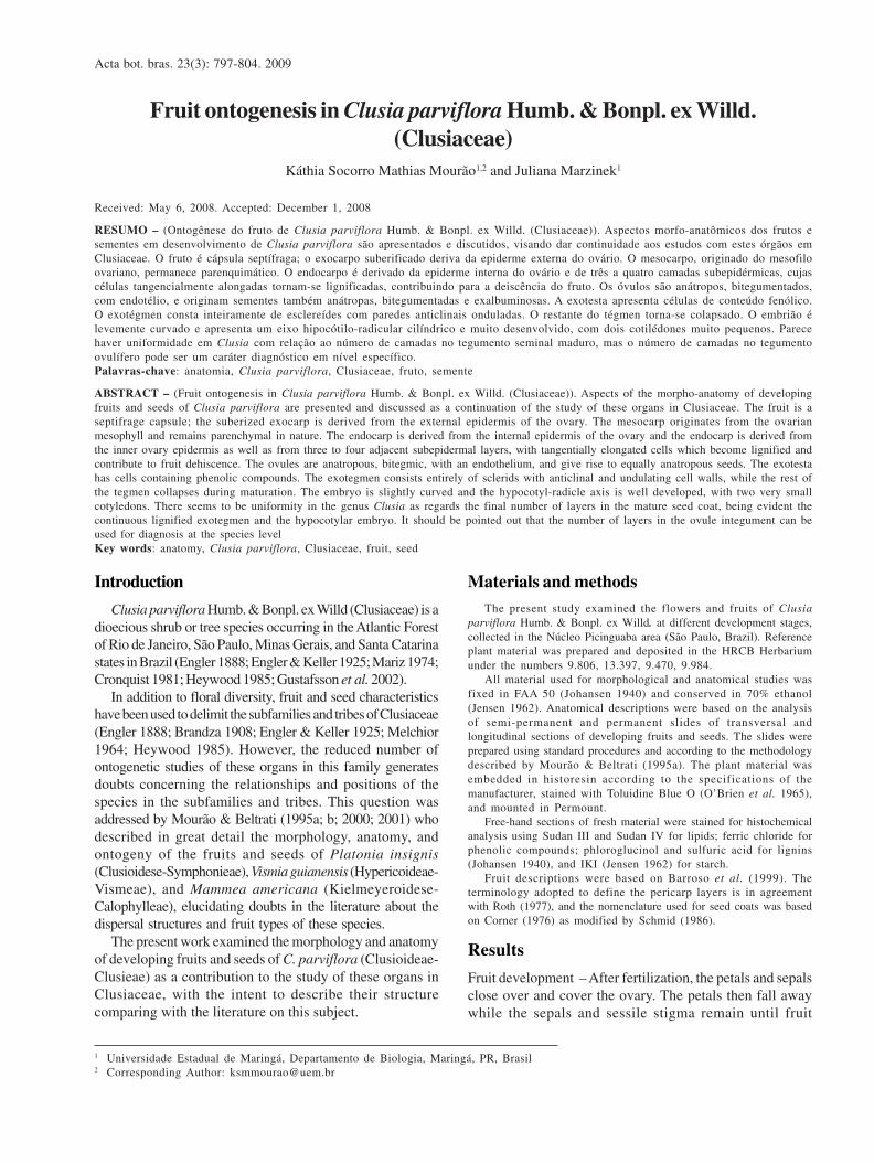
Figures 1-4. Clusia parviflora (Sald.) Engler. 1-2. General aspect of the mature fruit before and after dehiscence, respectively, showing the sessile stigma and seedscovered by the aril attached to the central column. 3-4. Medium longitudinal and transversal sections, respectively, showing tissue zonation in the pericarp. ar: aril,cl: columella, ec: endocarp, ep: exocarp, mp: mesocarp, sd: secretory duct, se: seed, sp: sepal, vv: valve (Bar scale = 1.5 cm).
maturation (Fig. 1). The fruits of C. parviflora are greenish-yellow spherical capsules approximately 25 mm in diameter.The seeds and aborted ovules, both covered by a sugar-rich pulp, are either attached to the central column or to theinner cavity of the fruit valves and become exposed whenthe fruit dehisces (Fig. 2).
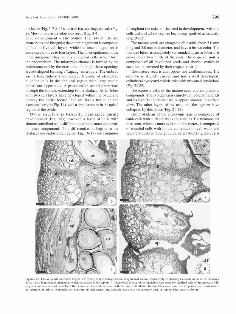
The 5-carpellary and 5-locular ovary of C. parviflora issuperior and has sessile stigmas. The ovules have axialplacentation. The outer epidermis of the ovary is uniseriateand is composed of cubic to slightly elongated cells coveredby a thin cuticle (Fig. 12). Five to six tangentially elongatedcells layers define the locule (Fig. 9). The ovarian mesophyllis composed of fundamental parenchyma, where a numberof branched secretory ducts and vascular bundles can beobserved. These ramified secretory ducts have a
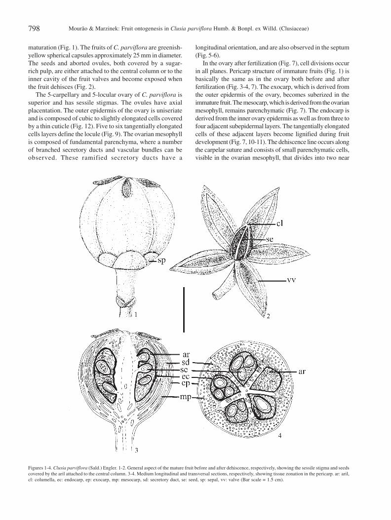
longitudinal orientation, and are also observed in the septum(Fig. 5-6).
In the ovary after fertilization (Fig. 7), cell divisions occurin all planes. Pericarp structure of immature fruits (Fig. 1) isbasically the same as in the ovary both before and afterfertilization (Fig. 3-4, 7). The exocarp, which is derived fromthe outer epidermis of the ovary, becomes suberized in theimmature fruit. The mesocarp, which is derived from the ovarianmesophyll, remains parenchymatic (Fig. 7). The endocarp isderived from the inner ovary epidermis as well as from three tofour adjacent subepidermal layers. The tangentially elongatedcells of these adjacent layers become lignified during fruitdevelopment (Fig. 7, 10-11). The dehiscence line occurs alongthe carpelar suture and consists of small parenchymatic cells,visible in the ovarian mesophyll, that divides into two near
v23n3_20.pmd 26/9/2009, 16:31798
Acta bot. bras. 23(3): 797-804. 2009. 799
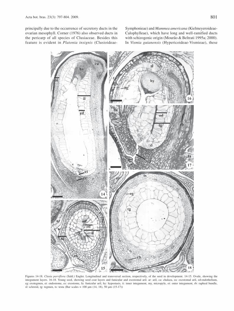
the locule (Fig. 5, 7-8, 13); the fruit is a septifrage capsule (Fig.2). Most of ovules develop into seeds (Fig. 3-4).Seed development – The ovules (Fig. 14-15, 19) areanatropous and bitegmic; the outer integument is composedof four to five cell layers, while the inner integument iscomposed of three to four layers. The inner epidermis of theinner integument has radially elongated cells, which formthe endothelium. The micropyle channel is formed by theendostome and by the exostome, although these openingsare not aligned forming a “zigzag” micropyle. The embryosac is longitudinally elongated. A group of elongatednucellar cells in the chalazal region with large nucleiconstitute hypostasis. A provascular strand penetratesthrough the funicle, extending to the chalaza. Arilar lobeswith two cell layers have developed within the ovule andoccupy the entire locule. The aril has a funicular andexostomal origin (Fig. 14), with a circular shape in the apicalregion of the ovule.
Ovule structure is basically maintained duringdevelopment (Fig. 18); however, a layer of cells withsinuous anticlinal walls differentiates in the outer epidermisof inner integument. This differentiation begins in thechalazal and endostomal region (Fig. 16-17) and continues
throughout the sides of the seed in development, with thecells walls of all exotegmen becoming lignified at maturity(Fig. 20-22).
The mature seeds are elongated ellipsoids about 5.0 mmlong and 3.0 mm in diameter, and have a brown color. Therounded hilum is completely surrounded by arilar lobes thatcover about two thirds of the seed. The dispersal unit iscomposed of all developed seeds and aborted ovules ineach locule, covered by their respective arils.
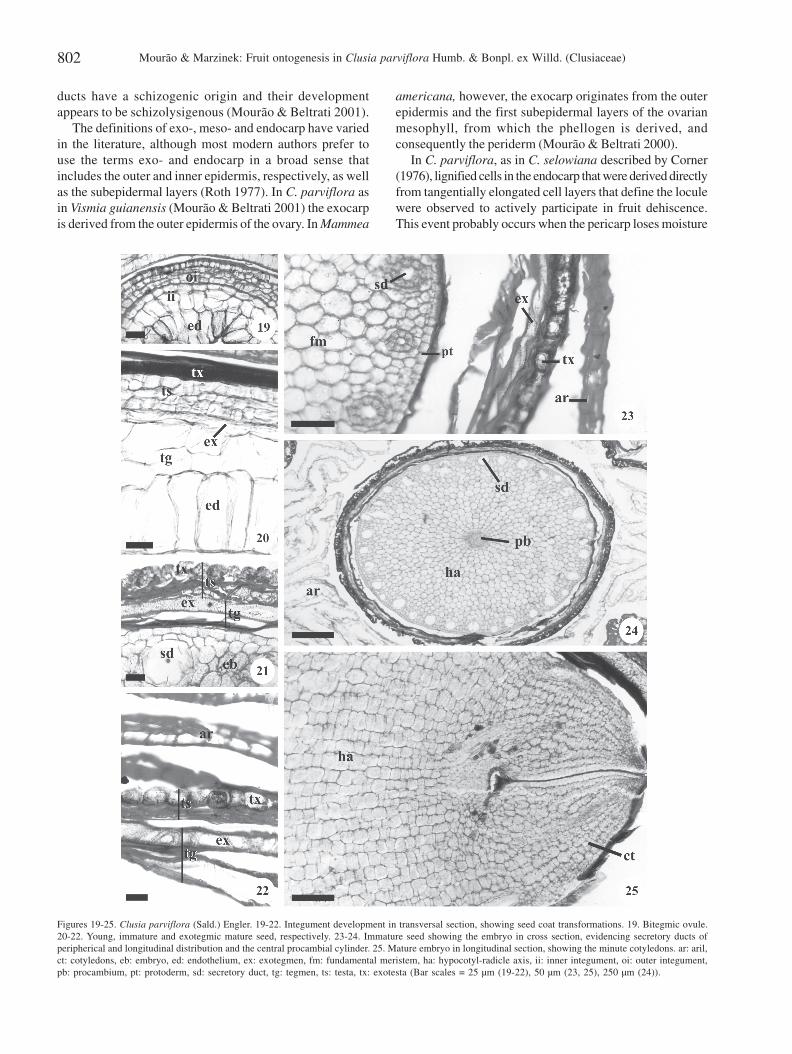
The mature seed is anatropous and exalbuminous. Theembryo is slightly curved and has a well developed,cylindrical hypocotyl-radicle axis, with two small cotyledons(Fig. 24-25).
The exotesta cells of the mature seed contain phenoliccompounds. The exotegmen is entirely composed of scleridsand its lignified anticlinal walls appear sinuous in surfaceview. The other layers of the testa and the tegmen havecollapsed by this phase (Fig. 21-22).
The protoderm of the embryonic axis is composed ofcubic cells with thick cell walls and cuticles. The fundamentalmeristem, which is more evident in the cortex, is composedof rounded cells with lipidic contents, thin cell walls andsecretory ducts with longitudinal orientation (Fig. 22-23). A
Figures 5-8. Clusia parviflora (Sald.) Engler. 5-6. Young fruit in transversal and longitudinal section, respectively, evidencing the seeds and ramified secretoryducts with a longitudinal orientation, which occur also in the septum. 7. Transversal section of the immature fruit (note the lignified cells of the endocarp withtangential orientation and the cells of the dehiscence line and mesocarp with thin walls). 8. Mature fruit in dehiscence (note that all pericarp cells lost water).ap: aperture, ar: aril, cl: columella, ec: endocarp, dl: dehiscence line, lo:locule, ov: ovule, sd: secretory duct, st: septum (Bar scale = 250 µm).
v23n3_20.pmd 26/9/2009, 16:31799
Mourão & Marzinek: Fruit ontogenesis in Clusia parviflora Humb. & Bonpl. ex Willd. (Clusiaceae)800
procambial cylinder extends the length of the embryonicaxis (Fig. 24).
Discussion
Engler (1888) and Mariz (1974) have classified the fruitsof Clusia as septicide capsules. However, Barroso et al.
(1999) and Stevens (2007a) described the fruits of this genusas septicidal or septifragal capsules. In C. parviflora theontogeny elucidated that the fruit is a septifragal capsule,but to affirm that is a condition to the genus studies withmore species are necessary.
In terms of fruit development, the ovarian structure ofC. parviflora resembles that of other species of Clusiaceae,
Figures 9-13. Clusia parviflora (Sald.) Engler. Transversal sections of the fruit: 9-11. Endocarp development, showing the tangencially elongated cells of the innerand subepidermical layers. 12. Ovarian outer epidermis and subepidermical layers. 13. Detail of the dehiscence line of an immature fruit, evidencing the spongytissue before the maturation. ar: aril, dl: dehiscence line, ec: endocarp, ie: inner epidermis, oe: outer epidermis, sd: secretory duct, sl: subepidermical layers(Bar scales = 25 µm (9), 50 µm (10-13)).
v23n3_20.pmd 26/9/2009, 16:31800
Acta bot. bras. 23(3): 797-804. 2009. 801
Figures 14-18. Clusia parviflora (Sald.) Engler. Longitudinal and transversal section, respectively, of the seed in development. 14-15. Ovule, showing theintegument layers. 16-18. Young seed, showing seed coat layers and funicular and exostomal aril. ar: aril, ca: chalaza, ea: exostomal aril, ed:endothelium,eg: exotegmen, et: endostome, es: exostome, fa: funicular aril, hy: hypostasis, ii: inner integument, my, micropyle, oi: outer integument, rb: rapheal bundle,sl: sclereid, tg: tegmen, ts: testa (Bar scales = 100 µm (14, 18), 50 µm (15-17))
principally due to the occurrence of secretory ducts in theovarian mesophyll. Corner (1976) also observed ducts inthe pericarp of all species of Clusiaceae. Besides thisfeature is evident in Platonia insignis (Clusioideae-
Symphonieae) and Mammea americana (Kielmeyeroideae-Calophylleae), which have long and well-ramified ductswith schizogenic origin (Mourão & Beltrati 1995a; 2000).In Vismia guianensis (Hypericoideae-Vismieae), these
v23n3_20.pmd 26/9/2009, 16:31801
Mourão & Marzinek: Fruit ontogenesis in Clusia parviflora Humb. & Bonpl. ex Willd. (Clusiaceae)802
ducts have a schizogenic origin and their developmentappears to be schizolysigenous (Mourão & Beltrati 2001).
The definitions of exo-, meso- and endocarp have variedin the literature, although most modern authors prefer touse the terms exo- and endocarp in a broad sense thatincludes the outer and inner epidermis, respectively, as wellas the subepidermal layers (Roth 1977). In C. parviflora asin Vismia guianensis (Mourão & Beltrati 2001) the exocarpis derived from the outer epidermis of the ovary. In Mammea
americana, however, the exocarp originates from the outerepidermis and the first subepidermal layers of the ovarianmesophyll, from which the phellogen is derived, andconsequently the periderm (Mourão & Beltrati 2000).
In C. parviflora, as in C. selowiana described by Corner(1976), lignified cells in the endocarp that were derived directlyfrom tangentially elongated cell layers that define the loculewere observed to actively participate in fruit dehiscence.This event probably occurs when the pericarp loses moisture
Figures 19-25. Clusia parviflora (Sald.) Engler. 19-22. Integument development in transversal section, showing seed coat transformations. 19. Bitegmic ovule.20-22. Young, immature and exotegmic mature seed, respectively. 23-24. Immature seed showing the embryo in cross section, evidencing secretory ducts ofperipherical and longitudinal distribution and the central procambial cylinder. 25. Mature embryo in longitudinal section, showing the minute cotyledons. ar: aril,ct: cotyledons, eb: embryo, ed: endothelium, ex: exotegmen, fm: fundamental meristem, ha: hypocotyl-radicle axis, ii: inner integument, oi: outer integument,pb: procambium, pt: protoderm, sd: secretory duct, tg: tegmen, ts: testa, tx: exotesta (Bar scales = 25 µm (19-22), 50 µm (23, 25), 250 µm (24)).
v23n3_20.pmd 26/9/2009, 16:31802
Acta bot. bras. 23(3): 797-804. 2009. 803
at maturation, generating opposing forces between thecarpelar sections due to contraction of the endocarp cellwalls.
According to Roth (1977), the mechanism of fruitdehiscence is mostly based on active movements broughtabout by dead or living tissues, whereby two fundamentaltypes may be distinguished: a hygroscopic and a turgormechanism. The hygroscopic movements generally rely onshrinkage and swelling of walls of dead cells, whereas theturgor mechanisms function with living cells which havehighly elastic walls.
In hygroscopic dehiscence Roth (1977) distinguishestwo types: hygrochastic and xerochastic. The acting forcesof hygrochastic movements rest on water absorption,which cause a swelling of the cell walls so that a swellingpressure is developed, and in xerochastic depend on waterlosses, resulting in shrinkage of the cell walls. Inhygrochastic mechanisms the source of energy thereforelies in the swelling power, whereas in xerechasticmovements, it is developed by cohesion forces. Toproduce a bending of certain pericarp parts, twoantagonistically acting layers are generally present whichmay show different structure of their composing elements,frequently fibers, that may be arranged in differentdirections so that the cells of one layer cross those of theother layer.
The dehiscence mechanism in C. parviflora seems to bea combination of hygrochastic and xerochastic movements,considering that the mostly pericarp cells don’t lose water,except to the shrinkage of the lignified cells of the endocarpwhich have a tangential orientation and can contribute withantagonistic forces responsible for fruit aperture.
Corner (1976) described the occurrence of anatropousand bitegmic ovules in Allanblackia, Calophyllum,Caraipa, Clusia, Garcinia, Havetiopsis, Mesua,Pentadesma, Septogarcinia, Symphonia, Tovomitopsis,Tsimatimia (Clusiaceae), Cratoxylon, Haronga, Hypericumand Vismia (Hypericaceae). These characteristics wereobserved in C. parviflora in the present study and were alsodescribed in Platonia insignis by Mourão & Beltrati (1995a)and in Vismia guianensis by Mourão & Beltrati (2001). InMammea americana, however, Mourão & Beltrati (2000)described anatropous and unitegmic ovules.
The ovule structure in C. parviflora is similar to thatdescribed for all species of Clusia described by Corner(1976), especially Clusia sp. This author was not certain ifthis species (which was not identified) really belonged tothe genus because of the characteristics of its fruits andseeds.
In C. parviflora the inner epidermis of the innerintegument has radially elongated cells, which form theendothelium. The presence of an endothelium in ovules ofClusiaceae and Hypericaceae was mentioned in Hypericumpatulum, H. mysorense (Rao 1957), Clusia sp. (Corner 1976),Platonia insignis (Mourão & Beltrati 1995a), Vismiaguianensis (Mourão & Beltrati 2001).
Kapil & Tiwari (1978) defined the integumentary tapetum,or endothelium, as being the inner epidermis of the innerintegument of bitegmic ovules, or the only integument inunitegmic ovules. In many plants it has differentiated into aspecialized layer of radially elongated cells with densecytoplasm and prominent and often polyploid nuclei. Theseauthors have recorded the presence of an endothelium in65 families of dicotyledons, but not in the Clusiaceae.
The presence of tanniniferous cells in the chalazal regionof C. parviflora is in agreement with the definition ofhypostasis sensu stricto proposed by Von Teichman & VanWyk (1991). Hypostasis also occurs in Platonia insignis(Mourão & Beltrati 1995a).
The seeds of C. parviflora are exotegmic. Corner (1976)affirms that seeds of the Hypericoideae (Hypericaceae)appear to be related to those of the Clusioideae-Clusieae,due to the presence of an exotegmen with star-shaped,undulating cells with thick cell walls; and the production ofphanerocotyledonous embryos. Basta & Basta (1984)described an entirely lignified tegument in the seeds ofKielmeyera coriacea (Kielmeyeroideae-Calophylleae)without distinguishing between the testa and tegmen but,likewise, they did not undertake a detailed anatomical studyof seed development in this species.
According to Corner (1976) the aril of Clusieae may beclassified in four types. As proven in the present study inC. parviflora occurs the second type, exostomal andfunicular aril, that is also present in Clusia rosea andC. sellowiana, Tovomita, Havetia and Havetiopsis (Corner1976). This author describes for Clusia sp. a fourth type, anexostomal aril combined with funicular lobes but withoutparticipation of the raphe-side of the funicle. If other typesof aril occur in Clusia only additional research with morespecies can provide the answer.
Stevens (2007a) comments that the seeds of arillate NewWorld Clusieae are dispersed by birds, although antssubsequently affect the distribution and germination ofprimarily bird-dispersed seeds. Studies on dispersal in thisgenus are important from an ecological point of view.
Detailed chemical analyses of the seeds of C. parviflorahave not yet been undertaken, although the present studyindicated that the main reserve substances in the embryoare lipids, as was also reported by Mourão & Beltrati (1999b)for Vismia guianensis. Lipidic substances and phenoliccompounds were detected in the embryos of M. americanain duct secretory cells as well as in parenchymal idioblasts,although starch was the most abundant reserve overall(Mourão & Beltrati 1999a).
The occurrence of lipidic substances in Clusiaceae seedshas often been reported in the literature (Brandza 1908; Earle& Jones 1962; Vaughan 1970; Basta & Basta 1984; Benteset al. 1986/1987; Adeyeye 1991).
The genus seems to be uniform in the final number oflayers in the mature seed coat, being evident the continuouslignified exotegmen and the hypocotylar embryo. In anothergenus of Clusieae, Corner (1976) described this exotegmic
v23n3_20.pmd 26/9/2009, 16:31803
Mourão & Marzinek: Fruit ontogenesis in Clusia parviflora Humb. & Bonpl. ex Willd. (Clusiaceae)804
contruction for Havetiopsis flexilis only. However, there isuniformity in Clusioideae in the hypocotylar embryo, aspointed out in the research descriptions of Brandza (1908),Guillaumin (1910) and Corner (1976), but occurring inClusioideae - Garcinieae and Symphonieae (Stevens 2007a).
It should be pointed out that the number of layers inovule integument, can be a diagnostic characteristic at thespecific level, varying in the inner integument from 2 to 3 inthe species of the present study, from 6 to 7 in C. rosea, from5 to 7 in C. selowiana and 3 in Clusia sp. described byCorner (1976). In relation of the outer integumentC. parviflora shows 3 to 4 layers, being different fromC. rosea (4 to 5), C. selowiana (6 to 8) and Clusia sp. (2 to 3).
Considering that few studies have closely examinedClusiaceae fruits and seeds, and in light of the characteristicscited in the literature as either ancestral or derived as relatedto these organs, it can be concluded from the infra-familialtreatment of this family (Stevens 2007a; b) thatKielmeyeroideae, Hypericoideae (Hypericaceae) andClusioideae (Clusieae) demonstrate more ancestralcharacteristics, as they have mostly dehiscent fruits(capsule type), arilate and exotegmic seeds, and embryoswith cotyledons varying from distinct to very reduced; whileClusioideae (Garcinieae and Symphonieae) havecharacteristics considered derived, including indehiscentfruits (berry or drupe), seeds without an aril, integumentsnot well differentiated or mesotestals, and hypocotylar orconferruminate embryos.
It should be pointed out that the establishment ofrelationships among the subfamilies of Clusiaceae usingfruit and seed characteristics is extremely difficult due toa lack of developmental studies in a majority of thespecies.
AcknowledgmentsThe authors would like to thank the Instituto Florestal, SP for
both allowing and aiding the collection of plant material in the area ofthe Núcleo Picinguaba.
ReferencesAdeyeye, A. 1991. Studies on seed oils of Garcinia kola and
Calophyllum inophyllum. Journal of the science of food andagriculture 57: 441-442.
Barroso, G.M.; Morim, M.P.; Peixoto, A.L. & Ichaso, C.L.F. 1999.Frutos e sementes: morfologia aplicada à sistemática dedicotiledôneas. Viçosa, Universidade Federal de Viçosa.
Basta, S.B.D. & Basta, F. 1984. Estudos morfológicos das sementes edo desenvolvimento das plântulas de Kielmeyera coriacea Mart.Brasil Florestal 58: 25-30.
Bentes, M.H.S.; Serruya; H.; Rocha Filho, G.N.; Godoy, R.L.O.; Cabral,J.A.S. & Maia, J.G.S. 1986/1987. Estudo químico das sementes debacuri. Acta Amazônica 16/17: 363-368.
Brandza, G. 1908. Recherches anatomiques sur la germination desHypéricacées et des Guttifères. Annales des SciencesNaturélles, Botanique Série 9: 221-300.
Corner, E.J.H. 1976. The seeds of dicotyledons. Cambridge,University Press.
Cronquist, A. 1981. An integrated system of classification offlowering plants. New York, Columbia University Press.
Earle, F.R. & Jones, Q. 1962. Analyses of seed samples from 113plant families. Economic Botany 16: 221-250.
Engler, A. 1888. Guttiferae. In: C.F.P. Martius (ed.). FloraBrasiliensis 12: 381-486
Engler, A. & Keller, R. 1925. Guttiferae. Pp. 154-237. In: A. Engler& K. Prantl (eds.). Die naturlichen Pflanzenfamilien.Leipzig, Verlag Wilhelm Engelmann.
Guillaumin, A. 1910. L’étude des germinations appliquée à laclassification des genres et a la phylogénie des groupes. RevueGénérale de Botanique 22: 449-468.
Gustafsson, M.H.G.; Bittrich, V. & Stevens, P.F. 2002. Phylogeny ofClusiaceae base on rbcL sequences. International Journal ofPlant Sciences 163: 1045-1054.
Heywood, V.H. 1985. Flowering plants of the world. London,Croom Helm.
Jensen, W.A. 1962. Botanical histochemistry: principles andpractice. San Francisco, W.H. Freeman.
Johansen, D.A. 1940. Plant microtechnique. New York, Mc Graw-Hill Book.
Kapil, N.R. & Tiwari, S.C. 1978. The integumentary tapetum. TheBotanical Review 44: 457-490.
Mariz, G. 1974. Chaves para as espécies de Clusia nativas no Brasil.Memórias do Instituto de Biociências da UniversidadeFederal de Pernambuco 1: 249-314.
Melchior, H. 1964. Guttiferae (Clusiaceae). In: A. Engler (ed.).Syllabus de Planzenfamilien 2: 170-173.
Mourão, K.S.M. & Beltrati, C.M. 1995a. Morfologia dos frutos,sementes e plântulas de Platonia insignis Mart. (Clusiaceae). I.Aspectos anatômicos dos frutos e sementes em desenvolvimento.Acta Amazônica 25: 11-32.
Mourão, K.S.M. & Beltrati, C.M. 1995b. Morfologia dos frutos,sementes e plântulas de Platonia insignis Mart. (Clusiaceae). II.Morfo-anatomia dos frutos e sementes maduros. Acta Amazonica25: 33-46.
Mourão, K.S.M. & Beltrati, C.M. 2000. Morphology and anatomy ofdeveloping fruits and seeds of Mammea americana L. (Clusiaceae).Revista Brasileira de Biologia 60: 701-711.
Mourão, K.S.M. & Beltrati, C.M. 2001. Morphology and anatomy ofdeveloping fruits and seeds of Vismia guianensis (Aubl.) Choisy(Clusiaceae). Revista Brasileira de Biologia 61: 147-158.
O’Brien, T.P.; Feder, N. & Mc Cully, M.E. 1965. Polychromaticstaining of plant cell walls by toluidine blue O. Protoplasma 59:368-373.
Rao, A.N. 1957. The embryology of Hypericum patulum Thunb. andH. mysorense Heyne. Phytomorphology 7: 36-45.
Roth, I. 1977. Fruits of Angiosperms. Berlin, Gebruder Borntreger.Schmid, R. 1986. On cornerian and other terminology of
angiospermous and gymnospermous seed coats: historicalperspective and terminological recommendations. Taxon 35:476-491.
Stevens, P.F. 2007a. Clusiaceae-Guttiferae. P. 48-66. In: K. Kubitzki(ed.). The families and genera of vascular plants 10: 48-66.Berlin, Springer-Verlag.
Stevens, P.F. 2007b. Hypericaceae, P. 194-201. In: K. Kubitzki (ed.).The families and genera of vascular plants 10: 194-201.Berlin, Springer-Verlag.
Vaughan, J.G. 1970. The structure and utilization of oil seeds.London, Chapman and Hall Ltda.
Von Teichman, I. & Van Wik, A.E. 1991. Trends in the evolution ofdicotyledonous seeds based on character association, with specialreference to pachychalazy and recalcitrance. Botanical Journalof Linnean Society of London 105: 211-237.
Versão eletrônica do artigo em www.scielo.br/abb e http://www.botanica.org.br/acta/ojs
v23n3_20.pmd 26/9/2009, 16:31804