fresh frozen plasma for succinylcholine apnoea - time to reconsider?
TRANSCRIPT

Correspondence
Anaesthetists and care of thecritically ill child 1
The recent editorial on anaesthetists and
care of the critically ill child (Tomlin-
son. Anaesthesia 2003; 58: 309–11) was
both timely and sensible. After the 1990
NCEPOD report on deaths in children
following surgery [1], there was an
entirely appropriate movement to raise
standards in the provision of anaesthesia
for children. However, an excess of
reforming zeal has led to the excellent
becoming the enemy of the good. In
pursuit of the highest standards, we have
managed to scare some previously com-
petent anaesthetists into perceived
incompetence.
As the editorial points out, in an
emergency involving the airway, any
anaesthetist is better than none. In these
circumstances, it is simply not accept-
able to say ‘I do not do children’. If one
is the most competent anaesthetist
available, one must do one’s best.
Indeed, the quoted guidance from the
GMC makes this amply clear [2]. The
corollary of this is that if things do not
go well (short of actual negligence),
support for the individual who did
his ⁄ her best must be forthcoming from
employers, colleagues, the medical de-
fence organisations, and from the bodies
who set the standards – the Royal
College, the Association of Anaesthet-
ists and the GMC itself. A clear state-
ment to this effect would be both
reassuring and helpful, and might do
something to temper the growing
reluctance of some anaesthetists to have
anything to do with the care of this
vulnerable group of patients.
A.-M. Rollin
Epsom General Hospital,
Epsom KT18 7EG, UK
E-mail: anna.rollin@epsom-
sthelier.nhs.uk
References1 Campling EA, Devlin HB, Lunn JN.
The Report of the National Enquiry into
Perioperative Deaths. London:
NCEPOD, 1989.
2 GMC. Good Medical Practice. London:
The General Medical Council, 2001.
Anaesthetists and care of thecritically ill child 2
I agree with the recent editorial (Tom-
linson. Anaesthesia 2003; 58: 309–11)
that there are a number of potential
ways to maintain skills and confidence
regarding the resuscitation and early
management of critically ill children in
the district general hospital. In our
region, South and West Wales, we have
initiated a number of developments to
facilitate this. District general hospital
consultants have visited for a ‘refresher’
week to attend a mix of paediatric
anaesthetic and intensive care sessions,
including retrievals. Also, we have
developed a ‘Stabilisation of the Critic-
ally Ill Child Study Day’ aimed at
medical and nursing staff who have first
contact with critically ill children. This
is aimed at filling the obvious deficit
between initial resuscitation, well cov-
ered by paediatric life-support courses,
and the arrival of the paediatric inten-
sive care retrieval team. This includes
extensive coverage of advanced paedi-
atric airway management, ventilatory
strategy and cardiovascular support.
This is part of a an extensive regional
[1] outreach programme that includes
basic life-support training and feedback
sessions.
Lastly, exposure to paediatric inten-
sive care in Specialist Registrar Training
should be viewed as an effective tool to
prepare for a district general consultant
post, not just for those pursuing the
speciality.
M. A. Price
University Hospital of Wales,
Cardiff CF14 4XN, UK
E-mail: mark.price@cardiffandvale.
wales.nhs.uk
Reference1 Anon. Improving Health in Wales. Caring
for Critically Ill Children, Standards.
Cardiff: Welsh Assembly Government.
Anaesthetists and care of thecritically ill child 3
The recent editorial (Tomlinson.
Anaesthesia 2003; 58: 309–11) is well
argued and puts into context the Royal
College of Anaesthetist’s flyer, which
may have been poorly understood by
All correspondence should be addressed to Professor M. Harmer, Editor of Anaesthesia, Department of Anaesthetics, University of
Wales College of Medicine, Heath Park, Cardiff CF14 4XN UK.
Letters (two copies) must be typewritten on one side of the paper only and double spaced with wide margins. In addition, please
include your letter as a Word for Windows or .rtf document on disk or alternatively submit as an e-mail attachment addressed to
[email protected]. Alternatively a response to a previously published article or letter can be submitted online at www.anaesthesia
correspondence.com. Copy should be prepared in the usual style and format of the Correspondence section. Authors must follow the
advice about references and other matters contained in the Notice to Contributors to Anaesthesia printed at the back of the January
and July issues. Author guidelines can also be found on www.blackwellpublishing.com/journals/ana/submiss.htm. The degree and diploma
of each author must be given in a covering letter personally signed by all the authors.
Correspondence presented in any other style or format may be the subject of considerable delay and may be returned to the author for
revision.
Anaesthesia, 2003, 58, pages 804–827.....................................................................................................................................................................................................................
804 � 2003 Blackwell Publishing Ltd
some. However, I would like to make
two additional points.
Whilst I applaud efforts to promote a
team approach to the care of sick
children, it is often the anaesthetist
who does the bulk of the work. Col-
leagues in acute paediatrics and surgery
must also keep their resuscitation skills
current and, importantly, be willing to
use these skills. As far as I am aware, the
Royal College of Paediatrics and Child
Health has not issued a similar flyer.
Units may wait many hours for
retrieval teams to arrive, albeit with
telephone advice in the interim. The
vast majority of our general ICUs have
not been specifically funded to perform
the task of delivering level 2 care to
children. Whilst we can reasonably
expect on-call consultant anaesthetists
to perform the initial resuscitation and
airway management in children, the
next 6 h of intensive care may be
something they feel ill prepared to lead.
These issues need to be addressed if
we are to make an impact on the quality
of care of very sick children in the UK.
K. Wilkinson
Norfolk and Norwich University
Hospital,
Norwich NR4, UK
E-mail: Kathy.Wilkinson@
nnuh.nhs.uk
Does the Internet provide safeinformation for pre-anaestheticpatients?
The importance of clear information for
patients was emphasised in the recent
article by Heidegger et al. [1]. Increas-
ingly, people are using the Internet to
further their knowledge of healthcare
subjects. Unfortunately, the quality of
this information is very variable and at
times can be incomplete.
We decided to type the exact search
phrase ‘having an anaesthetic’ into a
search engine and review the first
10 sites. Waldman [2] reported on the
dominance of the search engine
‘Google’, and hence this was used. A
recent study by Eysenbach & Kohler [3]
reported that people tend to look only
at the first couple of websites from any
search; thus, to obtain a brief snapshot
of the information patients would
access, this was the strategy used
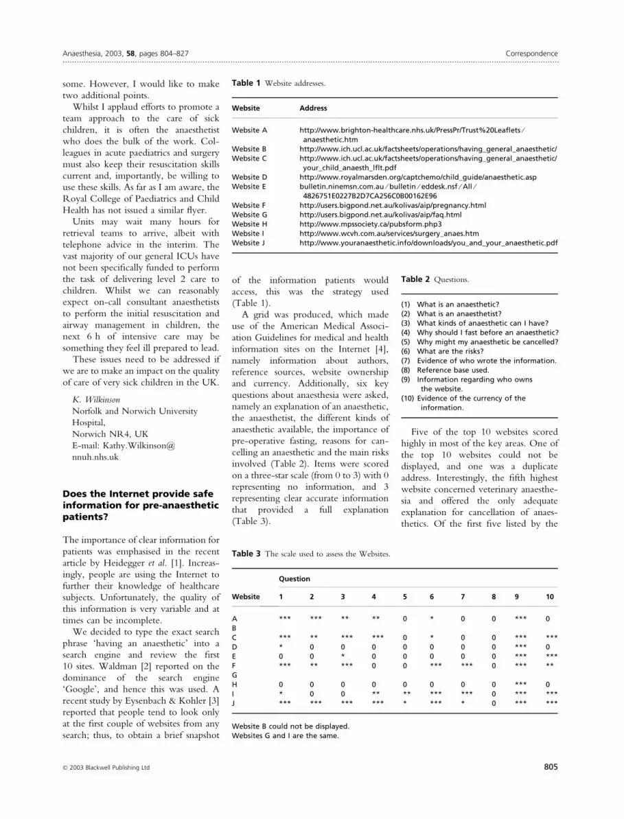
(Table 1).
A grid was produced, which made
use of the American Medical Associ-
ation Guidelines for medical and health
information sites on the Internet [4],
namely information about authors,
reference sources, website ownership
and currency. Additionally, six key
questions about anaesthesia were asked,
namely an explanation of an anaesthetic,
the anaesthetist, the different kinds of
anaesthetic available, the importance of
pre-operative fasting, reasons for can-
celling an anaesthetic and the main risks
involved (Table 2). Items were scored
on a three-star scale (from 0 to 3) with 0
representing no information, and 3
representing clear accurate information
that provided a full explanation
(Table 3).
Five of the top 10 websites scored
highly in most of the key areas. One of
the top 10 websites could not be
displayed, and one was a duplicate
address. Interestingly, the fifth highest
website concerned veterinary anaesthe-
sia and offered the only adequate
explanation for cancellation of anaes-
thetics. Of the first five listed by the
Table 1 Website addresses.
Website Address
Website A http://www.brighton-healthcare.nhs.uk/PressPr/Trust%20Leaflets ⁄anaesthetic.htm
Website B http://www.ich.ucl.ac.uk/factsheets/operations/having_general_anaesthetic/Website C http://www.ich.ucl.ac.uk/factsheets/operations/having_general_anaesthetic/
your_child_anaesth_lflt.pdfWebsite D http://www.royalmarsden.org/captchemo/child_guide/anaesthetic.aspWebsite E bulletin.ninemsn.com.au ⁄ bulletin ⁄ eddesk.nsf ⁄ All ⁄
4826751E0227B2D7CA256C0B00162E96Website F http://users.bigpond.net.au/kolivas/aip/pregnancy.htmlWebsite G http://users.bigpond.net.au/kolivas/aip/faq.htmlWebsite H http://www.mpssociety.ca/pubsform.php3Website I http://www.wcvh.com.au/services/surgery_anaes.htmWebsite J http://www.youranaesthetic.info/downloads/you_and_your_anaesthetic.pdf
Table 2 Questions.
(1) What is an anaesthetic?(2) What is an anaesthetist?(3) What kinds of anaesthetic can I have?(4) Why should I fast before an anaesthetic?(5) Why might my anaesthetic be cancelled?(6) What are the risks?(7) Evidence of who wrote the information.(8) Reference base used.(9) Information regarding who owns
the website.(10) Evidence of the currency of the
information.
Table 3 The scale used to assess the Websites.
Website
Question
1 2 3 4 5 6 7 8 9 10
A *** *** ** ** 0 * 0 0 *** 0BC *** ** *** *** 0 * 0 0 *** ***D * 0 0 0 0 0 0 0 *** 0E 0 0 * 0 0 0 0 0 *** ***F *** ** *** 0 0 *** *** 0 *** **GH 0 0 0 0 0 0 0 0 *** 0I * 0 0 ** ** *** *** 0 *** ***J *** *** *** *** * *** * 0 *** ***
Website B could not be displayed.Websites G and I are the same.
Anaesthesia, 2003, 58, pages 804–827 Correspondence......................................................................................................................................................................................................................
� 2003 Blackwell Publishing Ltd 805
search engine, only two provided com-
prehensive information. The areas that
were less thoroughly explained included
the questions: what is an anaesthetist,
why might my anaesthetic be cancelled
and what are the main risks? Reassur-
ingly, the top scoring website was
produced by the Royal College of
Anaesthetists and the Association of
Anaesthetists, but this was the 10th
address listed by Google.
To conclude, there is good informa-
tion available on this subject, and the
sites that scored poorly contained little
information rather than dangerous or
misleading facts.
C. A. Theodosiou
Victoria Hospital,
Kirkaldy KY2 5AH, UK
E-mail: [email protected]
L. J. Theodosiou
Prestwich Hospital,
Manchester, UK
E-mail: [email protected]
References1 Heidegger T, Husemann T, Nuebling
M et al. Patient satisfaction with anaes-
thesia care: development of a psycho-
metric questionnaire and benchmarking
among six hospitals in Switzerland and
Austria. British Journal of Anaesthesia
2002; 89: 863–72.
2 Waldman S. Google is the net domi-
nator. The Guardian Newspaper 2003:
Thursday February 27.
3 Eysenbach G, Kohler C. How do
consumers search for and appraise
health information on the world wide
web? Qualitative study using focus
groups, usability tests, and in-depth
interviews. British Medical Journal 2002;
324: 573–7.
4 Winker MA, Flanagin A, Chi-Lum B
et al. Guidelines for medical and health
information sites on the internet:
principles governing AMA web sites.
Journal of American Medical Association
2000; 283: 1600–6.
Towards needleless inductionof anaesthesia
I found the correspondence regarding
the necessity of premedication (Hames.
Anaesthesia 2003; 58: 297) especially
interesting as it draws attention to one
of two aspects of contemporary anaes-
thetic practice that are at variance with
traditional practice before the advent of
day surgery units (DSUs) and sevoflu-
rane. The popularisation of DSUs has
been accompanied by a change from the
traditional practice of giving premedi-
cant anaesthetic drugs intramuscularly,
orally or rectally some hours before
surgery, to the contemporary practices
of either omitting premedication or
administering an aliquot of benzo-
diazepine or opioid intravenously in
the DSU or operating room shortly
before induction of anaesthesia, whilst
the introduction of sevoflurane has been
accompanied by a resurgence of interest
in inhalational induction of anaesthesia
in adults [1].
The trend from traditional to con-
temporary practice regarding premedi-
cation appears to have developed as a
result of better premedicants and the
requirement for rapid patient turnover
in DSUs, rather than according to
standards of good practice rationalised
by outcome studies. Further, despite
the ready availability of sevoflurane, the
wishes of adult patients regarding their
preferred route of induction of anaes-
thesia (mask or needle) is not usually
canvassed during the pre-operative visit,
and the traditional intravenous route is
still routinely used by most anaesthetists.
In an attempt to rationalise both issues, I
undertook an audit to assess the opinion
of patients scheduled for ambulatory
surgery as regards their desire to receive
premedication, and their choice of
route for induction of anaesthesia.
Having liaised with the Hospital’s
Research Ethics Committee, 168 con-
secutive adults presenting for all types
of elective, ambulatory surgery under
general anaesthesia administered by
myself during a 4-month period were
visited in the DSU prior to surgery.
Each patient was asked whether they
felt anxious about their surgery and
whether they wished to receive sedation
prior to surgery. Those requesting
medication were asked whether they
wished to receive the medication
immediately or intravenously shortly
before surgery. Patients requesting
immediate medication were also asked
whether they wished it to be adminis-
tered by the oral, intramuscular or rectal
route. Enquiry was then made whether
they wished their anaesthetic to be
commenced ‘by a small needle on the
back of the hand, in the usual manner’
or ‘by breathing a new anaesthetic gas –
sevoflurane – via a fruit-scented mask,
as is popular with children because it
avoids the tiny needle’.
I questioned 190 patients (age range
16–89 years). Of these, 156 (82%) had
undergone previous surgery for which
anaesthesia had been induced by the
intravenous route in 148 (95%) patients,
by inhalation in six (4%) patients and by
a non-recalled route in two (1%)
patients. Of the 190 patients, 97 (51%)
declined and 93 (49%) requested pre-
medication, of whom two (1%) selected
the intramuscular, 60 (65%) the oral, 0
(0%) the rectal and 31 (35%) the
intravenous routes of administration.
Intravenous (needle) induction was
chosen by 49 (26%) and inhalational
(mask) induction by 100 (53%) patients,
the remaining 41 (22%) patients being
equivocal (‘I don’t mind’), and asking
that I ‘do whatever is best’. Of the
latter, 14 (34%) patients had an intra-
venous induction and the remaining
27 (66%) patients were induced using
sevoflurane. Sevoflurane was successful
and without incident, as the induction
agent in the 127 patients to whom it
was offered, and was administered via a
scented mask from a circle absorber
primed with sevoflurane 8% in equal
parts of nitrous oxide and oxygen.
Elucidation of these data has inter-
esting implications. The wish expressed
by 49% of patients to have pre-opera-
tive anxiolytic medication suggests that
the trends towards omission of pre-
medication or administration of an
aliquot of short-acting benzodiazepine
or opioid in the pre-operative holding
or anaesthetic room shortly before
induction of anaesthesia may be un-
der-treating pre-operative anxiety in
almost half of patients presenting for
ambulatory surgery. More interesting is
the wish expressed by 52% of patients
not to have premedication, which sug-
gests that the current popular practice
of inserting an intravenous cannula and
Correspondence Anaesthesia, 2003, 58, pages 804–827......................................................................................................................................................................................................................
806 � 2003 Blackwell Publishing Ltd
administering a subhypnotic dose of
midazolam shortly before induction of
anaesthesia may be considered unnec-
essarily invasive by approximately half
of patients. Most surprising was the
choice by 54% of patients to have
anaesthesia induced by inhalation of
sevoflurane via mask, with only 26%
opting for the conventional intravenous
route. This appears to contradict the
concept popular amongst many anaes-
thetists that most patients have a mask
phobia and suggests, to the contrary,
that needle phobia is more prevalent
amongst adults.
These data, though warranting ana-
lysis with respect to gender, age, ethni-
city, nationality, previous exposure to
anaesthesia ⁄ mask ⁄ needles, etc., which
may influence patient choice as regard-
ing premedication and route of induc-
tion of anaesthesia, suggests that such
inquiry should be routine at the pre-
operative visit. By so doing, the appro-
priate prescription of premedication and
avoidance of needles in conscious
patients as part of a patient-centred
philosophy [2] of ‘needleless induction
of anaesthesia’ may contribute to allay-
ing pre-operative anxiety and enhance
patient satisfaction with their care.
Although induction of anaesthesia by
mask in the absence of an intravenous
cannula is contrary to the view of
anaesthetists familiar with the arrhythm-
ogenic and vagal effects of halothane,
the remarkably rapid action and stable
cardio-respiratory profile of sevoflurane
suggests that its use for induction of
anaesthesia in adults, prior to insertion
of an intravenous cannula, as is usual
with children, is an acceptably safe
technique.
A. A. van den Berg
University of Texas,
Houston, USA
E-mail: [email protected]
References1 Muzi M, Robinson BJ, Bbert TJ,
O’Brian TJ. Induction of anesthesia and
tracheal intubation with sevoflurane
in adults. Anesthesiology 1996; 85:
536–43.
2 Levenstein JH, McCracken EC,
McWhinney IR, Stewart MA, Brown
JB. The patient-centred clinical
method. 1. A model for the doctor–
patient interaction in family medicine.
Family Practice 1986; 3: 24–30.
Major incident preparation
In light of increased concern of a
terrorist-related major incident, as well
as the ongoing risk of a domestic major
incident in the catchment area, I audi-
ted the knowledge of the anaesthetists
working in my local Trust on their
knowledge and preparation for man-
aging a major incident.
There were 33 responses, reflecting a
66% response to the audit questionnaire
circulated to anaesthetists of all grades
working within the two hospitals in the
Trust. Consultants were responsible for
55% of responses. A significant number
of responders (27%) did not know
where the Major Incident Plan was
kept, and 36% had never read the Major
Incident Plan. Many (37%) did not have
direct access at home to colleagues’
telephone numbers. In the event of a
major incident, most on-call staff would
either contact A & E, operating theatres
or begin clearing ICU beds, whilst most
staff not on-call would either attend A
& E, operating theatres or await tele-
phone instructions. Excluding the brief
lecture given in ATLS courses, only
two anaesthetists (6%) had received
formal training in major incident man-
agement. Six anaesthetists (18%) had
previous experience of major incidents,
and 54% had never been involved with
any planning involving scenarios of
major incidents.
A major incident, in medical terms, is
an incident involving a number of
casualties that can overwhelm the
resources normally available to patients.
As examples, a large number of admis-
sions with minor problems from an
accident or incident could swamp and
overcrowd a hospital, whilst a smaller
number of critically ill patients could
overload even a regionally based ICU
network. In light of this, most, if not all,
NHS hospitals with an A & E depart-
ment have a Major Incident Plan to
help cope with the potentially over-
whelming number of patients involved.
Anaesthetists have a key role in the plan
as they can be involved with resuscita-
tion, definitive operating theatre care,
provision of critical care in HDU ⁄ ICU,
safe transfer of patients within and
outside of the hospital, and may be part
of the Mobile Medical Teams des-
patched to the major incident site. Thus
it is essential that anaesthetists have a
clear idea about their role and action in
the event of a major incident being
declared in their hospital.
This audit has highlighted deficien-
cies in the knowledge of our Trust’s
Major Incident Plan, and this has been
remedied by heightening awareness of
the Plan coupled with presentations by
the A & E consultants. Home telephone
numbers have been circulated to per-
manent members of staff to facilitate a
cascade of telephoning other staff from
home so that reliance is not placed on
the hospital’s switchboards. Staff are
encouraged to undertake training such
as the MIMMS course (Major Incident
Medical Management and Support), and
we will continue to ensure that the
Anaesthetic Department is represented
at any major incident planning sessions.
Major incidents can occur at any
time. Is your Department prepared?
R. V. Johnson
Calderdale Royal Hospital,
Halifax HX3 0PW, UK
E-mail: [email protected]
Interrupted monitoring
We also have been interested in the
significance of monitoring or the lack
of it during transfers between the oper-
ating theatre and recovery area (Adek-
anye & Wali. Anaesthesia 2003; 58:
190–1). The gap in continuous monit-
oring from anaesthetic room to theatre
may have been closed by induction of
anaesthesia in the operating theatre but
in the postoperative phase, other than
transfers to an intensive-care or high-
dependency unit, it has so far been
neglected.
We had an opportunity to note the
continuity of monitoring on seven
consecutive cases in a day surgical unit
where an anaesthetic room is currently
employed. All patients were fit, aged
from 22 to 39 years and required
general anaesthesia for minor gynaeco-
Anaesthesia, 2003, 58, pages 804–827 Correspondence......................................................................................................................................................................................................................
� 2003 Blackwell Publishing Ltd 807
logical surgery by a consultant anaes-
thetist working normally. We were
confident that neither the consultant
(not myself), operating department
practitioner nor recovery staff were
aware of the observations being made.
The time to re-apply a pulse oximeter
and non-invasive blood pressure mon-
itor was measured to the nearest 5 s,
first when moving the patient from the
anaesthetic room through a door to the
adjacent operating theatre over a distance
of less than 5 m and again when moving
the same patient from the theatre to the
recovery room through another door
and around corners, a distance of
approximately 18 m. It is not surprising
there were some differences.
Except in one instance, the pulse
oximeter was always applied first. The
break in oximeter monitoring from the
anaesthetic room to theatre averaged
31 s (range 15–60 s) compared with the
much longer, again averaged, time of
105 s (65–200 s) to restore readings
between theatre and recovery. The
blood pressure cuff was re-applied after
55 s (25–90 s) when going into theatre
but after surgery it took very much
longer time – 125 s (60–250 s) before it
was re-applied in recovery, including
the occasion when replaced before the
oximeter. The extreme times are the
more interesting and arguably the more
important. Thus, we found that mon-
itoring was interrupted for a maximum
time of 1 min for the pulse oximeter or
1 min 30 s for blood pressure for trans-
fers between anaesthetic room and
theatre. When transferring to recovery,
the minimum times taken to re-apply
the monitors were similar to these.
However, in two patients, the delays
were in excess of 2 min. In one of these
cases, the pulse oximeter was not
re-applied for over 3 min and the blood
pressure cuff not replaced for more than
4 min.
Distances in the main operating the-
atres at this hospital are greater, through
two doors, around as many as five
corners and as far as 56 m to the furthest
bed bays in recovery so that any delay
before monitoring is re-established is
likely to be that much longer. It could
be said that routine surgery does not
represent a high risk. Nevertheless,
there is some risk and continuity of
monitoring is nowadays considered
important. Lightweight, portable pulse
oximeters are available (e.g. Nellcor
N-20 PA) and would be entirely suit-
able for postoperative use.
M. R. Nott
Royal West Sussex Hospital,
Chichester PO19 4SE, UK
E-mail: [email protected]
Are five minutes enough?
The trainees in our department are
again filling out diary cards this week.
They have been told that they must
adhere to set hours, which means that
they must not arrive for work before
07:45 hours and must not leave later
than 17:00 hours. Our lists commence
at 08:30 and finish at 17:00 hours. If we
are to be ready when the patient arrives
in the anaesthetic room, we need to be
there by about 08:15 hours to check the
anaesthetic machines and draw up our
anaesthetic agents. A quick calculation
reveals that we have to pick up the list,
get to the wards, see all our patients for
the morning and get changed, all in just
30 min. As there are, on average, about
three patients on each morning list, we
clearly have barely 5 min for the pre-
operative assessment and consenting of
each patient.
To get an idea of how much time we
are actually spending on pre-operative
assessment ⁄ consent, we recently sur-
veyed a group of 80 consultant ana-
esthetists working in the United
Kingdom and asked them to record
for each patient in a 1-week period how
long they spent on the pre-operative
visit. The results show that an average of
11 min are spent with each patient, the
minimum being 2 min and the maxi-
mum 30 min. We similarly surveyed a
group of 94 German and Austrian
consultant anaesthetists who also kept
a diary for a week detailing how long
they spent on each pre-operative visit.
The results revealed that our European
counterparts are spending significantly
longer on the pre-operative visit than
ourselves. The average visit lasted
18 min, ranging from 7 min to 45 min.
In the light of the following state-
ments taken from two recent Associ-
ation of Anaesthetists’ publications, is
the amount of time we are spending on
the pre-operative visit enough?
The aim in assessing patients before
anaesthesia and surgery is to improve
outcome. This is achieved by ‘… pro-
viding the opportunity for explanation
and discussion … allaying fear and
anxiety’ [1]. The anaesthetist should
explain the proposed anaesthetic proce-
dure. There is often a choice of anaes-
thetic technique and the anaesthetist
must ensure that the advantages and
complications of each anaesthetic are
explained to the patient [1]. All patients
should be given the opportunity to ask
questions and honest answers should be
given [2]. Are we really to believe that a
meaningful exchange between the
anaesthetist and patient can be achieved
during a pre-operative visit lasting only
5 min?
Once again our profession is caught
between adhering to standards set by
our own professional bodies, govern-
ment guidance and patient expecta-
tions on the one hand, and the
constraints of evermore cost-conscious
NHS hospitals on the other. We
wonder if further business planning
by trusts and anaesthetic departments
might enable the necessary time and
resources to be directed to the pre-
operative visit. In the meantime, I am
sure that many of our trainees will still
spend adequate time seeing their
patients pre-operatively, albeit in their
own time.
V. Fludder
C. Osmer
Royal Sussex County Hospital,
Brighton BN2 5BE, UK
E-mail: [email protected]
References1 AAGBI. Pre-operative Assessment,
The Role of the Anaesthetist. London:
Association of Anaesthetists of Great
Britain and Ireland, 2001.
2 AAGBI. Information and Consent for
Anaesthesia. London: Association of
Anaesthetists of Great Britain and
Ireland, 1991.
Correspondence Anaesthesia, 2003, 58, pages 804–827......................................................................................................................................................................................................................
808 � 2003 Blackwell Publishing Ltd
Alveolar recruitment strategyimproves arterial oxygenationafter cardiopulmonary bypass
In a stimulating report (Claxton et al.
Anaesthesia 2003; 58: 111–16) the
authors describe arterial oxygenation
after cardiopulmonary bypass. They
conclude that postbypass manual infla-
tion followed by a stepwise increase in
positive end-expiratory pressure (PEEP)
(the recruitment strategy) resulted in
better oxygenation indices (PaO2 ⁄ FiO2)
than postbypass application of no or low
(5 cmH2O) PEEP. Absolute values of
the inspiratory fraction of oxygen (FiO2)
in the postbypass period were not
reported.
High FiO2 may result in alveolar
instability and collapse if gas absorption
from the alveoli outweighs their venti-
lation. Atelectatsis and decreased arterial
oxygenation may result. After a vital
capacity manoeuvre, re-collapse of
re-opened alveoli may be partially pre-
vented by the use of PEEP, but this
effect also has been attributed to low
oxygen concentrations [1]. It is thus not
possible to deduce from Claxton et al.’s
data whether better oxygenation in the
recruitment group was an airway pres-
sure or an oxygen concentration issue.
The application of tidal volumes up
to 18 ml.kg)1 in some, but not all,
patients probably added an additional
variable. Tidal volume amplitude deter-
mines the diameter of terminal airways
in a model recently presented by Ron
Anafi and colleagues [2, 3]. This model
predicts that terminal airway have two
stable states: open at high tidal volumes
or nearly closed at low tidal volumes. It
is thus possible that better oxygenation
in the recruitment group was essentially
due to tidal-volume-dependent open-
ing of terminal airways and alveoli
rather than to PEEP itself.
A. Kleinsasser
Innsbruck Medical School,
Innsbruck, A-6020, Austria
E-mail: [email protected]
References1 Wagner PD, Laravuso RB, Uhl RR,
West JB. Continuous distributions of
ventilation-perfusion ratios in normal
subjects breathing air and 100 per cent
O2. Journal of Clinical Investigation 1974;
54: 54–68.
2 Anafi RC, Beck KC, Wilson TA.
Impedance, gas mixing, and bimodal
ventilation in constricted lungs. Journal
of Applied Physiology 2003; 94: 1003–11.
3 Anafi RC, Wilson TA. Airway stability
and heterogeneity in the constricted
lung. Journal of Applied Physiology 2001;
91: 1185–92.
A replyI would like to thank Dr Kleinsasser for
his comments on our alveolar recruit-
ment study. He is indeed correct that
there has been research done by
Wagner et al. [1], suggesting that the
use of very high-inspired oxygen con-
centrations can result in absorption
atelectasis. He is also correct that we did
not state in our original paper what the
inspired oxygen concentration was in the
postoperative period. I would like to
thank him for pointing out this omission.
We did, however, use an inspired
oxygen concentration of 40% for all
groups of patients whilst they were still
in the operating theatre, which in
practice for all patients included the
30-min and 1-h time periods. It was at
these time periods that we showed a
significant improvement in oxygenation
indices, and thus reached our conclu-
sions that the recruitment strategy
improved oxygenation up to this time
period.
In the 2- and 6-h time periods, the
patients were on the intensive care unit,
and for ease of data collection and to
allow for potentially higher oxygen
requirements, we allowed any oxygen
concentration to be used, and hence
calculated the oxygenation index
(PaO2 ⁄ FiO2). In these time periods,
I agree that the use of a higher oxygen
concentration in the control groups
could have allowed a better result in
the treatment group; however, this was
not the case as there was no statistically
significant difference in the average
oxygen concentration in the three
groups at any time period, and we also
failed to show any improvement in the
treatment group at 2 and 6 h post bypass
anyway.
Dr Kleinsasser’s second comment
regarding the possibility that better
oxygenation in the recruitment group
was essentially due to tidal-volume-
dependent opening of terminal airways
and alveoli rather than to PEEP itself is
interesting and indeed further research
into whether prolongation of any bene-
ficial effect of high tidal volumes and
then continuous PEEP to maintain
alveolar splinting can be achieved
would be worthwhile.
B. Claxton
Bradford Royal Infirmary,
Bradford BD9 6RJ, UK
E-mail: [email protected]
Reference1 Wagner PD, Laravuso RB, Uhl RR,
West JB. Continuous distributions of
ventilation-perfusion ratios in normal
subjects breathing air and 100 per cent
O2. Journal of Clinical Investigation
1974; 54: 54–68.
Capnography through thelumen of a tracheal tube guide
It was interesting reading correspon-
dence regarding the Portex Tracheal
Tube Guide (Sellers. Anaesthesia 2003;
58: 190). We have reported its use in
cases of difficult intubation utilising its
hollow lumen to ensure that the tip of
the Tube Guide is in the trachea [1].
In spontaneously breathing patients, a
16FG intravenous cannula is inserted
into the lumen of the introducer at its
proximal end. The cannula is connected
to the end-tidal CO2 sampling line. By
observing the end-tidal CO2 waveform,
one can ensure that the tip of the
introducer is in the trachea. The tra-
cheal tube can be slid over the intro-
ducer with continuous monitoring of
capnography to ensure correct position-
ing of the tracheal tube. A ‘clicking’
sensation has been described as a sign of
correct placement of the introducer [2].
We have used our technique on anaes-
thetised patients with success. The
presence of the capnography waveform
provides an objective sign for the cor-
rect placement of the tip of the intro-
ducer. We also reported its successful
use in introducer-aided tracheal intuba-
tion through the laryngeal mask, where
a Portex 15-mm swivel connecter with
a bronchoscope cap angle piece is used
Anaesthesia, 2003, 58, pages 804–827 Correspondence......................................................................................................................................................................................................................
� 2003 Blackwell Publishing Ltd 809
to ensure that the patient remains well
oxygenated and receives anaesthetic
vapour.
B. Al-Shaikh
William Harvey Hospital,
Ashford TN24 0LZ, UK
E-mail: [email protected]
References1 Al-Shaikh B, Phipps M. Tracheal tube
introducer and capnography in difficult
intubation. CPD Anaesthesia 1999; 1:
153.
2 Kidd JF, Dyson A, Latto IP. Successful
difficult intubation: use of gum elastic
bougie. Anaesthesia 1988; 43: 437.
New airway devices
I would like to highly commend
Dr Cook for his most thoughtful,
insightful and correct editorial on ‘new
airway devices’ (Cook. Anaesthesia
2003; 58: 107–10). I especially agree,
in principle, with his three-stage study
process for the introduction of new
devices.
As Dr Cook pointed out, many
supraglottic airways were marketed
without proper premarketing evalua-
tion; the public knows about these
devices because the devices were mar-
keted. However, there may be some
companies that chose not to market a
supraglottic airway because the device
failed sufficiently to clear a testing
process similar to that suggested by
Dr Cook; the public does not know
about these devices because they were
not marketed.
One such company and device was
Augustine Medical Inc.’s (AMI) GO2
airway (I participated in the develop-
ment and testing of the GO2 airway
almost from the beginning). The GO2
airway was first developed in a few
cadavers and two different mannequins.
Next, the device was tested (with
fibreoptic confirmation of position) in
hundreds of awake topically anaesthe-
tised volunteers and the control device
for this phase of the testing was
the laryngeal mask airway. Each time
the design of the device was altered, the
testing process was restarted (that is why
hundreds of awake volunteers were
studied). Next, the device was tested in
thousands of patients under general
anaesthesia by approximately a total of
50 users (in a Beta-site testing format).
Each time the design of the device was
altered the testing process was restarted
(that is why 50 users studied thousands of
patients). Ultimately, and to the great
credit of AMI, the company decided the
device did not perform well enough to
avoid being considered just another ‘me
too’ device trying to divide up the large
supraglottic airway market. I write this
letter to give credit where credit is due (to
AMI) for following Dr Cook’s advice;
without this letter few would know
about AMI’s exemplary behaviour.
J. L. Benumof
University of California,
San Diego, CA 92103–8812, USA
E-mail: [email protected]
The Proseal laryngeal mask
We recently had to administer anaes-
thesia for an emergency laparotomy to
a 78-year-old man. His past medical
history was extensive and included
treated hypertension, severe chronic
obstructive pulmonary disease (COPD),
a Roux en Y gastrectomy for peptic
ulcer disease and three different malig-
nancies. The first two malignancies (a
choroidal melanoma and acute myeloid
leukaemia) had been successfully treated
and were still in remission. The third
malignancy, a squamous cell bronchial
carcinoma, had been diagnosed 3 weeks
previously following episodes of haem-
optysis and subsequent biopsy on
bronchoscopy. A CT scan of his thorax
revealed a 25-mm spiculated soft tissue
mass in the right upper lobe, encasing
and compressing the right main bron-
chus. There was extension into the
mediastinum causing bowing and nar-
rowing of the trachea. He had com-
menced dexamethasone 4 mg twice
daily and been referred for palliative
radiotherapy of this malignancy. Three
days before the laparotomy, he had
been admitted under the care of the
general physicians with black stools,
dizziness and shortness of breath. A
gastroscopy was performed and revealed
an acute bleeding ulcer situated at the
site of the Roux en Y anastomosis.
Despite heater probe coagulation, the
ulcer continued to bleed and emergency
surgical intervention was deemed to be
vital.
Anaesthesia for this patient presented
a number of problems. His physiologi-
cal reserve was already reduced due to
severe COPD and the potentially vas-
cular tumour in the trachea meant
tracheal intubation would carry a signi-
ficant risk. The tumour might bleed
heavily or cause a physical barrier to
passage of the tracheal tube. There was
also the chance of complete airway
obstruction secondary to loss of smooth
muscle tone on induction of anaesthe-
sia. After a frank discussion with the
patient, it was decided to attempt
anaesthesia under a thoracic epidural.
We made a backup plan for conversion
to a general anaesthetic. An epidural
was inserted at approximately T8 and
tested with 5 ml lidocaine 1%. Follow-
ing further epidural administration of
8 ml bupivacaine 0.5% and diamor-
phine 3 mg, a bilateral sensory block
to pain was achieved between T4 and
L2. He was given ketamine 30 mg and
midazolam 2 mg and the laparotomy
proceeded. After the peritoneum was
incised, it became apparent that the
epidural was not going to provide
adequate analgesia and muscle relaxa-
tion. Therefore, propofol 130 mg and
atracurium 30 mg were administered
and a size 5 ProSeal laryngeal mask
airway [1] (PLMA, Laryngeal Mask
Airway Company, Henley on Thames,
UK) was inserted. Its position was
checked (as described by the manufac-
turer [2]) and a size 18 Ryles tube was
passed down the drainage tube of the
PLMA allowing the stomach contents
to be aspirated. The rest of the laparot-
omy proceeded uneventfully. The
PLMA was removed once the patient
had reached a light plane of anaesthesia
and he subsequently made a comfort-
able and otherwise satisfactory recovery
to commence his radiotherapy as an
outpatient.
Cases of irreversible tracheobronchial
obstruction during anaesthesia of
patients with mediastinal masses are
numerous and well described [3, 4]
and so a case such as this would, ideally,
Correspondence Anaesthesia, 2003, 58, pages 804–827......................................................................................................................................................................................................................
810 � 2003 Blackwell Publishing Ltd

be carried out in a centre equipped to
perform emergency cardiopulmonary
bypass [5]. A regional anaesthetic tech-
nique was considered as an alternative
that might have avoided the unfavour-
able changes in respiratory physiology
associated with intermittent positive
pressure ventilation (IPPV) [6]. When
the regional technique proved inad-
equate, a PLMA allowed the establish-
ment of IPPV. A trade-off was therefore
made between the risk of intubating the
trachea of this patient and the risk of not
providing him with the definitively
secured airway.
The PLMA is similar to the classic
laryngeal mask airway, but for this
anaesthetic it had two additional,
important advantages. Firstly, the drain-
age tube allows fluid in the oesophagus
to bypass the pharynx and mouth. This
has been demonstrated both in cadavers
[7] and in a clinical setting [8]. Further-
more, the passage of a Ryles tube
allowed us to keep the stomach empty
until the protective airway reflexes
returned. Secondly, the PLMA has a
more effective seal, which is produced
by the differently constructed cuff. This
provides an improved airway to admin-
ister more effective IPPV [9] compared
with the classic laryngeal mask.
D. Dalgleish
J. Bromilow
Poole Hospital, Poole, UK
References1 Brain AIJ, Verghese C, Strube PJ. The
LMA ‘ProSeal’ – a laryngeal mask with
an oesophageal vent. British Journal of
Anaesthesia 2000; 84: 650–4.
2 Verghese C. LMA Proseal Instruction
Manual. Henley-on-Thames: The
Laryngeal Mask Company Ltd, 2000.
3 Prakash AB, Abel MD, Hubmayer RD.
Mediastinal mass and tracheal obstruc-
tion during anaesthesia. Mayo Clinic
Proceedings 1988; 63: 1004–11.
4 Goh MH, Liu XY, Goh YS. Anterior
mediastinal masses: an anaesthetic
challenge. Anaesthesia 1999; 54: 670–
82.
5 Hall KD, Friedman M. Extra corporeal
oxygenation for induction of anaesthe-
sia in patients with intrathoracic
tumours. Anesthesiology 1975; 42:
493–5.
6 Neuman CG, Wiengarten AE,
Abramowitz Kushins LG, Abrabsom
AL, Ladner W. The anaesthetic man-
agement of the patient with an anterior
mediastinal mass. Anesthesiology 1984;
60: 144–7.
7 Keller C, Brimacombe J, Kleinsasser A,
Loeckinger A. Does the ProSeal laryn-
geal mask airway prevent aspiration of
regurgitated fluid? Anesthesia and Anal-
gesia 2000; 91: 1017–20.
8 Dalgleish DJ, Dolgner M. Regurgita-
tion, aspiration and the ProSeal laryn-
geal mask airway. Anaesthesia 2001; 56:
1013.
9 Brimacombe J, Keller C. The ProSeal
laryngeal mask airway. A randomized,
crossover study with the standard
laryngeal mask airway in paralyzed,
anaesthetized patients. Anesthesiology
2000; 93: 104–9.
Assessing the difficult airway
It is easy to see how your corres-
pondents (Ramachandran & Bhishma.
Anaesthesia 2003; 58: 392–3) were lulled
into a false sense of security by personal
familiarity with a previously straightfor-
ward airway and the stability of the
patient’s symptoms. Their letter illus-
trated how difficulties can arise despite
the lack of warning signs.
In the assessment of a potentially diffi-
cult airway, all information is useful. We
recently anaesthetised a patient for a
bilateral neck dissection and laryngec-
tomy who had been anaesthetised
2 weeks previously by a colleague. At
that time, he did not have stridor and
proved to be an easy intubation. At
microlaryngoscopy, the surgeons saw
and photographed a tumour sitting in
the right piriform fossa. The picture was
filed in the notes. The patient made an
uneventful recovery from this procedure.
The laryngectomy was scheduled for
a Monday morning. The patient had no
new symptoms. On admission on the
Sunday afternoon, the ENT senior
house officer performed a nasendoscopy
and was able to confirm that the
appearances were the same as the pic-
ture taken 2 weeks previously. With
this reassurance, we proceeded with an
intravenous induction and secured the
airway without problems.
We feel that nasendoscopy is easy to
perform, and well tolerated by patients,
and it provides a useful check of airway
anatomy immediately before each pro-
cedure, particularly when images are
kept in the notes. Perhaps if this inves-
tigation had been performed on the
patient described in the letter, the
difficulties would have been anticipated,
and an awake fibreoptic intubation
planned.
In addition, we would like to empha-
sise the importance of good liaison
between the ENT surgeons and anaes-
thetists involved in the care of these
difficult patients, particularly with
respect to changing airway appearance
over time.
E. Hosking
E. A. J. Morris
C. J. H. Johnson
Southmead Hospital,
Bristol BS10 5NB, UK
E-mail: [email protected]
Bougie trauma – it is stillpossible
We read with interest the recent letter
(Hodzovic et al. Anaesthesia 2003; 58:
192–3) commenting on the paucity of
reported cases of soft tissue trauma
unambiguously caused by a multiple-
use gum elastic bougie in the manage-
ment of unexpected difficult intubation.
As they correctly stated, so far only one
such case has been reported [1]. In the
same context, we want to report a
critical incident involving a multiple-
use gum elastic bougie that caused
trauma to the airway. We believe it is
a rare complication, especially when the
insertion of the bougie and railroading
of the tracheal tube has been successful
at the first attempt.
A 60-year-old, ASA II woman pre-
sented for elective gynaecological sur-
gery. She gave a history of bronchial
asthma treated with bronchodilators as
required. She had received no previous
anaesthetics. On airway examination,
she was assessed as Mallampati grade I.
After routine monitoring was estab-
lished, anaesthesia was induced with
fentanyl 100 lg, propofol 180 mg and
rocuronium 40 mg. Bag-mask ventila-
Anaesthesia, 2003, 58, pages 804–827 Correspondence......................................................................................................................................................................................................................
� 2003 Blackwell Publishing Ltd 811
tion was commenced with isoflurane
and oxygen 50% in nitrous oxide. At
laryngoscopy, the patient was seen to
have a Cormack and Lehane grade 2
view (partial view of the larynx). At the
initial intubation attempt, a diagnosis of
oesophageal intubation was made on
the basis of lack of chest movements and
no capnography trace. The tracheal
tube was removed and ventilation with
mask re-instituted. A second intubation
was attempted using a multiple-use gum
elastic bougie (Eschmann UK 15-Ch
60 cm). The bougie was advanced till
the ‘hold-up’ sign was elicited (at
50 cm). No ‘clicks’ were felt as the
bougie advanced. Insertion of the bou-
gie seemed easy and an 8-mm cuffed,
oral tracheal tube was railroaded suc-
cessfully over it. After intubation, some
fresh blood was noticed inside the
tracheal tube. Correct placement was
confirmed by capnography, equal chest
movements and bilateral air entry into
the lungs.
Soon after intubation, the oxygen
saturation fell to 89%. Increased resist-
ance to ventilation and high airway
pressures were experienced. Subse-
quently, frank blood was suctioned out
of the tracheal tube. On auscultation,
there was generalised wheezing on the
right side. Oxygen saturations remained
low (89–90%) despite increase in FiO2
to 1.0.
The operation was postponed and the
patient transferred to the intensive care
unit (ICU) for further management. In
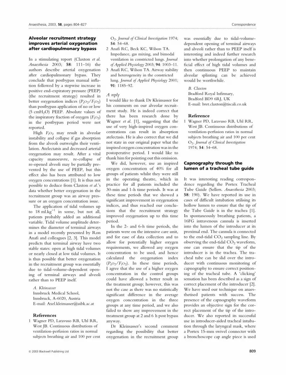
the ICU, a chest X-ray revealed exten-
sive opacification of the right hemitho-
rax with some loss of volume suggestive
of extensive collapse despite the tracheal
tube tip lying within the trachea
(Fig. 1). Fibreoptic bronchoscopy was
performed and several blood clots were
removed from the right main bronchus.
No obvious mucosal damage to the
trachea was seen. Postbronchoscopy,
the oxygen saturations improved to
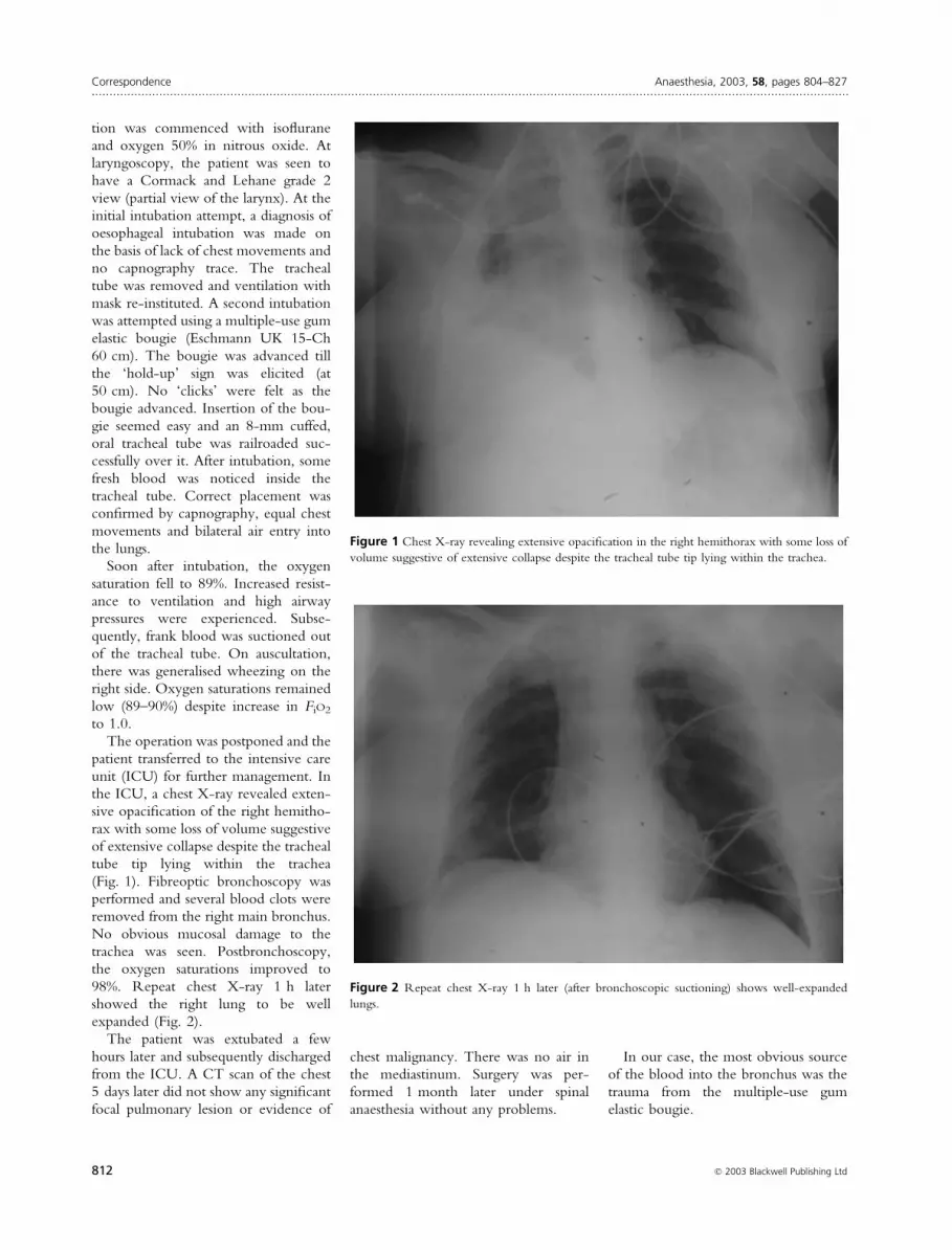
98%. Repeat chest X-ray 1 h later
showed the right lung to be well
expanded (Fig. 2).
The patient was extubated a few
hours later and subsequently discharged
from the ICU. A CT scan of the chest
5 days later did not show any significant
focal pulmonary lesion or evidence of
chest malignancy. There was no air in
the mediastinum. Surgery was per-
formed 1 month later under spinal
anaesthesia without any problems.
In our case, the most obvious source
of the blood into the bronchus was the
trauma from the multiple-use gum
elastic bougie.
Figure 1 Chest X-ray revealing extensive opacification in the right hemithorax with some loss of
volume suggestive of extensive collapse despite the tracheal tube tip lying within the trachea.
Figure 2 Repeat chest X-ray 1 h later (after bronchoscopic suctioning) shows well-expanded
lungs.
Correspondence Anaesthesia, 2003, 58, pages 804–827......................................................................................................................................................................................................................
812 � 2003 Blackwell Publishing Ltd

The gum elastic bougie is useful in
difficult intubation as long as the
epiglottis is visible (i.e. grade 2, grade 3)
at laryngoscopy [2]. It has been suggested
that blind placement of the bougie can be
confirmed by the ‘click’ and ‘hold-up’
signs [3]. In the United Kingdom,
the multiple-use bougie is widely used
by anaesthetists as the first choice method
in the event of unexpected difficult
intubation. Kadry and Popat reported
pharyngeal wall perforation caused
unambiguously by the bougie [1]. This
occurred during the initial attempt of
passing the multiple-use bougie. We feel
that in our case it was quite unusual for a
traumatic complication to happen as the
insertion of the bougie and the railroad-
ing of the tracheal tube were very smooth
and successful at the first attempt.
In the recent letter by Hodzovic
et al., the authors asked a worthwhile
question: ‘How many attempts (of
multiple-use bougie in the management
of difficult intubation) should be
recommended?’ We totally agree with
the relationship between the number of
attempts and traumatic complications in
the use of bougies. But we also believe
that it is worth considering the possi-
bility of a traumatic complication even
in an ‘easy’, first attempt, successful
insertion of the gum elastic bougie.
In our patient, a grade 2 view was
obtained on laryngoscopy. The ‘hold-
up’ sign was elicited after passing the
bougie. As some part of the cords was
visible, could we have avoided the
trauma by not eliciting the actual
‘hold-up’ by stopping once the bougie
had just passed beyond the cords?
A. Prabhu
P. Pradhan
R. Sanaka
A. Bilolikar
Kettering General Hospital,
Kettering NN16 8UZ, UK
E-mail: [email protected]
References1 Kadry M, Popat M. Pharyngeal wall
perforation – an unusual complication
of blind intubation with gum elastic
bougie. Anaesthesia 1999; 54: 404–5.
2 Wilson ME. Predicting difficult intu-
bation. British Journal of Anaesthesia
1993; 71: 333.
3 Sellers WYS, Jones GW. Difficult tra-
cheal intubation. Anaesthesia 1986;
41: 93.
Where is the narrowestsegment in the upper airway?
Your correspondents (Whyte et al.
Anaesthesia 2003; 58: 196) stated that,
as in children, the narrowest segment of
the upper airway in adults is not the
glottis, but at the cricoid cartilage, by
referring to a paper of Koufman and
colleagues [1]. In fact, this reference
states that ‘the subglottic cricoid is the
smallest, fixed cross section in the upper
airway’. Nevertheless, they only meas-
ured the diameter of the cricoid carti-
lage of cadavers [1], and compared it
with the anteroposterior diameter of the
glottis and trachea that were reported
previously by different researchers. The
anteroposterior distance is unlikely to
be narrowest in the glottis, and thus it
cannot be concluded whether the nar-
rowest diameter of the glottis is greater
or smaller than the narrowest length of
the cricoid lumen.
As far as we know, there has been only
one study that measured the length of
both the glottis and the cricoid lumen
[2]: Seymour & Prakash decided to
measure the glottic and cricoid lumen
in 134 cadavers because they sometimes
had difficulties in advancing a double-
lumen tube beyond the subglottis in
adults. By inserting a cylindrical nylon
sounds into the glottis and cricoid lumen,
they measured the maximum effective
size, and found that the diameter of the
glottis was always either similar to, or
greater than, the diameter of the cricoid
lumen. Therefore, it was just last year
(2002) when we finally obtained clear
evidence rejecting the orthodox opinion
that the narrowest segment in the upper
airway is the glottis in adults.
T. Asai
K. Shingu
Kansai Medical University,
Moriguchi City, Osaka, 570–8507,
Japan
E-mail: [email protected]
References1 Koufman JA, Fortson JK, Strong MS.
Predictive factors of cricoid ring size in
adults in relation to acquired subglottic
stenosis. Otolaryngology Head and Neck
Surgery 1983; 91: 177–82.
2 Seymour AH, Prakash N. A cadaver
study to measure the adult glottis and
subglottis: defining a problem associated
with the use of double-lumen tubes.
Journal of Cardiothoracic Vascular Anesthe-
sia 2002; 16: 196–8.
Haemophilus influenzae type B(HiB) is back
We would like to raise awareness of the
increasing frequency of occurrence of
the invasive Haemophilus influenzae type
B (HiB) which can result in life-threat-
ening epiglottitis. In October 1992, the
HiB conjugate vaccine was introduced
and the prevalence of HiB decreased
from 23.8 per 100 000 cases in 1991 to
0.92 cases per 100 000 in 1996. In the
last 4 years, its prevalence has started
rising again nationwide to 1.88 per
100 000 in 2001 and 2.81 per 100 000
in 2002 [1, 2].
The HiB vaccination programme is
based on three doses of conjugate
vaccine at 2, 3 and 4 months of age
without a further booster. In 1993, all
children under 4 years were offered an
additional single dose of vaccine (catch-
up campaign). The current rise of
invasive HiB disease in the UK could
be caused by the waning effect of the
1993 catch-up campaign, higher car-
riage rates amongst older children or
weaker immunisation effects related to
the use of a combination vaccine against
acellular pertussis, diphtheria, tetanus
and HiB use in 2000–01.
We encountered a case of acute
epiglottitis in a 6-year-old child, who
after a 2-day upper respiratory infection
developed stridor and respiratory dis-
tress. She was brought to the Emer-
gency Department drowsy, unable to
speak, using auxiliary respiratory mus-
cles, with a temperature of 37.0 �C.
The classic drooling of saliva was absent.
She had been fully vaccinated as usual in
the first year of life.
She was given two times 4 ml of
1 : 1000 epinephrine nebulised in 100%
oxygen. The on-call consultant anaes-
thetist and ENT surgeon were called.
A gas induction using sevoflurane 8% in
Anaesthesia, 2003, 58, pages 804–827 Correspondence......................................................................................................................................................................................................................
� 2003 Blackwell Publishing Ltd 813
oxygen was performed. At laryngos-
copy, a swollen bright red epiglottis was
seen and the trachea was intubated with
a size 4 uncuffed tracheal tube. The
lungs were ventilated with normal
compliance; however, there was no
leak from around the tracheal tube.
The child was treated with intraven-
ous cefotaxime and dexamethasone.
After 36 h of sedation and ventilation in
the intensive care unit, she was extubated
and made a full recovery. HiB was
isolated from her blood cultures.
For all those involved, this was the
first case of HiB epiglottitis that we have
seen. Given the above, it is possible that
we will have to deal with this condition
more often.
J. Hanousek
M. Swart
Torbay District General Hospital,
Torquay, UK
E-mail: [email protected]
References1 Garner D, Weston V. Effectiveness of
vaccination for Haemophilus influenzae
type B. Lancet 2003; 361: 395–6.
2 http://www.phls.co.uk/topics_az/
haemophilus/data_lab_age_qtr.htm.
Oral concretions – a potentialhazard for the airway
I would like to present the case of a
23-year-old fit and healthy female
patient for removal of an ingrown
toenail as a day case. A full history was
taken from this young traveller who
denied any medical problems. Physical
examination including airway assess-
ment was unremarkable.
The patient was anaesthetised fol-
lowing a routine intravenous induction
technique with propofol and fentanyl.
The airway was maintained throughout
the procedure by facemask allowing
spontaneous ventilation. Oxygen and
intermittent boluses of propofol were
administered at the same time. Routine
monitoring was used and recorded.
When the patient was anaesthetised,
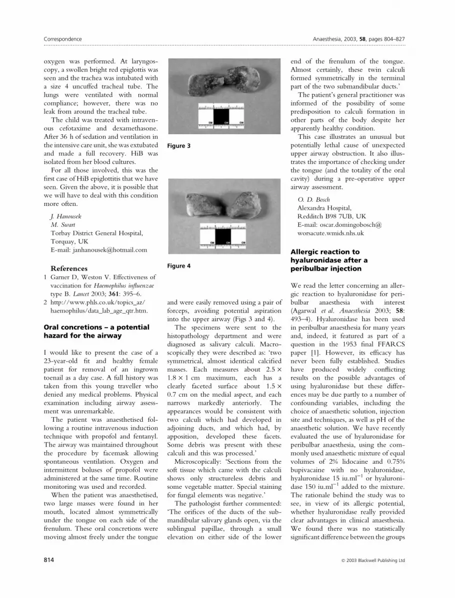
two large masses were found in her
mouth, located almost symmetrically
under the tongue on each side of the
frenulum. These oral concretions were
moving almost freely under the tongue
and were easily removed using a pair of
forceps, avoiding potential aspiration
into the upper airway (Figs 3 and 4).
The specimens were sent to the
histopathology department and were
diagnosed as salivary calculi. Macro-
scopically they were described as: ‘two
symmetrical, almost identical calcified
masses. Each measures about 2.5 ·1.8 · 1 cm maximum, each has a
clearly faceted surface about 1.5 ·0.7 cm on the medial aspect, and each
narrows markedly anteriorly. The
appearances would be consistent with
two calculi which had developed in
adjoining ducts, and which had, by
apposition, developed these facets.
Some debris was present with these
calculi and this was processed.’
Microscopically: ‘Sections from the
soft tissue which came with the calculi
shows only structureless debris and
some vegetable matter. Special staining
for fungal elements was negative.’
The pathologist further commented:
‘The orifices of the ducts of the sub-
mandibular salivary glands open, via the
sublingual papillae, through a small
elevation on either side of the lower
end of the frenulum of the tongue.
Almost certainly, these twin calculi
formed symmetrically in the terminal
part of the two submandibular ducts.’
The patient’s general practitioner was
informed of the possibility of some
predisposition to calculi formation in
other parts of the body despite her
apparently healthy condition.
This case illustrates an unusual but
potentially lethal cause of unexpected
upper airway obstruction. It also illus-
trates the importance of checking under
the tongue (and the totality of the oral
cavity) during a pre-operative upper
airway assessment.
O. D. Bosch
Alexandra Hospital,
Redditch B98 7UB, UK
E-mail: oscar.domingobosch@
worsacute.wmids.nhs.uk
Allergic reaction tohyaluronidase after aperibulbar injection
We read the letter concerning an aller-
gic reaction to hyaluronidase for peri-
bulbar anaesthesia with interest
(Agarwal et al. Anaesthesia 2003; 58:
493–4). Hyaluronidase has been used
in peribulbar anaesthesia for many years
and, indeed, it featured as part of a
question in the 1953 final FFARCS
paper [1]. However, its efficacy has
never been fully established. Studies
have produced widely conflicting
results on the possible advantages of
using hyaluronidase but these differ-
ences may be due partly to a number of
confounding variables, including the
choice of anaesthetic solution, injection
site and techniques, as well as pH of the
anaesthetic solution. We have recently
evaluated the use of hyaluronidase for
peribulbar anaesthesia, using the com-
monly used anaesthetic mixture of equal
volumes of 2% lidocaine and 0.75%
bupivacaine with no hyaluronidase,
hyaluronidase 15 iu.ml)1 or hyaluroni-
dase 150 iu.ml)1 added to the mixture.
The rationale behind the study was to
see, in view of its allergic potential,
whether hyaluronidase really provided
clear advantages in clinical anaesthesia.
We found there was no statistically
significant difference between the groups
Figure 3
Figure 4
Correspondence Anaesthesia, 2003, 58, pages 804–827......................................................................................................................................................................................................................
814 � 2003 Blackwell Publishing Ltd

in time taken to adequate anaesthesia
and a small but statistically significant
difference in ocular movement scores at
8 min in the group receiving the highest
concentration of hyaluronidase [2]. In
the light of these findings, we would
recommend that the potential hazards of
using hyaluronidase far outweigh any
clinical advantages and its continued
use for peribulbar anaesthesia must be
questioned.
G. Nicholson
G. M. Hall
St George’s Hospital Medical School,
London SW17 0RE, UK
E-mail: [email protected]
References1 Jorab JSM, Zuck D. Anniversaries – the
exam wot we took. The Royal College
of Anaesthetists’ Bulletin 2003; 19:
947–50.
2 Mantovani C, Bryant AE, Nicholson
G. Efficacy of varying concentrations of
hyaluronidase in peribulbar anaesthesia.
British Journal of Anaesthesia 2001; 86:
876–8.
Fresh frozen plasmafor succinylcholine apnoea –time to reconsider?
A 44-year-old woman weighing 80 kg,
with a history of mild asthma and reflux
oesophagitis is presented for exploration
of a parotid abscess. She had received
one uneventful previous anaesthetic for
varicose vein surgery. There was no
history of familial adverse reaction to
anaesthesia, although it only became
apparent on closer questioning after the
event that her parents were not her
biological relations. Pre-operative haem-
atological and biochemical parameters
were in the normal range.
She was premedicated with oral niz-
atidine 150 mg and metoclopramide
10 mg. After pre-oxygenation and appli-
cation of routine monitoring, anaesthesia
was induced using a rapid sequence
induction with thiopental 375 mg, fent-
anyl 100 lg and succinylcholine 100 mg
to facilitate tracheal intubation. Anaes-
thesia was maintained with nitrous
oxide, oxygen and isoflurane. Atracu-
rium 30 mg was administered 5 min
after induction. The procedure itself las-
ted approximately 20 min. An attempt
was made to reverse the neuromuscular
blockade with neostigmine 2.5 mg and
glycopyrronium 0.5 mg but there was
no return of respiratory effort. Peripheral
nerve stimulation monitoring train-of-
four activity showed two very feeble
twitches. At this point, a second dose of
neostigmine and glycopyrronium was
administered. The pupils appeared to be
somewhat constricted, and therefore
naloxone 400 lg was given. There was,
however, no spontaneous respiratory
activity despite normothermia, normal
serum electrolytes and permissive hyper-
capnoea. Ventilation was continued
with isoflurane whilst a depolarising
pattern of four equally depressed twit-
ches with no fade or post-tetanic aug-
mentation of contraction emerged,
suggesting a prolonged succinylcholine
blockade.
As an ITU bed was not available, the
patient was kept sedated and ventilated
in the recovery bay and the neuromus-
cular block was evaluated every
15 min. Approximately three and a half
hours passed without any further
improvement. At this juncture, we
decided to administer two units of fresh
frozen plasma (FFP). Within a few
minutes of infusion of the second unit,
there was a dramatic return of sponta-
neous respiratory activity. Eventually,
sufficient muscle strength to sustain a
head lift for 5 s returned, enabling
successful extubation of the trachea.
The patient was later transferred to the
ward and monitored overnight. The
remainder of the recovery period was
uneventful.
The blood sample taken prior to the
infusion of FFP showed reduced plasma
cholinesterase levels and a phenotype
E1a : E1a.
Atypical plasma cholinesterase activity
occurs in 1 : 2800 patients. These pati-
ents may remain paralysed for many
hours after routine doses of succinyl-
choline. Normal plasma cholinesterase
activity shows a wide range in healthy
adults and approximately 5% have levels
in the low range [1]. This includes
pregnant women, genotypically abnor-
mal patients and those with systemic
disease, e.g. liver disease, malignancy,
etc. This patient had normal liver func-
tion tests and benign tumour pathology
of her parotid gland. In this case, as
atracurium had been given before any
recovery from succinylcholine was
apparent, there was some confusion as
to the nature of the residual block at the
end of the procedure. This prompted the
administration of the second dose of
neostigmine, leading to further uncer-
tainties. It was only with the passage of
time and careful monitoring of neuro-
muscular function that the depolarising
nature of the block became apparent.
We waited for a considerable length of
time before infusing FFP as it transpired
that spontaneous recovery could poten-
tially take hours, necessitating prolonged
ventilation in the recovery bay.
The use of blood products as a
treatment of succinylcholine apnoea
has been well documented [2, 3]. The
plasma cholinesterase activity in bank
blood falls to 85% within 2 days of
donation, but remains at this level for at
least 30 days. FFP, however, shows no
decrease for 7 weeks and can provide a
useful therapeutic and diagnostic man-
oeuvre [4]. The chance of a unit of FFP
coming from a donor who also has
a plasma cholinesterase deficiency is
0.03%. A freeze-dried cholinesterase
concentrate is also available, but its cost
and storage time preclude its routine use
[3]. Despite its effectiveness, the use of
FFP has not been widely advocated due
to fear of disease transmission. How-
ever, in the UK, the chance of such
transmission now is negligible and has to
be balanced against the risks associated
with continuing anaesthesia and mor-
bidity such as infection associated with
ventilation and invasive procedures.
In addition, possible transfer out of
the hospital for a suitable ITU bed is
not without potential hazards and
complications. According to the SHOT
(Serious Hazards of Transfusion)
cumulative data from October 1995 –
September 2001 [5], there have been
three cases of HIV, eight of hepatitis B
and two of hepatitis C transmission in
total. Over this period, 2500 000 units
of blood and 400 000 units of FFP were
transfused each year, thus representing
a very small risk of transmission of
infection.
Anaesthesia, 2003, 58, pages 804–827 Correspondence......................................................................................................................................................................................................................
� 2003 Blackwell Publishing Ltd 815

Although prolonged respiratory insuf-
ficiency after succinylcholine adminis-
tration is rare, the above case reiterates
the need for monitoring neuromuscular
function and highlights the risks asso-
ciated with empirical use of muscle
relaxants. This case also revisits the use
of FFP to treat prolonged succinylcho-
line blockade. If used correctly, FFP
may act as a useful diagnostic and
therapeutic tool. Currently, there is an
ever-increasing constraint on ITU beds
necessitating out-of-hospital transfers.
The risks of transmission of infection
from FFP are miniscule, although it
should not be forgotten that adminis-
tration and cross-matching errors are all
possible. However, these risks may be
balanced against the risk of prolonged
anaesthesia, ventilatory support and
possibility of acquiring other hospital
infections.
R. Dabas
S. B. Vohra
City Hospital,
Birmingham B18 7QH, UK
E-mail: [email protected]
References1 Davis L, Britten JJ, Morgan M. Choli-
nesterase – its significance in anaesthetic
practice. Anaesthesia 1997; 52: 244–60.
2 Gill RS, O’Connell N, Scott RPF.
Reversal of prolonged suxamethonium
apnoea with fresh frozen plasma in a
6-week-old infant. Anaesthesia 1991;
46: 1036–8.
3 Smith DC, Ridley SA, Donaldson KF.
Fresh frozen plasma and edrophonium
in a patient with a plasma cholinesterase
deficiency. Anaesthesia 1993; 48:
511–13.
4 Epstein HM, Jarzemsky D, Zuckerman
L et al. Plasma cholinesterase activity in
bank blood. Anesthesia and Analgesia
1980; 59: 211–14.
5 Asher D, Atterbury CLJ, Chapman C
et al. Serious Hazards of Transfusion
Annual Report 2000 ⁄ 2001. Manchester:
SHOT Office, 2002.
Drug syringe labelling – doescomplacency come with age?
We were interested to read the review
of 81 cases of awareness from the
Australian Incident Monitoring Study
(AIMS) (Anaesthesia 2002; 57: 549–56),
which showed that drug errors were the
largest single cause of awareness during
anaesthesia.
As part of a wider review of risk
management in anaesthesia, we conduc-
ted an audit of drug syringe labelling in
our department. Without prior warning,
during a randomly selected operating
list, one of us noted how many syringes
in the anaesthetist’s tray were labelled.
This was only done once during that list
and the purpose of the audit was not
specified to the anaesthetist.
Over a 2-month period, 50 separate
lists were audited. In total, 80 of 233
syringes were unlabelled (35%). How-
ever, if propofol syringes were excluded,
the percentage was 24%. When the
figures were broken down by grade
of anaesthetist, an interesting trend
emerged. Excluding propofol again,
consultants did not label 33% of syringes,
staff grades 16% and SHOs only 11%.
This was despite SHOs using two more
syringes per case on average than
consultants.
It has been reported that 94% of
surveyed College Tutors feel a stand-
ardised colour code should be intro-
duced [1]. We currently use the
Medilabel colour-coded labels, so there
was no shortage of available labels.
However, the labels still have to be
placed on the syringe! There is
undoubtedly an element of compla-
cency that creeps in as experience
grows. We all tend to use particular
size syringes for different drugs but this
is hardly failsafe. Unfortunately, no one
is immune from drug administration
errors and consultants are no different.
Few can lay their hand on their heart
and say it has never happened to them.
We believe syringe labelling should be
encouraged and practised by all as part
of general risk management.
D. Amoako
N. G. Lavies
Worthing Hospital,
Worthing BN11 2DH, UK
E-mail: [email protected]
Reference1 Christie IW, Hill MR. Standardised
colour coding for syringe drug labels: a
national survey. Anaesthesia 2002; 57:
793.
Locking the K away
In an attempt to prevent accidental or
inadvertent intravenous administration
of concentrated potassium solutions [1],
it has become the policy of our Trust to
store such solutions under ‘lock and
key’ in all clinical areas. Administration
involves a process similar to that used
for controlled medicines; the signatures
of two authorised members of staff are
required in a dedicated register. Drugs
other than potassium, which have equal
potential to cause harm, are not subject
to such stringent measures. Our worry
is that by effectively singling potassium
out, a proportion of ward-based med-
ical and nursing staff perceive that any
intravenous administration of potassium
is inherently risky and best avoided. We
believe such stigmatisation may have
contributed to the events described
below.
A previously fit and well 77-year-old
male with a history of prior abdominal
surgery was admitted as an emergency
complaining of constipation, abdominal
pain and distension. A diagnosis of
bowel obstruction was made and he
underwent uneventful laparotomy the
following day when a sigmoid volvulus
was decompressed. His postoperative
course was prolonged and he was
managed on a general surgical ward
for 2 weeks on intravenous fluids. He
remained nil by mouth as he was unable
to tolerate oral administration due to a
persisting ileus. During this time, he
received 28 l of crystalloid ⁄ colloid but
only 8 g of potassium.
He was admitted to our critical care
facility obtunded, in respiratory failure
and in atrial fibrillation. Ultimately, he
required a short period of invasive ven-
tilation. Arterial blood gases after intu-
bation and ventilation revealed a severe
metabolic alkalosis with pH of 7.6 and
standard bicarbonate of 36 mmol.l)1.
Whilst intubated over the course of the
next 4 days, he required the administra-
tion of over 90 g of potassium, which
was associated with a return of pH and
standard bicarbonate to within normal
ranges.
Correspondence Anaesthesia, 2003, 58, pages 804–827......................................................................................................................................................................................................................
816 � 2003 Blackwell Publishing Ltd
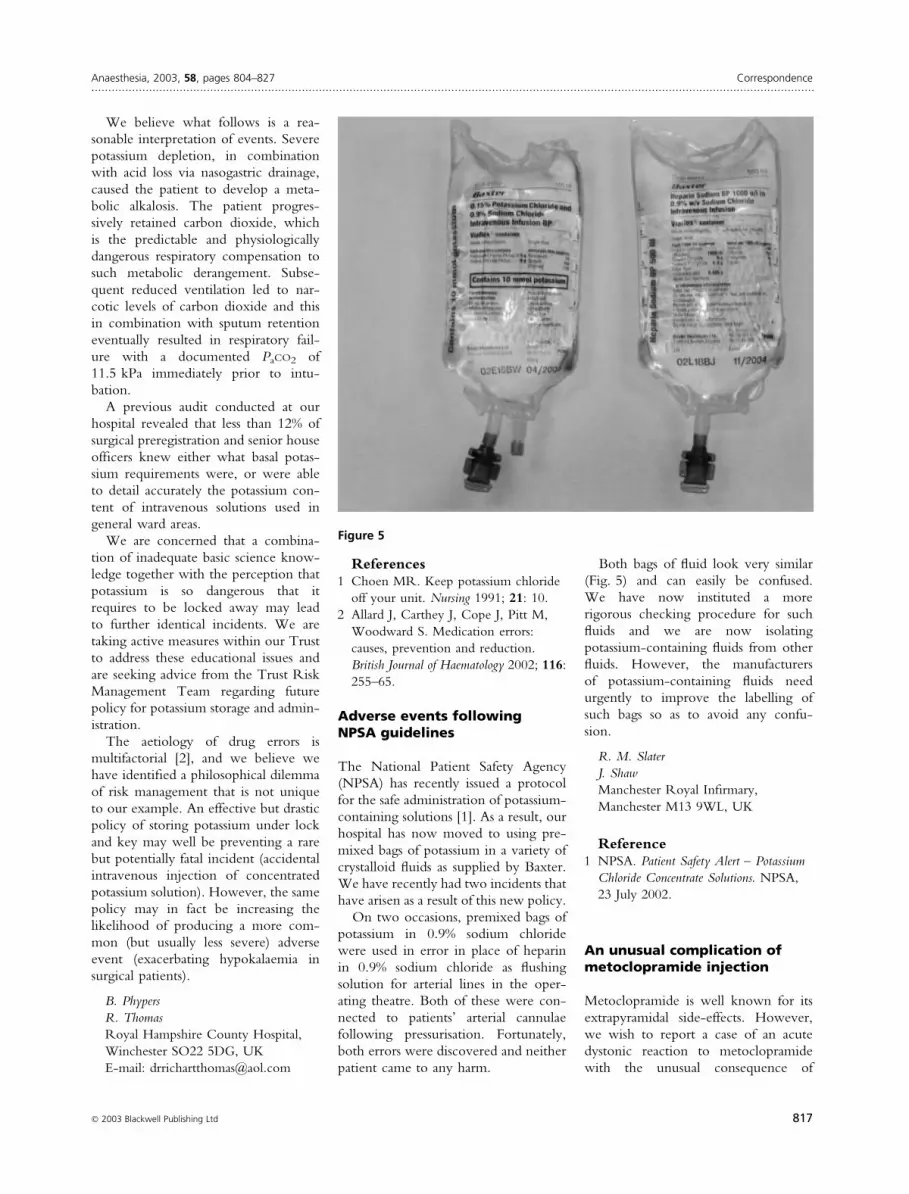
We believe what follows is a rea-
sonable interpretation of events. Severe
potassium depletion, in combination
with acid loss via nasogastric drainage,
caused the patient to develop a meta-
bolic alkalosis. The patient progres-
sively retained carbon dioxide, which
is the predictable and physiologically
dangerous respiratory compensation to
such metabolic derangement. Subse-
quent reduced ventilation led to nar-
cotic levels of carbon dioxide and this
in combination with sputum retention
eventually resulted in respiratory fail-
ure with a documented PaCO2 of
11.5 kPa immediately prior to intu-
bation.
A previous audit conducted at our
hospital revealed that less than 12% of
surgical preregistration and senior house
officers knew either what basal potas-
sium requirements were, or were able
to detail accurately the potassium con-
tent of intravenous solutions used in
general ward areas.
We are concerned that a combina-
tion of inadequate basic science know-
ledge together with the perception that
potassium is so dangerous that it
requires to be locked away may lead
to further identical incidents. We are
taking active measures within our Trust
to address these educational issues and
are seeking advice from the Trust Risk
Management Team regarding future
policy for potassium storage and admin-
istration.
The aetiology of drug errors is
multifactorial [2], and we believe we
have identified a philosophical dilemma
of risk management that is not unique
to our example. An effective but drastic
policy of storing potassium under lock
and key may well be preventing a rare
but potentially fatal incident (accidental
intravenous injection of concentrated
potassium solution). However, the same
policy may in fact be increasing the
likelihood of producing a more com-
mon (but usually less severe) adverse
event (exacerbating hypokalaemia in
surgical patients).
B. Phypers
R. Thomas
Royal Hampshire County Hospital,
Winchester SO22 5DG, UK
E-mail: [email protected]
References1 Choen MR. Keep potassium chloride
off your unit. Nursing 1991; 21: 10.
2 Allard J, Carthey J, Cope J, Pitt M,
Woodward S. Medication errors:
causes, prevention and reduction.
British Journal of Haematology 2002; 116:
255–65.
Adverse events followingNPSA guidelines
The National Patient Safety Agency
(NPSA) has recently issued a protocol
for the safe administration of potassium-
containing solutions [1]. As a result, our
hospital has now moved to using pre-
mixed bags of potassium in a variety of
crystalloid fluids as supplied by Baxter.
We have recently had two incidents that
have arisen as a result of this new policy.
On two occasions, premixed bags of
potassium in 0.9% sodium chloride
were used in error in place of heparin
in 0.9% sodium chloride as flushing
solution for arterial lines in the oper-
ating theatre. Both of these were con-
nected to patients’ arterial cannulae
following pressurisation. Fortunately,
both errors were discovered and neither
patient came to any harm.
Both bags of fluid look very similar
(Fig. 5) and can easily be confused.
We have now instituted a more
rigorous checking procedure for such
fluids and we are now isolating
potassium-containing fluids from other
fluids. However, the manufacturers
of potassium-containing fluids need
urgently to improve the labelling of
such bags so as to avoid any confu-
sion.
R. M. Slater
J. Shaw
Manchester Royal Infirmary,
Manchester M13 9WL, UK
Reference1 NPSA. Patient Safety Alert – Potassium
Chloride Concentrate Solutions. NPSA,
23 July 2002.
An unusual complication ofmetoclopramide injection
Metoclopramide is well known for its
extrapyramidal side-effects. However,
we wish to report a case of an acute
dystonic reaction to metoclopramide
with the unusual consequence of
Figure 5
Anaesthesia, 2003, 58, pages 804–827 Correspondence......................................................................................................................................................................................................................
� 2003 Blackwell Publishing Ltd 817

joint disruption requiring surgical inter-
vention.
A 44-year-old woman having taken
an overdose of nitrazepam and lofepr-
amine along with alcohol required
overnight airway management on the
intensive care unit. She was monitored
until she was judged fit to be discharged
to a general ward the following day.
Routine haematology, electrolytes and
chest X-ray were all normal. When able
to communicate, she revealed a past
history of depression and epilepsy,
although she had not had a seizure for
over 3 years and was no longer taking
anticonvulsant medication.
On the evening prior to her planned
discharge date, she complained to the
ward staff of mild nausea and was
administered an intramuscular injection
of metoclopramide 10 mg which had
been prescribed on the ‘as required’
section of her prescription chart. Forty-
five minutes later, the ward staff found
her unresponsive on her bed and sum-
moned the cardiac arrest team. On their
arrival, she was maintaining her own
airway with good respiratory effort
and was cardiovascularly stable. She
appeared unconscious with posturing
similar to the classically described tetan-
ic opisthotonus – she was rigid with an
extended arched neck, flexor posturing
of her arms and stiff legs. Oxygen was
administered via a facemask and intra-
venous access was established. Procyc-
lidine, to a total of 10 mg, was given
intravenously, which produced a rapid
regression of the rigidity and improve-
ment in her level of consciousness.
Blood gas analysis and electrolytes were
entirely normal. She had no recollection
of what had happened but complained
of a severely painful left foot, which
appeared clinically deformed, inverted
and acutely painful to palpation. Subse-
quent X-ray examination of her left
foot along with orthopaedic advice
revealed dislocation of the midfoot
joints. This required manipulation
under general anaesthesia, which was
achieved uneventfully. She was dis-
charged the next day and kept under
orthopaedic review, requiring crutches
and a below knee plaster for 6 weeks.
Although the literature is full of
reports of the adverse effects of meto-
clopramide, including tetanus-like
dystonic reactions [1], we have been
unable to find reference to an acute
dystonic reaction producing joint dis-
ruption as occurred in this case. We
would like to add this case to the long
list of reports of adverse events follow-
ing metoclopramide administration.
Whilst metoclopramide is rarely used
these days by anaesthetists as a first line
anti-emetic, it remains popular amongst
ward-based junior doctors. Our atten-
tion and theirs’ should be focused on
this case, where the clinically unneces-
sary use of a drug known to have
unpleasant side-effects culminated in
severe consequences. Perhaps it falls to
us anaesthetists, as relative experts in
applied pharmacology and frequent users
of anti-emetic medication, to promote
their safe and appropriate use through-
out the hospitals in which we work.
A. G. H. Stone
P. R. Howell
Homerton Hospital,
London, E9 6SR, UK
E-mail: [email protected]
Reference1 DellaValle R, Wielenga JL, Taylor O.
Metoclopramide induced tetanus-like
dystonic reaction. Clinical Pharmacology
1985; 4: 102–3.
Intranasal fentanyl forpostoperative analgesia afterelective Caesarean section
There are many methods for providing
postoperative analgesia, but each have
certain disadvantages. Patient-con-
trolled analgesia (PCA) requires intra-
venous access; epidural insertion may be
difficult or contraindicated; intramus-
cular injections are painful, relatively
inconvenient to administer, and have a
risk of infection and nerve damage. We
tested fentanyl administered as patient-
controlled intranasal analgesia (PCINA).
In this study, we used a nasal pump by
Go Medical from Anaesthetic Medical
Systems Ltd. The pump is a spray bottle
with flow-control tubing that, after
activating a spray, takes 3 min for the
reservoir to refill. This serves as a
lockout mechanism and prevents
continuous and over-administration of
fentanyl. The volume of the bolus is
0.18 ml.
Striebel et al. [1, 2] used a 27-lg
fentanyl dose for non-obstetric surgical
procedures but side-effects included
mild respiratory depression. We opted
for a 4.5-lg dose. The efficacy of nasal
fentanyl is comparable to intravenous
administration, because opioids are well
absorbed from the nasal mucosa, it
avoids first-pass metabolism and results
in a high bioavailability (71%) [1, 2].
Following Research Ethics Commit-
tee approval, we compared analgesia for
pain relief for 8 h after elective Caesar-
ean section under a standard spinal
anaesthetic. We administered fentanyl
via PCINA (a maximum of 90 lg of
fentanyl per hour) to 10 patients (Group
I), and intramuscular morphine 10 mg
on a 4-hourly PRN basis to 11 patients
(Group II). Physiological parameters
were measured at 30-min intervals,
including sedation, pulse oximetry,
heart rate, blood pressure, respiratory
rate and time for three-segment reg-
ression of the spinal block. Rescue
analgesia of 2–2.5 mg intravenous
morphine was provided to patients in
both groups. Pain relief was assessed
every 30 min using a 10-cm visual
analogue scale (VAS). All patients recor-
ded pain scores by 4 h.
In Group I, there was a mean of four
fentanyl dosages per 30-min interval.
The lowest number of dosages was 17
(76.5 lg) and the highest was 71
(319.5 lg) during the 8 h. Two patients
had nine dosages in 30-min intervals,
i.e. the highest possible number of
intranasal sprays allowed by the lockout
time; neither required rescue analgesia.
In Group II, most patients received one
intramuscular morphine dose during the
8 h. Two patients received two intra-
muscular doses. One patient did not
request any analgesia. The mean of the
highest VAS scores was 4.5 for Group I
and 5.1 for Group II.
Rescue analgesia was given as
2–2.5 mg intravenous morphine PRN
every 15 min if VAS scores were greater
than 5, or if the patient was too
distressed by pain. This occurred in six
patients: one in Group I (6 mg) and five
in Group II (6–20 mg).
Correspondence Anaesthesia, 2003, 58, pages 804–827......................................................................................................................................................................................................................
818 � 2003 Blackwell Publishing Ltd
No side-effects were reported from
any Group I patients. One patient from
Group II reported mild to moderate
pruritus.
Mean analgesic plasma concentration
of fentanyl ranges from 0.6 to 3 ng.ml)1
following intravenous administration
[3]. Blood samples from patients in
Group I were taken at 0, 5, 15, 30, 60
and 120 min after the first administration
of intranasal fentanyl. However, no
fentanyl was detected in any of the serum
samples.
We found no clinically relevant
intergroup difference in the measured
sedation scores, oxygen saturation, heart
rate, systolic blood pressure and respir-
atory rate (unpaired t-test with two-
sample equal variance test), VAS scores,
or amount of rescue analgesia (Fisher’s
exact test, p ¼ 0.149). However, for
this small sample of patients, the power
of this study was only 0.23.
In conclusion, we think that our
results are encouraging. The ease of
intranasal administration, the safety of a
controlled dose and the high patient
acceptability of this method of pain
relief deserve more attention. The lack
of clinical significance between the
two groups’ VAS scores suggests that
PCINA may be as effective as intra-
muscular morphine, or may be superior
in terms of the number of patients
requiring rescue analgesia. The high
patient satisfaction with the intranasal
pain relief was also remarkable. How-
ever, the PCINA patients had a much
more frequent rate of analgesic admin-
istration at their disposal. This may
contribute to a placebo effect. Further
work needs to be done with this novel
but, as yet, less established tool of
providing analgesia to determine opti-
mum dose without increasing side-
effects.
P. Wong
F. D. Chadwick
J. Karovits
Guy’s Hospital,
London SE1 9RT, UK
E-mail: [email protected]
References1 Striebel HW, Pommerening J, Rieger
A. Intranasal fentanyl titration for
postoperative pain management in an
unselected population. Anaesthesia
1993; 48: 753–7.
2 Striebel HW, Koenigs D, Kramer J.
Postoperative pain management by
intranasal demand-adapted fentanyl
titration. Anesthesiology 1992; 77:
281–5.
3 Peng PWH, Sandler AN. A review of
the use of fentanyl analgesia in the
management of acute pain in adults.
Anesthesiology 1999; 90: 576–99.
AcknowledgementsThis work was supported by funds from
The Friends of Guy’s Hospital, Guy’s
Hospital, London. The spray bottles
(manufactured by Go Medical) were
provided by Dr Tim Craft (Royal
United Hospital, Bath).
Cerebral hypoperfusion withsystemic hypotension duringcommon carotid ligation
We experienced a case of cerebral hyp-
operfusion during systemic hypotension,
which was detected by continuous
monitoring of regional cerebral oxygen
saturation (rCsO2) by near-infrared
spectroscopy in a patient whose collateral
circulation through the Circle of Willis
had been determined by a pre-operative
balloon occlusion test.
A 71-year-old woman with cancer of
the oropharynx was admitted to our
hospital. She had a history of varicose
veins in the lower extremities. Prior to
the scheduled operation, irradiation to
her tumour and chemotherapy were
performed effectively. Three months
later the patient underwent tumour
resection, radical neck dissection, recon-
struction using a forearm musclocutane-
ous flap and tracheostomy. During the
postoperative period, the patient suffered
from a refractory wound infection and
graft thrombosis. Ten days after the last
operation, when the dressing was being
changed, blood was found to be oozing
from both the neck and the oral cavity. A
cerebral angiogram was performed in
order to determine the most appropriate
therapeutic strategy. The patency of the
Circle of Willis was confirmed by cer-
ebral angiography, and left common
carotid artery (CCA) occlusion for
20 min induced no neurological symp-
toms. Systolic blood pressure (SBP)
was maintained between 110 and
120 mmHg throughout the test. Just
after conclusion of the test, the patient
suffered sudden and acute massive bleed-
ing from a branch of the left CCA. She
developed hypovolaemic shock (SBP
50–60 mmHg). A continuous infusion
of dopamine (5 lg.kg)1.min)1) was star-
ted to maintain her blood pressure and
the patient was immediately scheduled to
undergo ligation of the left CCA to
control the bleeding. Anaesthesia was
maintained with fentanyl, sevoflurane
and nitrous oxide. During the operation,
when the SBP decreased below
90 mmHg (mean blood pressure 75–
80 mmHg), the value of rCsO2 of the left
side decreased 10% more than that of the
right side. Throughout the operation, we
tried to maintain SBP at more than
90 mmHg. The operation time was
10 h 12 min and that of anaesthesia was
11 h 47min. The patient was extubated
in the operating room and was dis-
charged without any sequelae.
Carotid artery ligation is sometimes
performed to control bleeding from the
head and neck [1]. The most serious
complication of this procedure is cerebral
hypoperfusion with neurological deficit
[2]. In normal human subjects, the Circle
of Willis is the most important collateral
channel for circulation in the brain. This
system has many variations and 28% of
patients have an inadequate Circle of
Willis as a channel for collateral circula-
tion [3]. Therefore, it is important to
confirm the function of this system.
Cerebral angiography with unilateral
carotid artery balloon occlusion is a
popular method of testing the function
of the circle of Willis [4]. In our patient,
the test suggested normal collateral
circulatory function, yet during the
CCA ligation, transcranial cerebral
oximeter (INVOS 3100 A) indicated a
decrease in rCsO2 during systolic hypo-
tension suggesting cerebral hypoper-
fusion from an insufficient collateral
circulation. When severe systemic
hypotension is present, cerebral blood
flow decreases and cerebral hypoper-
fusion may occur even if the Circle of
Willis is intact. A decrease in rCsO2
correlates well with cerebral blood
flow [5].
Anaesthesia, 2003, 58, pages 804–827 Correspondence......................................................................................................................................................................................................................
� 2003 Blackwell Publishing Ltd 819
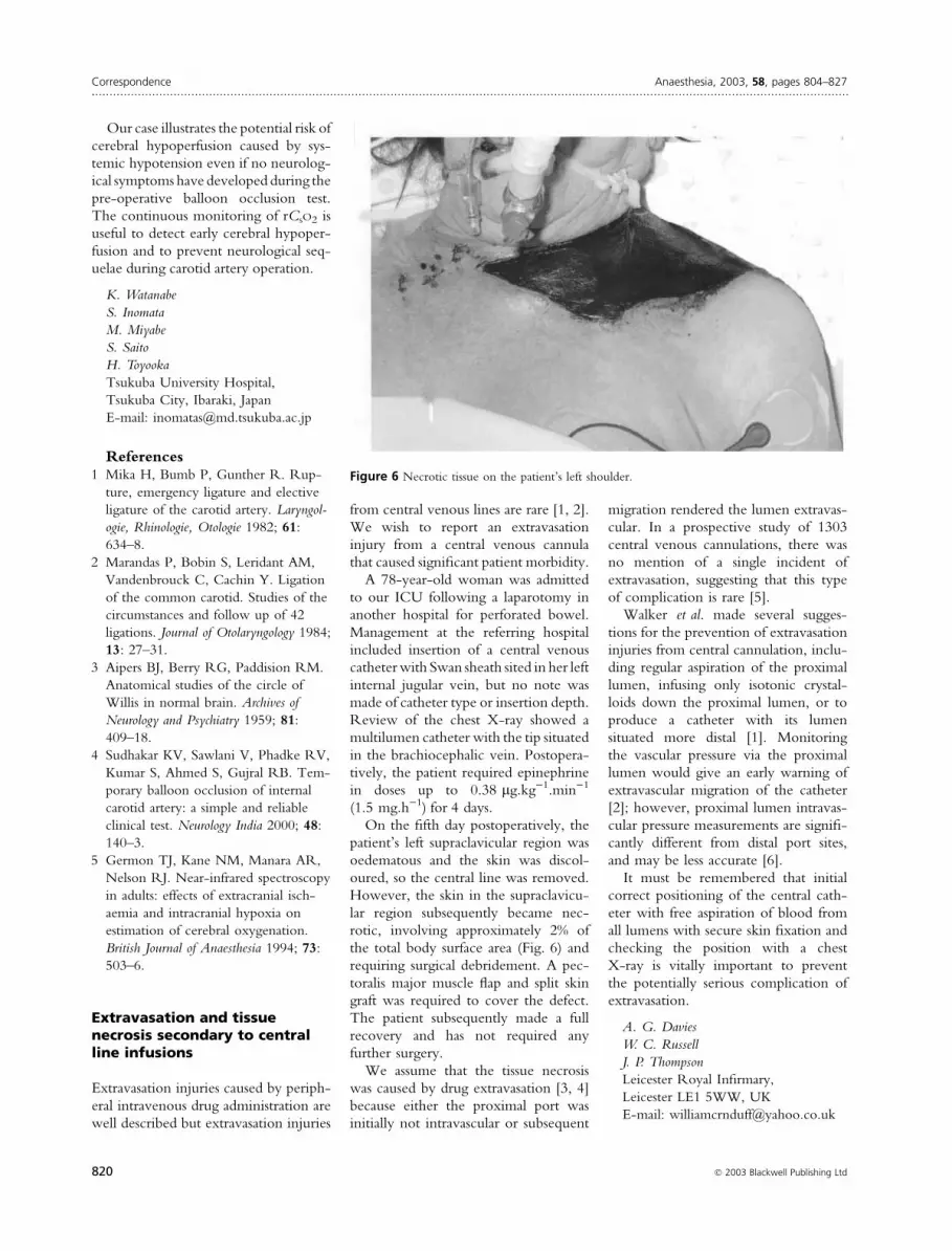
Our case illustrates the potential risk of
cerebral hypoperfusion caused by sys-
temic hypotension even if no neurolog-
ical symptoms have developed during the
pre-operative balloon occlusion test.
The continuous monitoring of rCsO2 is
useful to detect early cerebral hypoper-
fusion and to prevent neurological seq-
uelae during carotid artery operation.
K. Watanabe
S. Inomata
M. Miyabe
S. Saito
H. Toyooka
Tsukuba University Hospital,
Tsukuba City, Ibaraki, Japan
E-mail: [email protected]
References1 Mika H, Bumb P, Gunther R. Rup-
ture, emergency ligature and elective
ligature of the carotid artery. Laryngol-
ogie, Rhinologie, Otologie 1982; 61:
634–8.
2 Marandas P, Bobin S, Leridant AM,
Vandenbrouck C, Cachin Y. Ligation
of the common carotid. Studies of the
circumstances and follow up of 42
ligations. Journal of Otolaryngology 1984;
13: 27–31.
3 Aipers BJ, Berry RG, Paddision RM.
Anatomical studies of the circle of
Willis in normal brain. Archives of
Neurology and Psychiatry 1959; 81:
409–18.
4 Sudhakar KV, Sawlani V, Phadke RV,
Kumar S, Ahmed S, Gujral RB. Tem-
porary balloon occlusion of internal
carotid artery: a simple and reliable
clinical test. Neurology India 2000; 48:
140–3.
5 Germon TJ, Kane NM, Manara AR,
Nelson RJ. Near-infrared spectroscopy
in adults: effects of extracranial isch-
aemia and intracranial hypoxia on
estimation of cerebral oxygenation.
British Journal of Anaesthesia 1994; 73:
503–6.
Extravasation and tissuenecrosis secondary to centralline infusions
Extravasation injuries caused by periph-
eral intravenous drug administration are
well described but extravasation injuries
from central venous lines are rare [1, 2].
We wish to report an extravasation
injury from a central venous cannula
that caused significant patient morbidity.
A 78-year-old woman was admitted
to our ICU following a laparotomy in
another hospital for perforated bowel.
Management at the referring hospital
included insertion of a central venous
catheter with Swan sheath sited in her left
internal jugular vein, but no note was
made of catheter type or insertion depth.
Review of the chest X-ray showed a
multilumen catheter with the tip situated
in the brachiocephalic vein. Postopera-
tively, the patient required epinephrine
in doses up to 0.38 lg.kg)1.min)1
(1.5 mg.h)1) for 4 days.
On the fifth day postoperatively, the
patient’s left supraclavicular region was
oedematous and the skin was discol-
oured, so the central line was removed.
However, the skin in the supraclavicu-
lar region subsequently became nec-
rotic, involving approximately 2% of
the total body surface area (Fig. 6) and
requiring surgical debridement. A pec-
toralis major muscle flap and split skin
graft was required to cover the defect.
The patient subsequently made a full
recovery and has not required any
further surgery.
We assume that the tissue necrosis
was caused by drug extravasation [3, 4]
because either the proximal port was
initially not intravascular or subsequent
migration rendered the lumen extravas-
cular. In a prospective study of 1303
central venous cannulations, there was
no mention of a single incident of
extravasation, suggesting that this type
of complication is rare [5].
Walker et al. made several sugges-
tions for the prevention of extravasation
injuries from central cannulation, inclu-
ding regular aspiration of the proximal
lumen, infusing only isotonic crystal-
loids down the proximal lumen, or to
produce a catheter with its lumen
situated more distal [1]. Monitoring
the vascular pressure via the proximal
lumen would give an early warning of
extravascular migration of the catheter
[2]; however, proximal lumen intravas-
cular pressure measurements are signifi-
cantly different from distal port sites,
and may be less accurate [6].
It must be remembered that initial
correct positioning of the central cath-
eter with free aspiration of blood from
all lumens with secure skin fixation and
checking the position with a chest
X-ray is vitally important to prevent
the potentially serious complication of
extravasation.
A. G. Davies
W. C. Russell
J. P. Thompson
Leicester Royal Infirmary,
Leicester LE1 5WW, UK
E-mail: [email protected]
Figure 6 Necrotic tissue on the patient’s left shoulder.
Correspondence Anaesthesia, 2003, 58, pages 804–827......................................................................................................................................................................................................................
820 � 2003 Blackwell Publishing Ltd
References1 Walker C, Jackson D, Dolan S. The
potential for extravasation using a five-
lumen catheter. Anaesthesia 1997; 52:
716–17.
2 Bolton DT. Extravasation associated
with a multilumen central catheter.
Anaesthesia 1997; 52: 1119.
3 Dugger B. Peripheral dopamine infu-
sions: are they worth the risk of infil-
tration? Journal of Intravenous Nursing
1997; 20: 95–9.
4 Chen IL, O’Shea M. Extravasation
injury associated with low dose dop-
amine. Annals of Pharmacotherapy 1998;
32: 545–8.
5 Yilmazalar A, Bilgin H, Karfali G, Eren
A, Ozkan U. Complications of 1303
central venous cannulations. Journal of
the Royal Society of Medicine 1997; 90:
319–21.
6 Scott SS, Giuliano KK, Pysznick E,
Elliott S, Welsh K, Delbuono N. Influ-
ence of port site on central venous
pressure measurements from triple-
lumen catheters in critically ill adults.
American Journal of Critical Care 1998; 7:
60–3.
Anaesthetic management ofquinsy in a patient withShwachman–DiamondSyndrome
A 21-year-old female patient (40 kg,
128 cm tall) presented with a paraton-
sillar abscess, which required surgical
drainage. The patient had Shwachman–
Diamond Syndrome (SDS), diagnosed
at 4 months of age, a rare congenital
disorder characterised by bone marrow
dysfunction, exocrine pancreatic insuf-
ficiency and short stature.
The patient displayed severe trismus.
Computerised tomography had con-
firmed the diagnosis of a left paraton-
sillar abscess. Intravenous antibiotics had
been ineffective.
Pancreatic insufficiency had resulted
in relative malnutrition and poor skeletal
growth. She was pancytopaenic (haemo-
globin 8.1 g.dl)1, white cell count
1.7 · 109.l)1 and platelet count of only
11 · 109.l)1). Pre-operatively, she was
given 2 units of blood, 2 units of platelets
to cover nasal fibreoptic intubation and
intravenous vitamin K on the advice of
the haematologist. Previous general
anaesthetics had been uneventful.
After placement of standard monit-
oring (ECG, non-invasive blood pres-
sure and pulse oximetry), an 18G
intravenous cannula was inserted in
the dorsum of the left hand and intra-
venous midazolam 3 mg was adminis-
tered. The nasal mucosa was sprayed
with 3 ml of lidocaine 4%. A further
2 ml of lidocaine 4% was applied top-
ically to the oropharyngeal mucosa.
Oxygen 10 l.min)1 was administered
via a variable performance facemask
attached to a reservoir bag. Fibreoptic
laryngoscopy via the right nostril easily
visualised the vocal cords. A cuffed
5-mm Mallinckrodt tracheal tube was
correctly positioned under direct vision.
Capnography and auscultation con-
firmed correct tracheal tube placement
after removal of the fibrescope. General
anaesthesia was then induced with 8%
sevoflurane in 100% oxygen and main-
tained with 2–3% sevoflurane in a
50 : 50 mixture of oxygen and nitrous
oxide, with the patient breathing spon-
taneously. Fentanyl 50 lg was given to
supplement general anaesthesia. Extu-
bation, with the patient semi-erect, was
uneventful, as was her subsequent
recovery.
SDS was first reported in 1964 [1].
Only 200 cases have been reported
since. Shwachman, Diamond, Osaki
and Knaw reported this syndrome in a
group of five children attending a cystic
fibrosis clinic at Harvard Medical
School. SDS is inherited as an auto-
somal recessive. It mainly involves
pancreas, bone marrow and skeleton,
but the liver, kidneys, teeth and im-
mune system may also be affected [2].
Neutropaenia is the most common
haematological abnormality, occurring
in 88–100% of patients. In most cases,
neutropaenia occurs intermittently, the
neutrophils having defects in mobility,
migration and chemotaxis. Patients are
prone to recurrent bacterial infections
of the upper respiratory tract, otitis
media, pneumonia, osteomyelitis, bac-
teraemia and skin infections [3, 4].
Severe upper respiratory infections can
cause airway obstruction. Almost one
half of patients with SDS have pancy-
topaenia; however, some (24–88%) may
have variable degrees of thrombocyto-
paenia and up to 80% have anaemia,
which is usually mild and normochro-
mic–normocytic [2, 3, 5].
Varying severity of pancreatic dys-
function due to acinar maldevelopment
is a hallmark of SDS. This can lead to
malnutrition and fat-soluble vitamin
deficiencies. Its severity is not consistent
with either haematological or skeletal
abnormalities and usually improves with
advancing age.
Skeletal abnormalities are reported to
occur in more than 75% of patients with
SDS. These include rib cage abnormal-
ities, femoral head dystosis, kyphoscol-
iosis, genu and cubitus valgus,
syndactyly and clinodactyly [2, 3].
Patients with SDS appear emaciated
and frail with a tendency to easy
bruising [3] and require sympathetic
handling and positioning in the oper-
ating theatre.
P. Tamhane
N. I. Newton
S. White
St Thomas’ Hospital,
London SE1 7EH, UK
E-mail: [email protected]
References1 Shwachman H, Diamond L, Osaki F.
Knaw. The syndrome of pancreatic
insufficiency and bone marrow dys-
function. Journal of Pediatrics 1964; 65:
645–63.
2 Dror Y, Freedman MH. Review:
Shwachman–Diamond Syndrome.
British Journal of Haematology 2002; 118:
701–13.
3 Spoto-Cannons AC. Shwach-
man–Diamond Syndrome. http://
www.emedicine.com/ped/
topic2060.htm.
4 Dror Y, Ginzberg H, Dalal I et al.
Immune function in patients with
Shwachman–Diamond syndrome.
British Journal of Haematology 2001; 114:
712–17.
5 Smith OP, Hann IM, Chessells JM,
Reeves BR, Milla P. Haematological
abnormalities in Shwachman–Diamond
syndrome. British Journal of Haematology
1996; 94: 279–84.
Anaesthesia, 2003, 58, pages 804–827 Correspondence......................................................................................................................................................................................................................
� 2003 Blackwell Publishing Ltd 821
Trauma in pregnancy:anaesthetic management of thepregnant trauma victim withunstable cervical spine
Trauma in pregnancy is currently a
leading cause of non-obstetric maternal
death, and maternal death is the most
common cause of fetal death. Cervical
spine injury, respiratory failure and
haemorrhagic shock are the most fre-
quent causes of maternal death in preg-
nant trauma victims [1]. We present a
case of a parturient who presented for
an emergency posterior cervical fusion
following unstable traumatic cervical
spine fractures. The pregnancy was
known to be present pre-operatively.
A 40-year-old, previously healthy
female was admitted to the Emergency
Department with unstable, motor-vehi-
cle-related, cervical spine fractures (C1
and C2) and multiple rib fractures with
left-sided pneumothorax for an emer-
gency posterior cervical spinal fusion
under general anaesthesia. The patient
was haemodynamically stable. Her neu-
rological examination revealed a Glas-
gow Come Scale (GCS) of 15, free range
of motion in all four extremities and 5 ⁄ 5motor strength. An obstetric ultrasound
revealed a single fetus in breech presen-
tation with estimated gestational age of
15 weeks without any gross abnormal-
ities. Fetal heart rate (FHR) was
140 beat.min)1 and reactive.
Airway control with awake fibreoptic
tracheal intubationwasdeemednecessary
and therefore pharmacological prophy-
laxis against aspiration was accomplished
in a standard manner with intravenous
administration of a histamine 2 receptor
antagonist and metoclopramide com-
bined with oral non-particulate antacid.
Topical anaesthesia of the upper airway
mucosa via a mask nebuliser containing
lidocaine 4% for about 15–20 min was
conducted. The patient was taken to the
operating room and pre-oxygenation
was accomplished with 3–5 min of
normal tidal volume ventilation with
100% oxygen. Small amounts of mida-
zolam to a total dose of 2 mg were
titrated to effect to facilitate the patient’s
co-operation. Awake tracheal intuba-
tion was accomplished in a semisitting
position with gentle insertion of the
7-mm internal diameter tracheal tube
over the fibreoptic bronchoscope. The
patient was asked to assume the prone
position on the operating room table
and, following repeat neurological
examination documenting her ability
to move all four extremities, general
anaesthesia was induced in a standard
manner. Every effort was made to avoid
any direct pressure to her abdomen and
gravid uterus. Sensory evoked potentials
were monitored throughout surgery
and no changes in either amplitude or
latency were reported. Anaesthesia was
maintained with isoflurane, fentanyl,
propofol and vecuronium. Following
uneventful surgery, the patient was trans-
ported to the intensive care unit (ICU)
and extubated. FHR was 140 beat.min)1
and reactive. The postoperative hospital
course was uneventful and she accom-
plished full recovery.
The difficulty in airway management
in trauma victims increases from no
pregnancy present pre-operatively to
pregnancy present pre-operatively. The
difficulty in airway management in preg-
nant trauma victims increases from elec-
tive, to urgent, to emergency situations.
The anatomic and physiological changes
of pregnancy (mucosal oedema, in-
creased oxygen consumption, decreased
functional residual capacity) increase the
difficulty of airway management, while
decreasing the time available and the
margin of safety.
It is essential to titrate analgesic and
sedative drugs carefully to maintain con-
tinual meaningful verbal communication
between the anaesthetist and patient (i.e.
respond to commands in a clear, crisp and
unambiguous manner, and remain ra-
tional and alert). Respiratory depression
and aspiration of stomach contents dur-
ing the application of a local anaesthetic
agent is much less likely to occur if the
patient remains awake and alert. In
addition, a rational, alert mother mini-
mises the risk of neonatal depression.
Midazolam is the benzodiazepine
recommended for these purposes; how-
ever, it is highly unionised and very
lipophilic, and its fetal ⁄maternal ratio is
0.76 at 15–20 min after maternal admin-
istration. However, unlike other ben-
zodiazepines, the ratio falls rapidly. No
adverse fetal effects have been reported.
The literature documenting obstetric
and anaesthetic management of preg-
nant trauma victims is limited [2, 3]. We
are not aware of any reports document-
ing emergent anaesthetic management
(including awake fibreoptic intubation)
of the pregnant trauma victim present-
ing with unstable traumatic cervical
spine fractures for emergent surgery.
In summary, this case clearly demon-
strates that timely and aggressive surgical
and anaesthetic management (including
the airway management) of the pregnant
trauma victims may be life saving for
both the mother and her fetus.
K. M. Kuczkowski
S. A. Fouhy
M. Greenberg
J. L. Benumof
University of California San Diego,
San Diego, CA 92103–8770, USA
E-mail: [email protected]
References1 Baker BW. Trauma. In: Chestnut DH,
ed. Obstetric Anesthesia: Principles and
Practice. St Louis: Mosby, 1999: 1041–
50.
2 Aitokallio-Tallberg A, Halmesmaki E.
Motor vehicle accident during the
second or third trimester of pregnancy.
Acta Obstetrica Gynecologica Scandinavica
1997; 76: 313–17.
3 Kuczkowski KM, Ispirescu JS,
Benumof JL. Trauma in pregnancy:
anesthetic management of the parturi-
ent with multiple gun shot wounds to
the gravid uterus and fetal injury.
Journal of Trauma 2003; 54: 420.
Myocardial ischaemiacomplicating an electiveCaesarean section
The recent Confidential Enquiry into
Maternal Deaths in the UK [1] illustrated
possible problems associated with oxy-
tocin used to maintain a tonic uterus after
delivery of an infant by Caesarean sec-
tion. We describe a patient who suffered
myocardial ischaemia during Caesarean
section, temporally related to the admin-
istration of a bolus dose of oxytocin.
A healthy 31-year-old, non-smoking
tennis coach required an elective
Caesarean section, for which she had
Correspondence Anaesthesia, 2003, 58, pages 804–827......................................................................................................................................................................................................................
822 � 2003 Blackwell Publishing Ltd

straightforward spinal anaesthesia. After
delivery of the baby and subsequent
administration of intravenous synthetic
oxytocin 5 i.u., she developed clinical
symptoms and ECG changes of myo-
cardial ischaemia. Myocardial damage
was confirmed with a troponin T level of
0.19 lg.l)1, 12 h after the onset of
symptoms. A subsequent angiogram
demonstrated normal coronary arteries
and a diagnosis of acute coronary syn-
drome was made. Thereafter, the patient
made an unremarkable recovery.
It is not uncommon for patients
undergoing Caesarean section to experi-
ence symptoms of chest discomfort,
dyspnoea, nausea and vomiting, which
are often attributed to surgical stimula-
tion. Patients suffering from myocardial
ischaemia can also complain of very
similar symptoms, making it difficult to
differentiate between these two causes at
the time of surgery.
Myocardial infarction during preg-
nancy is unusual, with an incidence in
the region of 1 in 10 000 [2]. Hence,
transient symptoms of chest pain are
attributed to causes other than myocar-
dial ischaemia, particularly as patients
presenting for Caesarean section tend to
be young, healthy and have minimal
risk factors for coronary vascular disease.
In this case, it is not clear what preci-
pitated the myocardial ischaemia. Nei-
ther anaemia nor severe haemorrhage
were present, nor was there any appar-
ent cardiac disease or excessive use of
vasoactive drugs such as ephedrine.
Significant venous air emboli and
amniotic fluid emboli entering the cir-
culation can cause myocardial ischaemia
with potentially catastrophic conse-
quences. This does not seem likely in
our patient, as she remained haemo-
dynamically stable and well oxygenated
throughout.
The ischaemic episode was temporally
related to the administration of oxytocin.
This drug has been previously implicated
in causing myocardial ischaemia [3] for
which two mechanisms are postulated.
Coronary artery vasospasm has been
demonstrated by intracoronary adminis-
tration of oxytocin in an isolated dog’s
heart [4].Thismechanism is supportedby
a report of peripheral arterial spasm asso-
ciated with the use of synthetic oxytocin
[5]. Alternatively, intravenous oxytocin
produces vasodilatation of vascular
smooth muscle resulting in a reduction
in both systolic and diastolic blood pres-
sures. The diastolic hypotension reduces
coronary perfusion. This in combination
with the physiological anaemia of preg-
nancy may impair myocardial oxygen
supply sufficiently to induce myocardial
ischaemia. No decrease in blood pressure
was recorded, but even if it did occur
between the 3-min recordings, it would
still be difficult to explain how such a
transient effect could lead to myocardial
ischaemia in a healthy patient.
It seems likely that several factors
were involved, as the normal physiolo-
gical response to oxytocin (or indeed the
anxiety associated with childbirth) does
not result in such dramatic symptoms.
J. P. Chilvers
G. Cooper
University Hospital – Queen Elizabeth,
Birmingham. B15 2TH, UK
E-mail: [email protected]
M. Wilson
University of Washington Medical
Centre,
Seattle, USA
References1 Department of Health, Welsh Office,
Scottish Office Department of Health,
Department of Health and Social Services
Northern Ireland. Why Mothers Die.
Report on Confidential Enquiries into
Maternal Deaths in The United Kingdom
1997–9. London: RCOG Press, 2001.
2 Sutaria N, O’Toole L, Northridge D.
Postpartum acute MI following routine
ergometrine administration treated
successfully by primary PTCA. Heart
2000; 83: 97–8.
3 Spence A. Oxytocin during Caesarean
section. Anaesthesia 2002; 57: 722–3.
4 Fortner C, Hawley ES, Woodbury RA.
Effects of synthetic oxytocin with and
without preservatives upon coronary
blood flow in the dog. Journal of Phar-
macological Experimental Therapeutics
1969; 165: 258–66.
5 Evron S, Ariely S, Agasi M et al. Severe
peripheral arteriospasm following oxy-
tocin administration. American Journal of
Obstetric and Gynecology 1986; 155:
657–8.
Peripartum anaestheticmanagement of a parturientwith spinal cord injury andautonomic hyperreflexia
I report a case of a parturient with spinal
cord injury (SCI) who presented in early
preterm labour with uterine-contrac-
tions-induced acute onset autonomic
hyperreflexia and received combined
spinal epidural analgesia (CSEA) for
labour and an uneventful Caesarean
section.
A 17-year-old, 164-cm, 60-kg, grav-
ida 1, para 0 female with SCI was
admitted to the labour and delivery
(L & D) suite at 36 weeks’ gestation in
preterm labour with regular uterine
contractions, facial flushing, piloerec-
tion, headache and hypertension. She
did not report any pain associated with
her contractions. Her past medical
history was significant for motor-vehi-
cle-related cervical (C6)7) spinal cord
transsection at 28 weeks’ gestation.
Anterior cervical fusions were performed
under general anaesthesia in the imme-
diate post-trauma period. At the time of
the insult, the fetus suffered parenchymal
brain injury and subsequently developed
post-traumatic growth restriction.
The patient had no known drug
allergies. Her admission blood
pressure was 145 ⁄ 90 mmHg, heart rate
60 beat.min)1 and respiratory rate
19 breath.min)1. Fetal heart rate was
140 beat.min)1 and reactive. The diag-
nosis of eclampsia was ruled out by
routine laboratory studies (liver and
kidney function tests). The working
diagnosis of acute onset autonomic
hyperreflexia was established and
administration of labour analgesia was
indicated.
A CSEA with levobupivacane 2.5 mg
combined with fentanyl 5 lg was
promptly administered via a needle
through needle technique (18-G Tou-
hy-Schliff epidural needle, and a 27-G
Pencan spinal needle) at the L3)4 ver-
tebral interspace. A 20-G multi-orifice
epidural catheter was inserted 4 cm into
the epidural space. Aspiration from the
epidural catheter was negative for
blood and CSF. The decrease in the
patient’s blood pressure was recorded
in approximately 4–5 min after the
Anaesthesia, 2003, 58, pages 804–827 Correspondence......................................................................................................................................................................................................................
� 2003 Blackwell Publishing Ltd 823

induction of labour analgesia. Over the
next 5 min, her headache and other
early symptoms of autonomic hyperref-
lexia subsided. Labour epidural analgesia
consisted of a continuous infusion of
levobupivacaine 0.0625% with fentanyl
2 lg.ml)1 at a rate of 8 ml.h)1.
Six hours after the induction of
CSEA, emergency Caesarean section
was required for fetal distress. Surgical
anaesthesia was established with incre-
mental doses of chloroprocaine 3% to a
total of 15 ml (450 mg) and fentanyl
50 lg administered through an epidural
catheter. An uneventful Caesarean
delivery of a female fetus weighing
2050 g, who had Apgar scores of 6
and 8, after 1 and 5 min, respectively,
was accomplished. No postoperative
complications were reported.
Each year more than 2000 women of
childbearing age in the United States
alone sustain a SCI [1]. As a result of
increased survival rates in women with
SCI, the number of parturients with SCI
is increasing [2]. Vaginal delivery is
preferred. Any parturient with SCI
whose level of transsection is at T6 or
higher is at risk for acute autonomic
hyperreflexia as a result of uterine con-
tractions. Autonomic hyperreflexia in
patients with SCI may be mistaken for
pre-eclampsia at presentation [3]; conse-
quently, routine laboratory studies (liver
and kidney function tests) may be the key
differential between the two disorders.
Effective management includes prompt
administration of labour analgesia. Be-
cause of rapid onset of labour analgesia,
CSEA seems particularly suitable for
these patients. Occurrence of preterm
labour is not uncommon in parturients
with SCI [4]. Early diagnosis is hampered
by the inability of most quadriplegics to
sense uterine contractions in the usual
way. The incidence of pregnancy com-
plications in parturients with SCI is
increased, particularly when SCI occurs
in pregnancy.
The literature documenting peripar-
tum anaesthetic management of preg-
nant patients with SCI is limited [1–5]. I
am not aware of any reports document-
ing emergent administration of CSEA
for parturients with SCI presenting in
preterm labour with uterine-contrac-
tions-induced acute onset autonomic
hyperreflexia. This case clearly demon-
strates that expeditious induction of
labour analgesia may prevent or ameli-
orate peripartum complications (inclu-
ding autonomic hyperreflexia) in
labouring parturients with SCI.
K. M. Kuczkowski
University of California San Diego,
San Diego, California, USA
E-mail: [email protected]
References1 Cross LL, Meythaler JM, Tuel SM et al.
Pregnancy following spinal cord injury.
Western Journal of Medicine 1991; 154:
607–11.
2 Verduyn WH. Pregnancy and delivery
in tetraplegic women. Journal of Spinal
Cord Medicine 1997; 20: 371–4.
3 Ahmed AB, Bogod DG. Anaesthetic
management of a quadriplegic patient
with severe respiratory insufficiency
undergoing Caesarean section.
Anaesthesia 1996; 51: 1043–5.
4 Catanzarite VA, Ferguson JE 2nd,
Weinstein C et al. Preterm labor in the
quadriplegic parturient. American Journal
of Perinatolology 1986; 3: 115–18.
5 Plotz J, von Hugo R. Autonomic
hyperreflexia, pregnancy and delivery
in para-tetraplegia. The obstetric
anesthesiologic viewpoint on a case.
Anaesthetist 1996; 45: 1179–83.
Pushing water down thefemoral shaft
The orthopaedic surgeons in our hospital
are keen on using chlorhexidine irriga-
tion when treating wounds. Nowadays,
they even use it by means of a Pulse
Lavage system, which I have discovered
can produce pressures in excess of
400 mmHg if the outlet is restricted. I
do not know how well based their
routine is in respect of the antimicrobic
effect of this rather diluted, 0.02%,
chlorhexidine, but neither the anaesthet-
ists nor the surgeons in our hospital have
considered it to be much of a problem.
By accident, in relation to some testing
we wished to carry out, I discovered to
my surprise that these solutions are in
water, not in 0.9% saline. No one in
our department, or in the orthopaedic
department, was aware of this. We had all
assumed, which one should never do,
that it has been in saline.
For fear of effects along the lines of
the TUR syndrome, albeit of lesser
degree, I do not feel happy when any
significant amount of fluid is used as
irrigation in any body cavity unless it is
iso-osmotic. The solution in question
has an osmotic pressure of zero. The
raw surfaces down the femoral shaft
after reaming and before cementing a
prosthesis do not seem to me to be the
ideal cavities to irrigate under consid-
erable pressure with zero osmotic water.
However, as this has been going on for
a long time without any obvious side-
effects to our patients, our surgeons are
not happy abandoning their practice on
theoretic grounds only. We will, in
future, see if we can find signs of
increased haemolysis after total hip
replacements where a significant
amount of this chlorhexidine water has
been in use, but have as yet not done so.
This potential problem has surfaced
now because we want to expand our use
of the cell-saver techniques in orthopae-
dic surgery, and this irrigation solution
would of course totally destroy every
salvaged red cell. However, we have
done some in vitro tests in our haema-
tology laboratory using chlorhexidine
0.02% in 0.9% saline, and this solution
does not seem to have any effect on the
red cells, apart from a small increase in
clotting time. Indeed, the little literature
I have found suggests that chlorhexidine
itself is quite inert to red cells and the
vascular bed, and if it were to be used in
a cell saver, more than 90% would be
washed out before being retransfused to
the patient, so one would not expect to
see any significant toxic effect.
I would very much appreciate com-
ments from my colleagues in the UK
and elsewhere on this practice:
• Is it only in Cornwall that we have
not realised that the available chlorh-
exidine irrigation solutions are in
water rather than saline?
• Do the surgeons in your area use this
irrigation technique?
• If not, would you be happy if your
surgeon was to suggest power irriga-
tion of raw wound ⁄ bone surfaces
with zero osmotic water?
Correspondence Anaesthesia, 2003, 58, pages 804–827......................................................................................................................................................................................................................
824 � 2003 Blackwell Publishing Ltd

• If indeed there is a good case for
chlorhexidine irrigation, should we
not push the industry to produce it in
saline?
• Is there a suitable substitute for this
irrigation solution that we could offer
to our surgeons?
I invite you to contact me with your
answers to these questions and indeed
any comments.
L. Jakt
Royal Cornwall Hospital,
Truro TR1 3LJ, UK
E-mail: [email protected]
History of anaesthesia
John Zorab, in his recent letter (Zorab.
Anaesthesia 2002; 57: 1242), gives but a
brief explanation of Lethe. Were this
necessary for our learned readership, it
would be helpful if it were more
informative. Hades (Dis) was one of
the three sons of Cronos (Saturn) and
Rhea (Ops), youngest brother of Zeus
(Jupiter) and Poseidon (Neptune). In
the division of the world between the
three brothers, he took the lower
world, which became known as Hades
through association. The name Hades
was feared and rarely spoken, the Clas-
sical equivalent of Voldemort [1].
Lethe was the daughter of Eris
(Discordia). Through the nether world
ran a river named after her, but this
river was in the Elysian Fields, where
the souls of the blessed existed. They
would drink of the water of Lethe prior
to returning to the normal world as re-
incarnated souls so that they would have
no memory of the lower world. The
spirits of the guilty and evil-doers passed
into the judgement hall of Hades,
where their torment for eternity was
decided prior to their entering Tartarus
and joining the company of well-
known sufferers such as Tityus, Ixion,
Tantalus and Sisyphus. I am sure further
explanation is unnecessary to our
learned readership.
Finally, I think that it is most unlikely
that Adam is suffering from a postan-
aesthetic headache, as he is still ‘in a
deep sleep’ undergoing the procedure
of uxorectomy, and would one really
suggest that the Almighty is not able to
provide a perfect anaesthetic without
sequelae?
G. R. Harrison
Queen Elizabeth Hospital,
Birmingham B15 2TH, UK
E-mail: [email protected]
Reference1 Rowling JK. Harry Potter and the Philo-
sopher’s Stone. London: Bloomsbury,
1997.
James Matthews Duncan
There is an earlier account of Matthews
Duncan’s experiments with chloroform
than the one cited by your correspon-
dent (McKenzie. Anaesthesia 2003; 58:
488–9). This was communicated by
him to Sir Robert Christison, entered
in Christison’s journal on July 25, 1870,
and transcribed into his biography by
his sons [1, 2]. It reads as follows:
‘On asking Dr Matthews Duncan to
repeat a remarkable statement he made
to me a few months ago, relative to his
concern with the discovery of the
anaesthetic virtues of chloroform, he
gave it me thus. One day when Sir James
Simpson and he were in Dr Gregory’s
laboratory at the College, he (Dr Dun-
can) got possession of every liquid in the
laboratory which he imagined ‘‘would
breathe’’. Four or five bottles were thus
carried off, and chloroform was one. At
this time, the correspondence with
Mr Waldie about anaesthetics, and the
suggestion by that gentleman to try
chloroform, had not been heard of by
Dr Duncan. One forenoon Dr Duncan
made trial of the chloroform. He had
previously experimented on himself
with various substances, but found none
suitable. On trying chloroform, he was
convinced that the article sought for was
found. The same or next evening the
trial was repeated by Dr Keith, Sir James,
and himself. This was the trial which is
now a matter of history; but the previous
trial has never been noticed. Dr Duncan
was at that time assistant to Sir
J. Simpson in his scientific work, and
undertook this special inquiry under his
general guidance.’
A footnote adds the information,
received from Matthews Duncan, that
around 1885 nitrous oxide and ether
were much more used in London than
either chloroform or methylene, whereas
in Edinburgh chloroform still held first
place.
D. Zuck
London N12 8BA, UK
E-mail: [email protected]
References1 The Life of Sir Robert Christison, Bart.
Edited by his sons. Edinburgh: Black-
well, 1886, Vol. 2, pp. 352–3.
2 Zuck D. Simpson as a teacher – a
student’s notebook. British Journal of
Anaesthesia 1976; 48: 1103–10.
The Palm-LM (laryngeal airway)simulator
Laryngeal mask airway insertion is a
core skill for anaesthetists and those
involved in resuscitation. Commercially
available airway manikins are not ideal
for teaching laryngeal mask insertion.
We have therefore developed our
own hand-held simulator whilst teach-
ing anaesthetic trainees and candidates
on life-support courses. The use of
one’s own palm to teach and practise
laryngeal mask insertion is free and
effective. The Palm-LM simulator can
be used to mimic the hard palate, soft
palate and posterior pharyngeal wall as
the lubricated laryngeal mask slides
along them during insertion (Figs 7
and 8) and permits demonstration of
Figure 7
Anaesthesia, 2003, 58, pages 804–827 Correspondence......................................................................................................................................................................................................................
� 2003 Blackwell Publishing Ltd 825
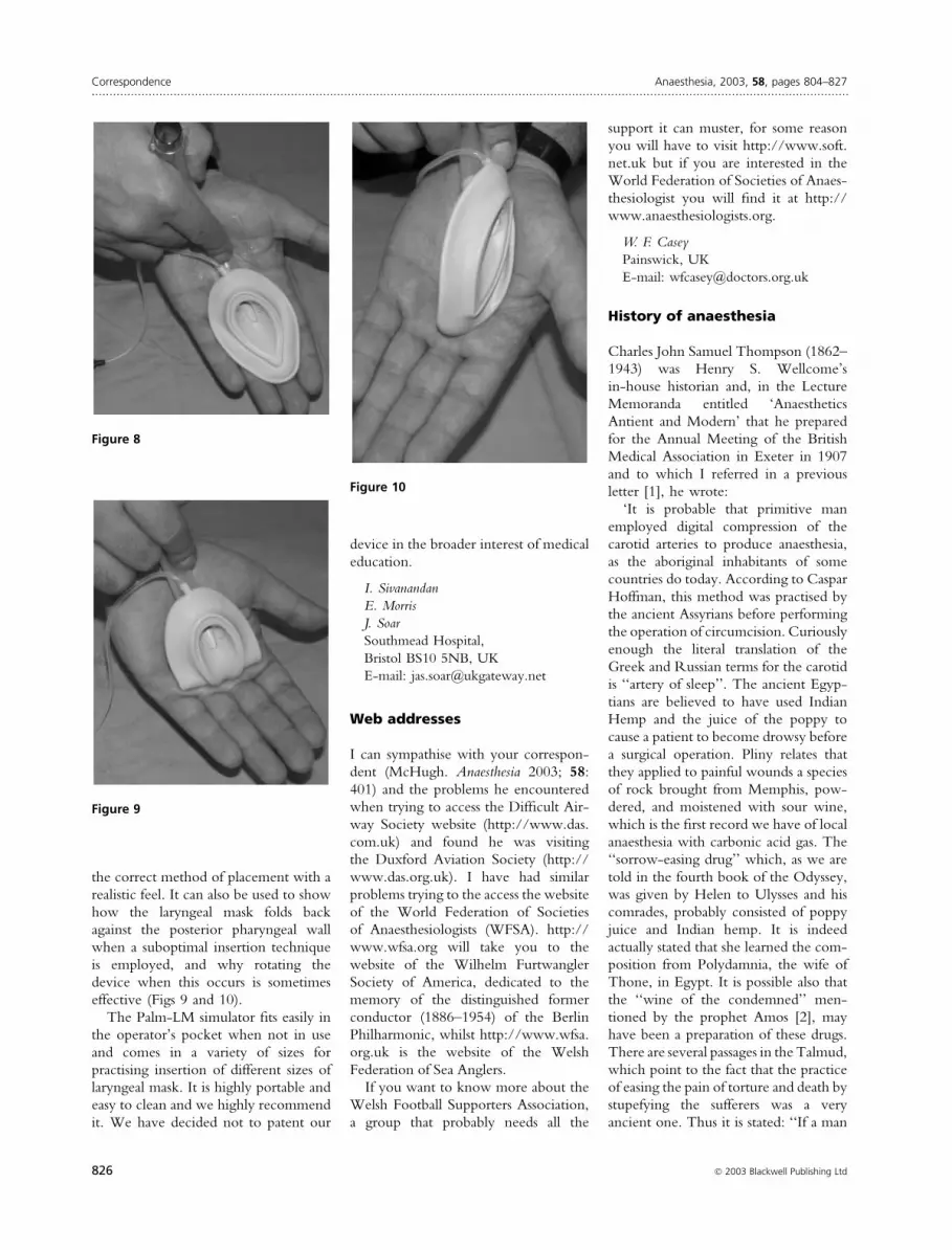
the correct method of placement with a
realistic feel. It can also be used to show
how the laryngeal mask folds back
against the posterior pharyngeal wall
when a suboptimal insertion technique
is employed, and why rotating the
device when this occurs is sometimes
effective (Figs 9 and 10).
The Palm-LM simulator fits easily in
the operator’s pocket when not in use
and comes in a variety of sizes for
practising insertion of different sizes of
laryngeal mask. It is highly portable and
easy to clean and we highly recommend
it. We have decided not to patent our
device in the broader interest of medical
education.
I. Sivanandan
E. Morris
J. Soar
Southmead Hospital,
Bristol BS10 5NB, UK
E-mail: [email protected]
Web addresses
I can sympathise with your correspon-
dent (McHugh. Anaesthesia 2003; 58:
401) and the problems he encountered
when trying to access the Difficult Air-
way Society website (http://www.das.
com.uk) and found he was visiting
the Duxford Aviation Society (http://
www.das.org.uk). I have had similar
problems trying to the access the website
of the World Federation of Societies
of Anaesthesiologists (WFSA). http://
www.wfsa.org will take you to the
website of the Wilhelm Furtwangler
Society of America, dedicated to the
memory of the distinguished former
conductor (1886–1954) of the Berlin
Philharmonic, whilst http://www.wfsa.
org.uk is the website of the Welsh
Federation of Sea Anglers.
If you want to know more about the
Welsh Football Supporters Association,
a group that probably needs all the
support it can muster, for some reason
you will have to visit http://www.soft.
net.uk but if you are interested in the
World Federation of Societies of Anaes-
thesiologist you will find it at http://
www.anaesthesiologists.org.
W. F. Casey
Painswick, UK
E-mail: [email protected]
History of anaesthesia
Charles John Samuel Thompson (1862–
1943) was Henry S. Wellcome’s
in-house historian and, in the Lecture
Memoranda entitled ‘Anaesthetics
Antient and Modern’ that he prepared
for the Annual Meeting of the British
Medical Association in Exeter in 1907
and to which I referred in a previous
letter [1], he wrote:
‘It is probable that primitive man
employed digital compression of the
carotid arteries to produce anaesthesia,
as the aboriginal inhabitants of some
countries do today. According to Caspar
Hoffman, this method was practised by
the ancient Assyrians before performing
the operation of circumcision. Curiously
enough the literal translation of the
Greek and Russian terms for the carotid
is ‘‘artery of sleep’’. The ancient Egyp-
tians are believed to have used Indian
Hemp and the juice of the poppy to
cause a patient to become drowsy before
a surgical operation. Pliny relates that
they applied to painful wounds a species
of rock brought from Memphis, pow-
dered, and moistened with sour wine,
which is the first record we have of local
anaesthesia with carbonic acid gas. The
‘‘sorrow-easing drug’’ which, as we are
told in the fourth book of the Odyssey,
was given by Helen to Ulysses and his
comrades, probably consisted of poppy
juice and Indian hemp. It is indeed
actually stated that she learned the com-
position from Polydamnia, the wife of
Thone, in Egypt. It is possible also that
the ‘‘wine of the condemned’’ men-
tioned by the prophet Amos [2], may
have been a preparation of these drugs.
There are several passages in the Talmud,
which point to the fact that the practice
of easing the pain of torture and death by
stupefying the sufferers was a very
ancient one. Thus it is stated: ‘‘If a man
Figure 10
Figure 8
Figure 9
Correspondence Anaesthesia, 2003, 58, pages 804–827......................................................................................................................................................................................................................
826 � 2003 Blackwell Publishing Ltd

is led forth to death, he is given a cup of
spiced wine to drink, whereby his soul is
wrapped in night’’; and again, ‘‘Give a
stupefying drink to him that loseth his
life, and wine to those that carry bitter-
ness in their heart. In connexion with
crucifixion, which was a common pun-
ishment for malefactors among the Jews
before the Christian era, with the sanc-
tion of the Sanhedrin, the women were
wont to ease the terrible death agony of
the sufferers by giving them some-
thing in the nature of a ‘‘wine of the
condemned’’ upon a sponge. It is prob-
able that the ‘‘wine mingled with
myrrh’’ which, according to St Mark
[3], was offered to Christ before nailing
Him upon the Cross, was indeed a
narcotic draught, given with the object
of lessening His sensibility to the agony.’
J. Zorab
Bristol, UK
E-mail: [email protected]
References1 Zorab J. History of anaesthesia.
Anaesthesia 2002; 57: 1472.
2 Holy Bible, Old Testament, Amos,
chapter 3, verse 8.
3 Holy Bible, New Testament, Gospel
According to St Mark, chapter 15,
verse 23.
Anaesthesia, 2003, 58, pages 804–827 Correspondence......................................................................................................................................................................................................................
� 2003 Blackwell Publishing Ltd 827