fractionation of mammalian liver cells by differential centrifugation
TRANSCRIPT
FRACTIONATION OF MAMMALIAN LIVER CELLS BY DIFFERENTIAL CENTRIFUGATION
II. EXl~rm~NTAL P~OCZDImES AND RESOr~TS
BY ALBERT CLAUDE, M.D.
(From the l_,aboralories of The Rockefeller Imtitute for Medical Reaearch)
(Received for publication, April 25, 1946)
The physiology of the cell cannot be fully understood unless we succeed in determining the constitution of its parts, and the relation which undoubtedly exists between its morphology and the distribution of its biochemical functions.
Previous work (1-4) has shown that a number of morphological constituents of the cell can be isolated and prepared in relatively large quantities, so that certain aspects of their composition and activities can be investigated directly by current chemical and biochemical methods. In the present work liver suspensions, representing the cytoplasmic content of the hepatic cells (5), have been fractionated into three main portions, morphologically distinct, by means of differential centrifugation: (1) a large granule fraction, essentially composed of mitochondria and secretory granules; (2) a microsorae fraction composed of particulate elements of submicroscopic size; and (3) a supernate fraction con- taining the elements of relatively small size which remained in the extract after the two first fractions had been removed. The chemical composition of the constituents of these fractions has been investigated, and a preliminary account of the distribution of certain enzymatic activities among the three different fractions has been published (6).
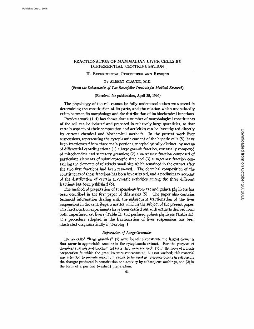
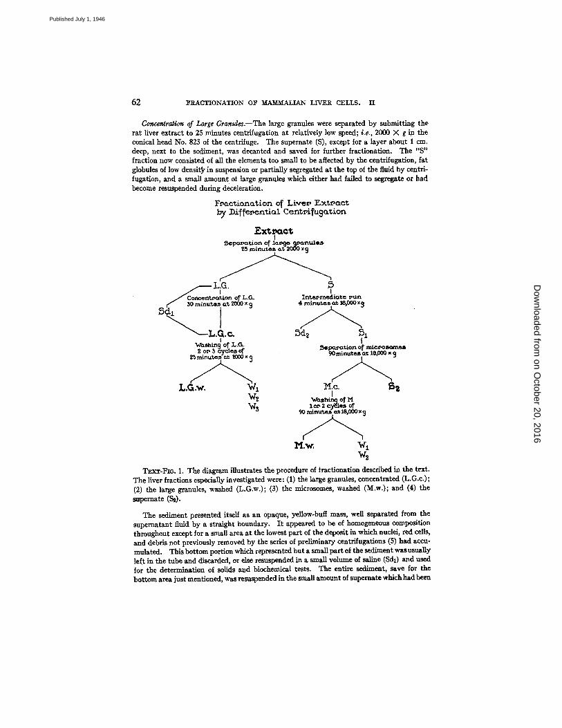
The method of preparation of suspensions from rat and guinea pig livers has been ddscribed in the first paper of this series (5). The paper also contains technical information dealing with the subsequent fractionation of the liver suspensions in the centrifuge, a matter which is the subject of the present paper. The fractionation experiments have been carried out with extracts derived from both unperfused rat livers (Table I), and perfused guinea pig livers (Table II). The procedure adopted in the fractionation of liver suspensions has been illustrated diagramatically in Text-fig. 1.
Separation of Large Granules
The So called 'qarge granules" (5) were found to constitute the largest elements that occur in appreciable amount in the cytoplasmic extract. For the purpose of chemical analysis and biochemical tests they were secured: (i) in the form of a crude preparation in which the granules were concentrated, but not washed; this material was intended to provide maximum values to be used as reference points in estimating the changes produced in constitution and activity by subsequent washings, and (2) in the form of a purified (washed) preparation.
61
on October 20, 2016
Dow
nloaded from
Published July 1, 1946
62 ]~RACTIONATION O]~ MAMMALIAN LIVER CELLS. II
Concentrat~ra of Large Granules.--The large granules were separated by submitting the rat liver extract to 25 minutes centrifugation at relatively low speed; i.e., 2000 × g in the conical head No. 823 of the centrifuge. The supemate (S), except for a layer about 1 era. deep, next to the sediment, was decanted and saved for further fractionation. The "S" fraction now consisted of all the elements too small to be affected by the centrifugation, fat globules of low densit~ in suspension or partially segregated at the top of the fluid by centri- fugation, and a small amount of large granules which either had failed to segregate or had become resuspended during deceleration.
F~actionation o~ Live~ ]Sxt~act by ])i~fepential Cent~i~ug~z~on
F - x t ~ c t I
25 s'ninute~, o.t 2000 ~
- . L.G. 5 I
S n c e n ~ o n of L,G. I
Inl~e~rnectiate ~,un -- ~ ~0 m~nute~ a~ 2000 x 9 4 ~
L..c ~dz 51 i !
~a~h~n 9 of L.G. ~u~tion of mlc~oB~n~ 2 o~ 5 cycle~ o~ 90mlnuteB a'c 18,000,
L.G w. Wx He ~ ~hin~ o T M
W~ I o~ 2 ~*,. of
l~I.w, wl w~
TExT-FIo. 1. The diagram illustrates the procedure of iractionation described in the text. The liver fractions especially investigated were: (1) the large granules, concentrated (L.G.c.); (2) the large granules, washed (L.G.w.); (3) the microsomes, washed (M.w.); and (4) the supemate (S~).
The sediment presented itself as an opaque, yellow-buff mass, well separated from the supernatant fluid by a straight boundary. I t appeared to be of homogeneous composition throughout except for a small area at the lowest part of the deposit in which nuclei, red cells, and debris not previously removed by the series of preliminary centrifugations (S) had accu- mulated. This bottom portion which represented but a small part of the sediment was usually left in the tube and discarded, or else resuspended in a small volume of saline (Sd,) and used for the determination of solids and biochemical tests. The entire sediment, save for the bottom area just mentioned, was resuspended in the small amount of supemate whichhad been
on October 20, 2016
Dow
nloaded from
Published July 1, 1946
ALBERT CLAUDE 63
left in the tube for that purpose and the material derived from the volume of liver extract employed, usually 250 to 350 cc., was combined into a single tube; no new solvent was added at this time: in this manner, the sedimented material was concentrated into a volume one- eighth to one-tenth that of the original extract.
The concentrated suspension was centrifuged again for 30 minutes at 2000 )< g. The purpose of this repetition in the concentration of the fraction was to obtain the material in a compact form and to permit complete removal of the supematant fluid without losing at the same time an appreciable amount of resuspended granules. The supernate was withdrawn by suction through a capillary pipette brought progressively as near as possible tothe surface of the deposit; it was then added to the supernate from the first centrifugation (S), or was discarded.
The voluminous, apparently homogeneous, sediment was taken up in alkaline saline, enough solvent being added to bring the total volume of the suspension to 1 : 12 that of the original liver extract; habitually this volume amounted to 20 or 25 cc. Again a small disc of substance composed of aggregated debris and a small number of nuclei was left in the tube and combined with the similar material derived from the first centrifugation (Sdl).
The main preparation, resuspended in saline, will be referred to as the "large granule concentrate" (L.G.c.). This fraction can be shown to contain most of the large granules originally present in the liver extract; in addition it contained in small amounts the other elements of the tx t rac t which had been carried along with, or had remained occluded between, the granules in the sediment.
A portion (5 to 10 cc.) of the large granule concentrate was set aside for dry weight determination and chemical tests. The dry weight, depending largely on the volume chosen for the fraction (25 cc. in the individual experiment), was 41.7 rag. per cc., giving a weight of 1.043 gm. for the total amount of substance separated by the procedure. From the data of the experiment illustrated in Table I it can be calculated that the liver extract contained, in terms of dry weights, a t least 3.3 nag. large granules (unwashed) per cc., or 12.2 per cent of its total solids. This large granule content of liver extract was found to vary, in twelve different experiments, from 2.4 to 3.9 rag. (average value, 3.3 rag.) per cc. representing from 9.3 to 13.5 (average value, 11.8) per cent of the total solids.
The remaining portion of the large granule concentrate was used for further purifi- cation and washing.
Washing of the Large Granules.--The volume of the large granule concentrate was increased from 15 to 20 cc., to 35 to 45 cc. (about 1:5 the volume of the original extract) by the addition of alkaline saline. The suspension was submitted to 30 minutes centrifugation at 2000 × g: the supernate (W1) was saved, and the entire sediment was redispersed in the same volume (35 to 45 co.) of saline. This cycle, consisting in 30 minute centrifugation followed by resus- pension of the sediment, was repeated two or three times. The deposit from the last centri- fugation was taken up in saline to a total volume equal to 1:12 that of the original extract: this preparation will be referred to as the "washed large granule fraction" (L.G.w.). At each centrifugation the entire sediment was resuspended so that any loss of substance or of activity which might be detected could be attributed to the washing of the granules, and not to the gross removal of sedimented material. Nevertheless a small amount of large granule material becomes resuspended during deceleration of the centrifuge; because of this the wash waters were usually recentrifuged and the small deposit returned, but not always, to the main large granule fraction.
on October 20, 2016
Dow
nloaded from
Published July 1, 1946
64 FRACTIONATION OF M.AM'IWALIAN LIVER CELLS. I1
The dry weight of the large granule preparation (washed), in the experiment illustrated in Table I, was 34.7 mg. per cc. From the data presented in Table I it can be calculated that the amount of large granules recovered after washing was, in terms of dry weights, 2.8 mg. per cc. of original extract, representing 10.3 per cent of its total solids. In different experiments the yield of large granules varied from 1.8 to 3.1 mg. per cc. of liver extract originally used, or 7.0 to 10.7 per cent of its total solids. This variation was due mostly to occasional deviations from the standard procedure and to a greater loss of granules during purification.
TABLE I
Fractionation of Mammalian Liver by Differential Centrifugation Solid Content and Proportion of Various Fractions in Rat Liver Extract
E S $1
S~ L.G.c. L.G.w. M.C. M.w. Sdl
Sd2
Wl W2
Fraction
Extract . . . . . . . . . . . . . . . . . . Large granules removed... Intermediate sediment (Sd~)
removed . . . . . . . . . . . . . . . . Microsomes removed . . . . . . Large granules concentrate. Large granules, washed ... . Microsomes, concentrate... Microsomes, washed . . . . . . . Cell ~debris removed from
L.G.c . . . . . . . . . . . . . . . . . . . [ 315 Intermediate sediment (L.G. 1
I
and M., mixed) . . . . . . . . . . I 252 First washings from L.G.c...I 189 Second washings from
L.G.c . . . . . . . . . . . . . . . . . . . 189
Volume Volume Dry of o~ original~ of. weight
__extract iractlon__ fractioz
CC. I CG. • ~er ¢~.
27.1 23;5
21.7 17.3
315 25 41.7 189 15 34.7 168 ' 15 48.2 140 10
5
18 35
35
23.5 1
34.7 [ 48.2 l 38.8
12.2
i 23.6 2.3
0.4
?otal I weightdry i fraction in
[ Amount of of [ extract
fraction
rag. rag. per ce~g
521 ~" ....
27.1 100.0 23.5 86.7
21.7 80.1 17.3 6318
043 3.3 12.2 2.8 10.3 f
723 4.3 15.8
Propor- tion of
liver pulp in extract
)er ¢¢n/~
47.5
388
61
425 81
14
2.8 10.2
0.2 0.7
1.7 6.3 0.4 1.5
0.1 0.4 i
The large granules of liver are metabolically active, and as already stated, the saline suspension is apt to become increasingly acid (5), the effect being more pro- nounced if in spite of washing some glycogen remains present. I t has not been estab- lished as yet whether the large granules themselves may produce and store glycogen, or whether they may become permeated by it during extraction. Because of this rela- tively abundant release of acid the large granule preparations were tested at intervals for possible changes in reaction and alkali was used to keep the solution neutral whenever necessary.
Separalion of Large Granules from Perfused Guinea Pig Liver.--Large granules were separated from perfused guinea pig liver, according to the procedure followed in the case of rat liver. The amount of large granule concentrate recovered from guinea
on October 20, 2016
Dow
nloaded from
Published July 1, 1946
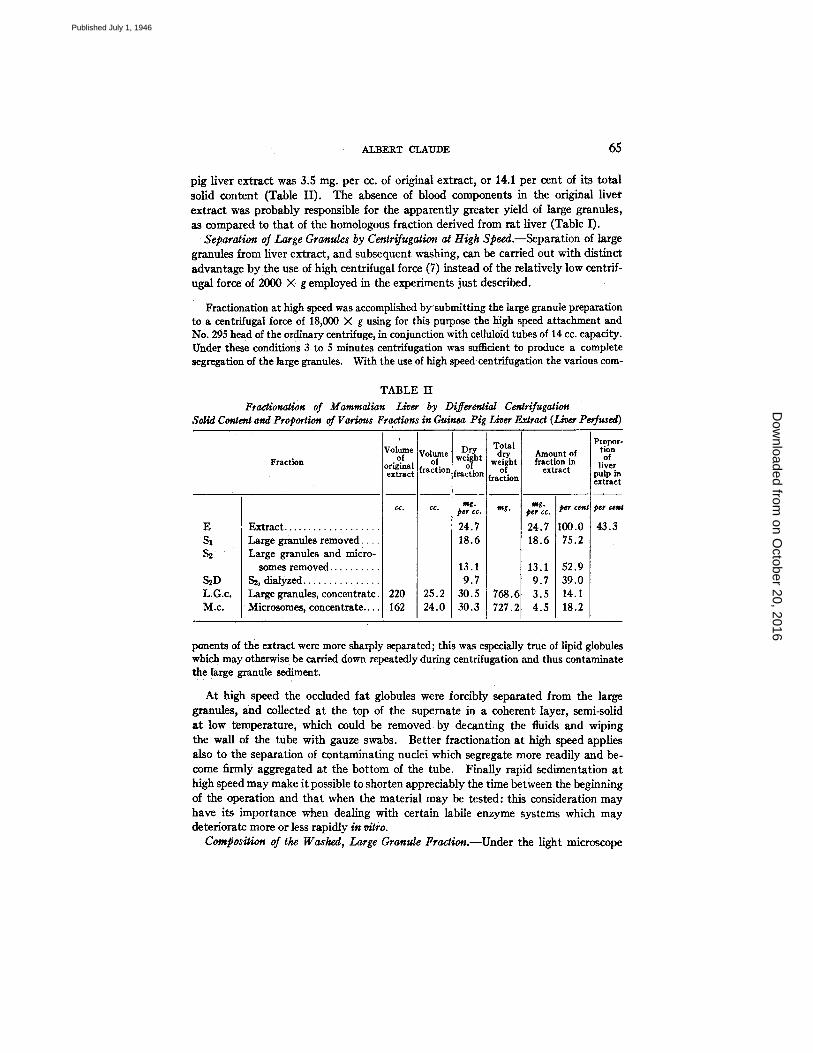
ALBEI~.T CLAUDE 6 5
pig liver extract was 3.5 nag. per cc, of original extract, or 14.1 per cent of its total solid content (Table I1). The absence of blood components in the original liver extract was probably responsible for the apparently greater yield of large granules, as compared to that of the homologous fraction derived from rat liver (Table 1).
Separation of Large Granules by Centrifugation at High Speed.--Separation of large granules from liver extract, and subsequent washing, can be carried out with distinct advantage by the use of high centrifugal force (7) instead of the relatively low centrif- ugal force of 2000 X g employed in the experiments just described.
Fractionation at high speed was accomplished by'submitting the large granule preparation to a centrifugal force of 18,000 X g using for this purpose the high speed attachment and No. 295 head of the ordinary centrifuge, in conjunction with celluloid tubes of 14 cc. capacity. Under these conditions 3 to 5 minutes centrifugatinn was sufficient to produce a complete segregation of the large granules. With the use of high speed centrifugation the various com-
TABLE II Fractionation of Mammalian Liver by Differential Cenlrifugation
Solid Content and Proportion of Various Fractions in Guinea Pig Liver Extract (Liver Pe fused)
Fraction
E Extract... S t
S2
S2D L.G.c. M . c ,
Large granules removed Large granules and micro-
somes removed. S~, dialyzed... Large granules, concentrate Microsomes, concentrate...
Volume of
original extract
• 220 • 162
Volume of
fractio~
25.2 24.0
Dry weight
of [faction
mg. peg CC.
24.7 18.6
13.1 9.7
30.5 30.3
Total dry
fract ion
mg.
768.~ 727.2
Propor- tion Amount of of
fraction in liver extract pulp in
extract
mr. per cent per cesj per co.
24.7 100.0 43.3 18.6 75.2
13.1 52.9 9.7 39.0 3.5 14.1 4.5 18.2
ponents of the extract were more sharply separated; this was especially true of lipid globules which may otherwise be carried down repeatedly during centrifugation and thus contaminate the large granule sediment.
At high speed the occluded fat globules were forcibly separated from the large granules, and collected at the top of the supemate in a coherent layer, semi-solid at low temperature, which could be removed by decanting the fluids and wiping the wall of the tube with gauze swabs. Better fractionation at high speed applies also to the separation of contaminating nuclei which segregate more readily and be- come firmly aggregated at the bottom of the tube. Finally rapid sedimentation at high speed may make it possible to shorten appreciably the time between the beginning of the operation and that when the material may be tested: this consideration may have its importance when dealing with certain labile enzyme systems which may deteriorate more or less rapidly in vitro.
Composition of the Washed, Large Granule Fraction.--Under the light microscope
on October 20, 2016
Dow
nloaded from
Published July 1, 1946
]~RACTIONATION 0]~ ~MM~LIAN LIVER CELLS. II
the large granule fraction prepared by the methods just described appears quite homogeneous, and to be composed almost exclusively of granules which may vary in size from 0.5 to 1 or 2 g in diameter. Elements of smaller size and similar density were not appreciably concentrated and were left in the supernate or discarded during washing. Microscopic examination showed free nuclei and red cells to be practically absent.
The elements which compose the washed granule fraction correspond to those referred to in preceding papers (7, 2) under the term "secretory granules" and by Bensley and Hoerr (8, 9) as mitochondria. In the living hepatic cell of normally fed animals (rats and guinea pigs) the granules are distributed mostly at the periphery of the cell but, in fasted animals, they seem to fill the cytoplasmic region completely. When the membrane of the liver cells is broken, these granules are released and dispersed into the surrounding medium where they appear to round up and persist morphologically intact.
In saline suspension, as in the sedimented condition, the large granules form opaque preparations of characteristic yellow-huff, somewhat greenish color. I t can be noted here that the yellow-buff color characteristic of a freshly perfused liver no longer colored by capillary blood is precisely that of the large cytoplasmic granules, now visible through the thin membrane of the hepatic cell.
The fact has already been mentioned that the large granule fraction of liver consists of at least two types of cytoplasmic elements; i.e., mitochondria and secretory granules. I t is conceivable that secretory granules, otherwise similar in size and appearance, may represent a variety of elements, each possessing differentiated composition and functions. In the first step of fractionation in the centrifuge, notably when dealing with guinea pig liver, the large granule sediment frequently presents two broad zones of slightly different color: one yellow, the other exhibiting a definite green tinge. I t is possible that a more refined technique might separate further specialized elements, each associated with definite cellular functions, for example the production of bile pigments, or the synthesis of glycogen.
Chemical Constitution of the Large Granules.--The large granules are complex structures composed largely of proteins and about 25 per cent lipids. Results of elementary analysis have appeared in preceding papers (7, 2, 4). The relatively low nitrogen content, 10 to 12 per cent depending on the mode of preparation, is the re- flection, in part, of the presence of lipids in the material. The phosphorus content, 0.9 to 1.3 per cent of the granules, is conditioned in part by the presence of phospho- lipids which constitute about two-thirds of the total lipids, and by the presence of appreciable amounts of ribose nueleotides. The possible significance of the presence of ribose nucleic'acid in cytoplasmic granules has been emphasized in preceding papers (2, 4). The sulfur content is relatively high, 0.82 to 1.16 per cent in large granules of guinea pig and rat liver; it is probable that part of this sulfur is in the form of sulfhydryl groups related to the active metabolism which the large granules have been found to possess (6). The large granule preparations gave a positive nitroprusside reaction characteristic of SH groups. The large granules were found to contain from 0.02 to 0.04 per cent iron, or roughly one-tenth the amount found in hemoglobin. I t can be assumed that part of this iron contributes to the cytochrome compounds detected in the granules by other means (6, 10). Copper is a relativelyabundant
on October 20, 2016
Dow
nloaded from
Published July 1, 1946
ALBERT CLAUDE 67
constituent, representing from 0.02 to 0.035 per cent of the granules; this quantity of copper corresponds to about 12 to 20 per cent of the amount known to be present in the respiratory pigment hemocyanin of Limulus (7). Inositol was found to occur in the large granules to the extent of about 0.5 per cent of the dry weight (2 b). I/ it is assumed that the entire inositol content exists in the granules in the form of lipositol (11), it can be calculated that the latter substance would account for as much as 12 per cent of the total lipids of the material. 1
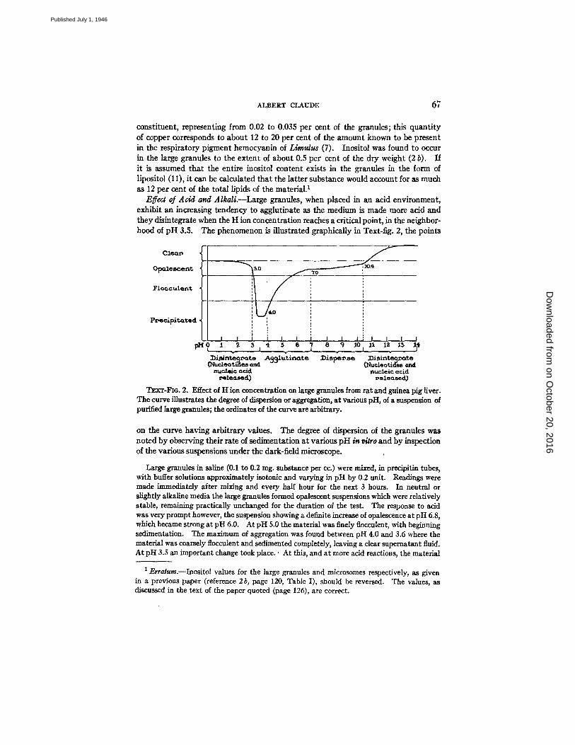
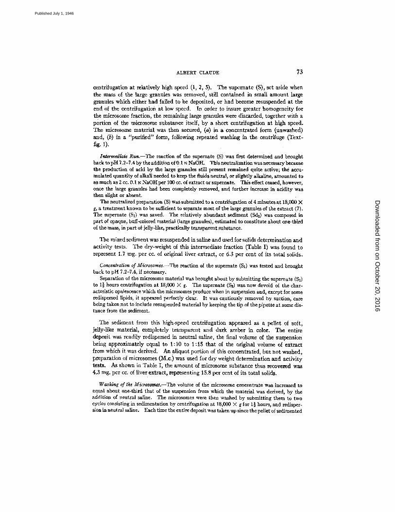
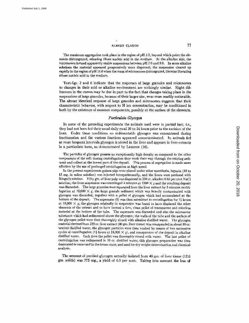
Effect of Acid and Alkali.--Large granules, when placed in an acid environment, exhibit an increasing tendency to agglutinate as the medium is made more acid and they disintegrate when the H ion concentration reaches a critical point, in the neighbor- hood of pH 3.5. The phenomenon is illustrated graphically in Text-fig. 2, the points
C 1 ~ f
FlocculerLt i 4.~0 ~ ',
P~ecAp~t~ed
~o I 2 3~ 5 s ~ 8 9 ~o ~I 12 l~ I~
: m S n t ~ t . A391Jtinate ~i,~e : m , i n t ~ m t e (~ucleoti'ae8 and ~Nucleoti(:[es and
n u c l e i c G¢~. ,'1 DUCIe~c (Icid ~,elealsec[) neleo.sed)
TEXT-FIG. 2. Effect of t t ion concentration on large granules from rat and ~ u e a pig liver. The curve illustrates the degree of dispersion or aggregation, at various pH, of a suspension of purified large granules; the ordinates of the curve are arbitrary.
on the curve having arbitrary values. The degree of dispersion of the granules was noted by observing their rate of sedimentation at various pH in r/~ro and by inspection of the various suspensions under the dark-field microscope.
Large granules in saline (0.1 to 0.2 rag. substance per cc.) were mixed, in precipitin tubes, with buffer solutions approximately isotonic and varying in pH by 0.2 unit. Readings were made immediately after mixing and every half hour for the next 3 hours. In neutral or slightly alkaline media the large granules formed opalescent suspensions which were relatively stable, remaining practically unchanged for the duration of the test. The response to acid was very prompt however, the suspension showing a definite increase of opalescence at pH 6.8, which became strong at pH 6.0. At pH 5.0 the material was finely flocculent, with beginning sedimentation. The maximum of aggregation was found between pH 4.0 and 3.6 where the material was coarsely flocculeut and sedimented completely, leaving a clear supematant fluid. At pH 3.S an important change took place.' At this, and at more acid reactions, the material
1 Erratum.--Inositol values for the large granules and microsomes respectively, as given in a previous paper (reference 2b, page 120, Table I), should be reversed. The values, as discussed in the text of the paper quoted (page 126), are correct.
on October 20, 2016
Dow
nloaded from
Published July 1, 1946
68 ~?RACTIONATION OF M.~M'MALIAN LIVER CELLS. II
ceased to aggregate and returned rapidly to a dispersed condition. At pH 3.0 the dispersion of the material was already complete, and the opalescence of the suspension was less than that shown at pH 7.0. On the alkaline side the opalescence of the suspensions remained apparently unchanged between pH 7.0 and 8.0. Between pH 8.0 and 10.0 there was a very slight but progressive decrease in the opalescence of the preparations. In the region of pH 10.4 there was a sudden clearing, the decrease in opalescence being thereafter rapid. At pH 13.0 (cor- responding approximately to a 0.1 N NaOH solution), the preparation was water-clear.
Examination of the suspensions under the dark-field microscope showed the granules, at pH 7.0 to be freely dispersed in the medium, with no tendency to adhere to each other; the individual granules presented Brownian movement of small amplitude. At pH 6.0 the gran- ules appeared agglutinated in small clusters; between these clusters, free granules or aggregates of 3 or 4 elements could still be iound. At pH 5.0 the preparation showed large masses of agglutinated material, while small aggregates and free granules were still present. At pH 4.0 all the granules in suspension appeared to have coalesced into large lumps, leaving the inter- vening fluid optically empty. At pH 3.0 a few free globules were again present, together with innumerable small particles, similar in size to the microsomes described in another part of the paper, and exhibiting active Brownlan motion. At pH 2.0 the field was occupied almost exclusively by small particles, possibly no more than 0.1 micron in diameter, and showing active Brownian movement. In alkaline media, between pH 7.0 and 10.0, the granules ap- peared freely dispersed and apparently unchanged. In the region ot pH 10.4 they rapidly disintegrated into smaller units, and at pH 13.0 the preparation was optically empty.
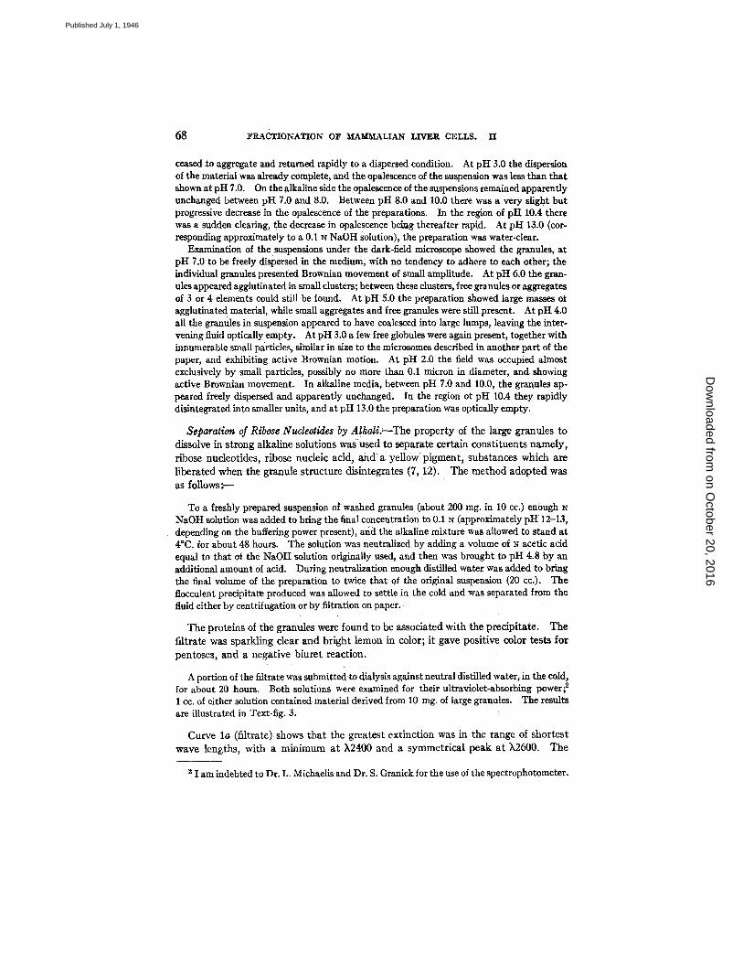
Separation of Ribose Nucleotides by AlkaIi.--The property of the large granules to dissolve in strong alkaline solutions was used to separate certain constituents namely, ribose nudeofides, ribose nucleic acid, and a yellow pigment~ substances which are liberated when the granule structure disintegrates (7, 12). The method adopted was as fol lows:--
To a freshly prepared suspension of washed granules (about 200 rag. in 10 co.) enough N NaOH solution was added to bring the final concentration to 0.1 N (approximately pH 12-13, depending on the buffeting power present); and the alkaline mixture was allowed to stand at 4°C. for about 48 hours. The solution was neutralized by adding a volume of N acetic acid equal to that oi the NaOH solution originally used, and then was brought to pH 4.8 by an additional amount of acid. During neutralization enough distilled water was added to bring the final volume of the preparation to twice that of the original suspension (20 cc.). The flocculent precipitate produced was aUowed to settle in the cold and was separated from the fluid either by centrffugation or by filtration on paper.
The proteins of the granules were found to be associated with the precipitate. The filtrate was sparkling clear and bright lemon in color; it gave positive color tests for pentoses, and a negative biuret reaction.
A portion of the filtrate was submitted to dialysis against neutral distilled water, in the cold, for about 20 hours. Both solutions were examined for their ultraviolet-absorbing power; ~ 1 cc. of either solution contained material derived from 10 rag. of large granules. The remits are illustrated in Text-fig. 3.
Curve l a (filtrate) shows that the greatest extinction was in the range of shortest wave lengths, with a minimum at k2400 and a symmetrical peak a t k2600. The
2 1 am indebted to Dr. L. Michaelis and Dr. S. Granick for the use of the spectrophotometer.
on October 20, 2016
Dow
nloaded from
Published July 1, 1946
~BERT CZ.A~rI~E 69
aspect of the absorption in this region suggests that ribose nucleotides were probably present. Absorption bands of lesser intensities are also indicated in the region of longer wave lengths but their significance can only be surmised at the moment (curve 1 b). Dialysis removed as much as 61 per cent of the absorbing power of the solution, at ),2600 (curve 2, a and b). A characteristic peak of absorption in the region of )`2600 w a s retained after dialysis but absorption at longer wave lengths was simplified and the secondary bands were for the most part abolished. I t can be
8.0 ' 0.8
'I.0 0.'I
6.0 , t~ ~ s _ 0.6 50 05
4.0 0.4
5.0 0.5
2,0 0.2
1.0 .0.1
3000 38oo 50o0 54o0
TExT-FIO. 3. Separation of ribose nucleotides and ribose nucleic acid from liver large granules (mostly mitochondria) by successive treatment at pH 13.0 and pH 4.7. The figure illustrates the light-absorbing power of the neutralized pH 4.7 filtrate before (curve lg), and after (curve 2a), dialysis. The coeflidents • represent the extinction produced by a filtrate, each cubic centimeter of which contained material originally derived from l0 rag. purified large granules. The parts of the curves between ~,2900 and X5400 were plotted again (curves lb and 2b) against the enlarged scale shown at the right-hand side of the graph.
tentatively concluded that ribose nucleotides of various types, and of relatively low molecular weight, were responsible for about 60 per cent of the absorbing power of the solution, a t )`2600, whereas the non-dialyzable absorbing portion was represented by nucleotides of simpler structure but of relatively high molecular weight, possibly in the form of ribose nucleic acid. The dialyzed solution was practically colorless indicating that in this case the yellow pigment (possibly riboflavin derivatives) was dialyzable. The fact that the pH 4.8 precipitate still gave strongly positive reactions for pentoses suggests that appreciable amounts of ribose nucleic acid, or ribose nucleo- tides may have remained attached, or recombined, with the protein moiety.
on October 20, 2016
Dow
nloaded from
Published July 1, 1946
7O FRACTIONATION OF MAMMALIAN LIVER CELLS. II
The view that the large granules contain both ribose nucleic acid and ribose nucleo- tides of relatively low molecular weight is supported by the results of a study of the products of disintegration of the large granules in distilled water, recorded in the paragraph which now follows.
Effect of Distilled Water on the Large Granules.--As shown in previous experiments (7, 2) distilled water can be used as medium for extracting and washing the large granules from liver, provided that the volume of solvent is kept relatively small, and that the time of contact between the granules and distilled water is not unduly prolonged. Under these conditions no appreciable difference was found in the ele- mentary composition of the granules prepared by means of either distilled water or isotonic saline. In hypotonic solutions, the granules absorb water with a resulting decrease in density, as shown by a lower rate of sedimentation in the centrifuge. Under dark-field illumination, the granules were observed to swell and increase in size enormously as the salt concentration was being decreased, reaching 3 and even 6 ~ or more in diameter until their, at first, bright outline became barely discernible, so that they resembled the ghosts of red cells. The fact that the large granules of liver take up water and increase proportionally in size when placed in a hypotonic environment suggests that these elements may constitute small osmotic systems by virtue of the presence at their surface of a semipermeable mem- brane. That such a membrane may exist is indicated by recent studies of mitochondria in the electron microscope (13, 14).
Large granules were observed to break up spontaneously into units of smaller size when suspensions were stored at 4°C. This process of disintegration was accelerated in hypotonic solutions, and it took place immediately on reducing the salt concentra- tion of the medium below a critical point either by repeated washing of the granules in distilled water or by dispersing the elements at once into a sufficiently large volume of distilled water. Following disintegration in distilled water, the large granules leave only a small residue sedimentable under the usual centrifugal force of 2000 X g; on the other hand, two fractions can be obtained from the water suspension: (a) an insoluble component composed of submicroscopic units, sedimentable at 18000 X g and resembling the microsomes to be described in another part of this paper, and, (b) soluble components, including nudeotides and a yellow pigment previouMy encountered in the case of dissolution of large granules by alkali. The process of disintegration of the large granules in distilled water was investigated in the following experiment.
Guinea pig livers were extracted by means of alkaline saline, and the large granules were separated from the liver extract in the usual manner. The large granules were then washed in saline through four successive cycles of centriiugation; the first three centrifugations were of 30 minutes at 2000 X g, the fourth of 7 minutes at 18,000 X g. Through centrifugation at high speed the last sediment was firmly packed and most of the salt solution was thus dis- carded. The washed large granules were then dispersed in a relatively large volume of ~lk~tline distilled water, the final suspension containing 10.7 mg. large granule material per co., and the suspension was stored at 2°C. for 48 hours. At the end of this period the water suspension was submitted to 30 minutes centrifugation at 2000 X g and the unbroken granules and insoluble debris, amounting to about 4 rag. per cc., were discarded.
The supemate was saved and two fractions were obtained from it, namely, (a) a particulate component, which will be referred to under the term "large granule
on October 20, 2016
Dow
nloaded from
Published July 1, 1946
ALBERT CLAUDE 71
microsomes," and (b) the "water supernate" containing granule constituents of relatively low molecular weights, and readily soluble in water.
Separation of the microsome-like component was accomplished by 1½ hours centrifugation at 18,000 X g. An additional centrifugation of 1 hour at 18,000 X g yielded no further sedi- ment indicating that practically all the microsome substance had been removed by the first run of 1½ hours at high speed.
The sedimented material was in the form of a completely transparent pellet, and amber-brown in color; in amount, it corresponded to 2.4 rag. of the original water suspension, or about 22 per cent of the large granule mass. On chemical analysis the large granule microsome fraction was found to contain 8.8 per cent nitrogen and 1.5 per cent phosphorus.
The remaining fluid or water supernate was clear and golden yellow in color; its solid content was 4.3 mg. per cc., a value representing 40 per cent of the dry weight of the large granules.
A portion of the water supernate was subjected to dialysis against distilled water, at 4 ° C., for 20 hours. Much of the color was lost during dialysis but the solution was still distinctly yellow. The dry weight of the dialyzed preparation was 2.6 rag. per cc., indicating that about 40 per cent of the solids had been removed.
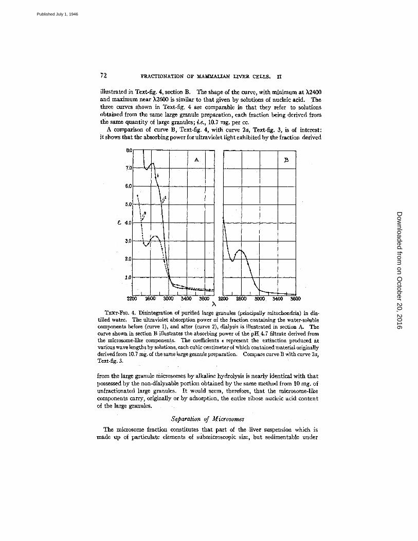
The water supernate was examined for its power to absorb ultraviolet light and the results are illustrated in Text-fig. 4 section A, curves 1 and 2 representing the absorbing power of the solution before and after dialysis, respectively. Absorption before dialysis (curve A-I) was relatively intense in the region of ~,2550 to 2650, indicating that nucleotides were probably present. Deflection of the curve at ~,2750 suggested the possible presence of proteins. Color tests for pentoses and proteins were positive. Values given in curve A-2 show that as much as 57 per cent of the absorbing power at ~,2600 was removed by dialysis (against 40 per cent of the solids). Furthermore, the maximum was displaced towards the longer wave lengths, suggesting that proteins or possibly nucleoproteins were probably responsible for the aspect of curve A-2 in the k2650 to 2750 region.
The foregoing observations show that the effect of distilled water on the large granules is to release from these elements substances which, according to their size fall into three distinct groups: (1) a dialyzable fraction apparently composed of nucleotides of relatively low molecular weight ; (2) a non-dialyzable fraction, con- taining proteins and possibly nucleoproproteins; and (3) a particulate, microsome- like component, itself a complex of ribose nucleic acid and lipids. Under the con- ditions of the experiments these various constituents accounted for 25, 39, and 36 per cent, respectively, of the dry weight of the large granule extract.
The fact that the microsome-like elements from large granules contain nucleic acid was investigated in the following tests:
The microsome-like material, isolated from the large granules by means of distilled water and saved from the preceding experiment, was treated with alkali according to the method pre- viously described. The pH 13.0 mixture was stored at 4°C. for 48 hours, then acidified to pH 4.7 with acetic acid, and filtered on paper.
The filtrate was clear and colorless; it gave positive tests for pentoses, and a negative biuret reaction. The property of the neutralized filtrate to absorb ultraviolet light is
on October 20, 2016
Dow
nloaded from
Published July 1, 1946
72 ~I~ACTIONATION O~F M A ~ LIVER CELLS. II
illustrated in Text-fig. 4, section B. The shape of the curve, with minimum at k2400 and maximum near ),2600 is similar to that given by solutions of nucleic acid. The three curves shown in Text-fig. 4 are comparable in that they refer to solutions obtained from the same large granule preparation, each fraction being derived from the same quantity of large granules; i.e., 10.7 rag. per cc.
A comparison of curve B, Text-fig. 4, with curve 2a, Text-fig. 3, is of interest: it shows that the absorbing power for ultraviolet light exhibited by the fraction derived
8.0 ~, I A
7.C f ~ ~ 5.C
5.o
, r F_, 4.0
t
i ,-', t \ ~.J ,,
', f
2200 ~ 3000 5400 ~ g~00 2~00 ~000 5400 5BOO
TExx-FIo. 4. Disintegration of purified large granules (principally mitochondria) in di~- tilled water. The ultraviolet absorption power of the fraction containing the water-soluble components before (curve 1), and alter (curve 2), dialysis is illustrated in section A. The curve shown in section B illustrates the absorbing power of the pH 4.7 filtrate derived from the microsome-like components. The coefficients ~ represent the extinction produced at various wave lengths by solutions, each cubic centimeter of which contained material originally derived from 10.7 rag. of the same large granule preparation. Compare curve B with curve 2a, Text-fig. 3.
from the large granule microsomes by alkaline hydrolysis is nearly identical with that possessed by the non-dialyzable portion obtained by the same method from 10 mg. of unfractionated large granules. I t would seem, therefore, that the microsome-like components carry, originally or by adsorption, the entire ribose nucleic acid content of the large granules.
Separation of Microsomes The microsome fraction constitutes that part of the liver suspension which is
made up of particulate elements of submicroscopic size, but sedimentable under
on October 20, 2016
Dow
nloaded from
Published July 1, 1946
ALBERT CLAUDE 73
centrifugation at relatively high speed (1, 2, 5). The supernate (S), set aside when the mass of the large granules was removed, still contained in small amount large granules which either had failed to be deposited, or had become resuspended at the end of the eentriiugation at low speed. In order to insure greater homogeneity for the microsome fraction, the remaining large granules were discarded, together with a portion of the microsome substance itself, by a short centrlfugation at high speed. The microsome material was then secured, (a) in a concentrated form (unwashed) and, (b) in a "purified" form, following repeated washing in the centrifuge (Text- fig. 1).
I ~ e d / a t e Ru~.--The reaction of the supernate (S) was first determined and brought back to pH 7.2-7.4 by the addition of 0.1 N Na0H. This neutralization was necessary because the production of acid by the large granules still present remained quite active; the accu- mulated quantity of alkali needed to keep the fluids neutral, or slightly alkaline, amounted to as much as 2 cc. 0.1 N NaOI-I per 100 ee. of extract or supernate. This effect ceased, however, once the large granules had been completely removed, and further increase in acidity was then slight or absent.
The neutralized preparation (S) was submitted to a centrifugation of 4 minutes at 18,000 X g, a treatment known to be sufficient to separate most of the large granules of the extract (7). The supemate ($1) was saved. The relatively abundant sediment (Sd~) was composed in part of opaque, buff-colored material (large granules), estimated to constitute about one-third of the mass, in part of jelly-like, practically transparent substance.
The mixed sediment was resuspended in saline and used for solids determination and activity tests. The dry-weight of this intermediate fraction (Table I) was found to represent 1.7 rag. per cc. of original liver extract, or 6.3 per cent of its total sohds.
Concentration of Microsomes.--Tbe reaction of the supernate (S0 was tested and brought back to pH 7.2-7.4, if necessary.
Separation of the microsome material was brought about by submitting the supernate (S1) to 1½ hours centrifugation at 18,000 × g. The supernate ($2) was now devoid of the char- acteristic opalescence which the microsomes produce when in suspension and, except for some redispersed lipids, it appeared perfectly clear. It was cautiously removed by suction, care being taken not to include resuspended material by keeping the tip of the pipette at some dis- tance from the sediment.
The sediment from this high-speed centrifugation appeared as a pellet of soft, jeUy-like material, completely transparent and dark amber in color. The entire deposit was readily redispersed in neutral saline, the final volume of the suspension being approximately equal to 1:10 to 1:15 that of the original volume of extract from which it was derived. An aliquot portion of this concentrated, but not washed, preparation of microsomes (M.c.) was used for dry weight determination and activity tests. As shown in Table I, the amount of microsome substance thus recovered was 4.3 rag. per cc. of liver extract, representing 15.8 per cent of its total solids.
Washing of the Microsome, s.--The volume of the microsome concentrate was increased to equal about one-third that of the suspension from which the material was derived, by the addition of neutral saline. The microsomes were then washed by submitting them to two cycles consisting in sedimentation by centrifugation at 18,000 X g for 1½ hours, and redisper- sion in neutral saline. Each time the entire deposit was taken up since the pellet of sedimented
on October 20, 2016
Dow
nloaded from
Published July 1, 1946
74 FRACTIONATION OF MAMMALIAN LIVER CELLS. II
material appeared perfectly homogeneous and was composed throughout of a transparent jelly. The wash waters were usually discarded. The pellet from the last centrifugation was resuspended in a small volume of saline usually one-tenth to one-fifteenth that of the original extract. The resulting suspension constituted the washed microsome preparation (M.w.).
The amount of purified microsomes thus recovered was 2.8 mg. per cc. of original liver extract, representing 10.2 per cent of its total solids. The considerable loss o f material (about 36 per cen0 incurred during the process of washing can be ascribed, for the large part, to the removal of incompletely sedimented microsome substance discarded along with the wash water.
Separation of Microsomes from Perfused Guinea Pig Liver.--A relatively low yield in microsome material may result when the liver extract happens to be unusuaUy rich in glycogen and blood proteins, the presence of the latter substances being responsible for a reduction in the rate of sedimentation of the microsomes. The considerable loss of microsome material suffered during washing, as noted in the preceding para- graph, may be ascribed in part to excessive hydration and dispersion of the substance when alkaline or hypotonic solutions are used. Conditions leading to a loss of micro- some substance can be corrected by using perfused liver and isotonic saline constantly buffered at nearly neutral pH.
Perfused guinea pig livers were extracted and fractionated following the usual procedure, but using as solvent a 0.85 per cent NaC1 solution buffered to p i t 7.3 with phosphates, the final concentration of the latter being 0.0025 M for extraction, and 0.005 M for washing. After concentration in the centrifuge, the microsome material was washed by three successive cycles of 1½ hour centrifugation at 18,000 × g and resuspension. At each centrifugation the microsome substance appeared sharply separated from the fluid. Submitting the super- nate to a test centrifugation of I hour at 18,000 X g yielded only small pellets of microsome- like material indicating that, under the conditions of the experiment, most of the substance was segregated by the original run at high speed.
The last pellet of purified microsome substance was completely transparent and amber-brown in color. The fact that the livers used in this experiment were thoroughly perfused until free of blood indicates that the dark amber color presented by the purified microsomes cannot be ascribed to contaminating pigments of blood or~m.
Data presented in Table I I show that the microsome fraction obtained from per- fused guinea pig liver amounted to 4.5 rag. per cc. of original extract, or 18.2 per cent of its total solids.
Composition o] M~rosomes.--In water or saline suspensions the microsome sub- stance forms strongly opalescent, colloidal preparations which are perfectly stable as long as the medium is kept neutral or slightly alkaline. From sedimentation rates it has been estimated that the washed microsome fraction is composed of elements ranging in size from 60 to 150 m~ in diameter, somewhat smaller and larger milts being discarded during purification, the average size of the majority being about 100 m~. The microsomes are thus beyond the power of resolution of the ordinary micro- scope; by means of intense lateral illumination however, as in the dark-field microscope, they appear as refractile bodies constantly agitated by active Brownian motion. Condensed in the pellet of centrifugation the microsome material presents
on October 20, 2016
Dow
nloaded from
Published July 1, 1946
A~BER~ CLAtn~ 75
itself as a transparent jelly which, under the light microscope, appears structureless and optically empty.
I t is not known whether the microsome substance exists in the cytoplasm in the dispersed condition, i.e. in the form of independent units of the size just mentioned, or as a continuous, homogeneous jelly. From observations to be reviewed in the discussion, it would appear that the particulate and the jelly-like states of the micro- some substance are reversible conditions which may be brought about in the cell by physiological or accidental changes.
Chemical Composition of Microsomes.--Average values for guinea pig liver gave 9.15 per cent nitrogen and 1.51 per cent phosphorus. Values for rat liver (average from 8 experiments) were 8.95 per cent nitrogen and 1.74 per cent phosphorus. The relatively low nitrogen content (about 9 per cent) of microsomes is largely determined by the abundance of lipids which constitute as much as 40 to 45 per cent of the structure. The lipid fraction itself is about two-thirds phospholipids, and contains about 2 per cent inositol (12 per cent lipositol)) The high phosphorus content (1.5 to 1.9 per cent) reflects in part the abundance of phospholipids and of ribose nucleotides in the material. The content of sulfur, about 0.75 per cent, is less than the amount found in the large granules. Copper occurs in the proportion of 0.01 to 0.02 per cent, iron in the proportion of 0.02 to 0.04 per cent.
The microsome substance from liver possesses a characteristic amber-brown color. This color was present even when the material was obtained from perfused liver, indicating that the pigment is probably not of blood origin. Treatment with sodium hydrosulfite caused the color of the microsome substance to turn pink and the appear- ance of a light-absorption band in the ),5500 region could be detected spectrophoto- metri6ally. This observation suggests that cytochrome c is responsible, at least for par t of the color exhibited by the microsomes.
As' in the case of the small particles derived from Chicken Tumor I (15), ribose nucleotides can be released from the liver microsomes by treatment with moderate heat (30 minutes at 35--60 ° C.), acid at pH 3.5, and 0.1 I~ NaOH. Ribose nucleic acid can be isolated from the microsomes of liver by a method used earlier (12) and outlined in this paper when dealing with the preparation of nucleic acid from large granules.
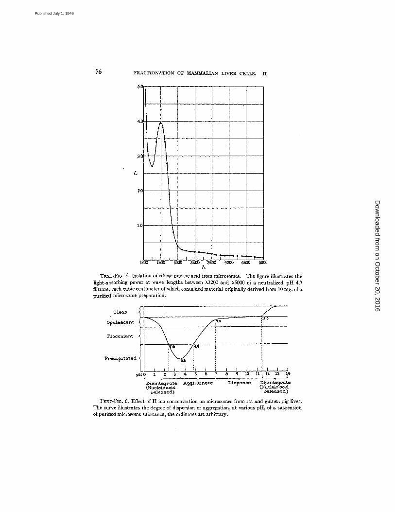
To a suspension of purified microsomes in water enough NaOI-I was added to bring the final concentration to 0.1 N and the alkaline mixture was stored overnight at 4°C. The solution was then diluted with water, the reaction brought to pH 4.7 by the addition of N acetic acid, and the floccullent preparation filtered on paper. The clear filtrate was neutralized and examined for its ultraviolet light-absorbing power. The results are illustrated in Text-fig. 5.
The absorption values given in Text-fig. 5 correspond to a filtrate, the content of which was derived from approximately 10 rag. microsomes per cc. Since the micro- some substance is about 45 per cent lipids it can be calculated, from the extinction coefficient of the filtrate at ~2600, that the nucleic acid recovered in this experiment represented about 3.5 per cent of the protein moiety of the microsomes.
Effect of Acid and Alkali on Suspensions of Microsomes.--In an acid environment the microsomes exhibited a strong tendency to agglutinate and, as illustrated in Text- fig. 6, aggregation was greater as the medium became more acid.
on October 20, 2016
Dow
nloaded from
Published July 1, 1946
75 FEACTIONATION OF MAMMALIAN LIVER CELLS. II
4.0
R0
1.0
t I I
2200 2600 3000 5 4 0 0 3800 4 2 0 0 4600 5000 A
TExT-FI6.5. Isolation of ribose nucleic acid from mlcrosomes. The figure illustrates the light-absorbing power at wave lengths between X2200 and kS000 of a neutralized pH 4.7 filtrate, each cubic centimeter of which contained material originally derived from 10 rag. of a purified microsome preparation.
CIeQ~ , f 11.,5
O p o . l e , s c e n t , ,'f.O
Flocculent
P r ~ e c i ~ i t a t e d :5.5
i I t I I I I I I I o 1 2 ~ . ~ 4 5 6 '1 8 9 lo z l , L l z 1~ ~,,
-,- v .... ~i~int~;~o.te A991utin~e ~i~pec~e DLsint~g~tc (NucleiEaciE ~i'¢udeic-ao/d
#ele~xsecl) 9e_lecL~ed)
TExT-FIG. 6. ]~ffect of H ion concentration on microsomes from rat and guinea pig liver. The curve illustrates the degree of dispersion or aggregation, at various pH, of a suspension of purified microsome substance; the ordinates are arbitrary.
on October 20, 2016
Dow
nloaded from
Published July 1, 1946
ALBERT CLAUDE 77
The maximum aggregation took place in the region of pH 3.5, beyond which point the ele- ments disintegrated, releasing ribose nucleic acid in the medium. At the alkaline side, the microsomes formed apparently stable suspensions between pH 7.0 and 9.0. In more alkaline solutions the material appeared progressively more dispersed; the suspension cleared up rapidly in the region of pH 11.0 when the mass of microsomes disintegrated, likewise liberating ribose nucleic acid in the medium.
Text-figs. 2 and 6 indicate that the responses of large granules and microsomes to changes in their acid or alkaline environment are strikingly similar. Slight dif- ferences in the curves may be due in part to the fact that changes taking place in the suspensions of large granules, because of their larger size, were more readily noticeable. The almost identical response of large granules and microsomes suggests that their characteristic behavior, with respect to H ion concentration, may be conditioned in both by the existence of common components, possibly at the surface of the elements.
Particulate Glycogen
In some of the preceding experiments the animals used were in partial fast; i.e., they had not been fed their usual daily meal 20 to 24 hours prior to the excision of the liver. Under these conditions no sedimentable glycogen was encountered during fractionation and the various fractions appeared uncontaminated. In animals fed at more frequent intervals glycogen is stored in the liver and appears in liver extracts in a particulate form, as demonstrated by Lazarow (16).
The particles of glycogen possess an exceptionally high density as compared to the other components of the cell: during centrifugatinn they work their way through the existing sedi- ment and collect at the lowest part of the deposit. This process of segregation is made more effective by the use of prolonged centrifugation at high speed.
In the present experiments guinea pigs were placed under ether anesthesia, heparin (10 to 15 mg. in saline solution) was injected intraperitoneally, and the livers were perinsed with Ringer's solution. Fifty gin. of liver pulp was dispersed in 250 ce. alkaline 0.85 per cent NaC1 solution; the liver suspension was centrifuged 3 minutes at 1500 X g and the resulting deposit was discarded. The large granules were separated from the liver extract by 5 minutes centri- fugation at 18,000 X g; the large granule sediment which was heavily contaminated with glycogen was discarded, together with a pellet of glycogen which had accumulated at the bottom of the deposit. The supernate (S) was then submitted to centrifugation for 1½ hours at 18,000 X g; the glycogen originally in suspension was found to have displaced the other elements of the extract and to have formed a firm, clean pellet of transparent and colorless material at the bottom of the tube. The supernate was discarded and also the microsome substance which had sedimented above the glycogen; the walls of the tube and the surface of the glycogen pellet were then thoroughly rinsed with alkaline distilled water. The glycogen material derived from 230 co. liver extract (40 gin. liver tissue) was resuspended in about 80 cc. neutral distilled water; the glycogen particles were then washed by means of two successive cycles of centrifugation (1½ hours at 18,000 X g), and resuspension of the deposit in alkaline distilled water. Each time the pellet was thoroughly rinsed with water. The last pellet of centrifugation was redispersed in 10 cc. distilled water; this glycogen preparation was then desiccated in vacuo and in the frozen state, and used for dry weight determination and chemical analysis.
The amount of purified glycogen actually isolated from 40 gm. of liver tissue (12.0 gm. solids) was 775 mg., a yield of 6.5 per cent. Taking into account the loss of
on October 20, 2016
Dow
nloaded from
Published July 1, 1946
78 FRACTIONATION OF MAMMALIAN LIVER CELLS. I I
substance sustained during fractionation, it was estimated that the glycogen content of liver in this experiment was approximately 10 to 15 per cent.
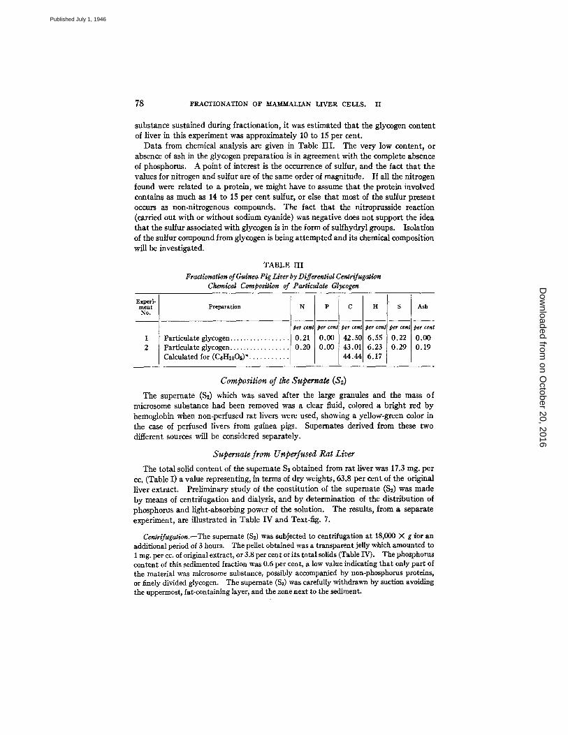
Data from chemical analysis are given in Table I I I . The very low content, or absence of ash in the glycogen preparation is in agreement with the complete absence of phosphorus. A point of interest is the occurrence of sulfur, and the fact that the values for nitrogen and sulfur are of the same order of magnitude. If all the nitrogen found were related to a protein, we might have to assume that the protein involved contains as much as 14 to 15 per cent sulfur, or else that most of the sulfur present occurs as non-nitrogenous compounds. The fact that the nitroprusside reaction (carried out with or without sodium cyanide) was negative does not support the idea that the sulfur associated with glycogen is in the form of sulfhydryl groups. Isolation of the sulfur compound from glycogen is being attempted and its chemical composition will be investigated.
TABLE I I I
Fractionation of Guinea Pig Liver by Differential Cenlrifugation Chemical Composition of Particulate Glycogen
Experi- ment Preparation No.
1 Particulate glycogen . . . . . . . 2 Particulate glycogen . . . . . . .
Calculated for (CeHloOt) ".
N
i per cent
0.21 0.20
P C
per cen~ ~er cent
0 . ~ 42.50 0.00 43.01
44.44
per ce~
6.55 6.23 6.17
S Ash
per cent per cem
0.22 0.00 0.29 0.19
Composition of the Supernate ($2) The supernate ($2) which was saved after the large granules and the mass of
microsome substance had been removed was a clear fluid, colored a bright red by hemoglobin when non-perfused rat livers were used, showing a yellow-green color in the case of perfused livers from guinea pigs. Supernates derived from these two different sources will be considered separately.
Supernate from Unperfused Rat Liver The total solid content of the supemate $2 obtained from rat liver was 17,3 rag. per
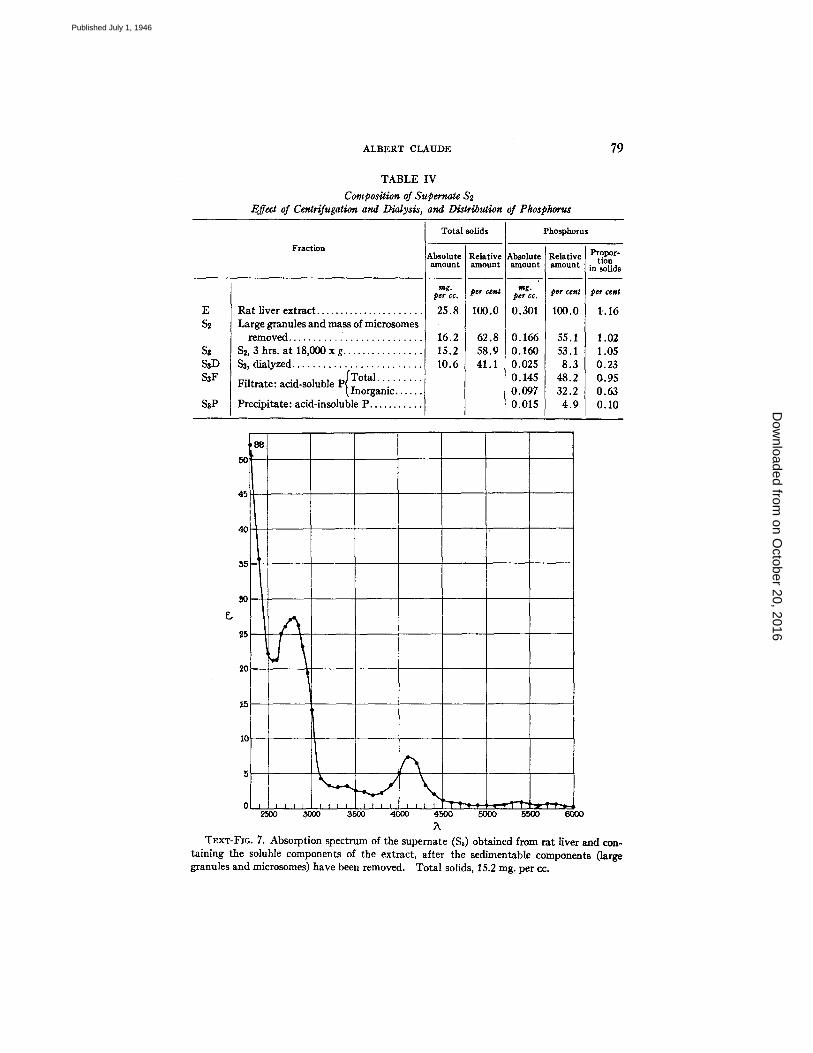
cc. (Table I) a value representing, in terms of dry weights, 63.8 per cent of the original liver extract. Preliminary study of the constitution of the supemate ($2) was made by means of centrifugation and dialysis, and by determination of the distribution of phosphorus and light-absorbing power of the solution. The results, from a separate experiment, are illustrated in Table IV and Text-fig. 7.
Cergrifugation.--The supernate ($2) was subjected to centrifugafion at 18,000 X g for an additional period of 3 hours. The pellet obtained was a transparent jelly which amounted to 1 rag. per cc. of original extract, or 3.8 per cent of its total solids (Table IV). The phosphorus content of this sedimented fraction was 0.6 per cent, a low value indicating that only part of the material was microsome substance, possibly accompanied by non-phosphorus proteins, or finely divided glycogen. The supernate ($8) was carefully withdrawn by suction avoiding the uppermost, fat-containing layer, and the zone next to the sediment.
on October 20, 2016
Dow
nloaded from
Published July 1, 1946
ALBERT CLAUDE
TABLE IV
Composition of Supernate $2 Effect of Centrifugation and Dialysis, and Distribution of Phosphorus
79
E $2
SaD SaF
SaP
Fraction
Ra t liver extract . . . . . . . . . . . . . . . . . . . . . . Large granules and mass of microsomes
removed . . . . . . . . . . i . . . . . . . . . . . . . . $2, 3 hrs. at 18,000 x g . . . . . . . . . . . . . . . Sa, dialyzed . . . . . . . . . . . . . . . . . . . . . . . .
p/ 'Total . . . . . . . . Fil trate: acid-soluble (Inorganic . . . . .
Precipitate: acid-insoluble P . . . . . . . . . .
Total solids
AbsoluU a n l o u n t
mg . peg CC.
25.8
16.2 15.2 10.6
elativ¢ mount
er cenl
LO0.O
62.8 58.9 41.1
Lbsolutl a l~o t ln |
mg . '
peg cc.
0.301
0.166 0.160 0.025 0.145 0.097 0.015
Phosphorus
P r o p o r - Re la t iv~ t i o n a m o u n t in so l i d s
per cem per cent
100.0 1..16
55.1 53.1 8.3
48.2 32.2
4 .9
1.02 1.05 0.23 0.95 0.63 0.10
50
45
40
55
20
~5
iO
5
0 i i 25OO
I I T I I I I f I I I I I ] ~ T ' I T ' ~ ' ~ I J ~ ~ ~ ' J P ' ~ ' ~ ' ~ I - ~ 3500 4000 4500 5000 5.~00 6000
T~.xT-Flo. 7. Absorption spectrum of the supemate ($3) obtained from rat liver and con- taining the soluble components of the extract, after the sedimentable components (large granules and microsomes) have been removed. Total solids, 15.2 rag. per cc.
on October 20, 2016
Dow
nloaded from
Published July 1, 1946
80 FRACTIONATION O:F M~AMM-AZIAN LIVER CELLS. II
Dialysis.--The supernate ($3) was permitted to dialyze in cellophane bags, against distilled water, for 48 hours. The dry weights of the preparation, before and after dialysis, were 15.2 rag. and 10.6 rag. per cc. respectively, a loss of 4.6 rag. per cc. (Table IV).
The findings indicate that about one-third (30.3 per cent) of the supernate (S~), and nearly one-fifth (in the present case 17.8 per cent) of the original liver extract was represented by dialyzable material.
Absorption Spectrum.--The power of the supernate $8 to absorb light selectively in the region between X2300 and ),6000 is illustrated in Text-fig. 7. 2 Characteristic absorption bands with maxima at ),4100, ),5350, and ),5750 indicate that hemoglobin was present in the solution in relatively large amounts, a situation explained by the fact that the material was originally prepared from non-peffused livers. An abun- dance of proteins in the solution is indicated by the high coefficient of absorption in the region of ),2800. The presence of nucleic acid or nucleotides in the supernate is not apparent except perhaps in the shoulder of the curve showing greatest absorption in the region of ),2650 to 2700.
Distribution of Phosphorus.--The total phosphorus content of the different liver fractions was determined. Data presented in Table IV show that about half (47 per cent) of the total phosphorus content of the original liver extract was removed by combined centrifugation at various speeds, and an equivalent amount (45 per cent) was removed by dialysis. Thus centrifugation and dialysis together removed as much as 92 per cent of the phosphorus present in the original liver suspension. The whole dialyzable fraction (4.6 rag. per ec.) contained 3.0 per cent phosphorus (0.135 rag. per c¢.). Treatment of the supernate $3 with trichloracetic acid showed that over 90 per cent of the non-sedimentable phosphorus present was acid-soluble, two- thirds of this amount being inorganic phosphorus (Table IV).
To sum up, it was found that of the total amount of phosphorus originally present in rat liver extract, 47 per cent was sedimentable in the form of large granules and microsome substance, 45 per cent was dialyzable and acid-soluble; as much as 32 per cent was in the form of inorganic phosphorus; and the remainder, (4.9 per cent), was represented by non-sedimentable, non-dialyzable, and acid-insoluble phosphorus.
I t may be doubted that the inorganic phosphorus found in such a high proportion in the liver extract existed originally in this form in the intact liver cell. Liver extracts are known to contain active phosphatases and it is possible that all, or part, of the in- organic phosphorus may have been set free, in the course of the preparation of the material, through enzymatic hydrolysis. The acid-soluble, but organic, phosphorus of the extract may have been represented by compounds of low molecular weight such as nucleotides or nucleosides, hexosephosphates, etc. On the other hand, the non-dialyzable residue may correspond to the presence in the solution of small amounts of nucleoproteins.
I t is of interest to compare the results just discussed and the data presented in Table IV with previous observations dealing with the fractionation of a rat lymphosarcoma (reference 4, page 25, Table II). In both cases the absolute values for phosphorus were almost identical, the only apparent difference residing, in the case of rat liver, in consistently higher values of the total solids of the various fractions, a fact reflected in correspondingly lower values in the percentage of phosphorus. The striking
on October 20, 2016
Dow
nloaded from
Published July 1, 1946
ALBERT CLAUDE 81
similarity in the phosphorus content of extracts from tissues as dissimilar as rat liver and lymphosarcoma, both in quantity and in characteristic distribution among the various cell fractions, suggests that phosphorus compounds may be related to a fundamental structure of protoplasm common to both types of tissues.
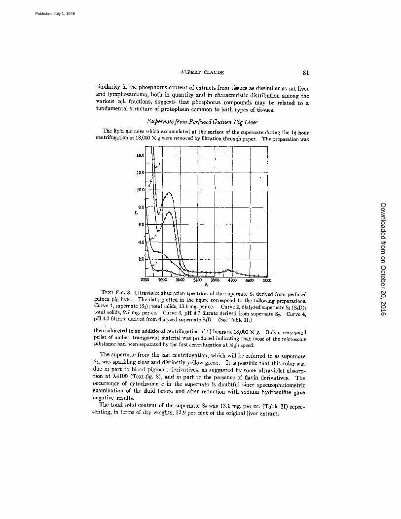
Supernate from Perfused Guinea Pig Liver The lipid globules which accumulated at the surface of the supernate during the 1½ hour
centrifugation at 18,000 X g were removed by filtration through paper. The preparation was
14.0
12.0
-/ 10.0 'I 8.0 r
g
6.0
4.0
2L~O0 ~o00 3000 3400 3800 4 ~ 0 4600 5000 k
T~xT-Fm. 8. Ultraviolet absorption spectrum of the supernate $2 derived from perfused guinea pig liver. The data plotted in the figure correspond to the following preparations. Curve 1, supernate ($2); total solids, 13.1 mg. per cc. Curve 2, dialyzed supernate S~ (S2D); total solids, 9.7 mg. per cc. Curve 3, pH 4.7 filtrate derived from supernate $2. Curve 4, pH 4.7 filtrate derived from dialyzed supernate S2D. (See Table II.)
thensubjected to an additional centrifugation of 1½ hours at 18,000 X g. Only a very small pellet of amber, transparent material was produced indicating that most of the microsome substance had been separated by the first centrifugation at high speed.
The supernate from the last centrifugation, which will be referred to as supernate $2, was sparkling clear and distinctly yellow-green. I t is possible that this color was due in part to blood pigment derivatives, as suggested by some ultraviolet absorp- tion at X4100 (Text-fig. 8), and in part to the presence of flavin derivatives. The occurrence of cytochrome c in the supernate is doubtful since spectrophotometric examination of the fluid before and after reduction with sodium hydrosulfite gave negative results.
The total solid content of the supernate $2 was 13.1 mg. per cc. (Table II) repre- senting, in terms of dry weights, 52.9 per cent of the original liver extract.
on October 20, 2016
Dow
nloaded from
Published July 1, 1946
82 FI~ACTIONATION OF ~LA~r~ALIAN LIVER CELLS. II
The supernate S~ was allowed to dialyze against neutral distilled water, at 4°C. for 48 hours. The total solid content of the dialyzed preparation (S~-D) was 9.7 mg. per cc. (Table II) or 39 per cent of the original liver extract. Thus the amount of sub- stance removed by dialysis was 3.4 mg. per cc. of the supernate $2, or about one-fourth of its total solid content.
The ultraviolet-absorbing power of the supernate before and after dialysis is illustrated in Text-fig. 8, curves 1 and 2 respectively. Both curves are similar, show- ing a minimum of absorption at ~2500, a broad band of absorption with maximum at k2750, and identical absorption in the k4100 region. Dialysis was responsible for the loss of 24 per cent of the absorbing power of the solution at ~2750, but the position of the maximum remained unchanged. Following dialysis, however, the peak of absorption at k2750 appeared more symmetrical, suggesting that substances possessing a somewhat greater absorbing power in the k2550 to 2650 region had been removed. The absorbing power in the wave length region between k3000 and ~,5000 was unaffected by dialysis.
Both the undialyzed and the dialyzed supemate S~ were subjected to fractionation by treat- ment in succession at pH 13 and pH 4.7, a procedure found to be effective in separating nucleo- tides and nucleic acid from large granules and microsomes.
To 9 cc. of the respective solutions 1 cc. of N NaOH was added and the alkaline mixtures were allowed to stand at 4°C. for about 20 hours. The preparations were then brought to pH 4.7 by the addition of N acetic acid and the flocculent precipitates were removed by filtra- tion on paper. The clear, nearly colorless filtrates were neutralized and examined for their power to absorb ultraviolet light. The values, recalculated for constant volume, are recorded in Text-fig. 8, curves 3 and 4 respectively.
The filtrate derived from the undialyzed supernate gave no definite absorption bands in the ultraviolet (curve 3) but there was some indication of greater absorption in the region of ~2600. I t is interesting to note that a curve nearly identical to curve 1 (Text-fig. 8) can be constructed by adding together the successive values of curves 2 and 3, at corresponding wave lengths. This observation seems to indicate that the absorbing substances of the supernate $2 which are dialyzable are also those which remain soluble in acid and which were responsible for the absorbing power of the pH 4.7 filtrate.
The filtrate derived from the dialyzed supernate S, showed a relatively low power of absorption in the ultraviolet (curve 4), suggesting that proteins were probably responsible for most of the absorbing power exhibited by the dialyzed fraction (curve 2).
Of the total absorbing power exhibited by the supernate S~ in the region of k2600 to 2750 about 75 per cent can be ascribed to proteins (curve 2), and 25 per cent to dialyz- able, acid-soluble substances (curve 3).
The observations just described seem to support the view (4) that the nucleic acid found in the liver extract occurred in association with its sedimentable constituents, hence with formed elements of the cytoplasm, i.e., the large granules (mitochondria and secretory granules) and the microsomes. Before this conclusion can be definitely reached, however, it will be necessary to investigate the distribution of nucleases in the cytoplasm and to give even greater consideration to the quantitative aspects of the problem.
on October 20, 2016
Dow
nloaded from
Published July 1, 1946
ALBERT CLAUDE 83
DISCUSSION
The hepatic cell appears to be relatively more fragile than the nucleus and during the process of extraction a high proportion of the cells are broken while the nuclei remain intact. I t was estimated that by the procedure adopted in the present work 50 to 50 per cent of the liver cells were broken and their entire cytoplasmic content dispersed in the medium; the remaining portion was found to be made up of seemingly undamaged liver cells, as a rule part of little tissue fragments. The persistence of apparently intact cells in the suspension may be due to the fact that they escaped mechanical injury, sufficient protection being afforded within the tissue fragments, or because there may exist among the cell population a gradient of resistance which would permit certain cells to with- stand the effect of injury while, under similar conditions, other cells would undergo disintegration.
Under the microscope the unbroken cells removed from the suspension ap- peared normal and undistinguishable from "unextracted" cells of liver tissue. The term "liver extract," therefore, should not be taken literally at least under the conditions of the present experiments, since it is not made up primarily of materials selectively removed from the cells, the latter retaining otherwise their integrity, but includes the entire cytoplasmic content of disintegrated ceils. In this respect our observations fail to support the conclusion of Bensley and Hoerr who have stated (17, 9, 18) that they were able to remove as much as 70 per cent of the cell substance by repeated extraction while producing hardly any detectable changes in the morphological make-up of the ceils. The as- surance was not given, however, that the ceils which, under microscopical inspection appeared unaltered, were the same cells from which the bulk of the extract had been derived. 3
The importance of neutral or slightly alkaline media in extraction and puri- fication has been emphasized. I t must be noted that the use of alkaline saline does not imply that the tissue elements were exposed to that reaction for any length of time, since the acid-producing capacity and the buffering power of the tissue and of the large granules constantly brought down the reaction towards a more acid pH and usually maintained it slightly above pH 7.0.
Both large granules and microsomes agglutinate strongly when in acid-media (see Text-figs. 2 and 6), and in this condition cannot be separated by differential centrifugation. Furthermore, on repeated centrifugation in slightly acid solutions the microsome substance, which normally forms completely trans- parent pellets, becomes increasingly opaque and finally can no longer be re- dispersed, even in alkaline medium. However, Bensley and Hoerr have ad- vocated the use of acid solutions for the fractionation of liver (8, 9), and some
s These remarks do not apply to cells in which the solubility of certain constituents has been modified by fixation or by drying. In such cases it is apparently possible to remove selectively, by appropriate solvents, those constituents which have remained soluble.
on October 20, 2016
Dow
nloaded from
Published July 1, 1946
84 FKACTIONATIOIq OF MAMMALIAN LIVER CELLS. II
of their results regarding the composition and the properties of the "mitochon- dria" fraction can be explained by the failure to separate the large granules from the microsome substance. Lazarow, who followed Bensley and Hoerr's direc- tions, experienced the type of difficulties just mentioned and noted that the microsome substance could not be properly purified by washing in acid solutions (29). It was found that the large granules also were better preserved when prepared and stored in slightly alkaline media (between pH 7.0 and 8.0), as shown by the fact that enzymatic activities associated with them were greater and could be maintained over longer periods of time in that range than when the reaction was permitted to become slightly acid (6, 10). Biochemical studies (6, I0) have shown that most of the succinoxidase activity exhibited by the liver extract was associated with the large granules, the activity possessed by the microsome fraction being practically negligible. In this respect our results contrast with those of Lazarow (29, 19) who reported that, on a cor- responding nitrogen basis, both mitochondria (large granules) and submi- croscopic particles (microsomes) from guinea pig liver possessed about equal succinoxidase activity. Both the low succinoxidase activity found by Lazarow and the fact that no difference was observed between the large granules and the microsome fraction would suggest that, with the procedure adopted by him, no effective separation of the two particulate components of liver extract had been accomplished.
Two types of inclusions, visible under the fight microscope, are generally recognized in the cytoplasm of glandular cells: (I) mitochondria, of general occurrence and present also in the cytoplasm of undifferentiated cells, and (2) secretory or zymogen granules associated with the specialized functions of the cell and concerned with the elaboration of products destined to be secreted into the blood stream or extruded into excretory ducts. In certain organs the duality of the cytoplasmic inclusions is obvious because of a sharp polarization in the arrangement of the cell elements, the mitochondria being found at the base of the cell, near the capillary bed, whereas the secretory granules are found accumulated at the opposite poles, in the neighborhood of the excretory canals. The situation is best illustrated in salivary glands, and in the pancreas. In mammalian liver, however, a similar segregation of cytoplasmic elements is not apparent and the large granules of the hepatic cell have been referred to gen- erally as mitochondria. That the two types of elements, corresponding to the classical concept of secretory granules and mitochondria, are both present in liver can be demonstrated by comparing sections of livers obtained from animals fasted for adequate periods, and animals recently fed (20). In am- phibians, in which the organization of the liver is of a simple, tubular type, the demonstration is facilitated by the fact that the hepatic cell presents a definite polarization of its elements with two types of inclusions, morphologically dis- tinct, segregating at the opposite poles of the cell. In Amphiuma tridactylum subjected to complete fasting, which in this animal can be of several months
on October 20, 2016
Dow
nloaded from
Published July 1, 1946
ALBERT CLAUDE 85
duration, granules accumulate in the region of the liver cells next to the biliary ducts, duplicating very closely the arrangement seen in mammalian pancreas, with "secretory granules" crowding at one pole and slender filamentous mito- chondria occupying th6 other end, or base, of the cell. Following a single meal practically all the secretory granules disappear, presumably through excretion into the biliary ducts, while the filamentous mitochondria retain their general position at the base of the cell. The same phenomenon, i.e. the removal of granules from the cell under the stimulus of feeding, and accumulation of granules in the cytoplasm during fasting can be demonstrated also, under proper conditions, in guinea pig and rat liver (20). I t has frequently been suggested that mitochondria and secretory granules are morphologically related and it is possible that they may constitute extreme forms in a continuous series of cy- toplasmic elements (21). Until this point can be ascertained, however, it would seem reasonable to continue to distinguish two types of elements, keeping in view the possibility that secretory granules, even if evolved originally from mitochondria may nevertheless differ from them ultimately in composition and function. Secretory granules and mitochondria do not differ greatly in size, and for this reason segregate together in the centrifuge. Because of these various considerations the general term "large granules" has been adhered to throughout this paper when referring to the large elements of liver extract, it being understood that the so called large granule fraction contains in various proportions, both mitochondria and secretory granules.
When using livers from rats and guinea pigs supplied at all times with an excess of food, as in many of the experiments described in this and subsequent papers, the large granules may be considered to be represented by mitochondria predominantly, and it may be possible to refer the observations made in such cases specifically to mitochondria. The distinction between secretory granules and mitochondria will become necessary when the work undertaken will be especially concerned with the specific function of these various cytoplasmic elements.
Evidence presented in previous papers (2, 4) indicates that the microsome fraction corresponds to the chromophilic ground substance of the cell. I t has been pointed out (2) that the microsome substance may present itself, in vitro, in two different forms: (a) in the dispersed condition it is composed of discrete elements roughly 80 to 150 m/~ in diameter and, therefore, not resolved as sepa- rate units by the light microscope, but readily detectable under the ultramicro- scope as brilliant objects exhibiting active Brownian motion; (b) in the con- centrated form, as in the pellet of centrifugation, it appears as an homogeneous, completely transparent jelly. The physical state that the microsome substance assumes in living cells is of particular interest since it may serve to explain some of the properties of protoplasm. When living cells are examined by dark- field microscopy the thinnest portions of their protoplasm, as in the case of extended tissue culture cells and the pseudopodia of ameba, often appear
on October 20, 2016
Dow
nloaded from
Published July 1, 1946
86 YRACTIOI~ATION OF MAM3KAIIAN LIVER CELLS. II
structureless and optically empty (22). This appearance, however, does not rule out the existence of physical heterogeneity since, under similar experimen- tal conditions the nucleus, known to contain highly complex chromosomes and nucleoli structures, frequently fails to reveal any sign of internal organization. The failure to demonstrate structural differences may be due to insufficient illumination, since the cytologist is usually, and rightly, concerned with the injurious effect that excessive light or heat may have on the condition of the cell. It must be remembered, however, that the amount of light reflected by a particle, hence its visibility in the ultramicroscope, is in inverse relation to the size of the particle and directly proportional to the intensity of the incident light. Bayliss (23) recognized the importance of intense illumination in the detection of submicroscopic structures and examined living cells under the filtered light of a carbon arc. Under these conditions he was able to observe that protoplasm, which appeared clear under transmitted light, contained an immense number of very minute particles." Bayliss found that the active Brownian movement exhibited by the particles would cease when a short elec- trical stimulus was applied, "as if the liquid protoplasm had been frozen," but "the Brownian movement and the flowing of the pseudopodial extrusion recommenced" as soon as the electrical stimulation was discontinued (23). Bayliss related the Brownian movement of the submicroscopic particles, or its absence, to the property of reversible gelation of protoplasm.
Experiments in which fragments of AmpMuma (2) and guinea pig (24) livers were centrifuged at high speed before fixation have indicated that the chromo- philic component of the ground substance, of structureless appearance in the light microscope, is nevertheless particulate in nature since it could be segre- gated from the hyaloplasm proper by 2 hours at 18,000 X g. Furthermore, examination of thin sections of guinea pig and rat liver in the electron micro- scope (25) revealed that, under certain conditions of fixation, the ground sub- stance can be resolved into discrete units of apparently uniform size and ap- proximately 100 m~ in average diameter.
The foregoing observations suggest that the property of the microsome sub- stance to appear in vitro in the form of particles of submicroscopic size may not be an artifact produced by extraction but that this particulate form may nor- mally exist in the cytoplasm of living cells. On the other hand, it is con- ceivable that, depending on the concentration of the substance in the cell and the state of gelation of the fibrous matrix of protoplasm (26), the microsome substance may participate in the formation of an apparently homogeneous jelly. One of the possible effects resulting from the extremely divided condition of the ground substance may be to offer an enormous surface capable of fixing substances which otherwise would accumulate in the cellular fluid.
The part played by the microsome substance in the physiology of the cell is not known. Of the various enzymes and enzyme systems so far detected in the liver extract, none were found to be associated with the microsomes in signifi-
on October 20, 2016
Dow
nloaded from
Published July 1, 1946
M~BERT C~UDE 87
cant amounts (6, 10). The fact that cytochrome c was found to be associated with the microsomes in appreciable amounts while the cytochrome-linked enzymes appear to be located in the mitochondria (6, 10) is of interest but the possibility that cytochrome c becomes attached to the microsomes secondarily during the preparation of the extract will have to be investigated. The thromboplastic activity of cell products has been found to reside in the micro- somes (27, 28).
The possibility of a relation between microsomes and mitochrondria has been discussed in previous papers (2). This point is supported by the fact that a particulate component similar to ordinary microsomes in size and composition can be separated from mitochondria (2, 4, and this paper), and by the fact that small elements, approximately 0.1 g in diameter, can occasionally be detected in mitochondria by electron microscopy (13, and unpublished data).
The data presented in this paper seem to support the view derived from previous work (4) that ribose nucleic acid occurs in the cytoplasm in association with preformed elements, namely, microsomes and mitochondria. The re- suits described (Text-fig. 4) suggest further that the ribose nucleic acid de- tected in unfractionated large granules may be localized within them on small particulate structures similar in size and composition to ordinary microsomes. The possibility that the ribose nucleic acid present in formed elements of the cytoplasm may be involved in the capacity for self-duplication possessed by the cytoplasmic substance has been discussed in previous papers (2).
Ribose nucleotides of various sorts occur in abundance (Text-fig. 3), if not exclusively, in the large granules. In apparent contrast with nucleic acid, nucleotides were found in a soluble, dialyzable form in the water preparation (Text-fig. 4) after the large granules had disintegrated. It is probable that the presence of nucleotides in the large granules is related to the high and varied metabolic activities which these elements have been found to possess (6, 10).
It is apparent that the isolation of cell components and the study of their properties in vitro may be of great value in elucidating certain aspects of the behavior of protoplasm, and may be of technical importance in the practice of tissue fixation: for example, aggregation and disintegration of large granules (secretory granules and mitochondria) and of microsomes in acid solutions (Text-figs. 2 and 6) might explain to some extent the disrupting effect that acid fixatives have on cytoplasm as also how the presence of relatively strong acids (below pH 3.5) may cause the displacement or the removal of ribose nucleo- tides from their original sites, thus making the residue of denatured proteins appear acidophilic.
SUMMARY
1. A method is described whereby the major components of liver suspensions are segregated according to size into three main fractions: (a) a large granule fraction composed of elements approximately 0.5 to 2 # in diameter; (b) a
on October 20, 2016
Dow
nloaded from
Published July 1, 1946
88 FRACTIONATION OF MAM2~ALIAN LIVER CELLS. II
microsome fraction composed of submicroscopic elements approximately 80 to 150 m/~ in diameter; and, (c) a supernate fraction containing the soluble components of the extract.
2. The nature and origin of the constituents of liver extract is discussed. The large granule fraction is deemed to consist mostly of mitochondria and liver secretory granules, whereas the microsome fraction corresponds to the chromophilic ground substance of the hepatic cell. Phosphorus distribution in the supernate fraction, and ultraviolet absorption of the solution suggests that practically all the ribose nucleoproteins of liver extract are sedimentable, and occur in association with the large granules and microsomes.
3. The method of fractionation of liver suspension by differential centrif- ugation is being used as a means to investigate the chemical constitution of the morphological constituents of cytoplasm, and the distribution of biochemical activities in the cytoplasm of the hepatic cell.
4. The method of differential centrifugation is found to be applicable not only to the fractionation of cells but also, with the aid of auxiliary techniques, to the fractionation of much smaller elements, such as mitochondria.
BIBLIOGRAPHY
1. Claude, A., (a) Proc. Soc. Exp. Biol. and Med., 1938, 39, 398, (b) Science, 1940, 91, 77.
2. Claude, A., (a) Science, 1943, 97,451. (b) Claude, A, in Frontiers in Cytochemis- try, (N. L. Hoerr, editor). Biological Symposia, Vol. 10, Lancaster, The Jaques Catell Press, 1943, 111.
3. Claude, A., and Potter, J. S., J. Exp. Med., 1943, 77, 345. 4. Claude, A., J. Exp. Med., 1944, 80, 19. 5. Claude, A., J. Exp. Med., 1946, 84, 51. 6. Claude, A., in A. A. A. S. Research Conference on Cancer, July 31-August 4,
1944, (F. R. Moulton, editor), Washington, D. C., The American Association for the Advancement of Science, 1945, 223.
7. Claude, A., in Cold Spring Harbor Symposia on Quantitative Biology, Cold Spring Harbor, Long Island Biological Association, 1941, 9, 263.
8. Bensley, R. R., and Hoerr, N. L., Anat. Rec., 1934, 60, 449. 9. Hoerr, N. L., in Frontiers in Cytochemistry, (N. L. Hoerr, editor). Biological
Symposia, Vol. 10, Lancaster, The Jaques CatteU Press, 1943, 185. 10. Hogeboom, G. H., Claude, A., and Hotchkiss, R., data to be published. 11. Woolley, D. W., J. Biol. Chem., 1943, 147, 581. 12. Claude, A., Science, 1939, 90, 213. 13. Claude, A., and Fullam, E. F., J. Exp. Meal., 1945, 81, 51. 14. Porter, K. R., Claude, A., and Fullam, E. F., J. Exp. Med., 1945, 81,233. 15. Claude, A., and Rothen, A., J. Exp. Med., 1940, 71,619. 16. Lazarow, A., Anat. Rec., 1942, 84, 31. 17. Bensley, R. R., and Hoerr, N. L., Anat. Rec., 1934, 60, 251. 18. Bensley, R. R., Anat. Rec., 1938, 72,351.
on October 20, 2016
Dow
nloaded from
Published July 1, 1946
ALBERT CLAUDE 89
19. Barron, E. S. G., in Frontiers in Cytochemistry, (N. L. Hoerr, editor). Bio- logical Symposia, Vol. 10, Lancaster, The Jaques Cattell Press, 1943, 27.
20. Claude, A., and Auger, C., data to be published. 21. No~l, R., Arch. anat. micr., 1923, 199 1. 22. Lewis, M. R., Bull. Johns Hopkins Hosp., 1923, 34, 373. 23. Bayliss, W. M. Proc. Roy. Soc. London, Series B, 1919-20, 91~ 196. 24. Claude, A., unpublished experiments. 25. Claude, A., and Fullam, E. F., J. Exp. Meal., 1946, 83, 499. 26. The Structure of Protoplasm, (W. Seifriz, editor), Ames, Iowa~ The Iowa State
College Press, 1942. 27. Chargaff, E., J. Biol. Chem., 1945, 160, 351. 28. Tagnon, H. J., and Claude, A., unpublished experiments. 29. Lazarow, A., in Frontiers in Cytochemistry, (N. L. Hoerr, editor). Biological
Symposia, Vol. 10, Lancaster, The Jaques Cattell Press, 1943, 9.
on October 20, 2016
Dow
nloaded from
Published July 1, 1946