forgetting, reminding, and remembering: the retrieval of lost spatial memory
TRANSCRIPT

Forgetting, Reminding, and Remembering:The Retrieval of Lost Spatial MemoryLivia de Hoz[*, Stephen J. Martin[, Richard G. M. Morris
Laboratory for Cognitive Neuroscience, Centre and Division of Neuroscience, University of Edinburgh, Edinburgh, Scotland, United Kingdom
Retrograde amnesia can occur after brain damage because this disrupts sites of storage, interrupts memoryconsolidation, or interferes with memory retrieval. While the retrieval failure account has been considered in severalanimal studies, recent work has focused mainly on memory consolidation, and the neural mechanisms responsible forreactivating memory from stored traces remain poorly understood. We now describe a new retrieval phenomenon inwhich rats’ memory for a spatial location in a watermaze was first weakened by partial lesions of the hippocampus to alevel at which it could not be detected. The animals were then reminded by the provision of incomplete and potentiallymisleading information—an escape platform in a novel location. Paradoxically, both incorrect and correct placeinformation reactivated dormant memory traces equally, such that the previously trained spatial memory was nowexpressed. It was also established that the reminding procedure could not itself generate new learning in either theoriginal environment, or in a new training situation. The key finding is the development of a protocol that definitivelydistinguishes reminding from new place learning and thereby reveals that a failure of memory during watermazetesting can arise, at least in part, from a disruption of memory retrieval.
Citation: de Hoz L, Martin SJ, Morris RGM (2004) Forgetting, reminding, and remembering: The retrieval of lost spatial memory. PLoS Biol 2(8): e225.
Introduction
For more than a century, the phenomenon of retrogradeamnesia (RA)—the loss of memory for events that occur priorto a variety of precipitating brain insults—has provided thefoundation for theories of memory consolidation and thelocus of trace storage (McGaugh 1966; Davis and Squire 1984;Dudai and Morris 2000). However, RA may also reflect theinability of a memory system to access a trace—a failure ofmemory retrieval (Warrington and Weiskrantz 1968). Thisvery dilemma was noted by Ribot (1883, p. 475) in his seminaldiscussion of RA:
‘‘Two suppositions are equally warranted, viz., that either theregistration of the prior states has been effaced; or that the retention ofthe anterior states persisting, their aptitude for being revived byassociations with the present is destroyed. We are not in a position todecide between these two hypotheses.’’
Studies of RA have favoured a memory-consolidationinterpretation in instances in which systematic variation ofthe time interval between experience or training and thesubsequent brain insult has revealed a temporal gradation ofRA (Squire 1992). Computational models also point to theneed for a rapid encoding and storage system, together with aslower interleaving mechanism that is thought to underliesystems-level consolidation and long-term storage in thecortex (e.g., McClelland et al. 1995). However, the existence ofsome amnesic patients with long, flat gradients of RAextending for years or decades into periods of their lifewhen memory function was normal provided some of the firstevidence that RA might be due to retrieval failure (Sandersand Warrington 1971). This perspective on RA was initiallysupported by studies indicating that, in the anterogradedomain, impaired memory could be alleviated by partial cues(Warrington and Weiskrantz 1968). However, these observa-tions were later construed as reflecting the operation of aseparate memory phenomenon called priming (Graf et al.
1984). Several animal studies have also indicated that a varietyof ‘reminder’ treatments delivered prior to retention testingcan realize the expression of lost memories (Gold et al. 1973;Miller and Springer 1973; Spear 1973; Gold and King 1974;Riccio and Richardson 1984; Sara 1999), but it is not easy todistinguish priming-induced memory from explicit recall andrecognition in animal studies. Experimental resolution of theconsolidation-versus-retrieval controversy has been notori-ously difficult, and no consensus has been achieved. A keymethodological issue, and the focus of the new techniquedescribed here, concerns the need to demonstrate that thememory observed after a reminder treatment results from thereactivation of an existing memory (Miller and Springer1972), rather than a facilitation of new learning (Gold et al.1973).In studies of spatial memory using the watermaze, amnesia
for the location of the escape platform in posttraining probetrials (PTs) has generally been interpreted as a failure oflearning, consolidation, or storage (D’Hooge and De Deyn2001). To investigate the alternative possibility of retrievalfailure, we deliberately created conditions that should max-imize the possibility of seeing such an effect. This involvedtraining rats to find an escape platform in a specific locationfollowed by partial lesioning of the hippocampus. We
Received March 3, 2004; Accepted May 14, 2004; Published August 17, 2004DOI: 10.1371/journal.pbio.0020225
Copyright: � 2004 de Hoz et al. This is an open-access article distributed underthe terms of the Creative Commons Attribution License, which permitsunrestricted use, distribution, and reproduction in any medium, provided theoriginal work is properly cited.
Abbreviations: ANOVA, analysis of variance; PT, probe trial; PTn, probe trial in anovel environment; RA, retrograde amnesia
Academic Editor: Howard B. Eichenbaum, Boston University
*To whom correspondence should be addressed. E-mail: [email protected]
[These authors contributed equally to this work.
PLoS Biology | www.plosbiology.org August 2004 | Volume 2 | Issue 8 | e2251233
Open access, freely available online PLoS BIOLOGY

reasoned that this would weaken but not completely disruptthe memory of the correct location by damaging a subset ofthe ensemble of stored traces. The animals’ memory wastested and observed to be undetectable. This same memorytest provided, however, the opportunity to remind animalsthat escape from the water was possible via an escapeplatform in the correct or incorrect location. One hour later,the animals’ memory was tested again. We observed thatmemory was now detectably above chance and was equallystrong when the animals had previously been given correct orpotentially misleading information about the current loca-tion of the platform. Additional control procedures, and theperformance of other groups with sham or complete hippo-campal lesions, established that the earlier failure of memorymust have been due, at least in part, to retrieval failure.
Results
A summary of the experimental design is provided inFigure 1 (see Materials and Methods).
Training Prior to the LesionsDuring cued pretraining, the rats quickly learned to search
for, and climb onto, the visually cued escape platform. In themain spatial training phase, the animals rapidly learned tolocate and raise the platform in order to escape from the pool(Figure 2), as indicated by the highly significant reduction inlatencies over trials (F[7.78, 412] = 30.4, p , 0.001). Onlyanimals that reached the acquisition criterion receivedlesions (69 out of 73 rats trained). The prospective lesiongroups, trained as normal animals, did not differ (F , 1, n =59; see Surgery below).
SurgeryOf the 69 animals that received lesions, one died after
surgery and nine were excluded based on strict histologicalcriteria, leaving a total of 59 animals (22 sham lesions, 19complete hippocampal lesions, and 18 partial hippocampallesions; see Figure 3).
Retention TestingThe key new findings are shown in Figures 4 and 5 using
two separate but related measures of memory retrieval:percentage time in quadrant (Figure 4) and a more sensitive
measure, percentage time in a zone centred on the platformlocation (Figure 5; see Materials and Methods). An overallanalysis of variance (ANOVA) of percentage time in thetraining (where the platform was located during training) andthe opposite quadrants of the pool revealed a significantquadruple interaction (F[2, 53] = 7.66, p , 0.01) involvingtwo between-subject factors: lesion group and platformlocation during the reminder treatment (original versusnovel), and two within-subject factors: PT (PT1 and PT2)and quadrant (training versus opposite). In both figures, theinitial memory expressed during PT1 is shown in the left lane.This reveals that the partially lesioned rats were at chance,whereas the sham-lesioned rats could remember the locationof the platform (t = 6.15, df = 21, p , 0.005, paired-sample t-test, training versus opposite quadrant). The complete-lesioned animals were at chance. Analysis of percentage timein zone (Figure 5) likewise confirmed that memory was
Figure 1. Experimental Design
Outline of the different phases of testing. The platform position used during training is indicated by a red circle in the NE quadrant of the pool(large blue circle), although in practice platform locations were counterbalanced between NE and SW locations. The novel location, to which asubset of rats was exposed during reminding, is indicated by a black circle in the SW quadrant. This position was always opposite to that usedduring training. PT1 and PT2: probe test 1 and 2. The hatched areas represent the original training quadrant irrespective of the position of theplatform (i.e., original or novel) during retention testing. PTn1 and PTn2: PTs during new context learning in the second pool.DOI: 10.1371/journal.pbio.0020225.g001
Figure 2. Training
Mean latencies to escape from the water and climb onto the hiddenplatform during task acquisition. Data are averaged in blocks of fivetrials and grouped according to the lesion made at the end oftraining; note that all animals were unoperated during acquisition.Only rats that reached criterion (mean escape latency less than 15 sover the last ten trials) and whose lesions were considered acceptable(see Results: Surgery) are presented. Animals rapidly learned to locatethe escape platform, and prospective lesion groups did not differ.DOI: 10.1371/journal.pbio.0020225.g002
PLoS Biology | www.plosbiology.org August 2004 | Volume 2 | Issue 8 | e2251234
Forgetting, Reminding, and Remembering

Figure 3. Lesion Analysis
Representative photomicrographs of cresyl-violet-stained coronal brain sections taken from subjects belonging to each of the three lesiongroups—partial hippocampal lesion (A), sham lesion (B), and complete hippocampal lesion (C). In each case, sections corresponding to anterior,middle, and posterior levels of the hippocampus are displayed. The mean area of spared hippocampal tissue in each group (see Materials andMethods for calculation) is plotted below in (D). Note that the volumes of spared tissue in the septal and temporal halves of the hippocampus areplotted separately, but these values are still expressed as percentages of the entire hippocampal volume—hence the value of 50% per half inshams. The cartoon hippocampi accompanying the graph indicate lesioned tissue in dark grey, and spared tissue in light cream. As intended,partially lesioned rats exhibited substantial sparing only in the septal (dorsal) half of the hippocampus, and rats with complete hippocampallesions exhibited minimal sparing (less than 5% at either pole).DOI: 10.1371/journal.pbio.0020225.g003
PLoS Biology | www.plosbiology.org August 2004 | Volume 2 | Issue 8 | e2251235
Forgetting, Reminding, and Remembering

detectable in the sham lesion group (t = 4.18, df = 21, p ,
0.005, one-sample t-test, comparison with chance = 50%),but not in the two lesion groups.
PT1 ended with the animals finding the platform in the
original training location, or in a novel location in the‘opposite’ quadrant of the pool (middle lane in Figures 4 and5; see Materials and Methods for explanation of terminology).These different events at the end of the swim trial potentially
Figure 4. Retention Testing: Quadrant
Analysis
Percentage time during PT1 and PT2spent in the training and oppositequadrants of the pool (left and rightlanes) and the reminder treatment (greycentral lane). The training location isrepresented as a red circle in the NEquadrant, and the novel location (novelsubgroups only) as a black circle in theSW quadrant. In practice, NE and SWquadrants were counterbalanced. Ratswith partial hippocampal lesions wereunable to remember the platform loca-tion on PT1 but could be reminded ofthe training location by exposure, at theend of PT1, to a platform in the originalor a novel location. (Note that the‘reminder’ lane simply refers to thisexposure to a platform—PT1 is itselfthe ‘reminder trial.’) The key finding isthat the improvement in PT2 occurredirrespective of the platform locationduring reminding. In contrast, sham-lesioned animals exhibited some reversallearning upon exposure to the platformin a novel location. Complete-lesionedrats did not remember the platformlocation during either PT1 or PT2. *p, 0.05; **p , 0.01; n.s. = nonsignificant;comparisons with chance = 50%; one-sample t-tests. Representative swimpaths are included.DOI: 10.1371/journal.pbio.0020225.g004
PLoS Biology | www.plosbiology.org August 2004 | Volume 2 | Issue 8 | e2251236
Forgetting, Reminding, and Remembering
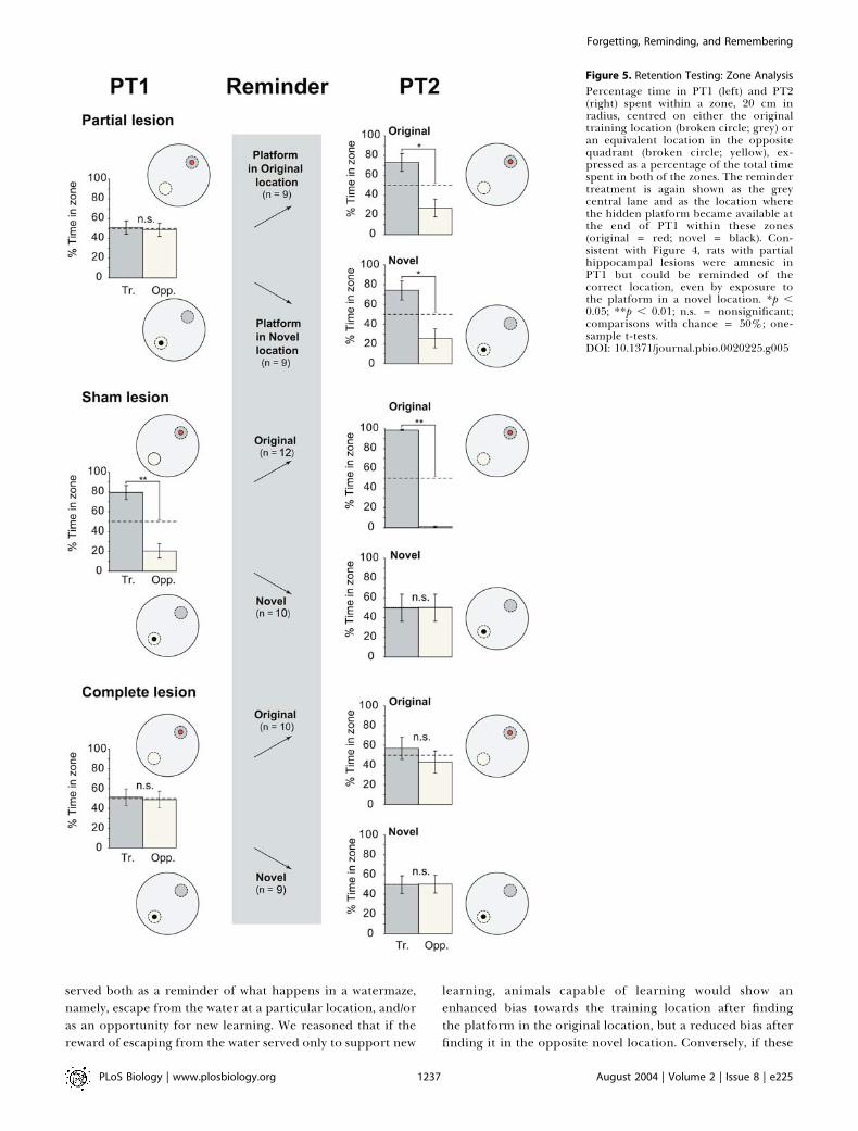
served both as a reminder of what happens in a watermaze,namely, escape from the water at a particular location, and/oras an opportunity for new learning. We reasoned that if thereward of escaping from the water served only to support new
learning, animals capable of learning would show anenhanced bias towards the training location after findingthe platform in the original location, but a reduced bias afterfinding it in the opposite novel location. Conversely, if these
Figure 5. Retention Testing: Zone Analysis
Percentage time in PT1 (left) and PT2(right) spent within a zone, 20 cm inradius, centred on either the originaltraining location (broken circle; grey) oran equivalent location in the oppositequadrant (broken circle; yellow), ex-pressed as a percentage of the total timespent in both of the zones. The remindertreatment is again shown as the greycentral lane and as the location wherethe hidden platform became available atthe end of PT1 within these zones(original = red; novel = black). Con-sistent with Figure 4, rats with partialhippocampal lesions were amnesic inPT1 but could be reminded of thecorrect location, even by exposure tothe platform in a novel location. *p ,0.05; **p , 0.01; n.s. = nonsignificant;comparisons with chance = 50%; one-sample t-tests.DOI: 10.1371/journal.pbio.0020225.g005
PLoS Biology | www.plosbiology.org August 2004 | Volume 2 | Issue 8 | e2251237
Forgetting, Reminding, and Remembering
events served only as reminder cues, they might be equallyeffective in reminding the rats of the original traininglocation.
The key new finding is that the partial lesion groupdisplayed a bias for the training quadrant that was equivalentwhether the animals had found the platform in the originaltraining location or in the novel opposite location, at the endof PT1. The overall ANOVA of the PT2 quadrant datarevealed a triple interaction of lesion group 3 quadrant(training versus opposite) 3 platform location duringreminding (novel versus original) (F[2, 53] = 19.28, p ,
0.001). With respect to the performance of the partial lesiongroup alone on this quadrant measure (see Figure 4, rightlane), there was a significant improvement between PT1 andPT2 (F[1, 16] = 7.98, p , 0.02) and no difference betweennovel and original reminding locations (F , 1). The partiallesion group also showed a highly significant preference forthe training quadrant versus the opposite quadrant on PT2(F[1, 16] = 16.83, p , 0.001). The same pattern of results isapparent in the zone data (see Figure 5) where, overall, thepartial lesion group displayed a significant improvementbetween PT1 and PT2 (F[1, 16] = 7.64, p , 0.02) that also didnot differ between ‘novel’ and ‘original’ groups (F , 1).Because a bias for the training location appeared even in theanimals that were exposed to a novel platform position,memory on PT2 cannot be attributed to relearning of theplatform location.
In contrast, sham-lesioned animals behaved quite differ-ently in PT2 as a function of whether the platform waspresented in the original or the novel location during thereminder treatment. Performance showed a further biastowards the training location between PT1 and PT2 followingthe event of climbing onto the escape platform in its originallocation, but exposure to the novel location resulted in areduction in time spent in the training zone—a partialreversal. Supported by significant interactions in the overallANOVA, analysis of time spent in the training quadrantrevealed that, as expected, sham-lesioned animals reexposedto the original location increased their time there betweenPT1 and PT2 (F[1, 11] = 12.41, p , 0.005). Conversely, sham-
lesioned animals exposed to the novel location exhibitedmodest reversal learning, increasing their time in theopposite quadrant (F[1, 9] = 9.35, p , 0.02). The samepattern of results was obtained from the analysis of time inthe training zone (Figure 5), for which a significantinteraction between PT (PT1 or PT2) and platform locationduring reminding (original versus novel) was observed (F[1,20] = 5.46, p , 0.05).Complete-lesioned rats performed at chance during all PTs
(see Figures 4 and 5, left and right lanes). That is, theirbehaviour during the retention tests before and after thereminder treatment showed no impact of that treatment.
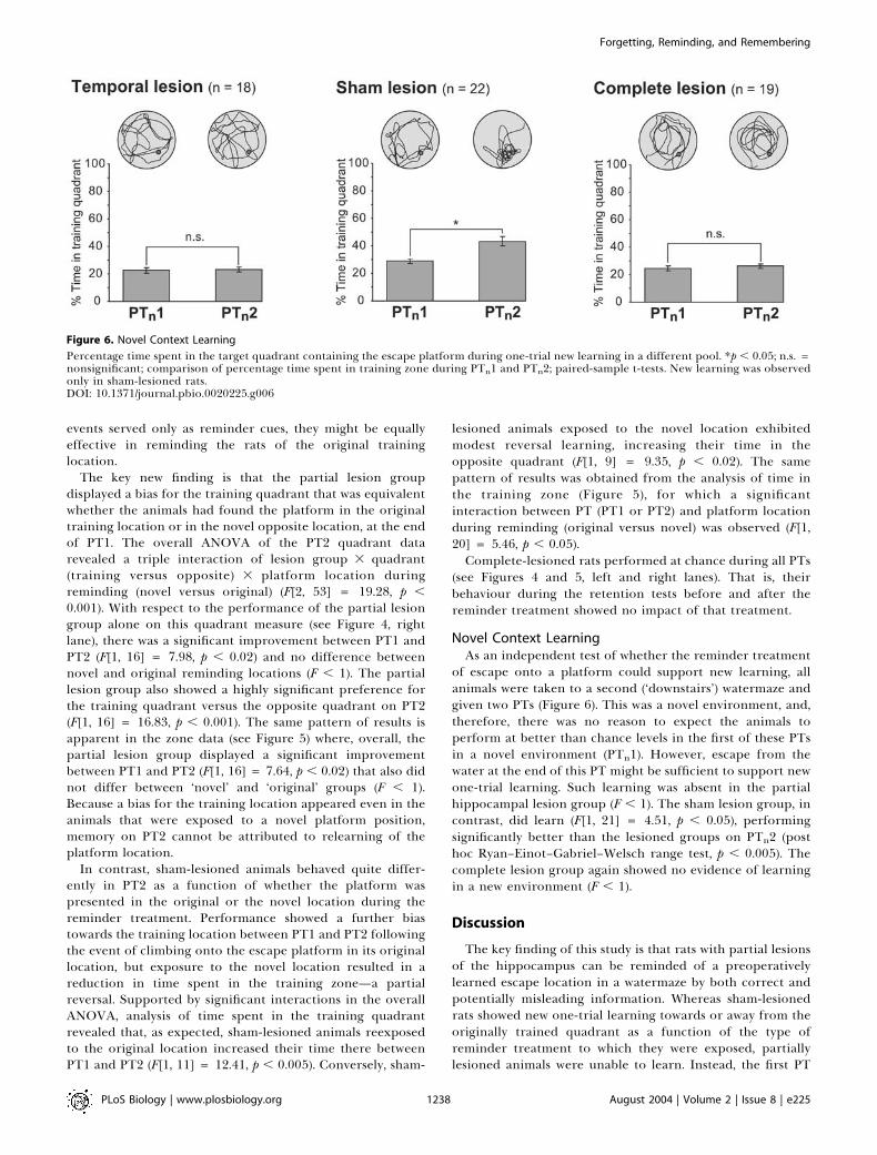
Novel Context LearningAs an independent test of whether the reminder treatment
of escape onto a platform could support new learning, allanimals were taken to a second (‘downstairs’) watermaze andgiven two PTs (Figure 6). This was a novel environment, and,therefore, there was no reason to expect the animals toperform at better than chance levels in the first of these PTsin a novel environment (PTn1). However, escape from thewater at the end of this PT might be sufficient to support newone-trial learning. Such learning was absent in the partialhippocampal lesion group (F , 1). The sham lesion group, incontrast, did learn (F[1, 21] = 4.51, p , 0.05), performingsignificantly better than the lesioned groups on PTn2 (posthoc Ryan–Einot–Gabriel–Welsch range test, p , 0.005). Thecomplete lesion group again showed no evidence of learningin a new environment (F , 1).
Discussion
The key finding of this study is that rats with partial lesionsof the hippocampus can be reminded of a preoperativelylearned escape location in a watermaze by both correct andpotentially misleading information. Whereas sham-lesionedrats showed new one-trial learning towards or away from theoriginally trained quadrant as a function of the type ofreminder treatment to which they were exposed, partiallylesioned animals were unable to learn. Instead, the first PT
Figure 6. Novel Context Learning
Percentage time spent in the target quadrant containing the escape platform during one-trial new learning in a different pool. *p , 0.05; n.s. =nonsignificant; comparison of percentage time spent in training zone during PTn1 and PTn2; paired-sample t-tests. New learning was observedonly in sham-lesioned rats.DOI: 10.1371/journal.pbio.0020225.g006
PLoS Biology | www.plosbiology.org August 2004 | Volume 2 | Issue 8 | e2251238
Forgetting, Reminding, and Remembering
served only as a reminder of the original platform locationirrespective of where in the pool the platform was raised atthe end of this trial. Rats with complete hippocampal lesionsshowed neither new learning nor reminding.
There is an extensive classic literature on the nature andeffectiveness of reminder treatments (Riccio and Richardson1984). Exposure to the training context, noncontingentstimuli, or additional training trials are just some examplesof methods successfully used to remind animals of a priortraining experience (Zinkin and Miller 1967; Miller andSpringer 1973; Mactutus et al. 1979; Gisquet Verrier andSchenk 1994; Przybyslawski and Sara 1997). Controversy did,however, surround studies that interpreted memory follow-ing a reminder treatment as evidence that the originalamnesia was the result of a retrieval deficit (Zinkin and Miller1967; Miller and Springer 1973). It was argued that areminding trial simply strengthens a weak memory that isbehaviourally unobservable, similar to what happens duringinitial learning (Cherkin 1972; Gold et al. 1973; Haycock et al.1973; Gold and King 1974), or that, when amnesia is complete,it results in one-trial learning or response generalization.However, manipulations that are unlikely to produce newlearning can also serve as effective reminders. Examplesinclude pharmacological manipulations of the internal state(Mactutus et al. 1980; Concannon and Carr 1982) andreexposure to the amnestic agent prior to retention testing(Thompson and Neely 1970; Hinderliter et al. 1975). In manysuch studies, however, the use of inhibitory avoidance as amemory test makes it difficult to determine the cognitive‘content’ (cf. Riccio and Richardson 1984) of the behaviourexpressed during retention testing. Although memory reac-tivation may have occurred when a rat inhibits movementthat previously led to electric shock, an alternative inter-pretation is that a generalized fear state has been induced.The issue of whether and when amnesia reflects a storage orretrieval deficit was, thus, left unresolved.
Two features are distinctive about our study. First, unlikein many previous studies, the reactivated memory involvesthe recall and expression of highly specific information—adiscriminable position in space, and not just a faster escapelatency, or greater freezing. Second, despite exposure to anovel platform location leading to reversal learning in thesham lesion group, the partial lesion group displayed onlyreminding of the original platform location. This distinctionis important because, with the current revival of interest inmemory retrieval, our protocol circumvents the ambiguitiesinvolved in the use of relearning as an index of retention.One example of a study that used a reacquisition rather thana true reminding protocol (Land et al. 2000) revealed that areminder prior to retention testing could alleviate amnesia inanimals with hippocampal lesions. However, it is difficult todistinguish between ‘pure’ reminding and the facilitation ofnew learning using reacquisition alone.
Nonetheless, the watermaze task is deceptively complex,and successful performance depends on the operation ofseveral distinct memory systems (Bannerman et al. 1995;Whishaw and Jarrard 1996; Warburton and Aggleton 1999;Eichenbaum 2000; White and McDonald 2002). Accordingly,while no new learning of the platform location occurs in thepartial and complete lesion groups, some ‘procedural’learning may take place during PT1; this may enhance aweak, subthreshold spatial memory to a point at which it can
be expressed in PT2. However, for this argument to beplausible, one would expect there to be minimal retention ofthe procedural components in PT1. This was clearly not thecase, as rats with both partial and complete hippocampallesions did not behave like naı̈ve animals during PT1. Theysearched at an appropriate distance from the pool walls andreadily climbed onto the escape platform when it waseventually made available. Procedural learning is also gen-erally well retained over time and, being slow, unlikely tochange much in one trial. We also doubt that the recovery ofmemory on PT2 reflects the emergence of latent memorymediated solely by an extrahippocampal structure, but notexpressed during PT1. For example, rats with completehippocampal lesions have been shown to learn a spatialconditioned-cued preference mediated by the amygdala(White and McDonald 1993), a form of memory that ispartially masked by hippocampus-dependent learning innormal rats (McDonald and White 1995). However, seeingreminding in partial but not complete hippocampus-lesionedanimals argues against this possibility in this case. Finally, therecovery of a simple stimulus–response strategy based onapproaching single cues is unlikely, as novel start locationswere always used during retention testing (cf. Eichenbaum etal. 1990; see Materials and Methods). Under these circum-stances, it is reasonable to interpret the apparently completeamnesia observed in PT1 as, at least in part, a failure ofspatial memory retrieval.Our use of partial hippocampal lesioning introduces
several other issues. First, it is a technique that is arguablymore relevant to human amnesia, in which damage to astructure is typically incomplete. Second, it is also relevant tothe many studies in which a pharmacological intervention isapplied at a single site within a brain region—microinfusioninto the dorsal hippocampus, for instance, is likely to haveminimal effects on ventral hippocampal tissue (see Steele andMorris 1999). Third, and perhaps most interesting, is thequestion of where memory traces are located. Given thatreminding only occurs in partially lesioned rats, it isreasonable to suppose that spatial memory traces are eitherlocated (and reactivated) within the hippocampus, or that thehippocampus is required for the process of reactivation orexpression of a reactivated memory stored elsewhere.According to the latter hypothesis, spatial memory tracesmight be stored in cortex but require fast synaptic trans-mission in the hippocampus to be retrieved (cf. Teyler andDiScenna 1986)—at least during the period after training andbefore the completion of systems-level consolidation. Alter-natively, some hippocampal tissue might be required forcortically expressed memory to gain access to striatal motorplanning and executive systems. Findings reported by Virleyet al. (1999) suggest that this retrieval hypothesis might not beimplausible. In this study, monkeys with CA1 pyramidal celllesions were amnesic for a preoperatively acquired visuospa-tial discrimination. Subsequent grafting of CA1 pyramidalcells resulted in the recovery of memory for a secondpreoperatively acquired discrimination. As the grafted tissuecannot contain specific memory traces, the implication is thatthe recovery of some aspects of CA1 cellular function issufficient for the information processing mediating theretrieval of memories stored elsewhere.In raising many more questions than they answer, the
present findings open a potential avenue of research into the
PLoS Biology | www.plosbiology.org August 2004 | Volume 2 | Issue 8 | e2251239
Forgetting, Reminding, and Remembering
neural dynamics of memory reactivation and retrieval.Specific interventions such as local AMPA receptor blockade(cf. Riedel et al. 1999) might be directed at the hippocampusor cortex during PT1 or PT2. Such a study could provideinformation about the role of these structures—and theirnetwork interactions—in the reactivation of apparently lostmemories, and in their subsequent retrieval. For example,hippocampal neural activity may be necessary for effectiveretrieval, but perhaps not for the reminding-inducedreactivation of memory, even for an ostensibly hippocam-pus-dependent task (cf. Land et al. 2000). Similarly, thenecessity for hippocampal neural activity during retrievalmight vary as a function of time after memory consolidation.In addition, the determinants of the reminder phenomenonitself remain unclear. It would be useful to establish whetherreinforcement in the form of an escape platform is, in fact,necessary during PT1, or indeed whether a reminder trial in aseparate pool would have been effective. Experimentsinvolving partial versus complete sets of cues might alsoprovide valuable insights into the reminding process (cf.O’Keefe and Conway 1978). These and related analyses will bethe subject of future studies.
Dissociating the storage and retrieval functions of thehippocampus in memory is central to our understanding ofthe role of hippocampo–cortical connections. Many theoriesof hippocampal function are based on the idea that thehippocampus acts as a mediating link between differentcortical regions during the interval before systems consol-idation is complete (Teyler and DiScenna 1986; Squire andAlvarez 1995). Paradoxically, the same features that point tothe alternative possibility—that the hippocampal formationis a site of encoding and long-term storage of complexmultimodal memories within its distributed intrinsic circui-try (Moscovitch and Nadel 1998)—also place this group ofstructures in an ideal position to help reactivate memoriesfrom traces distributed over several cortical structures,perhaps via a mechanism such as pattern completion (seeMarr 1971; Nakazawa et al. 2002). It is possible that, when thehippocampus is partially damaged and the cortico–hippo-campal network is therefore degraded, retrieval is onlypossible once a more complete recreation of the trainingsituation, possibly including reexposure to a platform, isprovided. Although comparisons across different species andforms of memory should be viewed with caution, this scenariois reminiscent of Tulving’s encoding specificity principle(Tulving and Pearlstone 1966; Thomson and Tulving 1970) inthat exposure to similar cues during encoding and retrievalphases permits the recovery of the original memory, despitethe provision of incorrect information about the targetlocation itself. Paradoxically, the poor learning abilities ofpartially lesioned rats might explain why a trial ending withexposure to a novel spatial location can serve as a reminderfor the original location—by limiting new learning of the newlocation, a reactivated memory for the old location isunmasked.
Materials and Methods
Subjects. We used a total of 73 male Lister Hooded rats obtainedfrom a commercial supplier (Charles River Laboratories, UnitedKingdom). They were pair-housed in plastic cages with sawdustbedding and ad libitum access to food and water. Their care and
maintenance and all experimental procedures were carried out inaccordance with United Kingdom Home Office Regulations.
Behavioural testing was conducted using two separate circularpools, 2.0 m in diameter and 60 cm high, each located in well-litrooms with numerous distal visual cues. One pool was used fortraining and retention (‘upstairs’) and the other for new contextlearning (‘downstairs’). The pools were filled with water at 25 8C 6 18C made opaque by the addition of 200 ml of latex liquid(Cementone-Beaver, Buckingham, United Kingdom). We used the‘Atlantis platform’ (Spooner et al. 1994), a polystyrene platform thatbecomes available by rising from the bottom of the pool only if theanimals swim to and stay within a specified ‘dwell radius’ centred onthe correct location for a predetermined ‘dwell time.’ When risen, thetop of the platform remained 1.5 cm below the water surface. Theanimals’ swimming was monitored by an overhead video cameraconnected to a video recorder and an online data acquisition system(Watermaze, Watermaze Software, Edinburgh, United Kingdom;Spooner et al. 1994) located in an adjacent room. This systemdigitizes the path taken by an animal and computes variousparameters such as escape latency, time spent in a zone overlyingthe platform, and other conventional measures of watermazeperformance.
Training protocol. Testing was carried out according to theschedule illustrated in Figure 1.
Cued pretraining. This phase consisted of a single day ofnonspatial cued training in the ‘upstairs’ watermaze (curtains drawnaround the pool to occlude extramaze cues, with ten trials in twosessions of five trials each (intertrial interval ’ 20 min; intersessioninterval ’ 3 h). The visible cue was suspended approximately 25 cmabove the platform, which was moved every two trials to one of fourpossible locations, according to a pseudorandom schedule; the dwellradius was set at 20 cm, and the dwell time was 1 s.
Training. Training on a spatial reference memory task began 3 dlater in the same watermaze. Rats received ten trials/day, in twosessions of five consecutive trials each (intersession interval ’ 2 h),for 4 d. The dwell time was set to 0.5 s throughout training, but thedwell radius was gradually reduced over days (day 1: 20 cm; day 2: 15cm; days 3 and 4: 13 cm). This schedule was intended to promoteaccurate and focused searching, but without generating the highlyperseverative strategy that typically results from the use of long dwelltimes (Riedel et al. 1999). Rats were given a maximum of 120 s to findan escape platform located at the centre of either the NE or SWquadrant, after which they remained on the platform for 30 s On therare trials in which a rat failed to escape within 2 min, theexperimenter placed a hand above the correct location in order toguide the animal to the platform. For each animal, the platformposition remained constant throughout training, but start locations(N, S, E, or W) were varied pseudorandomly across trials. Only thoseanimals achieving the acquisition criterion of mean escape latenciesof 15 s or less on day 4 of training proceeded to the next phase oftesting.
Surgery. Surgery took place 1–2 d after the end of training. Ratswere given either partial or complete bilateral neurotoxic lesions ofthe hippocampal formation (DG and CA fields), or sham surgery.Complete lesions were intended to remove 85% or more of the totalhippocampal volume. Partial lesions targeted the temporal two-thirdsof the hippocampus, sparing the septal (dorsal) third of the structure.The rats were assigned to groups of equivalent mean performance onthe basis of their escape latencies during the final day of training.Lesions were made with ibotenic acid (Biosearch Technologies,Novato, California, United States; dissolved in 0.1 M phosphate-buffered saline [pH 7.4] at 10 mg/ml) following the protocol of Jarrard(1989). The animals were anaesthetized with an intraperitonealinjection of tribromoethanol (avertin) and placed in a Kopf Instru-ments (Tujunga, California, United States) stereotaxic frame suchthat Bregma and Lambda lay on the same horizontal plane. Ratsreceived nine or 13 injections of ibotenic acid (partial and completelesion groups, respectively; 0.05 l1, 0.08 l1, or 0.1 l1 per injection) atdifferent rostrocaudal and dorsoventral levels via an SGE syringesecured to the stereotaxic frame (see de Hoz et al. 2003). Theinjection rate was 0.1 l1/min, and the needle was removed very slowly90 s after the injection. A total of 0.65 l1 or 0.91l1 per hemispherewas necessary for the partial and complete lesions, respectively. Thecoordinates were modified from Jarrard (1989) to suit the slightlydifferent brain size of Lister Hooded rats and to achieve the desiredamount of partial hippocampal damage (see de Hoz et al. 2003). Shamlesions were made in the same way, with the injections replaced by apiercing of the dura (intended to cause comparable neocorticaldamage).
Retention testing. This phase began 14 d after the end of training.
PLoS Biology | www.plosbiology.org August 2004 | Volume 2 | Issue 8 | e2251240
Forgetting, Reminding, and Remembering
It consisted of two PTs (PT1and PT2) spaced 1 h apart, with areminder treatment occurring at the end of PT1.
Each PT (PT1 and PT2) began with a standard 60-s swim with theplatform unavailable. In each PT, the rats were placed into the poolin either the adjacent right or the adjacent left quadrants withrespect to the training quadrant. Start positions were counter-balanced across PTs and across rats. At the end of the 60 s theplatform was raised and the animals were allowed to find and climbonto it. The rats were allowed a further 60 s to locate the platformonce risen (but still hidden just below the water surface); ifunsuccessful within this period, they were guided to the platform.They then remained on the platform for 30 s.
The raising of the platform at the end of PT1 constituted thereminder treatment; thus PT1 is sometimes referred to as the‘reminder trial.’ A key variable was that the platform was raised ineither the original training location (half the animals) or in a novellocation in the centre of the opposite quadrant of the pool (the otherhalf). Note that reminding using the original location always occurredin the training quadrant, and reminding using the novel locationalways occurred in the opposite quadrant. However, whereas theterms ‘training’ and ‘opposite’ are used to refer to physical areas ofthe pool, ‘novel’ and ‘original’ refer also to separate groups thatreceived each type of reminder.
For analysis of the different behavioural phases, several measuresof performance were assessed, including escape latency, swim speed,and time spent within defined regions of the pool. Memory retentionduring PTs is inferred from the time spent in each quadrant of thepool as a percentage of the 60-s duration of the PT. A more sensitivemeasure can be obtained by analysing percentage time spent within aspecified radius (zone) centred on the platform location (Moser andMoser 1998). When time in zone is presented, it is expressed as apercentage of the total time spent in both the original training zoneand the novel opposite zone. Statistical analysis (SPSS, Chicago,Illinois, United States) began with an ANOVA followed by appro-priate post hoc comparisons. Numerical data are reported as mean 6standard error (s.e.m.) throughout.
Novel context learning. New learning was assessed the next day in aseparate ‘downstairs’ watermaze that constituted a novel context. Theprotocol was identical to that used during ‘upstairs’ retention testing,i.e., two rewarded PTs (PTn1 and PTn2) spaced 1 h apart.
Lesion analysis. At the end of behavioural testing, rats wereperfused intracardially with saline followed by 10% formalin underterminal pentobarbitone anaesthesia (Euthatal, 1 ml). Their brainswere removed and stored in 10% formalin for 24 h before beingblocked and embedded in egg yolk. The embedding procedure is
described in de Hoz et al. (2003). Coronal, 30-lm sections through thehippocampus and other structures were cut using a cryostat: everyfifth section was recovered, mounted on a slide, and stained withcresyl violet (see Figure 3A–3C).
The relative volume of spared tissue was calculated by measuringthe area of hippocampus spared in each section of a particular brainaccording to the following protocol: Each coronal section containinghippocampus was placed under a photomacroscope (Wild, Heer-brugg, Switzerland), and the image taken by a mounted video camerawas imported into NIH Image 1.63 (National Institutes of Health,Bethesda, Maryland, United States). The area of spared hippocampaltissue in each section was then outlined and automatically calculated.Surrounding fibres such as the fimbria were excluded on the groundsthat they would not be considered in a section were all thehippocampal cells dead. The sections were spaced 150 lm apart,yielding up to 32 sections in a sham lesion animal, and fewer inanimals with acceptable partial lesions. For each rat, the totalhippocampal ‘volume’ was calculated by adding the area of hippo-campal tissue spared in each successive section. The proportion ofhippocampus spared for each lesioned animal was expressed as apercentage of the mean hippocampal ‘volume’ for sham-lesionedanimals. Values for the left and right hippocampi were initiallycalculated separately and then averaged (see Figure 3D).
Strict criteria for acceptance of a lesion were used. The lesion hadto be confined to the hippocampus in all cases, and leave intact tissuevolumes of 25%–50% in the septal hippocampus with minimalsparing (less than 10%) elsewhere in the structure in the case ofpartial lesions, or less than 15% total hippocampal sparing in the caseof complete lesions. Animals with minimal subicular damage,typically located at medial levels of the structure, were accepted.
Acknowledgments
We would like to thank Jane Knox for histology, Andrew Bernard foranimal care, and David Foster for helpful discussion. This researchwas supported by a Medical Research Council (MRC) ProgrammeGrant held by RGMM and an MRC Research Fellowship held by LdH.
Conflicts of interest. The authors have declared that no conflicts ofinterest exist.
Author contributions. LdH, SJM, and RGMM conceived anddesigned the experiments. LdH and SJM performed the experiments.LdH and SJM analysed the data. LdH, SJM, and RGMM wrote thepaper. &
ReferencesBannerman DM, Good MA, Butcher SP, Ramsay M, Morris RGM (1995) Distinct
components of spatial learning revealed by prior training and NMDAreceptor blockade. Nature 378: 182–186.
Cherkin A (1972) Retrograde amnesia in the chick: Resistance to the remindereffect. Physiol Behav 8: 949–955.
Concannon JT, Carr C (1982) Pre-test epinephrine injections reverse DDC-induced retrograde amnesia. Physiol Behav 9: 443–448.
Davis HP, Squire LR (1984) Protein synthesis and memory: A review. PsycholBull 96: 518–559.
deHoz L,Knox J,Morris RGM (2003) Longitudinal axis of the hippocampus: Bothseptal and temporal poles of the hippocampus support watermaze spatiallearning depending on the training protocol. Hippocampus 13: 587–603.
D’Hooge R, De Deyn PP (2001) Applications of the Morris water maze in thestudy of learning and memory. Brain Res Rev 36: 60–90.
Dudai Y, Morris RGM (2000) To consolidate or not to consolidate: What are thequestions? In: Bolhuis JJ, editor. Brain, perception, memory. Oxford: OxfordUniversity Press. pp. 149–162.
Eichenbaum H (2000) A cortical-hippocampal system for declarative memory.Nat Rev Neurosci 1: 41–50.
Eichenbaum H, Stewart C, Morris RGM (1990) Hippocampal representation inplace learning. J Neurosci 10: 3531–3542.
Gisquet Verrier P, Schenk F (1994) Selective hippocampal lesions in rats do notaffect retrieval processes promoted by prior cuing with the conditionedstimulus or the context. Psychobiology 22: 289–303.
Gold PE, King RA (1974) Retrograde amnesia: Storage failure versus retrievalfailure. Psychol Rev 81: 465–469.
Gold PE, Haycock JW, Marri J, McGaugh JL (1973) Retrograde amnesia and the‘‘reminder effect’’: An alternative interpretation. Science 180: 1199–1201.
Graf P, Squire LR, Mandler G (1984) The information that amnesic patients donot forget. J Exp Psychol Learn Mem Cogn 10: 164–178.
Haycock JW, Gold PE, Macri J, McGaugh JL (1973) Noncontingent footshockattenuation of retrograde amnesia: A generalization effect. Physiol Behav11: 99–102.
Hinderliter CF, Webster T, Riccio DC (1975) Amnesia induced by hypothermiaas a function of treatment-test interval and recooling in rats. Anim LearnBehav 2: 257–263.
Jarrard LE (1989) On the use of ibotenic acid to lesion selectively differentcomponents of the hippocampal formation. J Neurosci Methods 29: 251–259.
Land C, Bunsey M, Riccio DC (2000) Anomalous properties of hippocampallesion-induced retrograde amnesia. Psychobiology 28: 476–485.
Mactutus CF, Riccio DC, Ferek JM (1979) Retrograde amnesia for old(reactivated) memory: Some anomalous characteristics. Science 204: 1319–1320.
Mactutus CF, Smith RL, Riccio DC (1980) Extending the duration of ACTH-induced memory reactivation in an amnesic paradigm. Physiol Behav 24:541–546.
Marr D (1971) Simple memory: A theory for archicortex. Philos Trans R SocLond B Biol Sci 262: 23–81.
McClelland JL, McNaughton BL, O’Reilly RC (1995) Why there are comple-mentary learning systems in the hippocampus and neocortex: Insights fromthe successes and failures of connectionist models of learning and memory.Psychol Rev 102: 419–457.
McDonald R, White N (1995) Information acquired by the hippocampusinterferes with acquisition of the amygdala-based conditioned-cue prefer-ence in the rat. Hippocampus 5: 189–197.
McGaugh JL (1966) Time-dependent processes in memory storage. Science 153:1351–1358.
Miller RR, Springer AD (1972) Induced recovery of memory in rats followingelectroconvulsive shock. Physiol Behav 8: 645–651.
Miller RR, Springer AD (1973) Amnesia, consolidation, and retrieval. PsycholRev 80: 69–79.
Moscovitch M, Nadel L (1998) Consolidation and the hippocampal complexrevisited: In defense of the multiple-trace model. Curr Opin Neurobiol 8:297–300.
Moser M-B, Moser EI (1998) Distributed encoding and retrieval of spatialmemory in the hippocampus. J Neurosci 18: 7535–7542.
Nakazawa K, Quirk MC, Chitwood RA, Watanabe M, Yeckel MF, et al. (2002)
PLoS Biology | www.plosbiology.org August 2004 | Volume 2 | Issue 8 | e2251241
Forgetting, Reminding, and Remembering
Requirement for hippocampal CA3 NMDA receptors in associative memoryrecall. Science 297: 211–218.
O’Keefe J, Conway DH (1978) Hippocampal place units in the freely movingrat: Why they fire where they fire. Exp Brain Res 31: 573–590.
Przybyslawski J, Sara SJ (1997) Reconsolidation of memory after its reactiva-tion. Behav Brain Res 84: 241–246.
Ribot TH (1883) The diseases of memory. Fitzgerald J, translator. New York: JFitzgerald. 48 p.
Riccio DC, Richardson R (1984) The status of memory following experimentallyinduced amnesias: Gone, but not forgotten. Physiol Psychol 12: 59–72.
Riedel G, Micheau J, Lam AG, Roloff E, Martin SJ, et al. (1999) Reversible neuralinactivation reveals hippocampal participation in several memory pro-cesses. Nat Neurosci 2: 898–905.
Sanders HI, Warrington EK (1971) Memory for remote events in amnesicpatients. Brain 94: 661–668.
Sara SJ (1999) Retrieval and reconsolidation: Toward a neurobiology ofremembering. Learn Mem 7: 73–84.
Spear NE (1973) Retrieval of memory in animals. Psychol Rev 80: 163–194.Spooner RI, Thomson A, Hall J, Morris RGM, Salter SH (1994) The Atlantis
platform: A new design and further developments of Buresova’s on-demandplatform for the water maze. Learn Mem 1: 203–211.
Squire LR (1992) Memory and the hippocampus: A synthesis from findings withrats, monkeys, and humans. Psychol Rev 99: 195–231.
Squire LR, Alvarez P (1995) Retrograde amnesia and memory consolidation: Aneurobiological perspective. Curr Opin Neurobiol 5: 169–177.
Steele RJ, Morris RGM (1999) Delay-dependent impairment of a matching-to-place task with chronic and intrahippocampal infusion of the NMDA-antagonist D-AP5. Hippocampus 9: 118–136.
Teyler TJ, DiScenna P (1986) The hippocampal memory indexing theory. BehavNeurosci 100: 147–154.
Thompson C, Neely JE (1970) Dissociated learning in rats produced byelectroconvulsive shock. Physiol Behav 5: 783–786.
Thomson DM, Tulving E (1970) Associative encoding and retrieval: Weak andstrong cues. J Exp Psychol 86: 255–262.
Tulving E, Pearlstone Z (1966) Availability versus accessibility of information inmemory for words. J Verb Learn Verb Behav 5: 381–391.
Virley D, Ridley RM, Sinden JD, Kershaw TR, Harland S, et al. (1999) PrimaryCA1 and conditionally immortal MHP36 cell grafts restore conditionaldiscrimination learning and recall in marmosets after excitotoxic lesions ofthe hippocampus CA1 field. Brain 122: 2321–2335.
Warburton EC, Aggleton JP (1999) Differential deficits in the Morris watermaze following cytotoxic lesions of the anterior thalamus and fornixtransection. Behav Brain Res 98: 27–38.
Warrington EK, Weiskrantz L (1968) New method of testing long-termretention with special reference to amnesic patients. Nature 217: 972–974.
Whishaw IQ, Jarrard LE (1996) Evidence for extrahippocampal involvement inplace learning and hippocampal involvement in path integration. Hippo-campus 6: 513–524.
White NM, McDonald RJ (1993) Acquisition of a spatial conditioned placepreference is impaired by amygdala lesions and improved by fornix lesions.Behav Brain Res 55: 269–281.
White NM, McDonald RJ (2002) Multiple parallel memory systems in the brainof the rat. Neurobiol Learn Mem 77: 125–184.
Zinkin S, Miller AJ (1967) Recovery of memory after amnesia induced byelectroconvulsive shock. Science 155: 102–104.
PLoS Biology | www.plosbiology.org August 2004 | Volume 2 | Issue 8 | e2251242
Forgetting, Reminding, and Remembering