folding very short peptides using molecular dynamics
TRANSCRIPT
Folding Very Short Peptides UsingMolecular DynamicsBosco K. Ho
*, Ken A. Dill
Department of Pharmaceutical Chemistry, University of California San Francisco, San Francisco, California, United States of America
Peptides often have conformational preferences. We simulated 133 peptide 8-mer fragments from six differentproteins, sampled by replica-exchange molecular dynamics using Amber7 with a GB/SA (generalized-Born/solvent-accessible electrostatic approximation to water) implicit solvent. We found that 85 of the peptides have no preferredstructure, while 48 of them converge to a preferred structure. In 85% of the converged cases (41 peptides), thestructures found by the simulations bear some resemblance to their native structures, based on a coarse-grainedbackbone description. In particular, all seven of the b hairpins in the native structures contain a fragment in the turnthat is highly structured. In the eight cases where the bioinformatics-based I-sites library picks out native-likestructures, the present simulations are largely in agreement. Such physics-based modeling may be useful foridentifying early nuclei in folding kinetics and for assisting in protein-structure prediction methods that utilize theassembly of peptide fragments.
Citation: Ho BK, Dill KA (2006) Folding very short peptides using molecular dynamics. PLoS Comput Biol 2(4): e27. DOI: 10.1371/journal.pcbi.0020027
Introduction
Peptide fragments of proteins often have intrinsic propen-sities for the formation of their native conformations. Forexample, NMR experiments [1] show that long peptidefragments have native-like conformations [2–7]. Some shortpeptides in solution have also been shown to adopt their nativesecondary structures: a helices [8,9] and b hairpins [10–14].
As a consequence, peptide conformational propensitiesthat are taken from the protein databank (PDB) [1–17] arenow widely used in protein-structure prediction algorithms.A popular set of peptide fragment conformations is the I-siteslibrary of David Baker and his co-workers [18,19]. Extensivelibraries of peptide fragments have now been compiled [20–22] and have become essential elements in protein-predictionmethods [23]. From the recent CASP protein–structureprediction competition, it was noted that most of thesuccessful de novo methods use a fragment-based approach[23,24]. Typically, a candidate protein native structure isspliced together from fragments that are extracted from adatabase of conformations, and then treated to conforma-tional scoring and optimization.
Can physical models capture these conformational pro-pensities of peptides? There is good evidence that they can.First, simple physical models can reproduce the structuralbiases of certain peptide fragments [25–28]. To date, however,such studies have largely focused on selected peptides that areexpected to fold. Our interest here is to know whetherphysical models can also discriminate peptides that fold frompeptides that do not. Second, in molecular dynamicssimulations of small peptides, the ensemble of conformersdivides into well-defined clusters. This has been found for apenta–b peptide in explicit water [29,30], and for a small a-helical peptide [31]. Third, molecular dynamic simulations ofsmall peptides reproduce the a-helical propensities of certainfragments from the I-sites sequence-structure library [32].Many models of protein folding kinetics assume that peptidefragments of the chain that have preferred conformations areresponsible for nucleating the folding process [33–35].
Here, we study 133 peptide 8-mer fragments from sixdifferent proteins of different folds, using replica-exchangemolecular dynamics sampling [36] in Amber7, with theparm96 parameters and the GB/SA (generalized-Born/sol-vent-accessible electrostatic approximation to water) implicitsolvent model of Tsui and Case [37]. We chose this force fieldas it is the only implicit-solvation model that can adequatelyreproduce the native state of the b hairpin of protein G [38].We are interested in whether this physical model can
identify native-like secondary structures in peptide frag-ments. If so, it indicates the importance of local interactionsin those cases. Our study involves complete coverage of thoseproteins. For each protein, we systematically generate a seriesof 8-mer peptide fragments with overlapping sequences fromthe original protein sequence. Neighboring fragments have afive-residue overlap (and three-residue gap). We chose 8-mersbecause this length appears adequate to identify elements ofstructure in PDB studies [19] and because much longerfragments become too expensive for computer simulations.We simulate each peptide using 16 replicas for 5 ns/replica,and keep only the last 1 ns.In each case, we determine whether the peptide has
converged to its native conformation in the folded protein.We consider two measures of convergence. First, we monitorthe RMSD between the simulated conformations and theexperimental PDB structure of that peptide. However, for
Editor: Diana Murray, Weill Medical College of Cornell University, United States ofAmerica
Received December 22, 2005; Accepted February 20, 2005; Published April 14,2006
DOI: 10.1371/journal.pcbi.0020027
Copyright: � 2006 Ho and Dill. This is an open-access article distributed under theterms of the Creative Commons Attribution License, which permits unrestricteduse, distribution, and reproduction in any medium, provided the original authorand source are credited.
Abbreviations: GB/SA, generalized-Born solvent-accessible electrostatic approx-imation to water; PDB, protein databank; RMSD, root-mean-square deviation
* To whom correspondence should be addressed. E-mail: [email protected]
PLoS Computational Biology | www.ploscompbiol.org April 2006 | Volume 2 | Issue 4 | e270001

prediction purposes—determining whether a peptide has aconverged structure in the absence of knowledge of its nativestructure—we develop another measure based on the backbonemesostring, which is a coarse-grained description of thebackbone conformational ensemble.
A mesostring is a one-dimensional list of the mesostates ofeach residue in a peptide. A mesostate refers to a discreteregion of the /–w angles of the backbone of a residue.Mesostate [a] corresponds to a helical conformation, includ-ing the a helix, 310 helix, or p helix. Mesostate [b] correspondsto an extended b-strand conformation. Mesostate [l] corre-sponds to a left-handed helical conformation.
We use the mesostrings to cluster conformations in oursimulations. Based on the three mesostates described above,an 8-mer has 38 ¼ 6,561 possible mesostrings. When eachsimulation is completed, each 8-mer peptide will havedifferent populations for the 6,561 mesostrings, hencedifferent free energies. The mesostring that represents thehighest population (the lowest free energy) is called the groundmesostring. We use the properties of the ground mesostring todetermine structural bias in a peptide. The ground meso-strings are classified in terms of either a reverse-turn or ahelical-turn conformation (see Figure 1). We define a helical-turn as a mesostring that contains at least four [a] mesostatesin a row, and a reverse-turn as a mesostring that containseither the [bab] or [baab] motifs.
How do we know when a simulation has converged? Wecalculate the backbone entropy using the Boltzmann formulaS¼�k Ri pi ln pi, where pi is the probability that the peptide isin mesostring i. The backbone entropy is calculated over acertain window in a trajectory, where the sum is made overonly the mesostrings that are observed in the window. Thebackbone entropy S is useful for two purposes. First, itmeasures for a given peptide the sharpness of the distributionof probabilities of the mesostrings. The more peaked thedistribution is, and thus the more favored a mesostring is, thelower is the backbone entropy. In this way, the backboneentropy indicates whether any one conformation is substan-tially favored over the others, for the given peptide. Second,the backbone entropy should converge at equilibrium,approaching an asymptotic value with time in the simulation.
Even if a new mesostring emerges late within the sampling (asis often the case), it only changes the backbone entropy if ithas a significant population. We use the convergence of thebackbone entropy to indicate the convergence of thesimulation.We study peptide fragments extracted from a series of well-
characterized proteins: protein G, protein L, protein A, anda-spectrin, and chymotrypsin inhibitor. For each peptide, wesimulate the ensemble of states at equilibrium. We find thatsome of these peptides exhibit strong structural biases. Weanalyze the relationship of those structural biases to thetopology of the native structure.
Results/Discussion
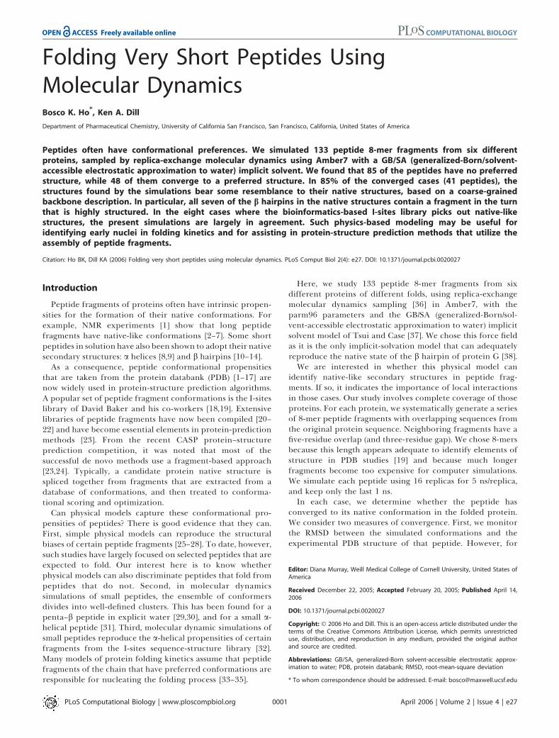
Structural Bias in the Peptide Conformation EnsembleDo peptides have native-like conformations? Figure 2
shows the simulated free-energy profiles of RMSD for thepeptides of protein G. We call the region of RMSD , 2 Anative-like. We find that some fragments spend a significantamount of time near their native structures (seq3, seq9, andseq10). Some peptides have a broad conformational distri-bution (seq14), while others have a narrow distribution(seq16). Narrow distributions indicate structural bias in thepeptide. To investigate this structural bias further, we list inTable 1 the lowest free-energy mesostrings of several protein
Figure 1. Representative Snapshots of Various Peptides from Protein G
The representative snapshot is the snapshot in the ground mesostringwith the lowest energy. The ground mesostring of seq1 and seq3 areclassed as reverse-turns, and the ground mesostring of seq9 and seq16are classed as helical-turns.DOI: 10.1371/journal.pcbi.0020027.g001
PLoS Computational Biology | www.ploscompbiol.org April 2006 | Volume 2 | Issue 4 | e270002
Synopsis
To carry out specific biochemical reactions, proteins must adoptprecise three-dimensional conformations. During the folding of aprotein, the protein picks out the right conformation out of billionsof other conformations. It is not yet possible to do this computa-tionally. Picking out the native conformation using physics-basedatomically detailed models, sampled by molecular dynamics, ispresently beyond the reach of computer methods. How can wespeed up computational protein-structure prediction? One idea isthat proteins start folding at specific parts of a chain that kink upearly in the folding process. If we can identify these kinks, we shouldbe able to speed up protein-structure prediction. Previous studieshave identified likely kinks through bioinformatic analysis of existingprotein structures. The goal of the authors here is to identify theseputative folding initiation sites with a physical model instead. In thisstudy, Ho and Dill show that, by chopping a protein chain intopeptide pieces, then simulating the pieces in molecular dynamics,they can identify those peptide fragments that have conformationalbiases. These peptides identify the kinks in the protein chain.
Short Peptides in Molecular Dynamics
G peptides. We show in Figure 1, a representative conforma-tion of the ground mesostrings of these peptides.
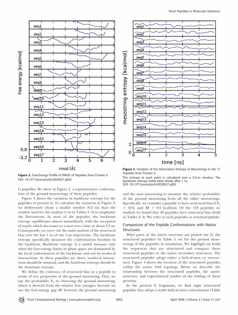
Figure 3 shows the variation in backbone entropy for thepeptides of protein G. To calculate the variation in Figure 3,we deliberately chose a smaller window (0.2 ns) than thewindow used for the analysis (1 ns in Tables 1–4) to emphasizethe fluctuations. In most of the peptides, the backboneentropy equilibrates almost immediately, with the exceptionof seq16, which decreases to a near zero value at about 3.5 ns.Consequently, we carry out the main analysis of the structuralbias over the last 1 ns of our 5-ns trajectories. The backboneentropy specifically measures the conformation freedom inthe backbone. Backbone entropy is a useful measure onlywhen the free-energy basins in phase space are dominated bythe local conformation of the backbone, and not by nonlocalinteractions. As these peptides are short, nonlocal interac-tions should be minimal, and the backbone entropy should bethe dominant entropy.
We define the existence of structural bias in a peptide interms of two properties of the ground mesostring. First, weuse the probability P1 in observing the ground mesostring,which is derived from the relative free energies. Second, weuse the free-energy gap DF between the ground mesostring
and the next mesostring to measure the relative probabilityof the ground mesostring from all the other mesostrings.Specifically, we consider a peptide to have structural bias if P1
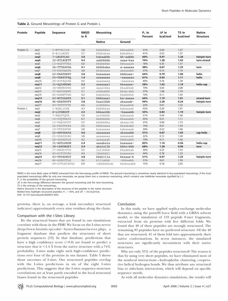
. 45% and DF . 0.6 kcal/mol. Of the 133 peptides westudied, we found that 48 peptides have structural bias (boldin Tables 2–4). We refer to such peptides as structured peptides.
Comparison of the Peptide Conformations with Native
StructuresWhat parts of the native structure are picked out by the
structured peptides? In Table 5, we list the ground meso-strings of the peptides in simulation. We highlight (in bold)the sequences that are structured and compare thesestructured peptides to the native secondary structures. Thestructured peptides adopt either a helical-turn or reverse-turn. Figure 4 shows the location of the structured peptideswithin the native fold topology. Below we describe therelationship between the structured peptides, the nativestructure, and experimental studies of the folding of theseproteins.In the protein G fragments, we find eight structured
peptides that adopt a stable helical-turn conformation (Table
Figure 2. Free-Energy Profile of RMSD of Peptides from Protein G
DOI: 10.1371/journal.pcbi.0020027.g002
Figure 3. Variation of the Information Entropy of Mesostrings in the 17
Peptides from Protein G
The entropy at each point is calculated over a 0.2-ns window. Thebackbone entropy holds fairly steady after 1 ns.DOI: 10.1371/journal.pcbi.0020027.g003
PLoS Computational Biology | www.ploscompbiol.org April 2006 | Volume 2 | Issue 4 | e270003
Short Peptides in Molecular Dynamics

2). Three of these helical-turns pick out the lone a helix in thenative structure, another helical-turn picks out the turnbetween the helix and N-terminal b hairpin, and theremaining two helical-turns pick out the turn in the C-terminal b hairpin. Another two structured peptides withoverlapping sequences adopt a stable reverse-turn conforma-tion, which both pick out the same N-terminal hairpin-turnin the native structure. The isolated C-terminal b hairpin hasbeen found experimentally to be stable [10], where thisstability is reflected in the structural bias found in the peptidefragments of the hairpin-turn. The structured peptidesprovide an explanation for an ingenious study of secondarystructure in protein G [39]. In that experiment, Minor andKim replaced the a-helix sequence with a sequence based onthe C-terminal hairpin. The mutant was able to fold into thesame topology, showing that there is a helical propensity inthe C-terminal hairpin. In the peptide studies, we find helical-turns in both the a helix and the turn of the C-terminalhairpin, which demonstrates the interchangeability of thesetwo sequences in our simulations.
In the protein L fragments, we find four structuredpeptides that adopt a stable helical-turn conformation (Table2). Two of the helical-turns pick out the a helix, while theother two helical-turns pick out the two hairpin-turns in thenative structure. Another structured peptide adopts areverse-turn conformation, which picks out the C-cap ofthe a helix.
In the fragments of the B domain of protein A, we foundthree structured peptides that adopt a stable helical-turnconformation (Table 3). These helical-turns pick out helix IIand helix III, and the turn between these helices. The stabilityof these pieces is consistent with experimental studies ofprotein A fragments, which show that helix II and helix IIIform a stable intermediate [40].
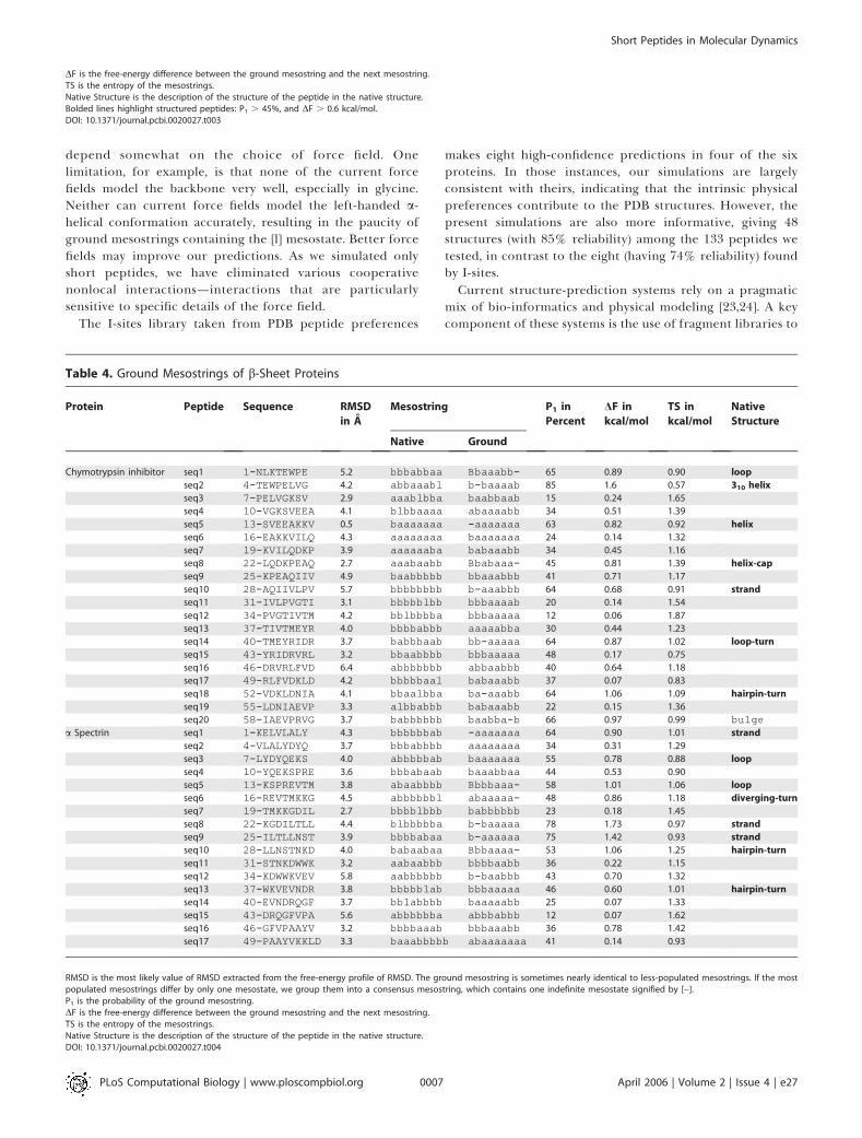
In the myoglobin fragments, 13 structured peptides adopt astable helical-turn conformation (Table 4). These helical-turnspick out six of the eight a helices in the native structure—withparticularly long helical-turns in helices A, G, and H. Anotherthree structured peptides adopt a stable reverse-turn con-formation. Two of the reverse-turns pick out the same turnbetween helices G–H. The large amount of structural biasfound in the fragments of helices G and H is consistent withexperimental studies, which show that helices G and H form astable intermediate [41]. Experimentally, helix F has theweakest helical propensity, and correspondingly we do notfind any structured peptides in fragments of helix F.In the chymotrypsin inhibitor fragments, we found eight
structured peptides that adopt a stable helical-turn con-formation (Table 4). One helical-turn picks out the 310 helixin the native structure, two helical-turns pick out the a helix,one helical-turn picks out a diverging turn, and one helical-turn picks out the turn in the b hairpin. Two helical-turnserroneously pick out b strands. We also found a structuredpeptide that adopts a reverse-turn conformation. Thisreverse-turn picks out the bulge in a b strand. Experimentalstudies find that only the a helix is stable [42].In the a-spectrin fragments, there are eight structured
peptides that adopt stable helical-turn conformations. Two ofthe helical-turns erroneously pick out the RT loop. Theconformation of the RT loop is somewhat indeterminate asboth experimental and simulation studies (unpublished data)show that the RT loop is unstable. Another helical-turnoverlaps with a diverging b-turn in the native structure.Three helical-turns erroneously pick out a b strand. Theother two helical-turns pick out the turns of the two bhairpins. Experimental studies find that only a fragment ofthe last b hairpins has structure in solution [43].Overall, of the 41 structured peptides that adopt a stable
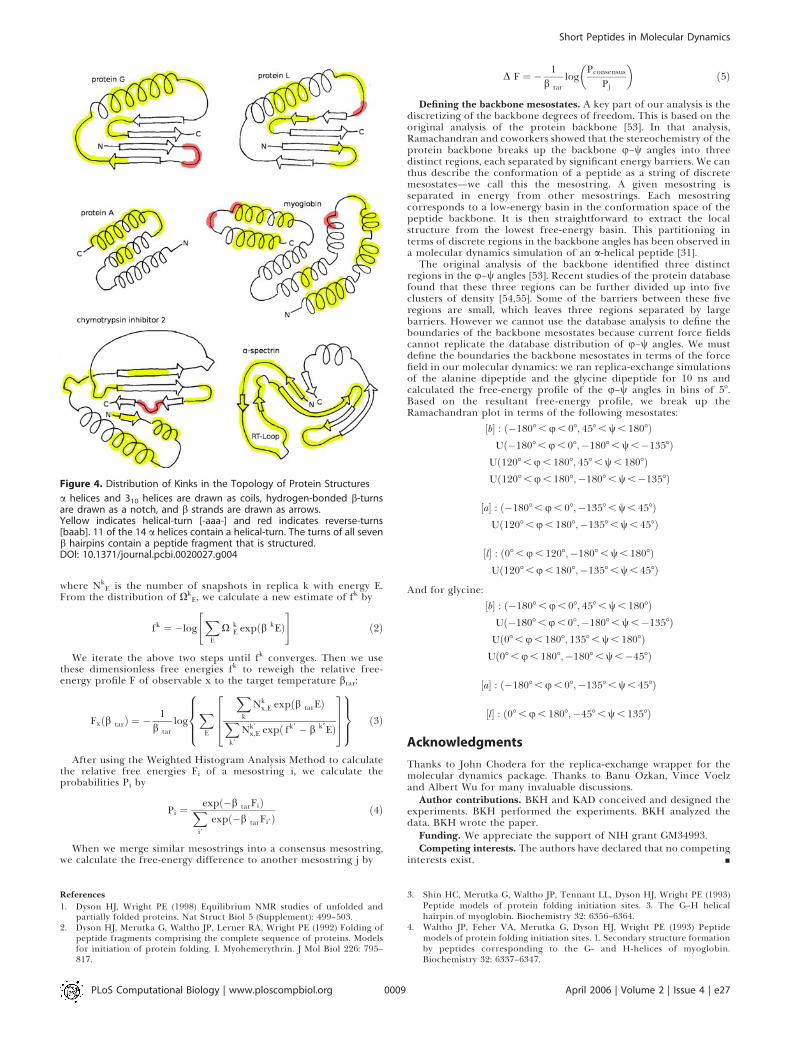
helical-turn conformation, 21 pick out a helices, three pickout 310 helices, and two overlap with diverging turns. Becausehelical motifs can be considered a continuum from divergingb-turns, to 310 helices, to a helices [44,45], we conclude that 26of the helical-turns pick out helical motifs in the nativestructures. Another seven helical-turns pick out b-hairpin–turns, and one helical-turn is found in a helix hairpin-turn.Five helical-turns erroneously pick out b strands and twoother helical-turns erroneously pick out the RT loop.We find six structured peptides that adopt a reverse-turn
conformation: one is found at a hairpin turn, two are foundat strand–helix turns, three are found at helix–helix turns,and one is found at a b-strand bulge.There is some debate [46] over whether b hairpins fold via
the turn [47] or through hydrophobic clustering [48]. Theresults here suggest that structural bias at the turn is veryimportant. We find that all seven b hairpins in the sixproteins contain a fragment in the turn that results in astructured peptide. If we interpret the structural bias in thepeptide as a kink in the full chain, then the formation ofstructure can be regarded as contacts coalescing around akinky chain. In terms of the b hairpin, this does notnecessarily mean that the turn forms first but that a kinkfavors the formation of nearby contacts.In summary, of the 48 structured peptides found in the
simulations, only five differ significantly from the nativestructure. Given that there are 436 residues in our six
Table 1. Mesostrings of Various Peptides from Protein G
Peptide Mesostring Free Energy of
the Mesostring
in kcal/mol
P in Percent
seq1: 1-MTYKLILN bbbaabbb �3.09 31
babaaaaa �2.59 12
baaaabba �2.54 11
baaaabbb �2.51 11
babaabbb �2.31 7
babaabba �2.26 7
seq3: 7-LNGKTLKG bbbaabbb �3.29 44
bblaabbb �2.77 16
abbaabbb �2.50 10
bbbaabbl �2.03 4
ablaabbb �1.88 3
seq9: 25-TAEKVFKQ baaaaaaa �3.16 39
aaaaaaaa �3.00 26
abaaaaaa �2.53 11
bbaaaaaa �2.09 5
aaaaaaab �1.97 4
bbblaaaa �1.87 3
seq16: 46-DDATKTFT abaaaabb �3.69 92
abaaaaba �1.71 2
aaaaabbb �1.42 1
baaaaaaa �1.39 1
DOI: 10.1371/journal.pcbi.0020027.t001
PLoS Computational Biology | www.ploscompbiol.org April 2006 | Volume 2 | Issue 4 | e270004
Short Peptides in Molecular Dynamics
proteins, there is, on average, a kink (secondary structuralindicator) approximately every nine residues along the chain.
Comparison with the I-Sites LibraryDo the structural biases that are found in our simulations
correlate with those in the PDB? We focus on the I-sites server(http://www.bioinfo.rpi.edu/;bystrc/hmmstr/server.php), afragment database that predicts the structures of shortprotein sequences [19]. In that database, predictions thathave a high confidence score (.0.8) are found to predict astructure that is ,1.4 A from the native structure with a 74%probability. I-sites make eight such high-confidence predic-tions over four of the proteins in our dataset. Table 5 showsthose successes of I-sites. Our structured peptides overlapwith the I-sites predictions in six of the eight I-sitespredictions. This suggests that the I-sites sequence-structurecorrelations are at least partly encoded in the local structuralbiases found in the structured peptides.
ConclusionIn this study, we have applied replica-exchange molecular
dynamics, using the parm96 force field with a GB/SA solventmodel, to the simulation of 133 peptide 8-mer fragments,extracted from six proteins with five different folds. Wefound that 48 of these peptides are strongly structured. Theremaining 85 peptides have no preferred structure. Of the 48that are structured, 41 of them fold into approximately theirnative conformations. In seven instances, the simulatedstructures are significantly inconsistent with their nativestructures.Why are only 35% of the peptides structured? The reason is
that by using very short peptides, we have eliminated most ofthe nonlocal interactions—hydrophobic clustering, coopera-tive helical hydrogen bonds. We thus attribute any structuralbias to sidechain interactions, which will depend on specificsequence motifs.As with all molecular dynamics simulations, the results will
Table 2. Ground Mesostrings of Protein G and Protein L
Protein Peptide Sequence RMSD
in A
Mesostring P1 in
Percent
DF in
kcal/mol
TS in
kcal/mol
Native
Structure
Native Ground
Protein G seq1 1-MTYKLILN 5.8 bbbbbbba bbbaabbb 31% 0.50 1.27
seq2 4-KLILNGKT 5.7 bbbbabaa Bababba- 41% 0.53 1.37
seq3 7-LNGKTLKG7-LNGKTLKG 3.0 babaabbbbabaabbb bb-aabbbbb-aabbb 60% 0.97 1.20 hairpin-turn
seq4 10-KTLKGETT10-KTLKGETT 4.5 aabbbbbbaabbbbbb aaaa-baaaaaa-baa 70% 1.28 1.03 turn-strand
seq5 13-KGETTTEA 6.0 bbbbbbbb bbbaaaab 18% 0.35 1.57
seq6 16-TTTEAVDA16-TTTEAVDA 4.1 bbbbbababbbbbaba a-aaaaaaa-aaaaaa 48% 0.67 1.33 turn
seq7 19-EAVDAATA 3.8 bbabaaaa abaaaabb 37% 0.15 1.19
seq8 22-DAATAEKV22-DAATAEKV 3.6 baaaaaaabaaaaaaa bbbbaaa-bbbbaaa- 64% 0.79 1.00 helix
seq9 25-TAEKVFKQ25-TAEKVFKQ 1.4 aaaaaaaaaaaaaaaa -aaaaaaa-aaaaaaa 61% 0.93 1.11 helix
seq10 28-KVFKQYAN 0.5 aaaaaaaa -aaaaaaa 40% 0.76 1.39
seq11 31-KQYANDNG31-KQYANDNG 2.5 aaaaaaalaaaaaaal bbaaaaa-bbaaaaa- 58% 1.02 1.19 helix-cap
seq12 34-ANDNGVDG 2.4 aaaalbba bbaabbab 19% 0.65 2.08
seq13 37-NGVDGEWT 5.1 albbabbb bbaa-abb 37% 1.08 1.74
seq14 40-DGEWTYDD 4.2 babbbbba bbabbbbb 10% 0.11 1.93
seq15 43-WTYDDATK43-WTYDDATK 3.1 bbbbaaalbbbbaaal ba-aaaaaba-aaaaa 64% 1.19 1.01 strand-turn
seq16 46-DDATKTFT46-DDATKTFT 3.8 baaalbbbbaaalbbb abaaaab-abaaaab- 94% 2.28 0.24 hairpin-turn
seq17 49-TKTFTVTE 6.0 albbbbba bbaaabbb 23% 0.01 1.25
Protein L seq1 1-KANLIFAN 4.3 bbbbbbaa babaaaab 42% 0.20 1.01
seq2 4-LIFANGST4-LIFANGST 2.5 bbbaalbbbbbaalbb -baaaabb-baaaabb 55% 0.83 1.19 hairpin-turn
seq3 7-ANGSTQTA 5.0 aalbbbbb babaaaab 31% 0.44 1.46
seq4 10-STQTAEFK 6.7 bbbbbbbb bbaaaabb 45% 0.51 1.11
seq5 13-TAEFKGTF 5.7 bbbbbbba abaaa-bb 37% 0.99 1.73
seq6 16-FKGTFEKA 2.5 bbbbaaaa bbaababb 14% 0.01 1.54
seq7 19-TFEKATSE 3.8 baaaaaaa babaaaab 39% 0.23 1.06
seq8 22-KATSEAYA22-KATSEAYA 4.6 aaaaaaaaaaaaaaaa abaaaabbabaaaabb 51% 0.67 1.03 cap-helix
seq9 25-SEAYAYAD 3.8 aaaaaaaa aaaaaaab 22% 0.13 1.44
seq10 28-YAYADTLK 4.2 aaaaaaab bbbbaabb 19% 0.11 1.42
seq11 31-ADTLKKDN31-ADTLKKDN 2.4 aaaabalaaaaabala baaaaaa-baaaaaa- 82% 1.16 0.56 helix-cap
seq12 34-LKKDNGEY34-LKKDNGEY 2.4 abalallbabalallb bbba-bbbbbba-bbb 68% 1.28 0.96 turn
seq13 37-DNGEYTVD 5.5 lallbbbb bbaabbab 30% 0.23 1.53
seq14 40-EYTVDVAD 4.4 lbbbbbbl baaaaaaa 44% 0.78 1.23
seq15 43-VDVADKGY43-VDVADKGY 3.8 bbbblllabbbbllla bbaaaa-bbbaaaa-b 51% 0.97 1.25 hairpin-turn
seq16 46-ADKGYTLN 3.2 blllabbb -bbbaabb 31% 0.61 1.59
seq17 49-GYTLNIKFAG 6.9 labbbbbbab bbabaaabb 15% 0.18 2.03
RMSD is the most likely value of RMSD extracted from the free-energy profile of RMSD. The ground mesostring is sometimes nearly identical to less-populated mesostrings. If the mostpopulated mesostrings differ by only one mesostate, we group them into a consensus mesostring, which contains one indefinite mesostate signified by [�].P1 is the probability of the ground mesostring.DF is the free-energy difference between the ground mesostring and the next mesostring.TS is the entropy of the mesostrings.Native Structure is the description of the structure of the peptide in the native structure.Bolded lines highlight structured peptides: P1 . 45%, and DF . 0.6 kcal/mol.DOI: 10.1371/journal.pcbi.0020027.t002
PLoS Computational Biology | www.ploscompbiol.org April 2006 | Volume 2 | Issue 4 | e270005
Short Peptides in Molecular Dynamics

Table 3. Ground Mesostrings of a-Helical Proteins
Protein Peptide Sequence RMSD
in A
Mesostring P1 in
Percent
DF in
kcal/mol
TS in
kcal/mol
Native
Structure
Native Ground
Protein A seq1 1-QQNAFYEI 3.7 aaaaaaaa baaaabbb 33 0.16 1.08
seq2 4-AFYEILHL 3.9 aaaaaaab aaaaaaaa 28 0.15 1.11
seq3 7-EILHLPNL 3.3 aaaabaab baaabbaa 37 0.34 1.10
seq4 10-HLPNLNEE 3.9 abaabbaa abaaaabb 30 0.33 1.21
seq5 13-NLNEEQRN 2.9 abbaaaaa baaaaaaa 29 0.28 1.14
seq6 16-EEQRNGFI16-EEQRNGFI 4.0 aaaaaaaaaaaaaaaa abaaa-bbabaaa-bb 80 1.66 0.96 helix
seq7 19-RNGFIQSL 3.5 aaaaaaaa ab-aabbb 19 0.53 1.83
seq8 22-FIQSLKDD 3.7 aaaaaaab baaaaaaa 30 0.24 1.24
seq9 25-SLKDDPSQ 3.7 aaaabaaa bababbbb 20 0.18 1.62
seq10 28-DDPSQSAN28-DDPSQSAN 1.6 abaaaaaaabaaaaaa -baaaaaa-baaaaaa 68 1.35 0.92 cap-helix
seq11 31-SQSANLLA 3.8 aaaaaaaa babaaaab 33 0.06 1.15
seq12 34-ANLLAEAK 3.5 aaaaaaaa bbaaaaaa 17 0.33 1.63
seq13 37-LAEAKKLNDA37-LAEAKKLNDA 1.9 aaaaaaaaaaaaaaaaaaaa bbaaaaaaa-bbaaaaaaa- 82 1.28 0.60 helix
mvoglobin seq1 1-MVLSEGEW 3.9 bbbbaaaa bb-aabbb 41 0.98 1.53
seq2 4-SEGEWQLV 3.9 baaaaaaa -bbaaaaa 43 0.88 1.41
seq3 7-EWQLVLHV7-EWQLVLHV 1.7 aaaaaaaaaaaaaaaa aaaaaaa-aaaaaaa- 83 1.58 0.76 helix
seq4 10-LVLHVWAK10-LVLHVWAK 0.6 aaaaaaaaaaaaaaaa -aaaaaaa-aaaaaaa 47 0.78 1.22 helix
seq5 13-HVWAKVEA 4.1 aaaaaaaa bbaabbbb 26 0.13 1.37
seq6 16-AKVEADVA 4.0 aaaaabaa baaabbbb 20 0.05 1.60
seq7 19-EADVAGHG 4.1 aabaaaaa baaaabbb 26 0.31 1.71
seq8 22-VAGHGQDI 3.4 aaaaaaaa bbbb-aab 38 0.57 1.62
seq9 25-HGQDILIR25-HGQDILIR 3.4 aaaaaaaaaaaaaaaa bb-aaaaabb-aaaaa 62 1.31 1.17 helix
seq10 28-DILIRLFK 4.5 aaaaaaaa aaaaaaaa 49 0.51 0.88
seq11 31-IRLFKSHP31-IRLFKSHP 4.1 aaaaaabaaaaaaaba b-aaaabbb-aaaabb 76 1.06 0.81 helix
seq12 34-FKSHPETL 2.0 aaabaaaa baabbaaa 30 0.12 1.23
seq13 37-HPETLEKF37-HPETLEKF 1.4 baaaaaabbaaaaaab baaaaaaabaaaaaaa 67 1.11 0.76 helix
seq14 40-TLEKFDRF40-TLEKFDRF 3.1 aaaabaaaaaaabaaa -aaaaaaa-aaaaaaa 62 0.66 0.93 helix
seq15 43-KFDRFKHL 3.5 abaaaaab bbblabbb 31 0.13 0.96
seq16 46-RFKHLKTE 4.3 aaaababa bbaaaaab 32 0.28 1.06
seq17 49-HLKTEAEM49-HLKTEAEM 3.1 ababaaaaababaaaa bb-aabbbbb-aabbb 56 1.07 1.18 turn
seq18 52-TEAEMKAS 2.0 baaaaaab abaaaaaa 21 0.16 1.41
seq19 55-EMKASEDL 3.0 aaaabaaa abaaabb- 30 0.49 1.55
seq20 58-ASEDLKKA 1.9 abaaaaaa aaaaaaaa 37 0.28 1.10
seq21 61-DLKKAGVT61-DLKKAGVT 3.9 aaaaaaaaaaaaaaaa baaaa-bbbaaaa-bb 59 1.00 1.30 helix
seq22 64-KAGVTVLT 4.3 aaaaaaaa babaabab 19 0.11 1.47
seq23 67-VTVLTALG 3.7 aaaaaaaa aaaaaaa- 40 0.75 1.48
seq24 70-LTALGAIL 4.1 aaaaaaaa baab-aab 31 0.69 1.80
seq25 73-LGAILKKK 3.5 aaaaaaal bba-aaaa 32 0.70 1.62
seq26 76-ILKKKGHH 3.9 aaaallba bbaaaabb 28 0.57 1.44
seq27 79-KKGHHEAE 3.5 allbaaaa ab-aabbb 23 0.54 1.66
seq28 82-HHEAELKP 4.7 baaaaaaa aaaaaabb 37 0.04 0.91
seq29 85-AELKPLAQ 3.8 aaaaaaaa bbbbbbbb 13 0.15 1.76
seq30 88-KPLAQSHA 4.3 aaaaaaaa bbbaabbb 29 0.25 1.29
seq31 91-AQSHATKH 3.3 aaaaaaaa babaaaab 29 0.44 1.40
seq32 94-HATKHKIP 2.5 aaaaalbb baaabbbb 25 0.30 1.34
seq33 97-KHKIPIKY 3.5 aalbbaaa aaabaaa- 40 0.61 1.39
seq34 100-IPIKYLEF100-IPIKYLEF 3.4 bbaaaaaabbaaaaaa bbbaaabbbbbaaabb 58 0.90 0.95 helix
seq35 103-KYLEFISE 4.2 aaaaaaaa ba-aabbb 45 0.53 1.25
seq36 106-EFISEAII 4.6 aaaaaaaa babaaaab 35 0.13 0.99
seq37 109-SEAIIHVL109-SEAIIHVL 0.6 aaaaaaaaaaaaaaaa b-aaaaaab-aaaaaa 73 1.12 0.90 helix
seq38 112-IIHVLHSR112-IIHVLHSR 4 aaaaaaaaaaaaaaaa aaaaaabbaaaaaabb 48 0.72 0.94 helix
seq39 115-VLHSRHPG115-VLHSRHPG 3.7 aaaaabaaaaaaabaa bbbaabbbbbbaabbb 49 0.81 1.14 turn
seq40 118-SRHPGNFG118-SRHPGNFG 3.1 aabaaabbaabaaabb bbbbba-bbbbbba-b 47 0.98 1.48 turn
seq41 121-PGNFGADA 3.9 aaabbaaa bbbbb-bb 10 0.43 2.31
seq42 124-FGADAQGA 3.6 bbaaaaaa bbaabb-b 18 0.72 2.14
seq43 127-DAQGAMNK 4.3 aaaaaaaa bbbbabb- 35 0.80 1.66
seq44 130-GAMNKALE 4.1 aaaaaaaa bbbaabbb 22 0.26 1.50
seq45 133-NKALELFR 3.7 aaaaaaaa baaabaab 36 0.28 1.13
seq46 136-LELFRKDI136-LELFRKDI 0.4 aaaaaaaaaaaaaaaa -aaaaaaa-aaaaaaa 78 0.88 0.73 helix
seq47 139-FRKDIAAK139-FRKDIAAK 0.4 aaaaaaaaaaaaaaaa b-aaaaaab-aaaaaa 71 1.28 0.95 helix
seq48 142-DIAAKYKE142-DIAAKYKE 3.9 aaaaaaaaaaaaaaaa aaaaaaaaaaaaaaaa 46 0.68 1.08 helix
seq49 145-AKYKELGYQG 3.7 aaaaaalala babaaaabb- 38 0.81 1.66
RMSD is the most likely value of RMSD extracted from the free-energy profile of RMSD. The ground mesostring is sometimes nearly identical to less-populated mesostrings. If the mostpopulated mesostrings differ by only one mesostate, we group them into a consensus mesostring, which contains one indefinite mesostate signified by [�].P1 is the probability of the ground mesostring.
PLoS Computational Biology | www.ploscompbiol.org April 2006 | Volume 2 | Issue 4 | e270006
Short Peptides in Molecular Dynamics
depend somewhat on the choice of force field. Onelimitation, for example, is that none of the current forcefields model the backbone very well, especially in glycine.Neither can current force fields model the left-handed a-helical conformation accurately, resulting in the paucity ofground mesostrings containing the [l] mesostate. Better forcefields may improve our predictions. As we simulated onlyshort peptides, we have eliminated various cooperativenonlocal interactions—interactions that are particularlysensitive to specific details of the force field.
The I-sites library taken from PDB peptide preferences
makes eight high-confidence predictions in four of the sixproteins. In those instances, our simulations are largelyconsistent with theirs, indicating that the intrinsic physicalpreferences contribute to the PDB structures. However, thepresent simulations are also more informative, giving 48structures (with 85% reliability) among the 133 peptides wetested, in contrast to the eight (having 74% reliability) foundby I-sites.Current structure-prediction systems rely on a pragmatic
mix of bio-informatics and physical modeling [23,24]. A keycomponent of these systems is the use of fragment libraries to
DF is the free-energy difference between the ground mesostring and the next mesostring.TS is the entropy of the mesostrings.Native Structure is the description of the structure of the peptide in the native structure.Bolded lines highlight structured peptides: P1 . 45%, and DF . 0.6 kcal/mol.DOI: 10.1371/journal.pcbi.0020027.t003
Table 4. Ground Mesostrings of b-Sheet Proteins
Protein Peptide Sequence RMSD
in A
Mesostring P1 in
Percent
DF in
kcal/mol
TS in
kcal/mol
Native
Structure
Native Ground
Chymotrypsin inhibitor seq1 1-NLKTEWPE 5.2 bbbabbaa Bbaaabb- 65 0.89 0.90 loop
seq2 4-TEWPELVG 4.2 abbaaabl b-baaaab 85 1.6 0.57 310 helix
seq3 7-PELVGKSV 2.9 aaablbba baabbaab 15 0.24 1.65
seq4 10-VGKSVEEA 4.1 blbbaaaa abaaaabb 34 0.51 1.39
seq5 13-SVEEAKKV 0.5 baaaaaaa -aaaaaaa 63 0.82 0.92 helix
seq6 16-EAKKVILQ 4.3 aaaaaaaa baaaaaaa 24 0.14 1.32
seq7 19-KVILQDKP 3.9 aaaaaaba babaaabb 34 0.45 1.16
seq8 22-LQDKPEAQ 2.7 aaabaabb Bbabaaa- 45 0.81 1.39 helix-cap
seq9 25-KPEAQIIV 4.9 baabbbbb bbaaabbb 41 0.71 1.17
seq10 28-AQIIVLPV 5.7 bbbbbbbb b-aaabbb 64 0.68 0.91 strand
seq11 31-IVLPVGTI 3.1 bbbbblbb bbbaaaab 20 0.14 1.54
seq12 34-PVGTIVTM 4.2 bblbbbba bbbaaaaa 12 0.06 1.87
seq13 37-TIVTMEYR 4.0 bbbbabbb aaaaabba 30 0.44 1.23
seq14 40-TMEYRIDR 3.7 babbbaab bb-aaaaa 64 0.87 1.02 loop-turn
seq15 43-YRIDRVRL 3.2 bbaabbbb bbbaaaaa 48 0.17 0.75
seq16 46-DRVRLFVD 6.4 abbbbbbb abbaabbb 40 0.64 1.18
seq17 49-RLFVDKLD 4.2 bbbbbaal babaaabb 37 0.07 0.83
seq18 52-VDKLDNIA 4.1 bbaalbba ba-aaabb 64 1.06 1.09 hairpin-turn
seq19 55-LDNIAEVP 3.3 albbabbb babaaabb 22 0.15 1.36
seq20 58-IAEVPRVG 3.7 babbbbbb baabba-b 66 0.97 0.99 bulgea Spectrin seq1 1-KELVLALY 4.3 bbbbbbab -aaaaaaa 64 0.90 1.01 strand
seq2 4-VLALYDYQ 3.7 bbbabbbb aaaaaaaa 34 0.31 1.29
seq3 7-LYDYQEKS 4.0 abbbbbab baaaaaaa 55 0.78 0.88 loop
seq4 10-YQEKSPRE 3.6 bbbabaab baaabbaa 44 0.53 0.90
seq5 13-KSPREVTM 3.8 abaabbbb Bbbbaaa- 58 1.01 1.06 loop
seq6 16-REVTMKKG 4.5 abbbbbbl abaaaaa- 48 0.86 1.18 diverging-turn
seq7 19-TMKKGDIL 2.7 bbbblbbb babbbbbb 23 0.18 1.45
seq8 22-KGDILTLL 4.4 blbbbbba b-baaaaa 78 1.73 0.97 strand
seq9 25-ILTLLNST 3.9 bbbbabaa b-aaaaaa 75 1.42 0.93 strand
seq10 28-LLNSTNKD 4.0 babaabaa Bbbaaaa- 53 1.06 1.25 hairpin-turn
seq11 31-STNKDWWK 3.2 aabaabbb bbbbaabb 36 0.22 1.15
seq12 34-KDWWKVEV 5.8 aabbbbbb b-baabbb 43 0.70 1.32
seq13 37-WKVEVNDR 3.8 bbbbblab bbbaaaaa 46 0.60 1.01 hairpin-turn
seq14 40-EVNDRQGF 3.7 bblabbbb baaaaabb 25 0.07 1.33
seq15 43-DRQGFVPA 5.6 abbbbbba abbbabbb 12 0.07 1.62
seq16 46-GFVPAAYV 3.2 bbbbaaab bbbaaabb 36 0.78 1.42
seq17 49-PAAYVKKLD 3.3 baaabbbbb abaaaaaaa 41 0.14 0.93
RMSD is the most likely value of RMSD extracted from the free-energy profile of RMSD. The ground mesostring is sometimes nearly identical to less-populated mesostrings. If the mostpopulated mesostrings differ by only one mesostate, we group them into a consensus mesostring, which contains one indefinite mesostate signified by [�].P1 is the probability of the ground mesostring.DF is the free-energy difference between the ground mesostring and the next mesostring.TS is the entropy of the mesostrings.Native Structure is the description of the structure of the peptide in the native structure.DOI: 10.1371/journal.pcbi.0020027.t004
PLoS Computational Biology | www.ploscompbiol.org April 2006 | Volume 2 | Issue 4 | e270007
Short Peptides in Molecular Dynamics
identify folding initiation sites. Here we have identified thephysical origin of the sequence–structure relations identifiedin the fragment libraries—local structural bias in shortpeptide sequences. The calculations are not exorbitant, aseach peptide takes ;160 CPU node hours, and, in many cases,our results go beyond the fragment libraries. By replacingfragment libraries with peptide simulations to identifyfolding initiation sites, we move closer to the goal ofpredicting protein structures using only physical models.
Materials and Methods
Replica-exchange simulations of the peptides. Replica-exchangesimulations were conducted using a PERL wrapper (http://www.dillgroup.ucsf.edu/;jchodera/code/rex) around the SANDER molec-ular dynamics program for the Amber7 molecular-modeling package[49]. We used 16 replicas exponentially spaced between 270K and690K, achieving an exchange–acceptance probability of approxi-mately 50%. Exchanges were attempted every 1 ps, with constant-energy dynamics conducted between exchanges. After each exchangeattempt, the velocities were redrawn from the appropriate Maxwell-
Boltzmann distribution to ensure proper thermostating. A 2-fs timestep was used, and bonds to hydrogens were constrained with SHAKE[50]. Configurations were stored every 1 ps for analysis. Simulationswere run for 5 ns per replica and the first 4 ns were used forequilibration. The peptides were capped with ACE and NME blockinggroups, and initialized in the extended state. Systems were set upusing the LEAP program. Peptide parameters were taken from theAmber Parm96 force field, and the GB/SA model of Tsui and Case wasused [37], along with a surface area penalty term of 5 cal � mol�1 � A�2.
Calculating thermodynamic observables. We use replica exchange[36] to simulate the equilibrium ensemble. It samples k parallelreplicas, each of which is at a different temperature. Hence, to extractthermodynamic observables for a given temperature, say T ¼ 300K,we must reweigh the configurations taken from the k differenttemperatures bk in order to combine them into a representativeensemble. We do this reweighing of the replicas with an implemen-tation [51] of the Weighted Histogram Analysis Method [52].
We first calculate the dimensionless free-energy fk for each replicak. Starting with a crude estimate of fk, we calculate Xk
E—the weight ofstates with energy E in replica k:
X kE ¼
NkE
Nk expðfk � b kEÞð1Þ
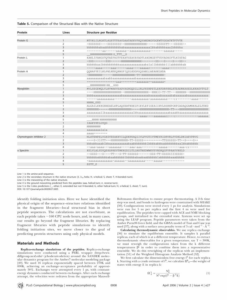
Table 5. Comparison of the Structural Bias with the Native Structure
Protein Lines Structure per Residue
Protein G 1 MTYKLILNGKTLKGETTTEAVDAATAEKVFKQYANDNGVDGEWTYDDATKTFTVTE2 -SSSSSSS----SSSSSSSS--HHHHHHHHHHHHH------SSSSSTT---SSSSS--3 bbbbbbbabaabbbbbbbbbabaaaaaaaaaaaaaaalbbabbbbbaaalbbbbba4 ---------aa------aaaaaa--aaaaaaaaaaaa--------aaaaaa-----5 __HHHHHHHHHHHHH S_TTT__S
Protein L 1 KANLIFANGSTQTAEFKGTFEKATSEAYAYADTLKKDNGEYTVDVADKGYTLNIKFAG2 -SSS-------SSS------HHHHHHHHHHHH--------SS---S----S---SS--3 bbbbbbaalbbbbbbbbbbaaaaaaaaaaaaaaabalallbbbbbblllabbbbbbab4 -----aaaa-----aaa------aaaa----aaaaar-------aaaa----------
Protein A 1 QQNAFYEILHLPNLNEEQRNGFIQSLKDDPSQSANLLAEAKKLNDA2 -HHHHHHHH------HHHHHHHHHHHHH-TT-HHHHHHHHHHHHH-3 aaaaaaaaaabaabbaaaaaaaaaaaaabaaaaaaaaaaaaaaaaa4 -----------------aaa---------aaaaaaa--aaaaaaa-5 __HHHHHHHHH HH__GGG
Myoglobin 1 MVLSEGEWQLVLHVWAKVEADVAGHGQDILIRLFKSHPETLEKFDRFKHLKTEAEMKASEDLKKAGVTVLT2 ----HHHHHHHHHHHHHHHH--HHHHHHHHHHHHHH--HHH33-TT-TT---HHHHHH--HHHHHHHHHHHH3 bbbbaaaaaaaaaaaaaaaabaaaaaaaaaaaaaaabaaaaaabaaaaababaaaaaabaaaaaaaaaaaa4 ------aaaaaaaaaa----------aaaaaaaaaa-aaaaaaaaaa----rr--------aaaa------5 HHHH_GGG1 ALGAILKKKGHHEAELKPLAQSHATKHKIPIKYLEFISEAIIHVLHSRHPGNFGADAQGAMNKALELFRKD2 HHHHHHH------HHHHHHHHHHHHH-----HHHHHHHHHHHHHHHHH-TT---HHHHHHHHHHHHHHHHH3 aaaaaaaallbaaaaaaaaaaaaaaaalbbaaaaaaaaaaaaaaaaaabaaabbaaaaaaaaaaaaaaaaa4 -------------------------------aaaa----aaaaaaarr---r-------------aaaaaa5 ___HHHH HHHHHHHHHH1 IAAKYKELGYQG2 HHHHHHHH3 aaaaaaaalala4 aaaa--------
Chymotrypsin inhibitor 2 1 NLKTEWPELVGKSVEEAKKVILQDKPEAQIIVLPVGTIVTMEYRIDRVRLFVDKLDNIAEVPRVG2 ----S-333TT---HHHHHHHHHH-TT-SSSSS-----------TTSSSSSS-TT--S----S--3 bbbabbaaablbbaaaaaaaaaaabaabbbbbbbblbbbbabbbaabbbbbbbaalbbabbbbbb4 --aaa-aaaa---aaaaaaa-----aaa-aaa----------aaaaa-------aaa-rr-----
a Spectrin 1 KELVLALYDYQEKSPREVTMKKGDILTLLNSTNKDWWKVEVNDRQGFVPAAYVKKLD2 --SSSS---S----TT---S-TT-SSSSSS-------SSS-TT-SSSSS333SSSS-3 bbbbbbabbbbbabaabbbbbblbbbbbabaabaabbbbbblabbbbbbaaabbbbb4 -aaaaaaaaaaaaaa-aaaaaa--aaaaaaaaaa-----aaaaa-------------5 EETT_E
Line 1 is the amino-acid sequence.Line 2 is the secondary structure in the native structure (3, 310 helix; H, a-helical; S, sheet; T, H-bonded–turn).Line 3 is the mesostring of the native structure.Line 4 is the ground mesostring predicted from the peptides (aaa, helical-turn; rr, reverse-turn).Line 5 is the I-sites predictions (_, other; E, extended but not H-bonded; G, other helical-turn; H, a-helical; S, sheet; T, turn).DOI: 10.1371/journal.pcbi.0020027.t005
PLoS Computational Biology | www.ploscompbiol.org April 2006 | Volume 2 | Issue 4 | e270008
Short Peptides in Molecular Dynamics
where NkE is the number of snapshots in replica k with energy E.
From the distribution of XkE, we calculate a new estimate of fk by
fk ¼ �logXE
X kE expðb
kEÞ" #
ð2Þ
We iterate the above two steps until fk converges. Then we usethese dimensionless free energies fk to reweigh the relative free-energy profile F of observable x to the target temperature btar:
Fxðb tarÞ ¼ �1
b tarlog
XE
Xk
Nkx;E expðb tarEÞX
k9
Nk9x;E expð fk9 � b k9EÞ
2664
3775
8>><>>:
9>>=>>; ð3Þ
After using the Weighted Histogram Analysis Method to calculatethe relative free energies Fi of a mesostring i, we calculate theprobabilities Pi by
Pi ¼expð�b tarFiÞX
i9
expð�b tarFi9Þð4Þ
When we merge similar mesostrings into a consensus mesostring,we calculate the free-energy difference to another mesostring j by
D F ¼ � 1b tar
logPconsensus
Pj
� �ð5Þ
Defining the backbone mesostates. A key part of our analysis is thediscretizing of the backbone degrees of freedom. This is based on theoriginal analysis of the protein backbone [53]. In that analysis,Ramachandran and coworkers showed that the stereochemistry of theprotein backbone breaks up the backbone u–w angles into threedistinct regions, each separated by significant energy barriers. We canthus describe the conformation of a peptide as a string of discretemesostates—we call this the mesostring. A given mesostring isseparated in energy from other mesostrings. Each mesostringcorresponds to a low-energy basin in the conformation space of thepeptide backbone. It is then straightforward to extract the localstructure from the lowest free-energy basin. This partitioning interms of discrete regions in the backbone angles has been observed ina molecular dynamics simulation of an a-helical peptide [31].
The original analysis of the backbone identified three distinctregions in the u–w angles [53]. Recent studies of the protein databasefound that these three regions can be further divided up into fiveclusters of density [54,55]. Some of the barriers between these fiveregions are small, which leaves three regions separated by largebarriers. However we cannot use the database analysis to define theboundaries of the backbone mesostates because current force fieldscannot replicate the database distribution of u–w angles. We mustdefine the boundaries the backbone mesostates in terms of the forcefield in our molecular dynamics: we ran replica-exchange simulationsof the alanine dipeptide and the glycine dipeptide for 10 ns andcalculated the free-energy profile of the u–w angles in bins of 58.Based on the resultant free-energy profile, we break up theRamachandran plot in terms of the following mesostates:
½b� : ð�1808 , u , 08; 458 , w , 1808ÞUð�1808 , u , 08;�1808 , w ,�1358Þ
Uð1208 , u , 1808; 458 , w , 1808ÞUð1208 , u , 1808;�1808 , w ,�1358Þ
½a� : ð�1808 , u , 08;�1358 , w , 458ÞUð1208 , u , 1808;�1358 , w , 458Þ
½l� : ð08 , u , 1208;�1808 , w , 1808ÞUð1208 , u , 1808;�1358 , w , 458Þ
And for glycine:
½b� : ð�1808 , u , 08; 458 , w , 1808ÞUð�1808 , u , 08;�1808 , w ,�1358ÞUð08 , u , 1808; 1358 , w , 1808ÞUð08 , u , 1808;�1808 , w ,�458Þ
½a� : ð�1808 , u , 08;�1358 , w , 458Þ
½l� : ð08 , u , 1808;�458 , w , 1358Þ
Acknowledgments
Thanks to John Chodera for the replica-exchange wrapper for themolecular dynamics package. Thanks to Banu Ozkan, Vince Voelzand Albert Wu for many invaluable discussions.
Author contributions. BKH and KAD conceived and designed theexperiments. BKH performed the experiments. BKH analyzed thedata. BKH wrote the paper.
Funding. We appreciate the support of NIH grant GM34993.Competing interests. The authors have declared that no competing
interests exist. &
References1. Dyson HJ, Wright PE (1998) Equilibrium NMR studies of unfolded and
partially folded proteins. Nat Struct Biol 5 (Supplement): 499–503.2. Dyson HJ, Merutka G, Waltho JP, Lerner RA, Wright PE (1992) Folding of
peptide fragments comprising the complete sequence of proteins. Modelsfor initiation of protein folding. I. Myohemerythrin. J Mol Biol 226: 795–817.
3. Shin HC, Merutka G, Waltho JP, Tennant LL, Dyson HJ, Wright PE (1993)Peptide models of protein folding initiation sites. 3. The G–H helicalhairpin of myoglobin. Biochemistry 32: 6356–6364.
4. Waltho JP, Feher VA, Merutka G, Dyson HJ, Wright PE (1993) Peptidemodels of protein folding initiation sites. 1. Secondary structure formationby peptides corresponding to the G- and H-helices of myoglobin.Biochemistry 32: 6337–6347.
Figure 4. Distribution of Kinks in the Topology of Protein Structures
a helices and 310 helices are drawn as coils, hydrogen-bonded b-turnsare drawn as a notch, and b strands are drawn as arrows.Yellow indicates helical-turn [-aaa-] and red indicates reverse-turns[baab]. 11 of the 14 a helices contain a helical-turn. The turns of all sevenb hairpins contain a peptide fragment that is structured.DOI: 10.1371/journal.pcbi.0020027.g004
PLoS Computational Biology | www.ploscompbiol.org April 2006 | Volume 2 | Issue 4 | e270009
Short Peptides in Molecular Dynamics

5. Ramirez-Alvarado M, Serrano L, Blanco FJ (1997) Conformational analysisof peptides corresponding to all the secondary structure elements ofprotein L B1 domain: Secondary structure propensities are not conservedin proteins with the same fold. Protein Sci 6: 162–174.
6. Eliezer D, Chung J, Dyson HJ, Wright PE (2000) Native and non-nativesecondary structure and dynamics in the pH 4 intermediate of apomyo-globin. Biochemistry 39: 2894–2901.
7. Mohana-Borges R, Goto NK, Kroon GJ, Dyson HJ, Wright PE (2004)Structural characterization of unfolded states of apomyoglobin usingresidual dipolar couplings. J Mol Biol 340: 1131–1142.
8. Marqusee S, Robbins VH, Baldwin RL (1989) Unusually stable helixformation in short alanine-based peptides. Proc Natl Acad Sci U S A 86:5286–5290.
9. Munoz V, Serrano L (1994) Elucidating the folding problem of helicalpeptides using empirical parameters. Nat Struct Biol 1: 399–409.
10. Blanco FJ, Rivas G, Serrano L (1994) A short linear peptide that folds into anative stable beta-hairpin in aqueous solution. Nat Struct Biol 1: 584–590.
11. Searle MS, Williams DH, Packman LC (1995) A short linear peptide derivedfrom the N-terminal sequence of ubiquitin folds into a water-stable non-native beta-hairpin. Nat Struct Biol 2: 999–1006.
12. Zerella R, Evans PA, Ionides JM, Packman LC, Trotter BW, Mackay JP,Williams DH (1999) Autonomous folding of a peptide corresponding to theN-terminal beta-hairpin from ubiquitin. Protein Sci 8: 1320–1331.
13. Espinosa JF, Munoz V, Gellman SH (2001) Interplay between hydrophobiccluster and loop propensity in beta-hairpin formation. J Mol Biol 306: 397–402.
14. Rotondi KS, Gierasch LM (2003) Role of local sequence in the folding ofcellular retinoic abinding protein I: Structural propensities of reverseturns. Biochemistry 42: 7976–7985.
15. Eisenberg D, Weiss RM, Terwilliger TC (1984) The hydrophobic momentdetects periodicity in protein hydrophobicity. Proc Natl Acad Sci U S A 81:140–144.
16. Kamtekar S, Schiffer JM, Xiong H, Babik JM, Hecht MH (1993) Proteindesign by binary patterning of polar and nonpolar amino acids. Science262: 1680–1685.
17. Han KF, Baker D (1996) Global properties of the mapping between localamino acid sequence and local structure in proteins. Proc Natl Acad Sci US A 93: 5814–5818.
18. Bystroff C, Simons KT, Han KF, Baker D (1996) Local sequence–structurecorrelations in proteins. Curr Opin Biotechnol 7: 417–421.
19. Bystroff C, Baker D (1998) Prediction of local structure in proteins using alibrary of sequence–structure motifs. J Mol Biol 281: 565–577.
20. Tsai CJ, Maizel JV Jr, Nussinov R (2000) Anatomy of protein structures:Visualizing how a one-dimensional protein chain folds into a three-dimensional shape. Proc Natl Acad Sci U S A 97: 12038–12043.
21. Kolodny R, Koehl P, Guibas L, Levitt M (2002) Small libraries of proteinfragments model native protein structures accurately. J Mol Biol 323: 297–307.
22. Tendulkar AV, Joshi AA, Sohoni MA, Wangikar PP (2004) Clustering ofprotein structural fragments reveals modular building block approach ofnature. J Mol Biol 338: 611–629.
23. Aloy P, Stark A, Hadley C, Russell RB (2003) Predictions without templates:New folds, secondary structure, and contacts in CASP5. Proteins 53(Supplement 6):: 436–456.
24. Moult J (2005) A decade of CASP: Progress, bottlenecks and prognosis inprotein structure prediction. Curr Opin Struct Biol 15: 285–289.
25. Avbelj F, Moult J (1995) Determination of the conformation of foldinginitiation sites in proteins by computer simulation. Proteins 23: 129–141.
26. Srinivasan R, Rose GD (1995) LINUS: A hierarchic procedure to predict thefold of a protein. Proteins 22: 81–99.
27. Gibbs N, Clarke AR, Sessions RB (2001) Ab initio protein structureprediction using physicochemical potentials and a simplified off-latticemodel. Proteins 43: 186–202.
28. Klepeis JL, Floudas CA (2002) Ab initio prediction of helical segments inpolypeptides. J Comput Chem 23: 245–266.
29. Daura X, van Gunsteren WF, Mark AE (1999) Folding–unfolding thermo-dynamics of a beta-heptapeptide from equilibrium simulations. Proteins34: 269–280.
30. de Groot BL, Daura X, Mark AE, Grubmuller H (2001) Essential dynamics
of reversible peptide folding: Memory-free conformational dynamicsgoverned by internal hydrogen bonds. J Mol Biol 309: 299–313.
31. Mu Y, Nguyen PH, Stock G (2005) Energy landscape of a small peptiderevealed by dihedral angle principal component analysis. Proteins 58: 45–52.
32. Bystroff C, Garde S (2003) Helix propensities of short peptides: Moleculardynamics versus bioinformatics. Proteins 50: 552–562.
33. Kim PS, Baldwin RL (1982) Specific intermediates in the folding reactionsof small proteins and the mechanism of protein folding. Annu RevBiochem 51: 459–489.
34. Dill KA, Fiebig KM, Chan HS (1993) Cooperativity in protein-foldingkinetics. Proc Natl Acad Sci U S A 90: 1942–1946.
35. Baldwin RL, Rose GD (1999) Is protein folding hierarchic? I. Local structureand peptide folding. Trends Biochem Sci 24: 26–33.
36. Sugita Y, Okamoto Y (1999) Replica-exchange molecular dynamics methodfor protein folding. Chemical Physics Letters 314: 141–151.
37. Tsui V, Case DA (2000) Theory and applications of the generalized Bornsolvation model in macromolecular simulations. Biopolymers 56: 275–291.
38. Zhou R (2003) Free energy landscape of protein folding in water: Explicitvs. implicit solvent. Proteins 53: 148–161.
39. Minor DL Jr, Kim PS (1996) Context-dependent secondary structureformation of a designed protein sequence. Nature 380: 730–734.
40. Bai Y, Englander SW (1994) . Bai Y, Englander SW (1994) Hydrogen bondstrength and beta-sheet propensities: The role of a side chain blockingeffect. Proteins 18, 262–266.
41. Jennings PA, Wright PE (1993) Formation of a molten globule intermediateearly in the kinetic folding pathway of apomyoglobin. Science 262: 892–896.
42. Itzhaki LS, Neira JL, Ruiz-Sanz J, de Prat Gay G, Fersht AR (1995) Search fornucleation sites in smaller fragments of chymotrypsin inhibitor 2. J MolBiol 254: 289–304.
43. Viguera AR, Jimenez MA, Rico M, Serrano L (1996) Conformationalanalysis of peptides corresponding to beta-hairpins and a beta-sheet thatrepresent the entire sequence of the alpha-spectrin SH3 domain. J Mol Biol255: 507–521.
44. Sundaralingam M, Sekharudu YC (1989) Water-inserted alpha-helicalsegments implicate reverse turns as folding intermediates. Science 244:1333–1337.
45. Soman KV, Karimi A, Case DA (1991) Unfolding of an alpha-helix in water.Biopolymers 31: 1351–1361.
46. Du D, Zhu Y, Huang CY, Gai F (2004) Understanding the key factors thatcontrol the rate of beta-hairpin folding. Proc Natl Acad Sci U S A 101:15915–15920.
47. Klimov DK, Thirumalai D (2000) Mechanisms and kinetics of beta-hairpinformation. Proc Natl Acad Sci U S A 97: 2544–2549.
48. Munoz V, Thompson PA, Hofrichter J, Eaton WA (1997) Folding dynamicsand mechanism of beta-hairpin formation. Nature 390: 196–199.
49. Pearlman DA, Case DA, Caldwell JW, Ross WS, Cheatham TE, et al. (1995)Amber, a package of computer-programs for applying molecular mechan-ics, normal-mode analysis, molecular-dynamics and free-energy calcula-tions to simulate the structural and energetic properties of molecules.Comput Physics Commun 91: 1–41.
50. Ryckaert JP, Ciccotti G, Berendsen HJC (1977) Numerical-integration ofCartesian equations of motion of a system with constraints—molecular-dynamics of N-alkanes. Journal of Computational Physics 23: 327–341.
51. Chodera JD, Swope WC, Pitera JW, Seok C, Dill KA (2006) Use of theWeighted Histogram Analysis Method for the analysis of simulated andparallel tempering simulations. J Chem Theory Comput: In press.
52. Kumar S, Bouzida D, Swendsen RH, Kollman PA, Rosenberg JM (1992) TheWeighted Histogram Analysis Method for free-energy calculations onbiomolecules. 1. The method. J Comput Chem 13: 1011–1021.
53. Ramachandran GN, Ramakrishnan C, Sasisekharan V (1963) Stereo-chemistry of polypeptide chain configurations. J Mol Biol 7: 95–99.
54. Karplus PA (1996) Experimentally observed conformation-dependentgeometry and hidden strain in proteins. Protein Sci 5: 1406–1420.
55. Ho BK, Thomas A, Brasseur R (2003) Revisiting the Ramachandran plot:Hard-sphere repulsion, electrostatics, and H-bonding in the alpha-helix.Protein Sci 12: 2508–2522.
PLoS Computational Biology | www.ploscompbiol.org April 2006 | Volume 2 | Issue 4 | e270010
Short Peptides in Molecular Dynamics