fluorescent super paramagnetic nanoparticles for medical diagnosis and treatment
TRANSCRIPT

Fluorescent, SuperparamagneticNanospheres forDrugStorage,Targeting, andImaging:AMultifunctionalNanocarrierSystem forCancerDiagnosis andTreatmentHoon-Sung Cho,† Zhongyun Dong,‡ Giovanni M. Pauletti,§ Jiaming Zhang,� Hong Xu,� Hongchen Gu,�
Lumin Wang,� Rodney C. Ewing,� Christopher Huth,† Feng Wang,† and Donglu Shi#,†,*†School of Energy, Environmental, Biological and Medical Engineering, University of Cincinnati, Cincinnati, Ohio 45221, ‡Department of Internal Medicine, College ofMedicine, University of Cincinnati, Cincinnati, Ohio 45221, §James L. Winkle College of Pharmacy, University of Cincinnati, Cincinnati, Ohio 45267, �Departments ofGeological Sciences, Nuclear Engineering & Radiological Sciences and Materials Science & Engineering, University of Michigan, Ann Arbor, Michigan 48109, �Med-X Institute,Shanghai Jiao Tong University, Shanghai 200030, China, and #The Institute for Advanced Materials and Nano Biomedicine, Tongji University, Shanghai, China 200092
One of the critical challenges inearly cancer diagnosis and treat-ment by nanotechnology is the
development of multifunctional particles at
the nanoscale that simultaneously serve as
sensitive, cell-specific bioprobes and local-
ized tumor treatment. Extensive efforts
have been devoted to designing nano-
carriers that combine cell targeting with ef-
ficient in vivo imaging, drug storage, and
controlled drug release capabilities.1�3 Spe-
cific material features required to imple-
ment these properties include surface func-
tional groups suitable for bioconjugation
and fluorescent imaging modules in addi-
tion to controlled drug storage and release
mechanisms. On the basis of specific medi-
cal requirements, the multifunctional nano-
carriers must also be biocompatible. For ef-
fective early cancer diagnosis and
treatment, the colloidal nanocarriers are re-
quired to be less than 200 nm in diameter4,5
and monodispersed to achieve efficient dis-
tributions in the targeted tumor lesions.
Spherical shape is preferred as it facilitates
uniform surface conjugation of cell-
targeting ligands (e.g., antibodies) and
imaging probes. Fluorescent emission near
800 nm is most suitable for deep tissue in
vivo imaging.6 Incorporation of superpara-
magnetic properties into nanocarriers fur-
ther benefits medical applications by facili-
tating multimodal imaging, hyperthermia
therapy, and magnetic manipulation.7�9
Previously, we reported on the design
of unique, fluorescent, magnetic nano-
spheres (MNSs) that allow effective in vivo
imaging due to their strong fluorescence
emissions of surface-conjugated quantum
dots (QDs). Our experimental results indi-
cated that these MNSs successfully induced
hyperthermia under an alternating mag-
netic field.10 The fabricated MNSs had an
average diameter around 150 nm and were
composed of Fe3O4 nanoparticles (�15�20
nm in diameter) embedded in a polysty-
rene matrix. TGA analysis shows that the
weight ratio of the embedded Fe3O4 nano-
particles to polystyrene in MNS is 4:1.11 QDs
were covalently conjugated to the surface
of these MNSs using standard carbodiimide
*Address correspondence [email protected].
Received for review May 7, 2010and accepted August 05, 2010.
Published online August 13, 2010.10.1021/nn101000e
© 2010 American Chemical Society
ABSTRACT For early cancer diagnosis and treatment, a nanocarrier system is designed and developed with
key components uniquely structured at nanoscale according to medical requirements. For imaging, quantum dots
with emissions in the near-infrared range (�800 nm) are conjugated onto the surface of a nanocomposite
consisting of a spherical polystyrene matrix (�150 nm) and the internally embedded, high fraction of
superparamagnetic Fe3O4 nanoparticles (�10 nm). For drug storage, the chemotherapeutic agent paclitaxel
(PTX) is loaded onto the surfaces of these composite multifunctional nanocarriers by using a layer of biodegradable
poly(lactic-co-glycolic acid) (PLGA). A cell-based cytotoxicity assay is employed to verify successful loading of
pharmacologically active drug. Cell viability of human, metastatic PC3mm2 prostate cancer cells is assessed in
the presence and absence of various multifunctional nanocarrier populations using the MTT assay. PTX-loaded
composite nanocarriers are synthesized by conjugating anti-prostate specific membrane antigen (anti-PSMA) for
targeting. Specific detection studies of anti-PSMA-conjugated nanocarrier binding activity in LNCaP prostate
cancer cells are carried out. LNCaP cells are targeted successfully in vitro by the conjugation of anti-PSMA on the
nanocarrier surfaces. To further explore targeting, the nanocarriers conjugated with anti-PSMA are intravenously
injected into tumor-bearing nude mice. Substantial differences in fluorescent signals are observed ex vivo between
tumor regions treated with the targeted nanocarrier system and the nontargeted nanocarrier system, indicating
considerable targeting effects due to anti-PSMA functionalization of the nanocarriers.
KEYWORDS: quantum dot · targeting · fluorescent imaging · drug storage ·magnetic nanosphere
ART
ICLE
VOL. 4 ▪ NO. 9 ▪ CHO ET AL. www.acsnano.org5398
chemistry. Our results demonstrated strong in vivo and
ex vivo fluorescence that facilitated biodistribution
monitoring of multifunctional, fluorescent MNSs in
mice. In addition, the high volume fraction of magne-
tite was sufficient to induce hyperthermia that can be
utilized for tumor cell suppression and drug release
control.
In this study, we have further advanced the multi-
functional nanosystem by tailoring the surfaces of MNSs
with therapeutic modality including cell targeting and
drug storage capabilities. In this way, the multifunc-
tional nanosystem will be fully developed specifically
for preclinical applications including in vivo imaging,
cell targeting, and drug storage. Note that these func-
tionalities can be potentially utilized simultaneously
within one nanosystem for cancer diagnosis and
chemotherapy.12,13
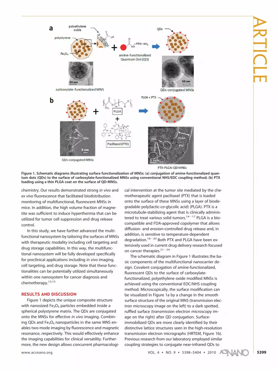
RESULTS AND DISCUSSIONFigure 1 depicts the unique composite structure
with nanosized Fe3O4 particles embedded inside aspherical polystyrene matrix. The QDs are conjugatedonto the MNSs for effective in vivo imaging. Combin-ing QDs and Fe3O4 nanoparticles in the same MNS en-ables two-mode imaging by fluorescence and magneticresonance, respectively. This would effectively enhancethe imaging capabilities for clinical versatility. Further-more, the new design allows concurrent pharmacologi-
cal intervention at the tumor site mediated by the che-motherapeutic agent paclitaxel (PTX) that is loadedonto the surface of these MNSs using a layer of biode-gradable poly(lactic-co-glycolic acid) (PLGA). PTX is amicrotubule-stabilizing agent that is clinically adminis-tered to treat various solid tumors.14�17 PLGA is a bio-compatible and FDA-approved copolymer that allowsdiffusion- and erosion-controlled drug release and, inaddition, is sensitive to temperature-dependentdegradation.18�20 Both PTX and PLGA have been ex-tensively used in current drug delivery research focusedon cancer therapies.21�24
The schematic diagram in Figure 1 illustrates the ba-sic components of the multifunctional nanocarrier de-sign. Covalent conjugation of amine-functionalized,fluorescent QDs to the surface of carboxylate-functionalized, polyethylene oxide modified MNSs isachieved using the conventional EDC/NHS couplingmethod. Microscopically, the surface modification canbe visualized in Figure 1a by a change in the smoothsurface structure of the original MNS (transmission elec-tron microscopy image on the left) to a dark spotted,ruffled surface (transmission electron microscopy im-age on the right) after QD conjugation. Surface-immobilized QDs are more clearly identified by theirdistinctive lattice structures seen in the high-resolutiontransmission electron micrographs (HRTEM, Figure 1b).Previous research from our laboratory employed similarcoupling strategies to conjugate near-infrared QDs to
Figure 1. Schematic diagrams illustrating surface functionalization of MNSs: (a) conjugation of amine-functionalized quan-tum dots (QDs) to the surface of carboxylate-functionalized MNSs using conventional NHS/EDC coupling method; (b) PTXloading using a thin PLGA coat on the surface of QD-MNSs.
ARTIC
LE
www.acsnano.org VOL. 4 ▪ NO. 9 ▪ 5398–5404 ▪ 2010 5399

polystyrene-Fe3O4 composite nanospheres for in vivofluorescent imaging.10
In this study, QDs with a visible emission wave-length of 655 nm were selected for the in vitro cell im-aging experiments, and QDs with a near-infraredemission wavelength of 800 nm were used for the invivo and ex vivo imaging. After conjugation of QDs toMNSs, PTX was incorporated into the fluorescent nano-carrier using a drug-loaded PLGA coating layer (PTX-PLGA-QD-MNSs). The transmission electron micrograph(TEM) in Figure 1b clearly shows a uniform, 7�10 mmthick PLGA layer surrounding the QD-MNS surfaceboundary. As PTX cannot be distinguished microscopi-cally from the PLGA matrix, a cell-based cytotoxicity as-say was employed to verify successful loading of a phar-macologically active drug.
Cell viability of human, metastatic PC3mm2 pros-tate cancer cells was assessed in the presence and ab-sence of various MNS populations using the MTT assay.Figure 2 shows that incubation of these tumor cellswith a broad dosing range of unmodified MNS, QD-labeled, fluorescent MNSs, and PLGA-coated, fluores-cent MNSs does not significantly affect mitochondrial
dehydrogenase activity as compared to vehicle con-trol. These results experimentally underline the safetyof drug-free, fluorescent nanocarriers up to 25 �g/mL.In contrast, PTX-PLGA-QD-MNSs dose-dependently de-creased cell viability of human PC3mm2 prostate cancercells in vitro after a 96 h incubation. The estimated PTX-PLGA-QD-MNS concentration associated with 50% inhi-bition of the mitochondrial enzyme activity (IC50) was125 ng/mL. Previously, we determined that the IC50
value of solubilized paclitaxel in the same cancer cellline is 5 ng/mL.25 As cytotoxic efficacy of PTX-PLGA-QD-MNS is associated with solubilized PTX, we estimatethat approximately 0.5 ng of PTX was released from thePLGA drug storage compartment leading to a free PTXconcentration of 5 ng/mL. Therapeutically, the relation-ship between drug loading capacity of colloidal nano-carriers and drug release rate is of utmost importance.Consequently, further experiments are planned toquantify drug loading efficiency and time-dependentrelease properties from these multifunctional MNSs us-ing different fabrication procedures, including doubleemulsion and organic coating methods.
To confirm the role of PLGA copolymer as a drug-holding and drug-releasing substance, the loading effi-cacy of paclitaxel on the MNSs according to the differ-ent ratio of PLGA was estimated in vitro by the MTTassay. For this purpose, the amount of PLGA wasdoubled or reduced by half from the original volume.It is shown that the cell viabilities are decreased by in-creasing the amount of PLGA used for paclitaxel load-ing (Figure 3). This indicates that the increased volumeof PLGA on the surface of MNSs may have enhancedpaclitaxel loading efficiency on the MNSs. However, itis observed that the variations of cell viabilities accord-ing to decreasing dosages of paclitaxel-loaded MNSsshow the same increasing rates regardless of the ratioof paclitaxel to PLGA, which means that in vitro drug re-lease behavior is not affected by the volume of PLGA,coated on the MNSs. It has been reported that the invitro drug release from the paclitaxel-loaded PLGAnanoparticles exhibit less than 30% of encapsulateddrug after 10 days.26 This suggests that the characteris-tics of PLGA copolymer, such as the variation of mono-mer ratio by breaking the ester linkages, or the glasstransition temperature, may have been changed dur-ing the process of a sequences of dissolving in dichlo-romethane (DCM) and reprecipitating in an aqueous so-lution, phosphate buffered saline (PBS).
Recently, it was reported that particle-associatedPTX accumulates more efficiently than solubilized drugin tumor models in vivo.27 Since intracellular PTX con-centration directly correlates with tumor cell kill, the cel-lular binding and transport studies were carried out inthis research to understand the mechanisms by whichpolymeric PTX particles are taken up into cells. The re-sults showed a significant contribution of caveolin-dependent, receptor-mediated endocytosis of intactparticles augmenting intracellular PTX concentrations.To determine whether similar endocytic pathways ex-
Figure 2. Dose-dependent effects of MNSs, QD-MNS, PLGA-QD-MNS,and PTX-PLGA-QD-MNSs on viability of human PC3mm2 prostatecancer cells. Tumor cells were treated for 96 h with various doses ofdrug-free or PTX-containing MNSs dispersed in EMEM/5% FBS. Cellviability was assessed using the MTT assay.
Figure 3. Drug loading effects on the MNSs according to the ratioof paclitaxel (PTX) and PLGA by viability of human PC3mm2 pros-tate cancer cells. Tumor cells were treated for 4 days with variousamounts of MNSs or drug-loaded MNSs dispersed in culture mediasupplemented with 5% fetal bovine serum for 4 days.
ART
ICLE
VOL. 4 ▪ NO. 9 ▪ CHO ET AL. www.acsnano.org5400

ist in human PC3mm2 prostate cancer cells, cellular up-take of PTX-PLGA-QD-MNSs was assessed by fluores-cence microscopy. The optical image in Figure 4a clearlyshows the cellular boundaries of PC3mm2 cancer cells.The same field visualized under fluorescence (EX �
560/40 nm, EM � 620/40 nm) demonstrates strongemission of MNS-immobilized QDs that appear to re-side in the cytosol (Figure 4b). Without z-stack images
acquired by confocal laser scanning mi-croscopy, it cannot be verified whetherPTX-PLGA-QD-MNSs successfully accumu-lated inside the cancer cells. However,even close association of drug-loaded,fluorescent MNSs with the cell membraneis predicted to enhance the therapeuticindex of PTX as a consequence of im-proved tumor targeting. Further studiesare required to delineate molecular eventsunderlying increased binding to and/oruptake of PTX-PLGA-QD-MNSs into hu-man PC3mm2 prostate cancer cells.
Targeted PTX-PLGA-QD-MNSs weresynthesized by conjugating anti-prostate
specific membrane antigen (anti-PSMA) on the PTX-PLGA-QD-MNSs for in vitro and in vivo targeting (Fig-ure 5). The surface of QD-MNSs was carboxyl-functionalized using carboxyl-terminated PLGA with in-corporation of PTX. The anti-PSMA was coupled to PTX-PLGA-QD-MNSs through ethylenediamine using theconventional EDC/NHS coupling method. In this fash-
Figure 4. Cellular interaction of PTX-PLGA-QD-MNSs with human PC3mm2prostate cancer cells evaluated by fluorescence microscopy: (a) opticalimage of live PC3mm2 cells incubated with PTX-PLGA-QD-MNSs, and(b) fluorescence image of PC3mm2 cells incubated with PTX-PLGA-QD-MNS (EX � 560/40 nm, EM � 620/40 nm).
Figure 5. Schematic diagrams illustrating surface functionalization of MNSs: (a) carboxyl functionalization using carboxyl-terminated PLGA on the surface of QD-MNSs with PTX loading; (b) amine functionalization by conjugation of ethylene-diamine to the surface of carboxylate-functionalized PTX-PLGA-QD-MNSs using conventional NHS/EDC coupling method;(c) conjugation of anti-PSMA to the PTX-PLGA-QD-MNSs, and (d) new multifunctional (fluorescent imaging, targeting, hyper-thermia, and chemotherapy) nanocarrier system.
ARTIC
LE
www.acsnano.org VOL. 4 ▪ NO. 9 ▪ 5398–5404 ▪ 2010 5401

ion, the novel multifunctional nanocarrier system (anti-PSMA-PTX-PLGA-QD-MNSs) was composed with mul-tiple components: targeted antibodies, fluorescentprobes, chemotherapeutic agents, and superparamag-netic nanoparticles, ideally suited for early cancer diag-nosis and treatment.
For cancer cell targeting, specific detection studieshave been carried out on the anti-PSMA-conjugatedMNSs’ binding activity in LNCaP prostate cancer cells.Anti-PSMA-PTX-PLGA-QD-MNSs and nontargeted PTX-PLGA-QD-MNS, as a control, were incubated with thefixed LNCaP and PC3mm2 prostate cancer cells, respec-tively (Figure 6). Substantially different behaviors areobserved between the targeted PTX-PLGA-QD-MNSsand the nontargeted PTX-PLGA-QD-MNSs. LNCaP cells,which express PSMA, are targeted successfully by theconjugation of anti-PSMA on the PTX-PLGA-QD-MNSssurface (Figure 6b). No fluorescent signals were de-tected in LNCaP cells exposed to nontargeted PTX-PLGA-QD-MNS (Figure 6a). In contrast, PC3mm2 hu-man prostate cells, which are known as PSMA-negative,show no fluorescent signals with and without anti-PSMA conjugation (Figure 6c,d). The binding activity ofanti-PSMA to LNCaP cancer cells was also confirmed by
immunocytochemical studies using Alexa Fluor 594F(ab=)2 fragment of goat anti-mouse IgG (H�L) as a sec-ondary antibody (data not shown). These results indi-cate that the specific targeting to cancer cells can be ef-fectively achieved by the multifunctional nanocarriersystem.
To further explore the effects of targeting, the bio-distribution of the nanocarriers administrated via tailvein was analyzed. The PTX-PLGA-QD-MNSs conjugatedwith anti-PSMA were intravenously injected into atumor-bearing nude mouse. This study was approvedby the Institutional Animal Use and Care Committee(IACUC) at the University of Cincinnati in compliancewith relevant State and Federal Regulations. Thirty min-utes after injection of the nanocarrier system, the PTX-PLGA-QD-MNSs were biodistributed in various organsof the mouse. These organs were then harvested andcompared with a nontreated mouse as a control. Ex vivoimages were initially taken under the condition for theoriginal emission wavelength of the quantum dot (exci-tation wavelength � 720 nm, emission wavelength �
790 nm). As shown in Figure 7, ex vivo fluorescence im-ages of the tumor indicate an accumulation of thenanocarrier system in this organ of the treated mouse.
Figure 6. In vitro targeting studies of anti-PSMA-conjugated PTX-PLGA-QD-MNSs’ binding activity in cultured LNCaP pros-tate cancer cells, which are PSMA-positive, and PC3mm2, which are known as PSMA-negative.
ART
ICLE
VOL. 4 ▪ NO. 9 ▪ CHO ET AL. www.acsnano.org5402

No significant fluorescence in the tumor was mea-sured in the untreated control animal. As can be seenin Figure 7a, there is a considerable fluorescent signal inthe tumor region. It was found that the fluorescent sig-nal was significantly enhanced by using a lower wave-length filter set (excitation wavelength � 625 nm, emis-sion wavelength � 700 nm). Under this condition, thetumor shows sharper contrast of fluorescent signals as-sociated with the nanocarrier system compared to theoriginal condition (Figure 7b). This is due to the emis-sion peak of quantum dot being blue-shifted duringsample preparation. This difference is a strong indica-tion that the fluorescent signals from the tumor regionare associated with the nanocarriers accumulated as aresult of targeting. The current research deals with asystematic in vivo targeting imaging study on tumor-bearing mice with intravenously injected anti-PSMA-PTX-PLGA-QD-MNSs.
CONCLUSIONSWe have developed a multifunctional nanocarrier
system for medical diagnosis and treatment. Thisunique system is composed of several key compo-nents, namely, fluorescent superparamagnetic nano-particles for multimodal imaging and hyperthermia,tumor-specific antibodies for cell targeting, and anti-cancer drugs for localized treatment. The unique nano-structures and surfaces are designed and developed ac-cording to biomedical requirements and procedures sothat these functionalities may be effectively utilized
clinically, allowing simultaneous cancer diagnosis andtherapy. Experimental evidence from in vitro studiessuggests acceptable safety profiles for drug-free, fluo-rescent PLGA-QD-MNSs. However, inclusion of PTX in-side a biocompatible PLGA copolymer layer transformsthis multifunctional nanocarrier into an effective thera-peutic strategy. In addition to the chemotherapeutictreatment, the nanocarrier system is able to target spe-cific cancer cells and ex vivo organs. Further clinical de-velopment of the PTX-PLGA-QD-MNS concept as anovel, targeted imaging and drug delivery system maybenefit cancer patients in the future due to significantlyreduced systemic toxicity and greatly improved nonin-vasive monitoring capabilities.
METHODSSurface Conjugation of QDs to MNSs: Carboxylate-functionalized
MNSs were washed three times with phosphate-bufferedsaline, pH 7.4 (PBS), and incubated for 20 min at room tempera-ture in 1-(3-dimethylaminopropyl)-3-ethylcarbodiimide hydro-chloride (EDC) solution (100 �L of 400 mM in PBS) andN-hydroxysuccinimide (NHS) solution (100 �L of 100 mM inPBS). Excess coupling reagent was removed by magnetic separa-tion. The NHS-activated MNSs were conjugated to amino-functionalized QDs [Qdot 655 ITK amino (PEG), Invitrogen Corp.,Carlsbad, CA] following procedures previously published by thislaboratory.5,18 QD-MNSs were washed three times with PBS andstored in 200 �L of PBS until used.
PTX Loading of QD-MNSs: A suspension of 1 mg of QD-MNSs,100 �g of PTX, and 100 �g of PLGA in 100 �L of acetonitrilewas mixed using a sonication bath. Drug-free nanocarriers werefabricated as controls. The organic suspension was added to 500mL of Milli-Q purified water and emulsified by sonication usingan energy output of 5 W. The organic solvent was removed fromthe oil-in-water emulsion under reduced pressure. PTX-PLGA-QD-MNSs and drug-free control carriers were washed threetimes in PBS and stored at 4 °C until used.
Synthesis of Amine-Functionalized PTX-PLGA-QD-MNSs: A suspensionof 1 mg of QD-MNSs in acetonitrile was mixed with 100 �g ofcarboxyl-terminated PLGA and 100 �g of PTX by sonication. Themixed organic suspension was emulsified in 500 mL of Milli-Qpurified water and then sonicated using an energy output of 5W. The organic solvent was removed from the oil-in-water emul-sion under reduced pressure. The suspension was applied by ap-plied external magnetic field, and carboxyl-functionalized PTX-PLGA-QD-MNSs were collected and washed three times withPBS. Amine reactive groups on the surface of the PTX-PLGA-QD-MNSs were obtained by amine coupling of ethylenediamine tocarboxyl-functionalized PTX-PLGA-QD-MNSs. A carboxyl-functionalized PTX-PLGA-QD-MNS suspension was treated with
ethylenediamine for 30 min at room temperature. Excess ethyl-enediamine was removed by external magnetic field, and amine-functionalized PTX-PLGA-QD-MNSs were collected and washedthree times with PBS.
Conjugation of Anti-PSMA on the Surface of PTX-PLGA-QD-MNSs: Amine-functionalized PTX-PLGA-QD-MNSs (500 �g) were treated with200 �L of 400 mM EDC and 200 �L of 200 mM NHS for 30 minat room temperature. The PTX-PLGA-QD-MNSs incorporatedwith amine-reactive NHS-esters were collected by applying mag-netic field and washed with Milli-Q purified water three timesto remove unreacted chemicals. An anti-PSMA solution wasadded with the NHS-activated PTX-PLGA-QD-MNSs for 2 h atroom temperature and then washed three times with Milli-Qpurified water.
In Vitro Cytotoxicity Assay: Human PC3mm2 prostate carcinomacells were cultured in Eagle’s minimal essential medium (EMEM)supplemented with 5% (v/v) fetal bovine serum (FBS), nonessen-tial amino acids, sodium pyruvate, vitamin A, and glutamine at37 °C in a humidified atmosphere of 5% CO2. Cells in exponen-tial growth phase were harvested using 0.25% (w/v) trypsin/0.02% EDTA (w/v) solution. For cell viability experiments,PC3mm2 cells were plated at a density of 1000 cells/well in 96-well plates. Untreated PC3 cells plating at 1000 cells/well willreach confluence 4 days later and give OD values of MTT stain-ing in linear range. After an overnight attachment period, cellswere exposed for 96 h at 37 °C to various MNS populations atdoses between 0.016 and 50 �g/mL. MNS suspensions were pre-pared in EMEM/5% FBS and dosed at 100 �L/well. During the fi-nal 2 h of incubation, 20 �L of 3-(4,5-dimethylthiazol-2-yl)-2,5-diphenyltetrazolium bromide (2 mg/mL in PBS) was added toeach well. At the end of the incubation period, the medium wascarefully aspirated, and the blue formazan complex dissolved in100 �L of dimethyl sulfoxide. Absorbance of each well was quan-tified at � � 570 nm using a FluoStar Optima microplate reader(BMG Labtechnologies, Durham, NC). Cell viability was normal-
Figure 7. Fluorescent images of excised organs in mice. The images oforgans and tumor from the sample mouse injected with anti-PSMA-PTX-PLGA-QD-MNSs are compared with those of the nontreated controlmouse.
ARTIC
LE
www.acsnano.org VOL. 4 ▪ NO. 9 ▪ 5398–5404 ▪ 2010 5403

ized to EMEM/5% FBS-treated vehicle control cells and expressedas %.
In Vitro Targeting: To determine anti-PSMA-PTX-PLGA-QD-MNSbinding to LNCaP cells, immunocytochemical studies were car-ried out. LNCap and PC3mm2 cells were fixed by incubatingthem in 4% (v/v) paraformaldehyde in PBS for 20 min at roomtemperature, blocked with 0.1% bovine serum albumin (5 minat room temperature), and then treated with anti-PSMA-conjugated PTX-PLGA-QD-MNSs and nontargeted PTX-PLGA-QD-MNS. After 30 min, the cells were washed with PBS threetimes and examined under the microscope (excitation � 560/40nm, emission � 620/40 nm).
Ex Vivo Targeting: Ex vivo fluorescence images of the mousewere taken with 720 nm excitation filter and 790 nm emission fil-ter set using a Kodak 4000MM Whole-Mouse Image Station.Nu/nu nude mice, obtained from the National Cancer Institute,were 6�8 weeks old and typically weighed 18 g. Mice were anes-thetized for imaging by beutal saline intraperitoneally, and the250 �g dosage of anti-PSMA-PTX-PLGA-QD-MNSs dispersed in100 �L of PBS was administrated via tail vein injection. After 30min postadministration, the mouse was sacrificed. The excisedorgans from the control mouse and the treated mouse wereplaced in a same place, and the fluorescent image of the or-gans was taken. This study was approved by Institutional Ani-mal Use and Care Committee (IACUC) at the University of Cincin-nati, OH.
Acknowledgment. This research was supported in part bygrants from the National Science Foundation (DGE-0333377)and the University of Cincinnati, Institute of Nanoscale Scienceand Technology.
REFERENCES AND NOTES1. Wood, K. C.; Azarin, S. M.; Arap, W.; Pasqualini, R.; Langer,
R.; Hammond, P. T. Tumor-Targeted Gene Delivery UsingMolecularly Engineered Hybrid Polymers Functionalizedwith a Tumor-Homing Peptide. Bioconjugate Chem. 2008,19, 403–405.
2. Inoue, Y.; Izawa, K.; Yoshikawa, K.; Yamada, H.; Tojo, A.;Ohtomo, K. In Vivo Fluorescence Imaging of theReticuloendothelial System Using Quantum Dots inCombination with Bioluminescent Tumour Monitoring.Eur. J. Nucl. Med. Mol. Imaging 2007, 34, 2048–2056.
3. Qian, X.; Peng, X.-H.; Ansari, D. O.; Yin-Goen, Q.; Chen, G. Z.;Shin, D. M.; Yang, L.; Young, A. N.; Wang, M. D.; Nie, S. InVivo Tumor Targeting and Spectroscopic Detection withSurface-Enhanced Raman Nanoparticle Tags. Nat.Biotechnol. 2008, 26, 83–90.
4. Moghimi, S. M.; Hunter, A. C.; Murray, J. C. Long-Circulatingand Target-Specific Nanoparticles: Theory to Practice.Pharmacol. Rev. 2001, 53, 283–318.
5. Shekunov, B. Y.; Chattopadhyay, P.; Tong, H. H. Y.; Chow,A. H. L. Particle Size Analysis in Pharmaceutics: Principles,Methods and Applications. Pharm. Res. 2007, 24, 203–227.
6. Weissleder, R. A Clearer Vision for In Vivo Imaging. Nat.Biotechnol. 2001, 19, 316–317.
7. Jordan, A.; Scholz, R.; Wust, P.; Fahling, H.; Roland, F.Magnetic Fluid Hyperthermia (MFH): Cancer Treatmentwith Ac Magnetic Field Induced Excitation ofBiocompatible Superparamagnetic Nanoparticles. J. Magn.Magn. Mater. 1999, 201, 413–419.
8. Hergt, R.; Dutz, S.; Muller, R.; Zeisberger, M. MagneticParticle Hyperthermia: Nanoparticle Magnetism andMaterials Development for Cancer Therapy. J. Phys.:Condens. Matter 2006, 18, S2919.
9. Neuberger, T.; Schopf, B.; Hofmann, H.; Hofmann, M.; vonRechenberg, B. Superparamagnetic Nanoparticles forBiomedical Applications: Possibilities and Limitations of aNew Drug Delivery System. J. Magn. Magn. Mater. 2005,293, 483–496.
10. Shi, D.; Cho, H. S.; Chen, Y.; Xu, H.; Gu, H.; Lian, J.; Wang, W.;Liu, G.; Huth, C.; Wang, L.; et al. Fluorescent Polystyrene-Fe3O4 Composite Nanospheres for In Vivo Imaging andHyperthermia. Adv. Mater. 2009, 21, 2170–2173.
11. Xu, H.; Cui, L.; Tong, N.; Gu, H. Development of HighMagnetization Fe3O4/Polystyrene/Silica Nanospheres via
Combined Miniemulsion/Emulsion Polymerization. J. Am.Chem. Soc. 2006, 128, 15582–15583.
12. Franchini, M. C.; Baldi, G.; Bonacchi, D.; Gentili, D.; Giudetti,G.; Lasciafari, A.; Corti, M.; Marmorato, P.; Ponti, J.; Micotti,E.; et al. Bovine Serum Albumin-Based MagneticNanocarrier for MRI Diagnosis and Hyperthermic Therapy:A Potential Theranostic Approach against Cancer. Small2010, 6, 366–370.
13. Pan, D.; Caruthers, S. D.; Hu, G.; Senpan, A.; Scott, M. J.;Gaffney, P. J.; Wickline, S. A.; Lanza, G. M. Ligand-DirectedNanobialys as Theranostic Agent for Drug Delivery andManganese-Based Magnetic Resonance Imaging ofVascular Targets. J. Am. Chem. Soc. 2008, 130, 9186–9187.
14. Liang, H.-F.; Chen, C.-T.; Chen, S.-C.; Kulkarni, A. R.; Chiu,Y.-L.; Chen, M.-C.; Sung, H.-W. Paclitaxel-Loaded Poly(L-Glutamic Acid)-Poly(Lactide) Nanoparticles as a TargetedDrug Delivery System for the Treatment of Liver Cancer.Biomaterials 2006, 27, 2051–2059.
15. Lu, Z.; Yeh, T.-K.; Tsai, M.; Au, J. L. S.; Wientjes, M. G.Paclitaxel-Loaded Gelatin Nanoparticles for IntravesicalBladder Cancer Therapy. Clin. Cancer Res. 2004, 10, 7677–7684.
16. Sahoo, S. K.; Ma, W.; Labhasetwar, V. Efficacy of Transferrin-Conjugated Paclitaxel-Loaded Nanoparticles in a MurineModel of Prostate Cancer. Int. J. Cancer 2004, 112,335–340.
17. Koziara, J. M.; Lockman, P. R.; Allen, D. D.; Mumper, R. J.Paclitaxel Nanoparticles for the Potential Treatment ofBrain Tumors. J. Controlled Release 2004, 99, 259–269.
18. Dunne, M.; Corrigan, O. I.; Ramtoola, Z. Influence ofParticle Size and Dissolution Conditions on theDegradation Properties of Polylactide-co-GlycolideParticles. Biomaterials 2000, 21, 1659–1668.
19. Grayson, A. C. R.; Cima, M. J.; Langer, R. Size andTemperature Effects on Poly(lactic-co-glycolic acid)Degradation and Microreservoir Device Performance.Biomaterials 2005, 26, 2137–2145.
20. Reed, A. M.; Gilding, D. K. Biodegradable Polymers for Usein SurgeryOPoly(glycolic)/Poly(lactic acid) Homo andCopolymers: 2. In Vitro Degradation. Polymer 1981, 22,494–498.
21. Dong, Y.; Feng, S.-S.Poly(D,L-lactide-co-glycolide)/MontmorilloniteNanoparticles for Oral Delivery of Anticancer Drugs.Biomaterials 2005, 26, 6068–6076.
22. Ong, B. Y. S.; Ranganath, S. H.; Lee, L. Y.; Lu, F.; Lee, H.-S.;Sahinidis, N. V.; Wang, C.-H. Paclitaxel Delivery from PLGAFoams for Controlled Release in Post-SurgicalChemotherapy against Glioblastoma Multiforme.Biomaterials 2009, 30, 3189–3196.
23. Fonseca, C.; Simos, S.; Gaspar, R. Paclitaxel-Loaded PLGANanoparticles: Preparation, PhysicochemicalCharacterization and In Vitro Anti-tumoral Activity. J.Controlled Release 2002, 83, 273–286.
24. Jin, C.; Bai, L.; Wu, H.; Song, W.; Guo, G.; Dou, K.Cytotoxicity of Paclitaxel Incorporated in PLGANanoparticles on Hypoxic Human Tumor Cells. Pharm. Res.2009, 26, 1776–1784.
25. Guo, Y.; Shi, D.; Cho, H.; Dong, Z.; Kulkami, A.; Pauletti,G. M.; Wang, W.; Lian, J.; Liu, W.; Ren, L.; et al. In VivoImaging and Drug Storage by Quantum-Dot-ConjugatedCarbon Nanotubes. Adv. Funct. Mater. 2008, 18,2489–2497.
26. Mu, L.; Seow, P. H. Application of TPGS in PolymericNanoparticulate Drug Delivery System. Colloids Surf., B2006, 47, 90–97.
27. Desai, N.; Trieu, V.; Yao, Z.; Louie, L.; Ci, S.; Yang, A.; Tao, C.;De, T.; Beals, B.; Dykes, D.; et al. Increased AntitumorActivity, Intratumor Paclitaxel Concentrations, andEndothelial Cell Transport of Cremophor-Free, Albumin-Bound Paclitaxel, Abi-007, Compared with Cremophor-Based Paclitaxel. Clin. Cancer Res. 2006, 12, 1317–1324.
ART
ICLE
VOL. 4 ▪ NO. 9 ▪ CHO ET AL. www.acsnano.org5404