first trimester vaginal bleeding - appropriateness criteria
TRANSCRIPT
ACR Appropriateness Criteria® First Trimester Vaginal Bleeding
EVIDENCE TABLE
* See Last Page for Key Revised 2017 Brown/Packard Page 1
Reference Study Type Patients/ Events
Study Objective (Purpose of Study) Study Results Study
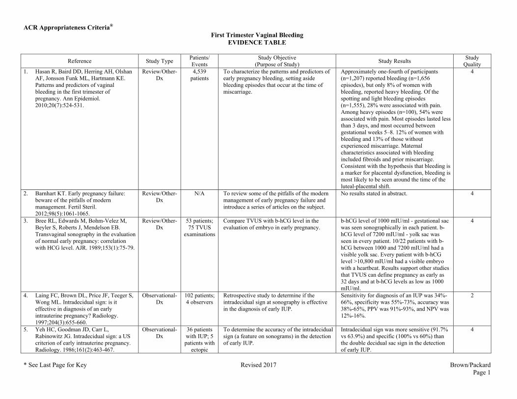
Quality 1. Hasan R, Baird DD, Herring AH, Olshan
AF, Jonsson Funk ML, Hartmann KE. Patterns and predictors of vaginal bleeding in the first trimester of pregnancy. Ann Epidemiol. 2010;20(7):524-531.
Review/Other-Dx
4,539 patients
To characterize the patterns and predictors of early pregnancy bleeding, setting aside bleeding episodes that occur at the time of miscarriage.
Approximately one-fourth of participants (n=1,207) reported bleeding (n=1,656 episodes), but only 8% of women with bleeding, reported heavy bleeding. Of the spotting and light bleeding episodes (n=1,555), 28% were associated with pain. Among heavy episodes (n=100), 54% were associated with pain. Most episodes lasted less than 3 days, and most occurred between gestational weeks 5–8. 12% of women with bleeding and 13% of those without experienced miscarriage. Maternal characteristics associated with bleeding included fibroids and prior miscarriage. Consistent with the hypothesis that bleeding is a marker for placental dysfunction, bleeding is most likely to be seen around the time of the luteal-placental shift.
4
2. Barnhart KT. Early pregnancy failure: beware of the pitfalls of modern management. Fertil Steril. 2012;98(5):1061-1065.
Review/Other-Dx
N/A To review some of the pitfalls of the modern management of early pregnancy failure and introduce a series of articles on the subject.
No results stated in abstract. 4
3. Bree RL, Edwards M, Bohm-Velez M, Beyler S, Roberts J, Mendelson EB. Transvaginal sonography in the evaluation of normal early pregnancy: correlation with HCG level. AJR. 1989;153(1):75-79.
Review/Other-Dx
53 patients; 75 TVUS
examinations
Compare TVUS with b-hCG level in the evaluation of embryo in early pregnancy.
b-hCG level of 1000 mIU/ml - gestational sac was seen sonographically in each patient. b-hCG level of 7200 mIU/ml - yolk sac was seen in every patient. 10/22 patients with b-hCG between 1000 and 7200 mIU/ml had a visible yolk sac. Every patient with b-hCG level >10,800 mIU/ml had a visible embryo with a heartbeat. Results support other studies that TVUS can define pregnancy as early as 32 days and at b-hCG levels as low as 1000 mIU/ml.
4
4. Laing FC, Brown DL, Price JF, Teeger S, Wong ML. Intradecidual sign: is it effective in diagnosis of an early intrauterine pregnancy? Radiology. 1997;204(3):655-660.
Observational-Dx
102 patients; 4 observers
Retrospective study to determine if the intradecidual sign at sonography is effective in the diagnosis of early IUP.
Sensitivity for diagnosis of an IUP was 34%-66%, specificity was 55%-73%, accuracy was 38%-65%, PPV was 91%-93%, and NPV was 12%-16%.
2
5. Yeh HC, Goodman JD, Carr L, Rabinowitz JG. Intradecidual sign: a US criterion of early intrauterine pregnancy. Radiology. 1986;161(2):463-467.
Observational-Dx
36 patients with IUP; 5 patients with
ectopic
To determine the accuracy of the intradecidual sign (a feature on sonograms) in the detection of early IUP.
Intradecidual sign was more sensitive (91.7% vs 63.9%) and specific (100% vs 60%) than the double decidual sac sign in the detection of early IUP.
4

ACR Appropriateness Criteria® First Trimester Vaginal Bleeding
EVIDENCE TABLE
* See Last Page for Key Revised 2017 Brown/Packard Page 2
Reference Study Type Patients/ Events
Study Objective (Purpose of Study) Study Results Study
Quality 6. Chiang G, Levine D, Swire M, McNamara
A, Mehta T. The intradecidual sign: is it reliable for diagnosis of early intrauterine pregnancy? AJR. 2004;183(3):725-731.
Observational-Dx
153 patients with IUP; 34 patients with
ectopic; 3 observers
Retrospective study to determine if intradecidual sign is accurate for the diagnosis of IUP and the exclusion of ectopic pregnancy.
Patients with IUP had sensitivity of 70%. Ectopic pregnancies had specificity of 100% for the intradecidual sign; the accuracy rate was 75%, PPV 100%, and NPV 43%. Sensitivity for diagnosis of an IUP increases when b-hCG levels are =2,000 mIU/ml or the mean sac diameter =3 mm.
2
7. Doubilet PM, Benson CB. Double sac sign and intradecidual sign in early pregnancy: interobserver reliability and frequency of occurrence. J Ultrasound Med. 2013;32(7):1207-1214.
Observational-Dx
199 sonographic
studies
To assess the interobserver agreement, frequency of occurrence, and prognostic importance of the double sac sign (DSS), intradecidual sign (IDS), and other sonographic findings in early intrauterine pregnancies.
Interobserver agreement was poor for the DSS (kappa= 0.24) and IDS (kappa= 0.23). Scans frequently demonstrated neither sign: 150 cases (75.4%) if we considered a sign to be present when both investigators graded it as present and 69 cases (34.7%) using the looser criterion that either graded it as present. The presence of a DSS or an IDS was unrelated to the beta-human chorionic gonadotropin (beta-hCG) value (P > .05, t test, all comparisons). An inner echogenic ring was present in 158 cases (79.4%), and the decidua was brighter peripherally than centrally in 102 (51.3%). The first-trimester outcome was unrelated to the presence of a DSS or an IDS, presence of an inner echogenic ring, or decidual appearance (P > .05, chi(2), all comparisons).
3
8. Richardson A, Gallos I, Dobson S, Campbell BK, Coomarasamy A, Raine-Fenning N. Accuracy of first-trimester ultrasound in diagnosis of intrauterine pregnancy prior to visualization of the yolk sac: a systematic review and meta-analysis. Ultrasound Obstet Gynecol. 2015;46(2):142-149.
Meta-analysis 17 studies To evaluate the diagnostic accuracy of ultrasound in predicting the location of an intrauterine pregnancy before visualization of the yolk sac is possible.
Seventeen studies including 2564 women were selected from 19 959 potential papers. Following meta-analysis, the presence of a gestational sac on ultrasound examination was found to predict an intrauterine pregnancy with a sensitivity of 52.8% (95% CI, 38.2-66.9%) and specificity of 97.6% (95% CI, 94.3-99.0%). The corresponding performance of the double decidual sac sign, intradecidual sign, chorionic rim sign and yolk sac were: 81.8% (95% CI, 68.1-90.4%) and 97.3% (95% CI, 76.1-99.8%); 66.1% (95% CI, 58.9-72.8%) and 100% (95% CI, 91.0-100%); 79.9% (95% CI, 73.0-85.7%) and 97.1% (95% CI, 89.9-99.6%); and 42.2% (95% CI, 27.7-57.9%) and 100% (95% CI, 54.1-100%), respectively.
M
ACR Appropriateness Criteria® First Trimester Vaginal Bleeding
EVIDENCE TABLE
* See Last Page for Key Revised 2017 Brown/Packard Page 3
Reference Study Type Patients/ Events
Study Objective (Purpose of Study) Study Results Study
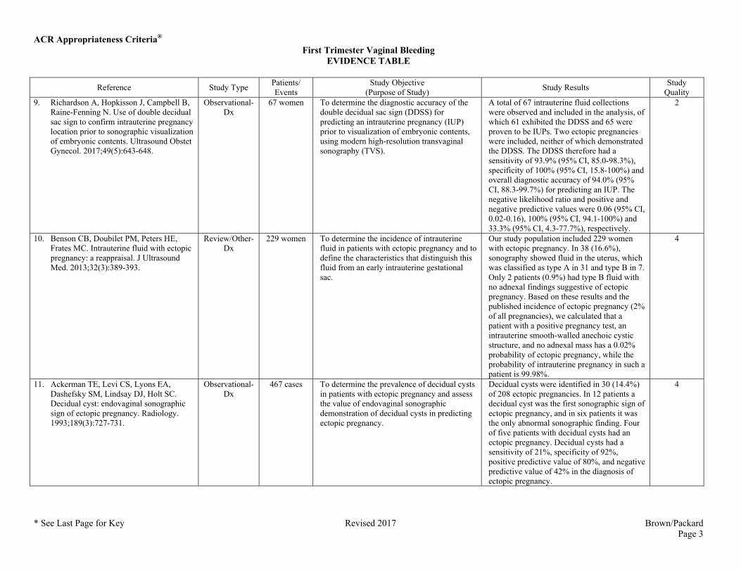
Quality 9. Richardson A, Hopkisson J, Campbell B,
Raine-Fenning N. Use of double decidual sac sign to confirm intrauterine pregnancy location prior to sonographic visualization of embryonic contents. Ultrasound Obstet Gynecol. 2017;49(5):643-648.
Observational-Dx
67 women To determine the diagnostic accuracy of the double decidual sac sign (DDSS) for predicting an intrauterine pregnancy (IUP) prior to visualization of embryonic contents, using modern high-resolution transvaginal sonography (TVS).
A total of 67 intrauterine fluid collections were observed and included in the analysis, of which 61 exhibited the DDSS and 65 were proven to be IUPs. Two ectopic pregnancies were included, neither of which demonstrated the DDSS. The DDSS therefore had a sensitivity of 93.9% (95% CI, 85.0-98.3%), specificity of 100% (95% CI, 15.8-100%) and overall diagnostic accuracy of 94.0% (95% CI, 88.3-99.7%) for predicting an IUP. The negative likelihood ratio and positive and negative predictive values were 0.06 (95% CI, 0.02-0.16), 100% (95% CI, 94.1-100%) and 33.3% (95% CI, 4.3-77.7%), respectively.
2
10. Benson CB, Doubilet PM, Peters HE, Frates MC. Intrauterine fluid with ectopic pregnancy: a reappraisal. J Ultrasound Med. 2013;32(3):389-393.
Review/Other-Dx
229 women To determine the incidence of intrauterine fluid in patients with ectopic pregnancy and to define the characteristics that distinguish this fluid from an early intrauterine gestational sac.
Our study population included 229 women with ectopic pregnancy. In 38 (16.6%), sonography showed fluid in the uterus, which was classified as type A in 31 and type B in 7. Only 2 patients (0.9%) had type B fluid with no adnexal findings suggestive of ectopic pregnancy. Based on these results and the published incidence of ectopic pregnancy (2% of all pregnancies), we calculated that a patient with a positive pregnancy test, an intrauterine smooth-walled anechoic cystic structure, and no adnexal mass has a 0.02% probability of ectopic pregnancy, while the probability of intrauterine pregnancy in such a patient is 99.98%.
4
11. Ackerman TE, Levi CS, Lyons EA, Dashefsky SM, Lindsay DJ, Holt SC. Decidual cyst: endovaginal sonographic sign of ectopic pregnancy. Radiology. 1993;189(3):727-731.
Observational-Dx
467 cases To determine the prevalence of decidual cysts in patients with ectopic pregnancy and assess the value of endovaginal sonographic demonstration of decidual cysts in predicting ectopic pregnancy.
Decidual cysts were identified in 30 (14.4%) of 208 ectopic pregnancies. In 12 patients a decidual cyst was the first sonographic sign of ectopic pregnancy, and in six patients it was the only abnormal sonographic finding. Four of five patients with decidual cysts had an ectopic pregnancy. Decidual cysts had a sensitivity of 21%, specificity of 92%, positive predictive value of 80%, and negative predictive value of 42% in the diagnosis of ectopic pregnancy.
4
ACR Appropriateness Criteria® First Trimester Vaginal Bleeding
EVIDENCE TABLE
* See Last Page for Key Revised 2017 Brown/Packard Page 4
Reference Study Type Patients/ Events
Study Objective (Purpose of Study) Study Results Study
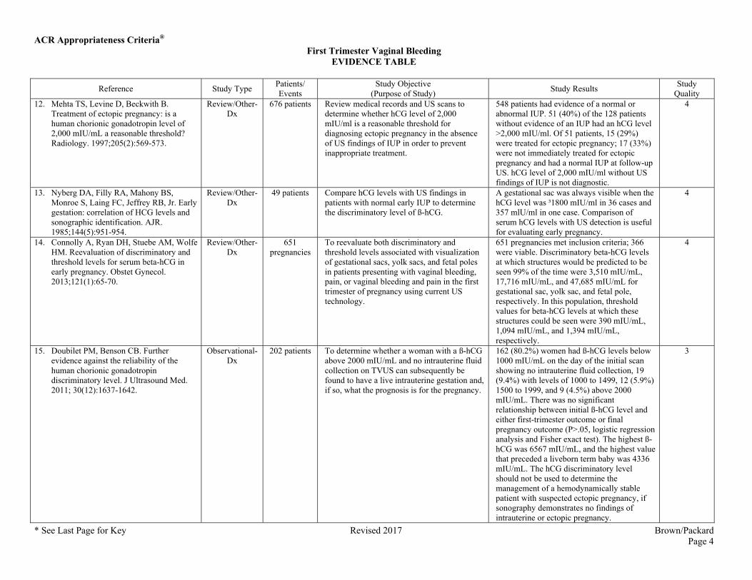
Quality 12. Mehta TS, Levine D, Beckwith B.
Treatment of ectopic pregnancy: is a human chorionic gonadotropin level of 2,000 mIU/mL a reasonable threshold? Radiology. 1997;205(2):569-573.
Review/Other-Dx
676 patients Review medical records and US scans to determine whether hCG level of 2,000 mIU/ml is a reasonable threshold for diagnosing ectopic pregnancy in the absence of US findings of IUP in order to prevent inappropriate treatment.
548 patients had evidence of a normal or abnormal IUP. 51 (40%) of the 128 patients without evidence of an IUP had an hCG level >2,000 mIU/ml. Of 51 patients, 15 (29%) were treated for ectopic pregnancy; 17 (33%) were not immediately treated for ectopic pregnancy and had a normal IUP at follow-up US. hCG level of 2,000 mIU/ml without US findings of IUP is not diagnostic.
4
13. Nyberg DA, Filly RA, Mahony BS, Monroe S, Laing FC, Jeffrey RB, Jr. Early gestation: correlation of HCG levels and sonographic identification. AJR. 1985;144(5):951-954.
Review/Other-Dx
49 patients Compare hCG levels with US findings in patients with normal early IUP to determine the discriminatory level of ß-hCG.
A gestational sac was always visible when the hCG level was ³1800 mIU/ml in 36 cases and 357 mlU/ml in one case. Comparison of serum hCG levels with US detection is useful for evaluating early pregnancy.
4
14. Connolly A, Ryan DH, Stuebe AM, Wolfe HM. Reevaluation of discriminatory and threshold levels for serum beta-hCG in early pregnancy. Obstet Gynecol. 2013;121(1):65-70.
Review/Other-Dx
651 pregnancies
To reevaluate both discriminatory and threshold levels associated with visualization of gestational sacs, yolk sacs, and fetal poles in patients presenting with vaginal bleeding, pain, or vaginal bleeding and pain in the first trimester of pregnancy using current US technology.
651 pregnancies met inclusion criteria; 366 were viable. Discriminatory beta-hCG levels at which structures would be predicted to be seen 99% of the time were 3,510 mIU/mL, 17,716 mIU/mL, and 47,685 mIU/mL for gestational sac, yolk sac, and fetal pole, respectively. In this population, threshold values for beta-hCG levels at which these structures could be seen were 390 mIU/mL, 1,094 mIU/mL, and 1,394 mIU/mL, respectively.
4
15. Doubilet PM, Benson CB. Further evidence against the reliability of the human chorionic gonadotropin discriminatory level. J Ultrasound Med. 2011; 30(12):1637-1642.
Observational-Dx
202 patients To determine whether a woman with a ß-hCG above 2000 mIU/mL and no intrauterine fluid collection on TVUS can subsequently be found to have a live intrauterine gestation and, if so, what the prognosis is for the pregnancy.
162 (80.2%) women had ß-hCG levels below 1000 mIU/mL on the day of the initial scan showing no intrauterine fluid collection, 19 (9.4%) with levels of 1000 to 1499, 12 (5.9%) 1500 to 1999, and 9 (4.5%) above 2000 mIU/mL. There was no significant relationship between initial ß-hCG level and either first-trimester outcome or final pregnancy outcome (P>.05, logistic regression analysis and Fisher exact test). The highest ß-hCG was 6567 mIU/mL, and the highest value that preceded a liveborn term baby was 4336 mIU/mL. The hCG discriminatory level should not be used to determine the management of a hemodynamically stable patient with suspected ectopic pregnancy, if sonography demonstrates no findings of intrauterine or ectopic pregnancy.
3
ACR Appropriateness Criteria® First Trimester Vaginal Bleeding
EVIDENCE TABLE
* See Last Page for Key Revised 2017 Brown/Packard Page 5
Reference Study Type Patients/ Events
Study Objective (Purpose of Study) Study Results Study
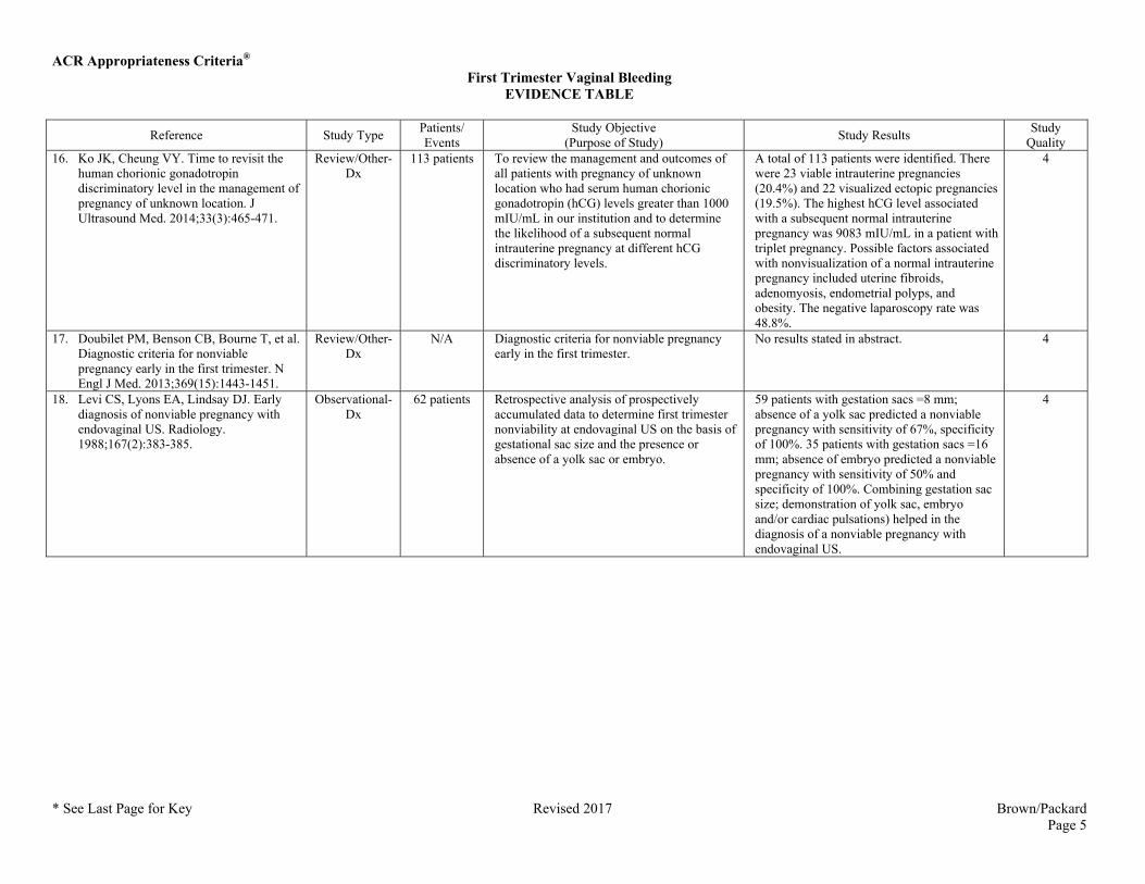
Quality 16. Ko JK, Cheung VY. Time to revisit the
human chorionic gonadotropin discriminatory level in the management of pregnancy of unknown location. J Ultrasound Med. 2014;33(3):465-471.
Review/Other-Dx
113 patients To review the management and outcomes of all patients with pregnancy of unknown location who had serum human chorionic gonadotropin (hCG) levels greater than 1000 mIU/mL in our institution and to determine the likelihood of a subsequent normal intrauterine pregnancy at different hCG discriminatory levels.
A total of 113 patients were identified. There were 23 viable intrauterine pregnancies (20.4%) and 22 visualized ectopic pregnancies (19.5%). The highest hCG level associated with a subsequent normal intrauterine pregnancy was 9083 mIU/mL in a patient with triplet pregnancy. Possible factors associated with nonvisualization of a normal intrauterine pregnancy included uterine fibroids, adenomyosis, endometrial polyps, and obesity. The negative laparoscopy rate was 48.8%.
4
17. Doubilet PM, Benson CB, Bourne T, et al. Diagnostic criteria for nonviable pregnancy early in the first trimester. N Engl J Med. 2013;369(15):1443-1451.
Review/Other-Dx
N/A Diagnostic criteria for nonviable pregnancy early in the first trimester.
No results stated in abstract. 4
18. Levi CS, Lyons EA, Lindsay DJ. Early diagnosis of nonviable pregnancy with endovaginal US. Radiology. 1988;167(2):383-385.
Observational-Dx
62 patients Retrospective analysis of prospectively accumulated data to determine first trimester nonviability at endovaginal US on the basis of gestational sac size and the presence or absence of a yolk sac or embryo.
59 patients with gestation sacs =8 mm; absence of a yolk sac predicted a nonviable pregnancy with sensitivity of 67%, specificity of 100%. 35 patients with gestation sacs =16 mm; absence of embryo predicted a nonviable pregnancy with sensitivity of 50% and specificity of 100%. Combining gestation sac size; demonstration of yolk sac, embryo and/or cardiac pulsations) helped in the diagnosis of a nonviable pregnancy with endovaginal US.
4

ACR Appropriateness Criteria® First Trimester Vaginal Bleeding
EVIDENCE TABLE
* See Last Page for Key Revised 2017 Brown/Packard Page 6
Reference Study Type Patients/ Events
Study Objective (Purpose of Study) Study Results Study
Quality 19. Abdallah Y, Daemen A, Kirk E, et al.
Limitations of current definitions of miscarriage using mean gestational sac diameter and crown-rump length measurements: a multicenter observational study. Ultrasound Obstet Gynecol. 2011;38(5):497-502.
Observational-Dx
1,060 consecutive
women
Observational cross-sectional study to define the false-positive rate for the diagnosis of miscarriage associated with different CRL and MSD measurements with or without a yolk sac in a large study population of patients attending early pregnancy clinics. The authors also aimed to define cut-off values for CRL and MSD that, on the basis of a single measurement, can definitively diagnose a miscarriage and so exclude possible inadvertent termination of pregnancy.
Of the 1,060 women with a diagnosis of IUP of uncertain viability, 473 remained viable and 587 were non-viable by the time of the 11-14-week scan. In the absence of both embryo and yolk sac, the false-positive rate for miscarriage was 4.4% when an MSD cut-off of 16 mm was used and 0.5% for a cut-off of 20 mm. There were no false-positive test results for miscarriage when a cut-off of MSD =21 mm was used. If a yolk sac was present but an embryo was not, the false-positive rate for miscarriage was 2.6% for an MSD cut-off of 16 mm and 0.4% for a cut-off of 20 mm, with no false-positive results when a cut-off of MSD =21 mm was used. When an embryo was visible with an absent heartbeat, using a CRL cut-off of 4 mm the false-positive rate for miscarriage was 8.3%, and for a CRL cut-off of 5 mm it was also 8.3%. There were no false-positive results using a CRL cut-off of =5.3 mm. These data show that some current definitions used to diagnose miscarriage are potentially unsafe. An MSD cut-off of >25 mm and a CRL cut-off of >7 mm could be introduced to minimize the risk of a false-positive diagnosis of miscarriage.
3

ACR Appropriateness Criteria® First Trimester Vaginal Bleeding
EVIDENCE TABLE
* See Last Page for Key Revised 2017 Brown/Packard Page 7
Reference Study Type Patients/ Events
Study Objective (Purpose of Study) Study Results Study
Quality 20. Rowling SE, Coleman BG, Langer JE,
Arger PH, Nisenbaum HL, Horii SC. First-trimester US parameters of failed pregnancy. Radiology. 1997;203(1):211-217.
Review/Other-Dx
2,655 first-trimester US
scans in 2,285
patients
Retrospective review of US scans to test the reliability of established US parameters in predicting the outcome of first-trimester pregnancy.
30 (22%) of 135 patients without yolk sacs and with an 8 mm mean sac diameter developed live embryos: 24 had normal follow-up or delivery; six were lost to follow-up. 5(8%) of 59 patients with no depiction of embryos and with a 16 mm mean sac diameter developed live embryos: Two delivered, one spontaneously aborted, one had death of one twin embryo before being lost to follow-up, and one was lost to follow-up. 17 (0.74%) of 2,285 patients had early oligohydramnios: 6 (35%) had normal follow-up scans or delivery, two (12%) spontaneously aborted, and nine (53%) were lost to follow-up. Established parameters predictive of early pregnancy failure potentially result in misdiagnosis of nonviability or poor prognosis when applied to a large, unselected patient population. Close follow-up is necessary in cases with borderline abnormal findings.
4

ACR Appropriateness Criteria® First Trimester Vaginal Bleeding
EVIDENCE TABLE
* See Last Page for Key Revised 2017 Brown/Packard Page 8
Reference Study Type Patients/ Events
Study Objective (Purpose of Study) Study Results Study
Quality 21. Preisler J, Kopeika J, Ismail L, et al.
Defining safe criteria to diagnose miscarriage: prospective observational multicentre study. BMJ. 2015;351:h4579.
Observational-Dx
2845 women To validate recent guidance changes by establishing the performance of cut-off values for embryo crown-rump length and mean gestational sac diameter to diagnose miscarriage with high levels of certainty. Secondary aims were to examine the influence of gestational age on interpretation of mean gestational sac diameter and crown-rump length values, determine the optimal intervals between scans and findings on repeat scans that definitively diagnose pregnancy failure.)
The following indicated a miscarriage at initial scan: mean gestational sac diameter >/= 25 mm with an empty sac (364/364 specificity: 100%, 95% confidence interval 99.0% to 100%), embryo with crown-rump length >/= 7 mm without visible embryo heart activity (110/110 specificity: 100%, 96.7% to 100%), mean gestational sac diameter >/= 18 mm for gestational sacs without an embryo presenting after 70 days' gestation (907/907 specificity: 100%, 99.6% to 100%), embryo with crown-rump length >/= 3 mm without visible heart activity presenting after 70 days' gestation (87/87 specificity: 100%, 95.8% to 100%). The following were indicative of miscarriage at a repeat scan: initial scan and repeat scan after seven days or more showing an embryo without visible heart activity (103/103 specificity: 100%, 96.5% to 100%), pregnancies without an embryo and mean gestational sac diameter <12 mm where the mean diameter has not doubled after 14 days or more (478/478 specificity: 100%, 99.2% to 100%), pregnancies without an embryo and mean gestational sac diameter >/= 12 mm showing no embryo heartbeat after seven days or more (150/150 specificity: 100%, 97.6% to 100%).
3
22. Huchon C, Deffieux X, Beucher G, et al. Pregnancy loss: French clinical practice guidelines. Eur J Obstet Gynecol Reprod Biol. 2016;201:18-26.
Review/Other-Dx
N/A To provide clinical practice guidelines for pregnancy loss.
No results stated in abstract. 4
23. Benson CB, Doubilet PM. Slow embryonic heart rate in early first trimester: indicator of poor pregnancy outcome. Radiology. 1994;192(2):343-344.
Review/Other-Dx
37 patients Examine US scans to determine the outcome of early first-trimester pregnancies with slow embryonic heart rates.
An embryonic heart rate =90 bpm in the first trimester has a high likelihood of fetal loss before the end of the first trimester. Loss occurred in all embryos with heart rates <70 bpm.
4
ACR Appropriateness Criteria® First Trimester Vaginal Bleeding
EVIDENCE TABLE
* See Last Page for Key Revised 2017 Brown/Packard Page 9
Reference Study Type Patients/ Events
Study Objective (Purpose of Study) Study Results Study
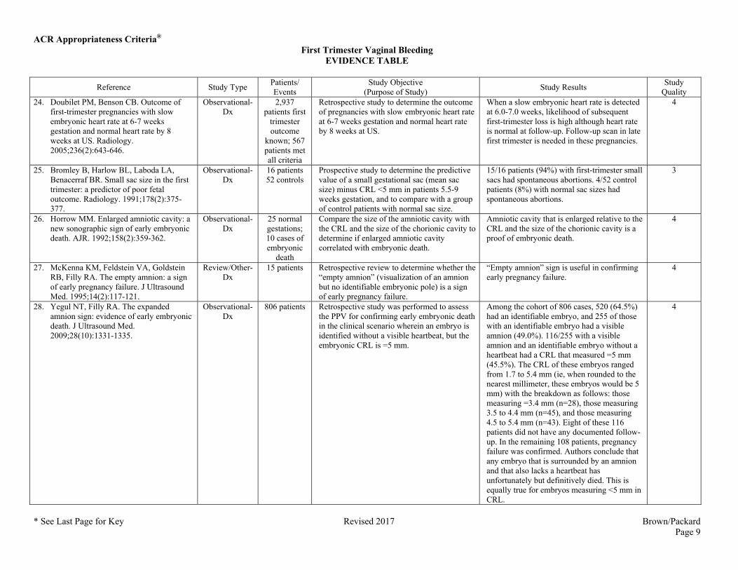
Quality 24. Doubilet PM, Benson CB. Outcome of
first-trimester pregnancies with slow embryonic heart rate at 6-7 weeks gestation and normal heart rate by 8 weeks at US. Radiology. 2005;236(2):643-646.
Observational-Dx
2,937 patients first
trimester outcome
known; 567 patients met all criteria
Retrospective study to determine the outcome of pregnancies with slow embryonic heart rate at 6-7 weeks gestation and normal heart rate by 8 weeks at US.
When a slow embryonic heart rate is detected at 6.0-7.0 weeks, likelihood of subsequent first-trimester loss is high although heart rate is normal at follow-up. Follow-up scan in late first trimester is needed in these pregnancies.
4
25. Bromley B, Harlow BL, Laboda LA, Benacerraf BR. Small sac size in the first trimester: a predictor of poor fetal outcome. Radiology. 1991;178(2):375-377.
Observational-Dx
16 patients 52 controls
Prospective study to determine the predictive value of a small gestational sac (mean sac size) minus CRL <5 mm in patients 5.5-9 weeks gestation, and to compare with a group of control patients with normal sac size.
15/16 patients (94%) with first-trimester small sacs had spontaneous abortions. 4/52 control patients (8%) with normal sac sizes had spontaneous abortions.
3
26. Horrow MM. Enlarged amniotic cavity: a new sonographic sign of early embryonic death. AJR. 1992;158(2):359-362.
Observational-Dx
25 normal gestations; 10 cases of embryonic
death
Compare the size of the amniotic cavity with the CRL and the size of the chorionic cavity to determine if enlarged amniotic cavity correlated with embryonic death.
Amniotic cavity that is enlarged relative to the CRL and the size of the chorionic cavity is a proof of embryonic death.
4
27. McKenna KM, Feldstein VA, Goldstein RB, Filly RA. The empty amnion: a sign of early pregnancy failure. J Ultrasound Med. 1995;14(2):117-121.
Review/Other-Dx
15 patients Retrospective review to determine whether the “empty amnion” (visualization of an amnion but no identifiable embryonic pole) is a sign of early pregnancy failure.
“Empty amnion” sign is useful in confirming early pregnancy failure.
4
28. Yegul NT, Filly RA. The expanded amnion sign: evidence of early embryonic death. J Ultrasound Med. 2009;28(10):1331-1335.
Observational-Dx
806 patients Retrospective study was performed to assess the PPV for confirming early embryonic death in the clinical scenario wherein an embryo is identified without a visible heartbeat, but the embryonic CRL is =5 mm.
Among the cohort of 806 cases, 520 (64.5%) had an identifiable embryo, and 255 of those with an identifiable embryo had a visible amnion (49.0%). 116/255 with a visible amnion and an identifiable embryo without a heartbeat had a CRL that measured =5 mm (45.5%). The CRL of these embryos ranged from 1.7 to 5.4 mm (ie, when rounded to the nearest millimeter, these embryos would be 5 mm) with the breakdown as follows: those measuring =3.4 mm (n=28), those measuring 3.5 to 4.4 mm (n=45), and those measuring 4.5 to 5.4 mm (n=43). Eight of these 116 patients did not have any documented follow-up. In the remaining 108 patients, pregnancy failure was confirmed. Authors conclude that any embryo that is surrounded by an amnion and that also lacks a heartbeat has unfortunately but definitively died. This is equally true for embryos measuring <5 mm in CRL.
4
ACR Appropriateness Criteria® First Trimester Vaginal Bleeding
EVIDENCE TABLE
* See Last Page for Key Revised 2017 Brown/Packard Page 10
Reference Study Type Patients/ Events
Study Objective (Purpose of Study) Study Results Study
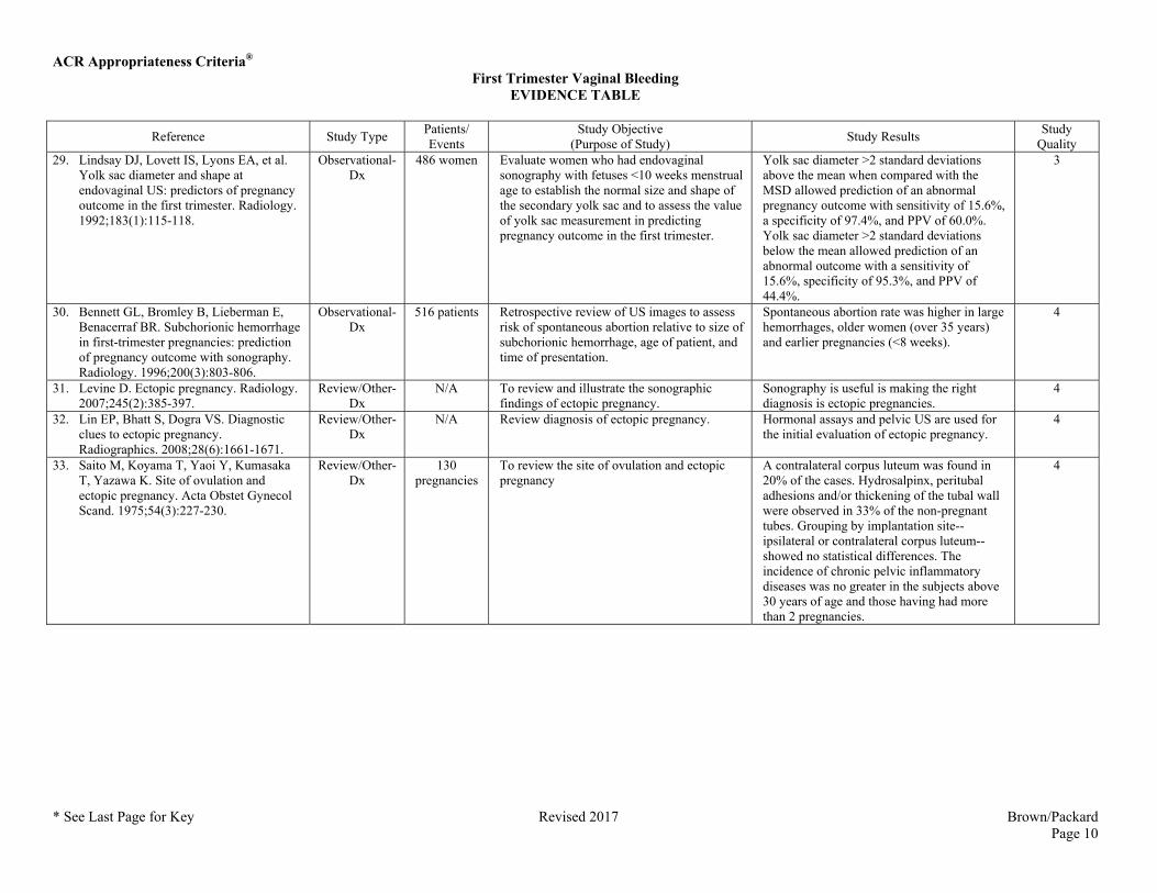
Quality 29. Lindsay DJ, Lovett IS, Lyons EA, et al.
Yolk sac diameter and shape at endovaginal US: predictors of pregnancy outcome in the first trimester. Radiology. 1992;183(1):115-118.
Observational-Dx
486 women Evaluate women who had endovaginal sonography with fetuses <10 weeks menstrual age to establish the normal size and shape of the secondary yolk sac and to assess the value of yolk sac measurement in predicting pregnancy outcome in the first trimester.
Yolk sac diameter >2 standard deviations above the mean when compared with the MSD allowed prediction of an abnormal pregnancy outcome with sensitivity of 15.6%, a specificity of 97.4%, and PPV of 60.0%. Yolk sac diameter >2 standard deviations below the mean allowed prediction of an abnormal outcome with a sensitivity of 15.6%, specificity of 95.3%, and PPV of 44.4%.
3
30. Bennett GL, Bromley B, Lieberman E, Benacerraf BR. Subchorionic hemorrhage in first-trimester pregnancies: prediction of pregnancy outcome with sonography. Radiology. 1996;200(3):803-806.
Observational-Dx
516 patients Retrospective review of US images to assess risk of spontaneous abortion relative to size of subchorionic hemorrhage, age of patient, and time of presentation.
Spontaneous abortion rate was higher in large hemorrhages, older women (over 35 years) and earlier pregnancies (<8 weeks).
4
31. Levine D. Ectopic pregnancy. Radiology. 2007;245(2):385-397.
Review/Other-Dx
N/A To review and illustrate the sonographic findings of ectopic pregnancy.
Sonography is useful is making the right diagnosis is ectopic pregnancies.
4
32. Lin EP, Bhatt S, Dogra VS. Diagnostic clues to ectopic pregnancy. Radiographics. 2008;28(6):1661-1671.
Review/Other-Dx
N/A Review diagnosis of ectopic pregnancy. Hormonal assays and pelvic US are used for the initial evaluation of ectopic pregnancy.
4
33. Saito M, Koyama T, Yaoi Y, Kumasaka T, Yazawa K. Site of ovulation and ectopic pregnancy. Acta Obstet Gynecol Scand. 1975;54(3):227-230.
Review/Other-Dx
130 pregnancies
To review the site of ovulation and ectopic pregnancy
A contralateral corpus luteum was found in 20% of the cases. Hydrosalpinx, peritubal adhesions and/or thickening of the tubal wall were observed in 33% of the non-pregnant tubes. Grouping by implantation site--ipsilateral or contralateral corpus luteum--showed no statistical differences. The incidence of chronic pelvic inflammatory diseases was no greater in the subjects above 30 years of age and those having had more than 2 pregnancies.
4

ACR Appropriateness Criteria® First Trimester Vaginal Bleeding
EVIDENCE TABLE
* See Last Page for Key Revised 2017 Brown/Packard Page 11
Reference Study Type Patients/ Events
Study Objective (Purpose of Study) Study Results Study
Quality 34. Walters MD, Eddy C, Pauerstein CJ. The
contralateral corpus luteum and tubal pregnancy. Obstet Gynecol. 1987;70(6):823-826.
Review/Other-Dx
114 cases To review the corpus luteum and tubal pregnancy.
A corpus luteum was found ipsilateral to the tubal pregnancy in 80 cases (70%) and contralateral in 18 (16%). In 16 cases (14%), the position of the corpus luteum could not be identified by inspection. No differences were noted among the groups in days from last normal menstrual period or the incidence of irregular bleeding. Of the 98 cases in which a corpus luteum was identified, 53 women (54%) had at least one condition that could be considered etiologic for tubal pregnancy, including 38 (39%) who had microscopic evidence of chronic salpingitis. No association was found between the laterality of the corpus luteum and the presence of risk factors, including mechanical factors.
4
35. Bonde AA, Korngold EK, Foster BR, et al. Radiological appearances of corpus luteum cysts and their imaging mimics. Abdom Radiol (NY). 2016;41(11):2270-2282.
Review/Other-Dx
N/A To review the radiological appearances of corpus luteum cysts and their imaging mimics.
No results listed in abstract. 4
36. Baerwald AR, Adams GP, Pierson RA. Form and function of the corpus luteum during the human menstrual cycle. Ultrasound Obstet Gynecol. 2005;25(5):498-507.
Observational-Dx
50 women To characterize the growth and regression of the corpus luteum (CL) during an interovulatory interval (IOI) using serial transvaginal ultrasonography.
Corpora lutea were of two morphological types: those with a central fluid-filled cavity (CFFC) (78%) and those without (22%). Eighty-eight percent of women exhibited a CL containing a CFFC 2 days after ovulation, followed by 34% 13 days after ovulation and 2% 27 days after ovulation. Luteal area, progesterone concentration and estradiol concentration increased for approximately the first 6 days following ovulation followed by a subsequent decline. Luteal NPV decreased from days 1 to 11 and increased during days 11-16. Changes in luteal area, NPV, progesterone and estradiol concentrations did not differ in women with two versus three waves of follicular development.
3

ACR Appropriateness Criteria® First Trimester Vaginal Bleeding
EVIDENCE TABLE
* See Last Page for Key Revised 2017 Brown/Packard Page 12
Reference Study Type Patients/ Events
Study Objective (Purpose of Study) Study Results Study
Quality 37. Durfee SM, Frates MC. Sonographic
spectrum of the corpus luteum in early pregnancy: gray-scale, color, and pulsed Doppler appearance. J Clin Ultrasound. 1999;27(2):55-59.
Review/Other-Dx
160 patients To describe the gray-scale and Doppler sonographic features of the corpus luteum during the first trimester of pregnancy.
The corpus luteum was identified in 157 (98%) of 160 patients. The mean diameter was 1.9 +/- 0.6 cm. The most common appearance was a round hypoechoic structure, found in 54 patients (34%). Other appearances included a cyst with a thick wall and anechoic center (43 patients, 27%), a cyst containing internal debris (36 patients, 23%), and a thin-walled simple cyst (24 patients, 15%). Corpus luteal blood flow was visualized with color Doppler imaging in 92% (145/157) of patients in whom the corpus luteum was found. Color Doppler imaging typically revealed a circumferential rim surrounding part or all of the corpus luteum. Low-resistance blood flow was seen with pulsed Doppler interrogation, with a mean resistance index of 0.49 +/- 0.08 and mean peak systolic velocity of 17 +/- 10 cm/second.
4
38. Frates MC, Doubilet PM, Durfee SM, et al. Sonographic and Doppler characteristics of the corpus luteum: can they predict pregnancy outcome? J Ultrasound Med. 2001;20(8):821-827.
Observational-Dx
201 patients To determine whether there is a relationship between gray scale or Doppler characteristics of the corpus luteum and first-trimester pregnancy outcome.
There were 201 study patients. The corpus luteum could be visualized in 197 (98%) and had a mean +/- SD size of 1.9 +/- 0.6 cm, a mean resistive index of 0.50 +/- 0.08, and a peak systolic velocity of 20.5 +/- 11.2 cm/s. There were 151 first-trimester survivors (75.1 %) and 50 spontaneous losses (24.9%). In a comparison of the survivors and losses, there was no significant difference in mean corpus luteum size (1.9 versus 1.7 cm; P = .10, t test), mean resistive index (0.50 versus 0.50; P = .71, t test), peak systolic velocity (21 versus 19 cm/s; P = .29, t test), or sonographic appearance (P = .78, chi2 test). The lack of association between corpus luteum characteristics and outcome persisted when cases were stratified by progesterone use and the presence or absence of a heartbeat on the study sonogram.
3

ACR Appropriateness Criteria® First Trimester Vaginal Bleeding
EVIDENCE TABLE
* See Last Page for Key Revised 2017 Brown/Packard Page 13
Reference Study Type Patients/ Events
Study Objective (Purpose of Study) Study Results Study
Quality 39. Frates MC, Doubilet PM, Peters HE,
Benson CB. Adnexal sonographic findings in ectopic pregnancy and their correlation with tubal rupture and human chorionic gonadotropin levels. J Ultrasound Med. 2014;33(4):697-703.
Observational-Dx
231 pregnancies
To determine whether the distribution of transvaginal sonographic findings of ectopic pregnancy has changed since the studies done 20 years ago and to explore the correlation of tubal rupture with transvaginal sonographic findings and human chorionic gonadotropin (hCG) levels.
Our study included 231 ectopic pregnancies. A positive sonographic adnexal finding was present in 219 cases (94.8%): adnexal mass in 218 (94.4%) and a moderate-to-large amount of free fluid in 84 (36.4%). The adnexal masses were graded as follows: 1, nonspecific mass (125 cases [54.1% of total]); 2, tubal ring without a yolk sac or embryo (57 [24.7%]); 3, yolk sac but no embryonic heartbeat (19 [8.3%]); and 4, embryo with cardiac activity (17 [7.4%]). The mean hCG level increased as the grade ascended from 1 to 4. Thirty-six patients had tubal rupture at surgery within 24 hours of the sonogram. A moderate-to-large amount of free fluid was significantly associated with tubal rupture (P < .05) but had low sensitivity, specificity, and positive predictive value for rupture. Other sonographic findings and hCG levels were not significantly related to tubal rupture.
3

ACR Appropriateness Criteria® First Trimester Vaginal Bleeding
EVIDENCE TABLE
* See Last Page for Key Revised 2017 Brown/Packard Page 14
Reference Study Type Patients/ Events
Study Objective (Purpose of Study) Study Results Study
Quality 40. Rottem S, Thaler I, Timor-Tritsch IE.
Classification of tubal gestations by transvaginal sonography. Ultrasound Obstet Gynecol. 1991;1(3):197-201.
Observational-Dx
191 patients To identify the classification of tubal gestations by transvaginal sonography
The following classifications were made: Type Ia (n = 43) A well-defined 'tubal ring' and a beating heart with or without discrete embryonic or extra-embryonic structures. Type Ib (n = 48) A 'tubal ring' containing embryonic and/or extra-embryonic structures without heart beats. Type II (n = 64) An ill-defined or thin tubal wall containing sonolucent or an irregularly echogenic core but not embryonic or extra-embryonic structures. Type III (n = 28) Free pelvic fluid and an empty uterus in patients with positive serum beta-hCG levels. The outline of the tube cannot be visualized. Surgery revealed unruptured tubal pregnancies in 90 patients of the combined groups of Types Ia and Ib. In one additional patient, a tubal rupture was found. In the Type II patients, 26 tubal pregnancies with blood clots in the tube but no evidence of bleeding into the pelvis, and 38 tubal ruptures or abortions were diagnosed. All Type III patients had ruptured tubal pregnancies or bleeding tubal abortions. In eight patients (4.2%), the only sonographic finding was an empty uterus, and these cases were erroneously diagnosed as not having an ectopic pregnancy (false negatives). There were two false-positive cases in which a tubal ring was detected, and this was related to an hemorrhagic corpus luteum. When used for diagnosing tubal pregnancy, the transvaginal scanning technique (together with beta-hCG in Type III cases) carries a sensitivity of 95.8% and specificity of 99.9%.
3

ACR Appropriateness Criteria® First Trimester Vaginal Bleeding
EVIDENCE TABLE
* See Last Page for Key Revised 2017 Brown/Packard Page 15
Reference Study Type Patients/ Events
Study Objective (Purpose of Study) Study Results Study
Quality 41. Brown DL, Doubilet PM. Transvaginal
sonography for diagnosing ectopic pregnancy: positivity criteria and performance characteristics. J Ultrasound Med. 1994;13(4):259-266.
Meta-analysis 10 studies/2216
patients
To identify original studies presenting suitable data on the use of TVS for the diagnosis of EP.
Ten studies involving a total of 2216 patients, 565 with EP and 1651 without EP, were included in our analysis. Based on the combined data from these studies, criteria A, B, and C all have high specificities (99.5-100%) and positive predictive values (97.8-100%) but low sensitivities (20.1-64.6%) and mediocre negative predictive values (78.5-89.1%). Criterion D, the most lax criterion, has the most uniformly excellent characteristics, with only slightly lower specificity (98.9%) and positive predictive value (96.3%) but considerably higher sensitivity (84.4%) and negative predictive value (94.8%).
M
42. Dart R, McLean SA, Dart L. Isolated fluid in the cul-de-sac: how well does it predict ectopic pregnancy? Am J Emerg Med. 2002;20(1):1-4.
Observational-Dx
1ST Group - 38 patients with cul-de-
sac fluid; 2nd Group - 523 patients with indeterminate
US
Retrospective cohort study to examine the risk of ectopic pregnancy among patients with isolated abnormal cul-de-sac fluid at TVUS. Moderate volume of anechoic fluid was compared with either a large volume of anechoic fluid or any echogenic fluid.
Ectopic pregnancy was diagnosed in 16/38: 42% (95% CI: 26%-59%) of patients with isolated cul-de-sac fluid, 5/23: 22% (95%. CI: 7%-42%) of patients with moderate amount of anechoic fluid, and 11/15: 73% (95%, CI: 45%-92%) of patients with a large volume of fluid or any echogenic fluid. Patients with isolated abnormal cul-de-sac fluid are at moderate risk for ectopic pregnancy. The risk increases if the fluid is echogenic or the volume is large.
3
43. Nyberg DA, Hughes MP, Mack LA, Wang KY. Extrauterine findings of ectopic pregnancy of transvaginal US: importance of echogenic fluid. Radiology. 1991;178(3):823-826.
Review/Other-Dx
232 total patients
Prospective study of TVUS studies to determine the significance of different extrauterine findings, including echogenic fluid in the cul-de-sac in patients with positive serum pregnancy tests considered to be at risk for ectopic pregnancy.
Intraperitoneal fluid was detected in 43 (63%) group 1 patients and in 81 (31%) group 3 patients. Echogenic fluid was the only abnormal finding at US in 10 (15%) group 1 patients and added confidence to the diagnosis of ectopic pregnancy in many others. Echogenic fluid correlated with hemoperitoneum at the time of surgery. Presence of echogenic fluid shows a high risk for ectopic pregnancy.
4
44. Wachsberg RH, Levine CD. Echogenic peritoneal fluid as an isolated sonographic finding: significance in patients at risk of ectopic pregnancy. Clin Radiol. 1998;53(7):520-522.
Observational-Dx
12 consecutive symptomatic
patients
Retrospective study of patients with positive pregnancy test in whom sonography revealed echogenic fluid as an isolated finding without evidence of IUP.
Small-to-moderate amount of echogenic fluid noted as an isolated finding may not be highly predictive of ectopic pregnancy.
4

ACR Appropriateness Criteria® First Trimester Vaginal Bleeding
EVIDENCE TABLE
* See Last Page for Key Revised 2017 Brown/Packard Page 16
Reference Study Type Patients/ Events
Study Objective (Purpose of Study) Study Results Study
Quality 45. Tanaka Y, Mimura K, Kanagawa T, et al.
Three-dimensional sonography in the differential diagnosis of interstitial, angular, and intrauterine pregnancies in a septate uterus. J Ultrasound Med. 2014;33(11):2031-2035.
Review/Other-Dx
N/A To demonstrate the differences in diagnostic imaging findings and emphasize the importance of 3D sonography in differentiating these entities.
No results stated in abstract. 4
46. Talbot K, Simpson R, Price N, Jackson SR. Heterotopic pregnancy. J Obstet Gynaecol. 2011;31(1):7-12.
Review/Other-Dx
N/A Review diagnosis and management of heterotopic pregnancy.
In the majority (71%) of cases reviewed, risk factors for a heterotopic pregnancy were present. However, in several instances (33%), previous sonographic reports of a normal IUP gave false reassurance. These results highlight the complexity of diagnosis. In addition, the findings were compared with two previous reviews covering cases from 1971 to 2004. This comparison highlighted two important trends: first, the increasing role of US in the definitive diagnosis of a heterotopic pregnancy, and second, the development of conservative approaches to management.
4
47. Barnhart K, van Mello NM, Bourne T, et al. Pregnancy of unknown location: a consensus statement of nomenclature, definitions, and outcome. Fertil Steril. 2011;95(3):857-866.
Review/Other-Dx
N/A To improve the interpretation of future studies in women who are initially diagnosed with a pregnancy of unknown location, the authors proposed a consensus statement with definitions of population, target disease, and final outcome.
Careful definition of populations and classification of outcomes should optimize objective interpretation of research, allow objective assessment of future reproductive prognosis, and hopefully lead to improved clinical care of women initially identified to have a pregnancy of unknown location.
4
48. Kirk E, Condous G, Bourne T. Pregnancies of unknown location. Best Pract Res Clin Obstet Gynaecol. 2009;23(4):493-499.
Review/Other-Dx
N/A To discuss the various aspects of management of women with a pregnancy of unknown location (PUL).
No results stated in abstract. 4
ACR Appropriateness Criteria® First Trimester Vaginal Bleeding
EVIDENCE TABLE
* See Last Page for Key Revised 2017 Brown/Packard Page 17
Reference Study Type Patients/ Events
Study Objective (Purpose of Study) Study Results Study
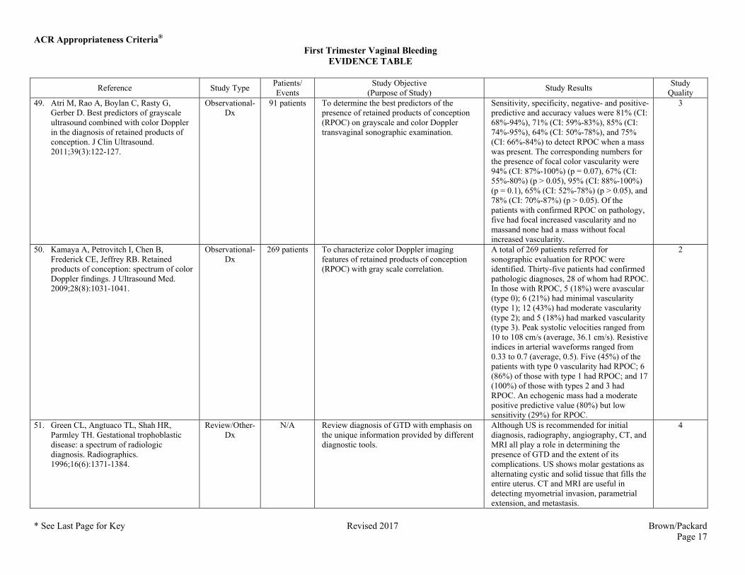
Quality 49. Atri M, Rao A, Boylan C, Rasty G,
Gerber D. Best predictors of grayscale ultrasound combined with color Doppler in the diagnosis of retained products of conception. J Clin Ultrasound. 2011;39(3):122-127.
Observational-Dx
91 patients To determine the best predictors of the presence of retained products of conception (RPOC) on grayscale and color Doppler transvaginal sonographic examination.
Sensitivity, specificity, negative- and positive-predictive and accuracy values were 81% (CI: 68%-94%), 71% (CI: 59%-83%), 85% (CI: 74%-95%), 64% (CI: 50%-78%), and 75% (CI: 66%-84%) to detect RPOC when a mass was present. The corresponding numbers for the presence of focal color vascularity were 94% (CI: 87%-100%) (p = 0.07), 67% (CI: 55%-80%) (p > 0.05), 95% (CI: 88%-100%) (p = 0.1), 65% (CI: 52%-78%) (p > 0.05), and 78% (CI: 70%-87%) (p > 0.05). Of the patients with confirmed RPOC on pathology, five had focal increased vascularity and no massand none had a mass without focal increased vascularity.
3
50. Kamaya A, Petrovitch I, Chen B, Frederick CE, Jeffrey RB. Retained products of conception: spectrum of color Doppler findings. J Ultrasound Med. 2009;28(8):1031-1041.
Observational-Dx
269 patients To characterize color Doppler imaging features of retained products of conception (RPOC) with gray scale correlation.
A total of 269 patients referred for sonographic evaluation for RPOC were identified. Thirty-five patients had confirmed pathologic diagnoses, 28 of whom had RPOC. In those with RPOC, 5 (18%) were avascular (type 0); 6 (21%) had minimal vascularity (type 1); 12 (43%) had moderate vascularity (type 2); and 5 (18%) had marked vascularity (type 3). Peak systolic velocities ranged from 10 to 108 cm/s (average, 36.1 cm/s). Resistive indices in arterial waveforms ranged from 0.33 to 0.7 (average, 0.5). Five (45%) of the patients with type 0 vascularity had RPOC; 6 (86%) of those with type 1 had RPOC; and 17 (100%) of those with types 2 and 3 had RPOC. An echogenic mass had a moderate positive predictive value (80%) but low sensitivity (29%) for RPOC.
2
51. Green CL, Angtuaco TL, Shah HR, Parmley TH. Gestational trophoblastic disease: a spectrum of radiologic diagnosis. Radiographics. 1996;16(6):1371-1384.
Review/Other-Dx
N/A Review diagnosis of GTD with emphasis on the unique information provided by different diagnostic tools.
Although US is recommended for initial diagnosis, radiography, angiography, CT, and MRI all play a role in determining the presence of GTD and the extent of its complications. US shows molar gestations as alternating cystic and solid tissue that fills the entire uterus. CT and MRI are useful in detecting myometrial invasion, parametrial extension, and metastasis.
4

ACR Appropriateness Criteria® First Trimester Vaginal Bleeding
EVIDENCE TABLE
* See Last Page for Key Revised 2017 Brown/Packard Page 18
Reference Study Type Patients/ Events
Study Objective (Purpose of Study) Study Results Study
Quality 52. Lazarus E, Hulka C, Siewert B, Levine D.
Sonographic appearance of early complete molar pregnancies. J Ultrasound Med. 1999;18(9):589-594; quiz 595-586.
Review/Other-Dx
21 cases To review the ultrasonographic reports and clinical data of 21 cases of histologically diagnosed complete molar pregnancies with a mean gestational age at sonography of 10.5 weeks (range, 4 to 18 weeks)
The diagnosis of molar pregnancy was made on ultrasonography in 12 (57%) cases, was second in the differential diagnosis of one (4.8%) case, and was not considered in eight (38%) cases. No theca-lutein cysts were identified. Five of five (100%) molar pregnancies of 13 weeks or over were diagnosed prospectively, while only eight of 16 (50%) earlier pregnancies were correctly diagnosed prospectively. In a retrospective review of the available images of 16 patients, only nine of 16 (56%) images demonstrated the classic appearance, and no theca-lutein cysts were seen.
4
53. Benson CB, Genest DR, Bernstein MR, Soto-Wright V, Goldstein DP, Berkowitz RS. Sonographic appearance of first trimester complete hydatidiform moles. Ultrasound Obstet Gynecol. 2000;16(2):188-191.
Review/Other-Dx
24 patients To study the sonographic appearance of first trimester moles and the ability of ultrasound to detect them.
Of the 24 patients in our study, the mean gestational age at time of the sonogram was 8.7 +/- 2.0 weeks (mean +/- SD) with a range of 5.7-12.3 weeks. The initial sonographic interpretation was a complete mole in 17 (71%) cases, partial mole versus failed pregnancy in two (8%), and failed pregnancy in five (21%) cases. Of the 22 patients with sonograms available for review, interpretation on review of the images was a complete mole in 18 (82%) cases, partial mole versus failed pregnancy in one (5%), and failed pregnancy in three (14%) cases. The typical sonographic appearance of a first trimester complete mole was a complex, echogenic, intra-uterine mass containing many small cystic spaces.
4

ACR Appropriateness Criteria® First Trimester Vaginal Bleeding
EVIDENCE TABLE
* See Last Page for Key Revised 2017 Brown/Packard Page 19
Reference Study Type Patients/ Events
Study Objective (Purpose of Study) Study Results Study
Quality 54. Kirk E, Papageorghiou AT, Condous G,
Bottomley C, Bourne T. The accuracy of first trimester ultrasound in the diagnosis of hydatidiform mole. Ultrasound Obstet Gynecol. 2007;29(1):70-75.
Observational-Dx
90 women To assess the first-trimester ultrasonographic findings in all women suspected of having hydatidiform mole on ultrasound and those subsequently diagnosed with hydatidiform mole after histological examination of removed products of conception after surgical evacuation of the uterus.
The study group consisted of 90 women; 56 were suspected of having hydatidiform mole on ultrasound, and of these 27 (48%) had hydatidiform mole confirmed after histopathological examination of the products of conception, while no changes suggestive of hydatidiform mole were present in the other 29 cases. Overall, 61 women had hydatidiform mole confirmed on histology-41 (67%) partial hydatidiform moles (PHM) and 20 (33%) complete hydatidiform moles (CHM). The ultrasound findings in the 34 cases not suspected of hydatidiform mole were an empty sac in 8/34 (24%) women and a delayed miscarriage in the other 26/34 (76%). The overall sensitivity and positive predictive value for the ultrasound diagnosis of hydatidiform mole was 44% and 48%, respectively. For PHMs the respective values were 20% and 22% and for CHMs they were 95% and 40%.
3

ACR Appropriateness Criteria® First Trimester Vaginal Bleeding
EVIDENCE TABLE
* See Last Page for Key Revised 2017 Brown/Packard Page 20
Reference Study Type Patients/ Events
Study Objective (Purpose of Study) Study Results Study
Quality 55. Savage JL, Maturen KE, Mowers EL, et
al. Sonographic diagnosis of partial versus complete molar pregnancy: A reappraisal. J Clin Ultrasound. 2017;45(2):72-78.
Observational-Dx
70 women To assess the prospective sonographic diagnosis of molar pregnancy and compare sonographic features of complete versus partial molar pregnancy.
Mean age of patients was 30.5 +/- 7.0 (SD) years (range, 16-49 years) with a mean gravidity of 3.2 +/- 2.3 (SD) (range 1-11). Mean gestational age was 74.0 +/- 19.1 day (range 39-138) and serum beta-human chorionic gonadotropin was 131 +/- 156 mIU/ml (range 447-662,000). Pathologic results showed 48 partial and 22 complete molar pregnancies. Sonographically, partial moles more commonly showed a yolk sac (56.3% versus 0%, p < 0.0001), fetal pole (62.5% versus 4.6%, p < 0.0001), fine septa within the sac (25.0% versus 4.6%, p = 0.05), and normal (31.3% versus 0%, p = 0.002) or minimally cystic placenta (27.1% versus 4.6%, p = 0.49), while complete moles had larger gestational sacs (612 versus 44 mm, p = 0.005), were more often avascular on color Doppler imaging (45.5% versus 18.8%, p = 0.02), had more often abnormal tissue in the uterus (82.6% versus 20.8%, p < 0.0001) and placental masses (86.9% versus 16.7%, p < 0.0001), and were more often diagnosed prospectively (86.4% versus 41.7%, p = 0.0005).
2
56. Frates MC, Brown DL, Doubilet PM, Hornstein MD. Tubal rupture in patients with ectopic pregnancy: diagnosis with transvaginal US. Radiology. 1994;191(3):769-772.
Observational-Dx
132 consecutive
patients
Retrospective review of US scans and medical records to determine whether sonography can help diagnose tubal rupture in patients with ectopic pregnancy.
Adnexal masses were seen in 93 patients at US. Thirty-four patients had a tubal ring, and 59 had a complex mass. The frequency of tubal rupture was similar for both groups. The adnexal mass was significantly smaller in patients without a ruptured tube, but there was considerable overlap. Rupture was present in 21% of patients with no sign of or a trace of intraperitoneal fluid, increasing steadily to 63% in patients with a large amount of free fluid. Even though the amount of fluid was the best predictor of rupture, it was not completely reliable, as 37% of patients in whom a large amount of fluid was found had intact tubes. Findings at TVUS cannot reliably determine whether tubal rupture is present.
3

ACR Appropriateness Criteria® First Trimester Vaginal Bleeding
EVIDENCE TABLE
* See Last Page for Key Revised 2017 Brown/Packard Page 21
Reference Study Type Patients/ Events
Study Objective (Purpose of Study) Study Results Study
Quality 57. World Federation for Ultrasound in
Medicine and Biology. WFUMB/ISUOG Statement on the Safe Use of Doppler Ultrasound During 11-14 week scans (or earlier in pregnancy) http://www.wfumb.org/safety-statements/.
Review/Other-Dx
N/A No abstract available. No abstract available. 4
58. Abramowicz JS. Ultrasound in Assisted Reproductive Technologies and the First Trimester: Is There a Risk? Clin Obstet Gynecol. 2017;60(1):121-132.
Review/Other-Dx
N/A To review the use of ultrasound in assisted reproductive technologies and the first trimester.
No results listed in abstract. 4
59. Bly S, Van den Hof MC. Obstetric ultrasound biological effects and safety. J Obstet Gynaecol Can. 2005;27(6):572-580.
Review/Other-Dx
N/A To review the biological effects and safety of obstetric ultrasound.
No results stated in abstract. 4
60. Zhou Q, Lei XY, Xie Q, Cardoza JD. Sonographic and Doppler imaging in the diagnosis and treatment of gestational trophoblastic disease: a 12-year experience. J Ultrasound Med. 2005;24(1):15-24.
Observational-Dx
355patients Retrospective analysis of cases of GTD in two hospitals to evaluate the clinical utility of sonography with Doppler examination in the diagnosis and treatment of GTD.
106/355 cases had hydatidiform mole, 33 had a partial hydatidiform mole, 184 had an invasive hydatidiform mole, and 32 had choriocarcinoma. US showed abnormal molar tissue confined to the endometrial cavity in all cases of hydatidiform mole. Doppler waveforms showed resistive indices of 0.55 for hydatidiform mole, 0.56 for partial hydatidiform mole, 0.28 for invasive hydatidiform mole, 0.25 for choriocarcinoma, and 0.66 for normal pregnancies. Sonography and Doppler imaging were helpful in diagnosing GTD, in determining whether invasive disease was present, in detecting recurrence of disease, and in following the effectiveness of chemotherapy.
4
61. Lin LH, Bernardes LS, Hase EA, Fushida K, Francisco RP. Is Doppler ultrasound useful for evaluating gestational trophoblastic disease? Clinics (Sao Paulo). 2015;70(12):810-815.
Review/Other-Dx
28 articles To summarize data found in the literature regarding the applications of Doppler ultrasound in managing patients with gestational trophoblastic neoplasia.
No results listed in abstract. 4

ACR Appropriateness Criteria® First Trimester Vaginal Bleeding
EVIDENCE TABLE
* See Last Page for Key Revised 2017 Brown/Packard Page 22
Reference Study Type Patients/ Events
Study Objective (Purpose of Study) Study Results Study
Quality 62. Timor-Tritsch IE, Haynes MC,
Monteagudo A, Khatib N, Kovacs S. Ultrasound diagnosis and management of acquired uterine enhanced myometrial vascularity/arteriovenous malformations. Am J Obstet Gynecol. 2016;214(6):731 e731-731 e710.
Observational-Tx
27 patients To evaluate the role of transvaginal ultrasound scanning in the diagnosis and treatment of acquired enhanced myometrial vascularity/arteriovenous malformations to outline the natural history of conservatively followed vs treated lesions.
Twenty-seven patients met the diagnostic criteria of uterine enhanced myometrial vascularity/arteriovenous malformation. Mean age was 31.8 years (range, 18-42 years). Clinical diagnoses of the patients included 10 incomplete abortions, 6 missed abortions, 5 spontaneous complete abortions, 5 cesarean scar pregnancies, and 1 molar pregnancy. Eighty-nine percent of patients had bleeding (n = 24/27), although 1 patient was febrile, and 2 patients were asymptomatic. Recent surgical procedures were performed in 55.5% patients (15/27) that included curettage (n = 10), cesarean deliveries (n = 5), or both (n = 1); 4 patients had a remote history of uterine surgery that included myomectomy. Treatment was varied and included expectant treatment alone in 48% of the patients with serial ultrasound scans and serum human chorionic gonadotropin until resolution (n = 13/27 patients), uterine artery embolization (29.6%; 8/27 patients), methotrexate administration (22.2%; 6/27 patients), hysterectomy (7.4%; 2/27 patients), and curettage (3.7%; 1/27 patients). Three patients required a blood transfusion. Of the 9 patients whose condition required embolization, the conditions of 7 patients resolved after the procedure although 1 patient's condition required operative hysteroscopy and 1 patient's condition required hysterectomy for intractable bleeding. Average peak systolic velocity after embolization in the 9 patients was 85.2 cm/sec (range, 35-170 cm/sec); the average peak systolic velocity of the 16 patients with spontaneous resolution was 58.5 cm/sec (range, 23-90 cm/sec).
3

ACR Appropriateness Criteria® First Trimester Vaginal Bleeding
EVIDENCE TABLE
* See Last Page for Key Revised 2017 Brown/Packard Page 23
Reference Study Type Patients/ Events
Study Objective (Purpose of Study) Study Results Study
Quality 63. Rufener SL, Adusumilli S, Weadock WJ,
Caoili E. Sonography of uterine abnormalities in postpartum and postabortion patients: a potential pitfall of interpretation. J Ultrasound Med. 2008;27(3):343-348.
Observational-Dx
29 patients To identify misleading imaging features that leads to inclusion of a uterine AVM in the differential diagnosis of a uterine abnormality because consideration of this diagnosis can potentially alter patient treatment.
Interobserver agreement was as follows: the presence of a uterine mass, 90%; myometrial involvement, 83%; the presence of an associated vascular abnormality, 72%; and inclusion of a uterine AVM in the differential diagnosis, 86%. Myometrial involvement showed a statistically significant relationship to inclusion of a uterine AVM in the differential diagnosis (P<.05). Final pathologic diagnoses included retained products of conception (n=26), an endometrial polyp (n=1), chronic endometritis (n=1), and an exogenous progestational effect (n=1). No uterine AVMs were found. Despite high interobserver agreement in characterizing uterine abnormalities on sonography, readers still include uterine AVMs in the differential diagnosis of uterine masses that are ultimately proven to be retained products of conception. A myometrial location of a uterine mass is a particularly misleading imaging feature of retained products of conception.
3
64. Kamaya A, Ro K, Benedetti NJ, Chang PL, Desser TS. Imaging and diagnosis of postpartum complications: sonography and other imaging modalities. Ultrasound Q. 2009;25(3):151-162.
Review/Other-Dx
N/A To reivew the imaging and diagnosis of postpartum complications
No results listed in abstract. 4
65. Lee TY, Kim SH, Lee HJ, et al. Ultrasonographic indications for conservative treatment in pregnancy-related uterine arteriovenous malformations. Acta Radiol. 2014;55(9):1145-1152.
Observational-Dx
75 patients To assess the predictive value of ultrasonography for patients requiring conservative treatment for pregnancy related to uterine arteriovenous malformations (AVMs).
Features strongly associated with conservative management and their accuracy were PSV 89.6%, hemoglobin 84.7%, RI 83.1%, TAMXV 79.3%, and PI 78.6%. The overall accuracy for correct outcome classification was 64 (85.3%) of 75 patients. Most patients with conservative management had quicker improvement of symptoms and spontaneous regression at follow-up.
3
ACR Appropriateness Criteria® First Trimester Vaginal Bleeding
EVIDENCE TABLE
* See Last Page for Key Revised 2017 Brown/Packard Page 24
Reference Study Type Patients/ Events
Study Objective (Purpose of Study) Study Results Study
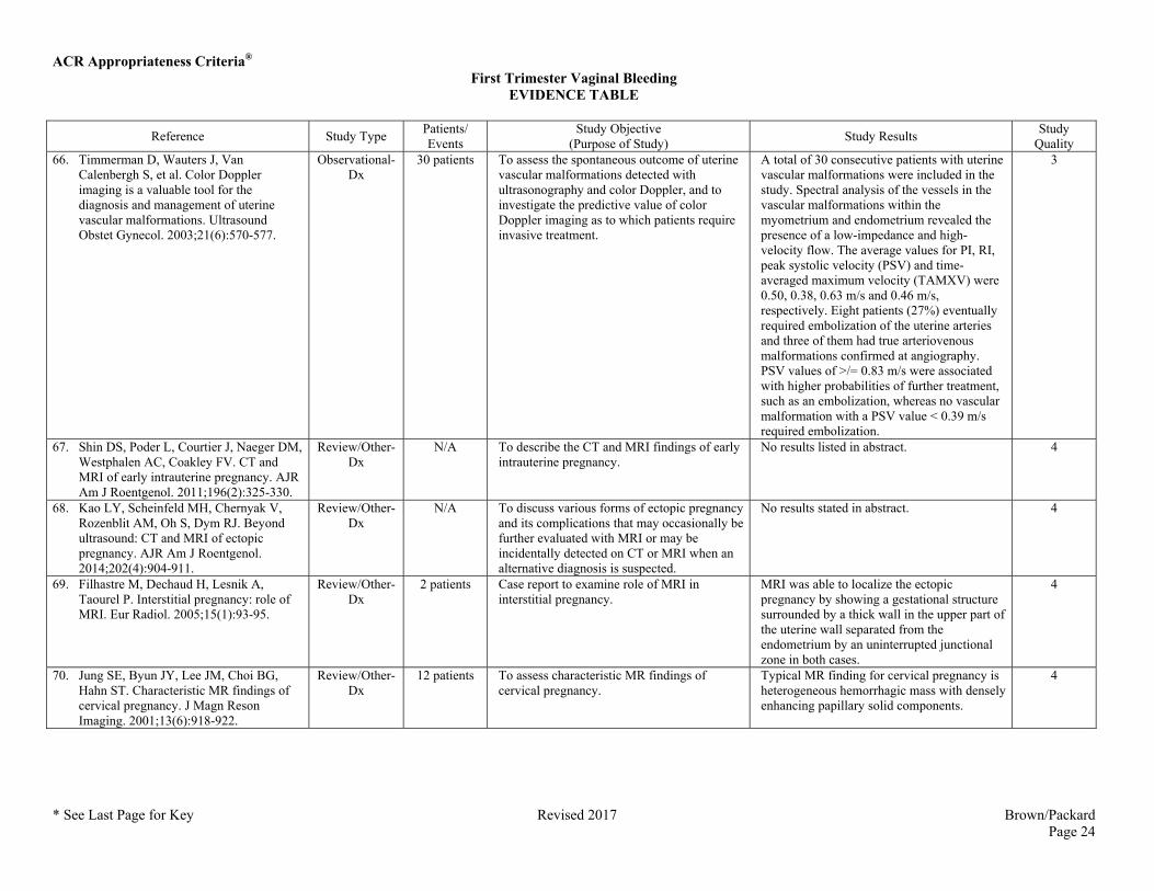
Quality 66. Timmerman D, Wauters J, Van
Calenbergh S, et al. Color Doppler imaging is a valuable tool for the diagnosis and management of uterine vascular malformations. Ultrasound Obstet Gynecol. 2003;21(6):570-577.
Observational-Dx
30 patients To assess the spontaneous outcome of uterine vascular malformations detected with ultrasonography and color Doppler, and to investigate the predictive value of color Doppler imaging as to which patients require invasive treatment.
A total of 30 consecutive patients with uterine vascular malformations were included in the study. Spectral analysis of the vessels in the vascular malformations within the myometrium and endometrium revealed the presence of a low-impedance and high-velocity flow. The average values for PI, RI, peak systolic velocity (PSV) and time-averaged maximum velocity (TAMXV) were 0.50, 0.38, 0.63 m/s and 0.46 m/s, respectively. Eight patients (27%) eventually required embolization of the uterine arteries and three of them had true arteriovenous malformations confirmed at angiography. PSV values of >/= 0.83 m/s were associated with higher probabilities of further treatment, such as an embolization, whereas no vascular malformation with a PSV value < 0.39 m/s required embolization.
3
67. Shin DS, Poder L, Courtier J, Naeger DM, Westphalen AC, Coakley FV. CT and MRI of early intrauterine pregnancy. AJR Am J Roentgenol. 2011;196(2):325-330.
Review/Other-Dx
N/A To describe the CT and MRI findings of early intrauterine pregnancy.
No results listed in abstract. 4
68. Kao LY, Scheinfeld MH, Chernyak V, Rozenblit AM, Oh S, Dym RJ. Beyond ultrasound: CT and MRI of ectopic pregnancy. AJR Am J Roentgenol. 2014;202(4):904-911.
Review/Other-Dx
N/A To discuss various forms of ectopic pregnancy and its complications that may occasionally be further evaluated with MRI or may be incidentally detected on CT or MRI when an alternative diagnosis is suspected.
No results stated in abstract. 4
69. Filhastre M, Dechaud H, Lesnik A, Taourel P. Interstitial pregnancy: role of MRI. Eur Radiol. 2005;15(1):93-95.
Review/Other-Dx
2 patients Case report to examine role of MRI in interstitial pregnancy.
MRI was able to localize the ectopic pregnancy by showing a gestational structure surrounded by a thick wall in the upper part of the uterine wall separated from the endometrium by an uninterrupted junctional zone in both cases.
4
70. Jung SE, Byun JY, Lee JM, Choi BG, Hahn ST. Characteristic MR findings of cervical pregnancy. J Magn Reson Imaging. 2001;13(6):918-922.
Review/Other-Dx
12 patients To assess characteristic MR findings of cervical pregnancy.
Typical MR finding for cervical pregnancy is heterogeneous hemorrhagic mass with densely enhancing papillary solid components.
4

ACR Appropriateness Criteria® First Trimester Vaginal Bleeding
EVIDENCE TABLE
* See Last Page for Key Revised 2017 Brown/Packard Page 25
Reference Study Type Patients/ Events
Study Objective (Purpose of Study) Study Results Study
Quality 71. Peng KW, Lei Z, Xiao TH, et al. First
trimester caesarean scar ectopic pregnancy evaluation using MRI. Clin Radiol. 2014;69(2):123-129.
Observational-Dx
39 patients To determine the features of caesarean scar ectopic pregnancy (CSP) by using magnetic resonance imaging (MRI) in the first trimester.
The CSPs were categorized into three groups: type I, in which a thin-walled diverticulum is present at the caesarean section scar (CSS) defect and the gestational sac (GS) is embedded in the diverticulum; type II, in which a thin-walled diverticulum is present at the CSS defect and the GS is partially embedded in the diverticulum; type III, in which a niche is present in the CSS defect and the GS is mainly embedded in the isthmus. Types I, II, and III CSP occurred in 40, 46, and 14% of the women, respectively. There was no significant difference between the three types in the minimum thickness of the CSS defect. In types I and II, there was a positive correlation in the maximum inlet diameter of the CSS defect and the approximate area of the GS.
3
72. American College of Radiology. Manual on Contrast Media. Available at: http://www.acr.org/Quality-Safety/Resources/Contrast-Manual
Review/Other-Dx
N/A Guidance document on contrast media to assist radiologists in recognizing and managing risks associated with the use of contrast media.
N/A 4

ACR Appropriateness Criteria® First Trimester Vaginal Bleeding
EVIDENCE TABLE
* See Last Page for Key Revised 2017 Brown/Packard Page 26
Reference Study Type Patients/ Events
Study Objective (Purpose of Study) Study Results Study
Quality 73. Ray JG, Vermeulen MJ, Bharatha A,
Montanera WJ, Park AL. Association Between MRI Exposure During Pregnancy and Fetal and Childhood Outcomes. JAMA. 2016;316(9):952-961.
Observational-Dx
1,424,105 children
To evaluate the long-term safety after exposure to MRI in the first trimester of pregnancy or to gadolinium at any time during pregnancy.
Of 1424105 deliveries (48% girls; mean gestational age, 39 weeks), the overall rate of MRI was 3.97 per 1000 pregnancies. Comparing first-trimester MRI (n = 1737) to no MRI (n = 1418451), there were 19 stillbirths or deaths vs 9844 in the unexposed cohort (adjusted relative risk [RR], 1.68; 95% CI, 0.97 to 2.90) for an adjusted risk difference of 4.7 per 1000 person-years (95% CI, -1.6 to 11.0). The risk was also not significantly higher for congenital anomalies, neoplasm, or vision or hearing loss. Comparing gadolinium MRI (n = 397) with no MRI (n = 1418451), the hazard ratio for NSF-like outcomes was not statistically significant. The broader outcome of any rheumatological, inflammatory, or infiltrative skin condition occurred in 123 vs 384180 births (adjusted HR, 1.36; 95% CI, 1.09 to 1.69) for an adjusted risk difference of 45.3 per 1000 person-years (95% CI, 11.3 to 86.8). Stillbirths and neonatal deaths occurred among 7 MRI-exposed vs 9844 unexposed pregnancies (adjusted RR, 3.70; 95% CI, 1.55 to 8.85) for an adjusted risk difference of 47.5 per 1000 pregnancies (95% CI, 9.7 to 138.2).
3
74. Sellmyer MA, Desser TS, Maturen KE, Jeffrey RB, Jr., Kamaya A. Physiologic, histologic, and imaging features of retained products of conception. Radiographics. 2013;33(3):781-796.
Review/Other-Dx
N/A To review the physiologic, histologic, and imaging features of retained products of conception
No results listed in abstract. 4
75. Dhanda S, Ramani S, Thakur M. Gestational trophoblastic disease: a multimodality imaging approach with impact on diagnosis and management. Radiol Res Pract. 2014;2014:842751.
Review/Other-Dx
N/A To review gestational trophoblastic disease and a multimodality imaging approach with impact on diagnosis and management
No results listed in abstract. 4
ACR Appropriateness Criteria® First Trimester Vaginal Bleeding
EVIDENCE TABLE
* See Last Page for Key Revised 2017 Brown/Packard Page 27
Reference Study Type Patients/ Events
Study Objective (Purpose of Study) Study Results Study
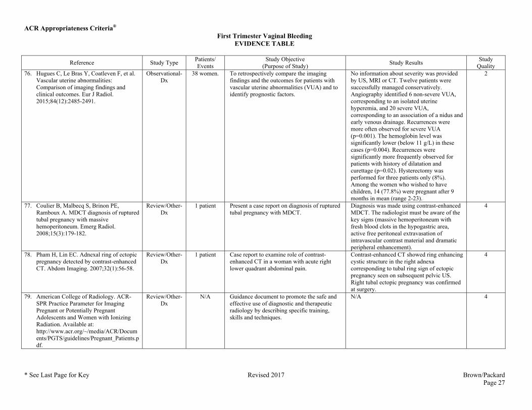
Quality 76. Hugues C, Le Bras Y, Coatleven F, et al.
Vascular uterine abnormalities: Comparison of imaging findings and clinical outcomes. Eur J Radiol. 2015;84(12):2485-2491.
Observational-Dx
38 women. To retrospectively compare the imaging findings and the outcomes for patients with vascular uterine abnormalities (VUA) and to identify prognostic factors.
No information about severity was provided by US, MRI or CT. Twelve patients were successfully managed conservatively. Angiography identified 6 non-severe VUA, corresponding to an isolated uterine hyperemia, and 20 severe VUA, corresponding to an association of a nidus and early venous drainage. Recurrences were more often observed for severe VUA (p=0.001). The hemoglobin level was significantly lower (below 11 g/L) in these cases (p=0.004). Recurrences were significantly more frequently observed for patients with history of dilatation and curettage (p=0.02). Hysterectomy was performed for three patients only (8%). Among the women who wished to have children, 14 (77.8%) were pregnant after 9 months in mean (range 2-23).
2
77. Coulier B, Malbecq S, Brinon PE, Ramboux A. MDCT diagnosis of ruptured tubal pregnancy with massive hemoperitoneum. Emerg Radiol. 2008;15(3):179-182.
Review/Other-Dx
1 patient Present a case report on diagnosis of ruptured tubal pregnancy with MDCT.
Diagnosis was made using contrast-enhanced MDCT. The radiologist must be aware of the key signs (massive hemoperitoneum with fresh blood clots in the hypogastric area, active free peritoneal extravasation of intravascular contrast material and dramatic peripheral enhancement).
4
78. Pham H, Lin EC. Adnexal ring of ectopic pregnancy detected by contrast-enhanced CT. Abdom Imaging. 2007;32(1):56-58.
Review/Other-Dx
1 patient Case report to examine role of contrast-enhanced CT in a woman with acute right lower quadrant abdominal pain.
Contrast-enhanced CT showed ring enhancing cystic structure in the right adnexa corresponding to tubal ring sign of ectopic pregnancy seen on subsequent pelvic US. Right tubal ectopic pregnancy was confirmed at surgery.
4
79. American College of Radiology. ACR-SPR Practice Parameter for Imaging Pregnant or Potentially Pregnant Adolescents and Women with Ionizing Radiation. Available at: http://www.acr.org/~/media/ACR/Documents/PGTS/guidelines/Pregnant_Patients.pdf.
Review/Other-Dx
N/A Guidance document to promote the safe and effective use of diagnostic and therapeutic radiology by describing specific training, skills and techniques.
N/A 4

ACR Appropriateness Criteria® First Trimester Vaginal Bleeding
EVIDENCE TABLE
* See Last Page for Key Revised 2017 Brown/Packard Page 28
Reference Study Type Patients/ Events
Study Objective (Purpose of Study) Study Results Study
Quality 80. American College of Radiology. ACR-
ACOG-AIUM-SRU Practice Paramater for the Performance of Obstetrical Ultrasound. Available at: http://www.acr.org/~/media/ACR/Documents/PGTS/guidelines/US_Obstetrical.pdf.
Review/Other-Dx
N/A Guidance document to promote the safe and effective use of diagnostic and therapeutic radiology by describing specific training, skills and techniques.
N/A 4
81. Kanal E, Barkovich AJ, Bell C, et al. ACR guidance document on MR safe practices: 2013. J Magn Reson Imaging. 2013;37(3):501-530.
Review/Other-Dx
N/A Guidance document on MR safety practices to help guide MR practitioners regarding MR safety issues and provide a basis for them to develop and implement their own MR policies and practices.
N/A 4
82. American College of Radiology. ACR Appropriateness Criteria® Radiation Dose Assessment Introduction. Available at: http://www.acr.org/~/media/ACR/Documents/AppCriteria/RadiationDoseAssessmentIntro.pdf.
Review/Other-Dx
N/A Guidance document on exposure of patients to ionizing radiation.
N/A 4

ACR Appropriateness Criteria®
ACR Appropriateness Criteria® Evidence Table Key
Evidence Table Key
Study Quality Category Definitions
Category 1: The study is well-designed and accounts for common biases.
Category 2: The study is moderately well-designed and accounts for most common biases.
Category 3: There are important study design limitations.
Category 4: The study is not useful as primary evidence. The article may not be a clinical study or the study design is invalid, or conclusions are based on expert consensus. For example:
a) the study does not meet the criteria for or is not a hypothesis-based clinical study (e.g., a book chapter or case report or case series description);
b) the study may synthesize and draw conclusions about several studies such as a literature review article or book chapter but is not primary evidence;
c) the study is an expert opinion or consensus document.
M = Meta-analysis
Dx = Diagnostic
Tx = Treatment