first detailed reconstruction of the karyotype of trachypithecus cristatus (mammalia:...
TRANSCRIPT
Xiaobo et al. Molecular Cytogenetics 2013, 6:58http://www.molecularcytogenetics.org/content/6/1/58
RESEARCH Open Access
First detailed reconstruction of the karyotype ofTrachypithecus cristatus (Mammalia:Cercopithecidae)Fan Xiaobo1, Krit Pinthong2, Hasmik Mkrtchyan3, Pornnarong Siripiyasing4, Nadezda Kosyakova1,Weerayuth Supiwong5, Alongkoad Tanomtong5, Arunrat Chaveerach5, Thomas Liehr1, Marcelo de Bello Cioffi6
and Anja Weise1,7*
Abstract
Background: The chromosomal homologies of human (Homo sapiens = HSA) and silvered leaf monkey(Trachypithecus cristatus = TCR) have been previously studied by classical chromosome staining and by fluorescencein situ hybridization (FISH) applying chromosome-specific DNA probes of all human chromosomes in the 1980s and1990s, respectively.
Results: However, as the resolution of these techniques is limited we used multicolor banding (MCB) at an ~250-bandlevel, and other selected human DNA probes to establish a detailed chromosomal map of TCR. Therefore it waspossible to precisely determine evolutionary conserved breakpoints, orientation of segments and distribution ofspecific regions in TCR compared to HSA. Overall, 69 evolutionary conserved breakpoints including chromosomalsegments, which failed to be resolved in previous reports, were exactly identified and characterized.
Conclusions: This work also represents the first molecular cytogenetic one characterizing a multiple sex chromosomesystem with a male karyotype 44,XY1Y2. The obtained results are compared to other available data for old worldmonkeys and drawbacks in hominoid evolution are discussed.
Keywords: Evolutionary conserved breakpoints, Multicolor banding, Old world monkeys, XY1Y2 sex system
BackgroundTrachypithecus cristatus (TCR) [1], also known as silveredlutung, silvered leaf monkey or the silvery langur, belongsto superfamily Cercopithecoidea, family Cercopithecidae,subfamily Colobinae. The colobines divided during evo-lution into an African clade and an Asian clade [2]. TCRis widely distributed in continental Southeast Asiaincluding Myanmar, West-central Thailand, Cambodia,Laos, Vietnam and Southern China [2-4]. Recently, fourspecies groups were recognized (T. pileatus, T. francoisi,T. obscurus and T. cristatus), including 18 species onlyin the Asian colobine of Trachypithecus. Genus TCR wasinitially denominated with various Latin names between1821 and 1962, like Simia cristata, Semnopithecus cristata,
* Correspondence: [email protected] of Human Genetics, Jena University Hospital, Friedrich SchillerUniversity, Kollegiengasse 10, Jena D-07743, Germany7Institut für Humangenetik, Postfach, Jena D-07740, GermanyFull list of author information is available at the end of the article
© 2013 Xiaobo et al.; licensee BioMed CentralCommons Attribution License (http://creativecreproduction in any medium, provided the orwaiver (http://creativecommons.org/publicdomstated.
Pygathrix cristata, Presbytis cristata, before the currentname Trachypithecus cristatus was introduced [2].The karyotype of TCR was described in 1970 as 2n = 44
[5]. In the 1980s, chromosome banding analysis were usedin TCR [6-8], including comparative R-banding of threedifferent species of Colobus genus [6]. In 1983, G and Qbanding were applied to analyze the banding patterns of afemale TCR [7]. One year later, male TCR was character-ized as carrying an evolutionary conserved translocationinvolving the Y chromosome and two autosomes [9]. Fur-thermore, since 1997 chromosomal homologies betweenhuman chromosomes and TCR has been established byfluorescence in situ hybridization (FISH) applying humanwhole chromosome paintings. Thus, up to now, uniquereciprocal translocations corresponding to HSA Y & 5,HSA 1 & 19, and HSA 6 & 16 as well as fusions of HSA14 & 15 and HSA 21 & 22, were characterized [10]. How-ever, as whole chromosome paints have only a limited
Ltd. This is an Open Access article distributed under the terms of the Creativeommons.org/licenses/by/2.0), which permits unrestricted use, distribution, andiginal work is properly cited. The Creative Commons Public Domain Dedicationain/zero/1.0/) applies to the data made available in this article, unless otherwise
Xiaobo et al. Molecular Cytogenetics 2013, 6:58 Page 2 of 9http://www.molecularcytogenetics.org/content/6/1/58
resolution [11], we established a detailed comparativechromosome map of TCR primarily based on multicolorbanding (MCB). The potential of this approach in order toclarify and to resolve evolutionary conserved chromosomalrearrangements was already shown by our group for otherprimates [12-14].
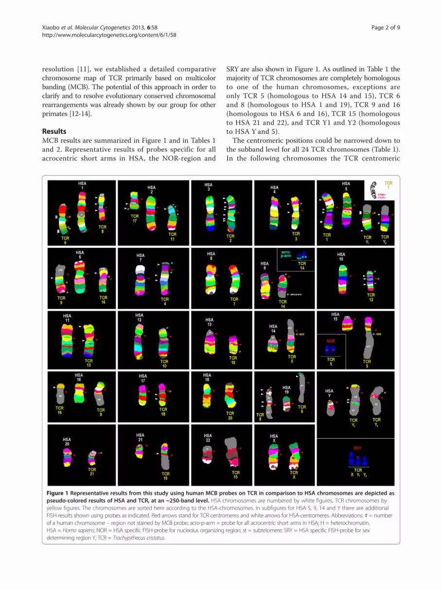
ResultsMCB results are summarized in Figure 1 and in Tables 1and 2. Representative results of probes specific for allacrocentric short arms in HSA, the NOR-region and
Figure 1 Representative results from this study using human MCB prpseudo-colored results of HSA and TCR, at an ~250-band level. HSA cyellow figures. The chromosomes are sorted here according to the HSA-chFISH-results shown using probes as indicated. Red arrows stand for TCR centroof a human chromosome – region not stained by MCB probe; acro-p-arm = pHSA = Homo sapiens; NOR = HSA specific FISH-probe for nucleolus organizingdetermining region Y; TCR = Trachypithecus cristatus.
SRY are also shown in Figure 1. As outlined in Table 1 themajority of TCR chromosomes are completely homologousto one of the human chromosomes, exceptions areonly TCR 5 (homologous to HSA 14 and 15), TCR 6and 8 (homologous to HSA 1 and 19), TCR 9 and 16(homologous to HSA 6 and 16), TCR 15 (homologousto HSA 21 and 22), and TCR Y1 and Y2 (homologousto HSA Y and 5).The centromeric positions could be narrowed down to
the subband level for all 24 TCR chromosomes (Table 1).In the following chromosomes the TCR centromeric
obes on TCR in comparison to HSA chromosomes are depicted ashromosomes are numbered by white figures, TCR chromosomes byromosomes. In subfigures for HSA 5, 9, 14 and Y there are additionalmeres and white arrows for HSA-centromeres. Abbreviations: # = numberrobe for all acrocentric short arms in HSA; H = heterochromatin,region; st = subtelomere; SRY = HSA specific FISH-probe for sex

Table 1 Homologous regions, centromere position and heterochromatic inserts observed in this study of TCRcompared to HSA chromosomes
TCR chromosome Homologous HSA-regions (for rearrangements see Table 2) Centromere position Heterochromatic inserts in
TCR 1 HSA 5pter-5qter like in HSA 5 n.d.i.t.s.
TCR 2 HSA 3pter-3qter HSA 3q26 n.d.i.t.s.
TCR 3 HSA 4pter-4qter like in HSA 4 n.d.i.t.s.
TCR 4 HSA 7pter-7qter like in HSA 7 - end of HSA 7p
- 7q11.1
TCR 5 HSA 15q11.2-15qter, HSA 14q11.2-14qter HSA 15q26.1 ~ 26.2 - fus HSA 14q11.2 / 15q26.3
TCR 6 HSA 1p22-1qter, HSA 19pter-19p13.2 fus HSA 1q22 / 1p14 - fus HSA 1q22 / 1q41;
- fus HSA 1q41 / 1p22;
- 1q24
TCR 7 HSA 8pter-8qter like in HSA 8 n.d.i.t.s.
TCR 8 HSA 1pter-1p22, HSA 19p13.2-19qter like in HSA 19 - end of HSA 19q
TCR 9 HSA 6pter-6q15, HSA 16p13.1-16qter like in HSA 16 - end of HSA 6p;
- 6p21
TCR 10 HSA 12pter-12qter like in HSA 12 - end of HSA 12p
TCR 11 HSA 2q14.1-2qter HSA 2q24.2 - distal from HSA 2q14.1;
- 2q21;
- 2q31
TCR 12 HSA 10pter-10qter like in HSA 10 - end of HSA 10p;
- fus HSA 10q21.1 / 10q22.3
- HSA 10p11.1
- HSA 10q11.1
TCR 13 HSA 11pter-11qter HSA 11p15.3 n.d.i.t.s.
TCR 14 HSA 9pter-9qter HSA 9q33 ~ 34.1 - end of HSA 9q
- end of HSA 9p
TCR 15 HSA 21q11.2-21qter, HSA 22q11.21-22qter fus 21q11.2 / 22q11.21 n.d.i.t.s.
TCR 16 HSA 6q15-6qter, 16pter-16p13.1 HSA 6q21 - HSA 6q21
TCR 17 HSA 2pter-2q14.1 like in HSA 2 - end of HSA 2p
TCR 18 HSA 17pter-17qter like in HSA 17 - HSA 17p11.1
TCR 19 HSA 13q12.1-13qter HSA 13q14 - HSA 13q32 ~ 33
TCR 20 HSA 18pter-18qter HSA 18q21 n.d.i.t.s.
TCR 21 HSA 20pter-20qter HSA 20p12 - HSA 20p11.1
- HSA 20q11.1
TCR X HSA Xpter-Xqter like in HSA X n.d.i.t.s.
TCR Y1 HSA 5p12-5q31.2, HSA Ypter-Yp11.2 like in HSA 5 - HSA 5p11
- HSA 5q11.1
TCR Y2 HSA 5pter-5p12, HSA 5q31.2-5qter,HSA Yp11.2-Yq11.23 like in HSA Y - distal from HSA Yp11.23
Abbreviations: fus = fusion of; n.d.i.t.s. = none detected in this study.
Xiaobo et al. Molecular Cytogenetics 2013, 6:58 Page 3 of 9http://www.molecularcytogenetics.org/content/6/1/58
positions were the same as in HSA: TCR 1 ( = HSA 5),TCR 3 ( = HSA 4), TCR 4 ( = HSA 7), TCR 7 ( = HSA 8),TCR 8 ( = HSA 19), TCR 9 ( = HSA 16), TCR 10 ( = HSA12), TCR 12 ( = HSA 10), TCR 17 ( = HSA 2), TCR 18( = HSA 17), TCR X ( = HSA X), TCR Y1 ( = HSA 5),and TCR Y2 ( = HSA Y). Centromere positions chan-ged compared to HSA in TCR 2 (HSA 3q26), TCR 5
(HSA 15q26.1 ~ 26.2), TCR 6 (HSA 1q22/ 1p14), TCR11 (HSA 2q24.2), TCR 13 (HSA 11p15.3), TCR 14(HSA 9q33 ~ 34.1), TCR 15 (HSA 21q11.2/ 22q11.2),TCR 6 (HSA 6q21), TCR 19 (HSA 13q14), TCR 20(HSA 18q21) and TCR 21 (HSA 20p12).None of the aforementioned TCR centromeric regions
that kept their position during evolution compared to

Table 2 Evolutionary conserved breakpoints in TCR chromosomes compared to HSA; the positions are analyzedconcerning their location in GTG-light bands, colocalization with human fragile sites and breakpoints observed in HLAand GGO using MCB-approach
HSA chr. Evolutionary conserved breakpoints includingneo-centromere and heterochromatin positions
GTG- lightband
Fragile sitein same band
Same breakpoint in HLA[Mrasek et al., 2003 [13]]
Same breakpoint in GGO[Mrasek et al., 2001 [12]]
1 1p22 + FRA1D - -
1q22 - - - -
1q24 - - + -
1q41 - FRA1R - -
2 2p25.3 - FRA2M - -
2q14.1 - FRA2 - +
2q21 + FRA2F - ?+
2q24.2 + FRA2T + -
2q31 + FRA2G - -
3 3p26.3 - FRA3E - -
3p25 + FRA3F - -
3p23 + - - -
3p21.3 + FRA3H ?+ -
3p14 - FRA3B ?+ -
3q22 - FRA3N ?+ -
3q25 + FRA3D + -
3q26 - FRA3O - -
3q28 - FRA3P - -
4 4p12 + FRA4H - +
4q22 - FRA4F - -
5 5p15.2 - FRA5H - -
5q11.2 + FRA5I ?+ -
5q21 - FRA5F - -
5q31.2 - FRA5C - -
5q35.3 (a) + FRA5G - +
5q35.3 (b) + FRA5G - +
6 6p25.3 + - - -
6p21 + FRA6H - -
6q15 + FRA6G + -
6q21 + FRA6F - -
7 7p22.3 + FRA7B + -
7p15.3 + - - -
7q11.1 - FRA7A - -
7q11.23 + FRA7J - +
9 9p34.2 - - - -
9q24.3 + - - -
9q33 ~ 34.1 - FRA9M - -
10 10p15.3 + FRA10H - -
10p11.2 + FRA10J - ?+
10p11.1 - - - -
10q11.1 - FRA10G - -
10q21.1 - FRA10C + -
Xiaobo et al. Molecular Cytogenetics 2013, 6:58 Page 4 of 9http://www.molecularcytogenetics.org/content/6/1/58

Table 2 Evolutionary conserved breakpoints in TCR chromosomes compared to HSA; the positions are analyzedconcerning their location in GTG-light bands, colocalization with human fragile sites and breakpoints observed in HLAand GGO using MCB-approach (Continued)
10q22.3 + FRA10D ?+ +
11 11p15.4 - - - -
11p15.3 + FRA11J - -
11q12 - - + -
12 12p13.33 + FRA12F - -
13 13q12.1 + - - -
13q14 + FRA13G ?+ -
13q32 ~ 33 + FRA13D + -
14 14q11.2 + FRA14D ?+ +
15 15q11.2 + FRA15C ?+ -
15q26.1 ~ 26.2 + FRA15G ?+ -
16 16p13.1 + FRA16H + -
17 17p11.1 - FRA17C ?+ -
17q21.3 + FRA17D ?+ -
17q24 - FRA17E + -
18 18q21 + FRA18B - -
19 19p13.2 - - ?+ -
19q13.2 - - + -
19q13.43 - - ?+ -
20 20p12 - FRA20B - -
20p11.1 - - - -
20q11.1 - FRA20D - -
21 21q11.2 + FRA21 ?+ -
22 22q11.21 + - ?+ -
Y Yp11.31 - - - -
Yp11.2 + - - -
Yq11.23 + - - -
Abbreviations: - = no; + = yes; ? + = most likely yes; a and b in 5q35.3 = break within subtelomere region.
Xiaobo et al. Molecular Cytogenetics 2013, 6:58 Page 5 of 9http://www.molecularcytogenetics.org/content/6/1/58
human showed positive FISH-signals with any of theused HSA centromere specific probes (data not shown).Also, none of the other human heterochromatin specificprobes from HCM probe set gave any specific signalsin TCR, with 2 exceptions: NOR-specific signals wereobserved in TCR 5 (at fusion of HSA 14q11.2 and 15q26.3)(Figure 1) and the probes midi54 (Figure 1) and midi 36(data not shown); the latter two located on the distal endof the long arm of TCR 14.In the literature there were 81 TCR specific heterochro-
matic insertions and/ or additions to chromosomes reported(Figure 2). In this study only 25 of them were confirmedand mapped (Figure 2 and Table 1, last column).Table 2 summarizes the 69 evolutionary conserved
breakpoints observed in TCR in this study; they are givenaccording to the homologous regions in HSA. Only HSAchromosomes X (TCR X) and 8 (TCR 7) are completely
unaltered during evolution from a common ancestor ofHSA and TCR. All other homologous of TCR chromo-somes have undergone one (HSA 12, 14, 16, 18, 21 and 22),two (HSA 4 and 15), three (HSA Y, 11, 13, 17, 19, and 20),four (HSA 1 and 6), five (HSA 2), six (HSA 5, and 10), ornine (HSA 3) evolutionary conserved break events duringspeciation in respect to the human karyotype.Besides, the characterized breakpoints of TCR are
compared with such previously mapped in Hylobates lar(HLA) and Gorilla gorilla (GGO) using MCB approach(Table 2). Again, an alignment of the breakpoint and theirpositioning in GTG-light bands and their spatial proximityto human fragile sites was done.
DiscussionThe present study represents the first one that compre-hensively characterizes the karyotype of TCR. In general,

Figure 2 A summary of the obtained results on TCR idiograms. The homologous regions of HSA and their orientation are shown as coloredarrows on the right of each ideogram. Also the evolutionary conserved breakpoints in respect to HSA nomenclature are inscribed. Consider alsolegend on top of the figure itself.
Xiaobo et al. Molecular Cytogenetics 2013, 6:58 Page 6 of 9http://www.molecularcytogenetics.org/content/6/1/58
previous homologies of HSA and TCR chromosomes couldbe confirmed [10]. However, in this study, homologousregions for TCR chromosomes 4, 10, 11, 14, 17, 18 and21 (that were not studied before) [10] were specificallyaligned to their HSA-homologous. In contrast to [10]NOR was mapped here to the fusion points of HSA 14and HSA 15, i.e. TCR 5 and not TCR 15. In our twostudied individuals derived from Thailand, no differences inTCR 1 banding pattern were seen, which is in accordancewith the literature [10].For the first time, the exact breakpoints could be deter-
mined for the extremely rearranged karyotype of TCR,
in comparison to HSA. In fact, 69 evolutionary conservedbreakpoints were determined in a male TCR and con-firmed in a female individual, excluding Y1 and Y2 chro-mosomes, obviously.In this study no special attention was given to the
centromeric regions of TCR, i.e. they were not detailedcharacterized as in other studies e.g. by [15] or [16].However, a first impression is provided in which centro-meres kept their positions during evolution from commonancestors to HSA and TCR and were neo-centromeres(Table 1), i.e. ~50% of them stayed at the same positionsand ~50% moved either in one of the two species or in

Xiaobo et al. Molecular Cytogenetics 2013, 6:58 Page 7 of 9http://www.molecularcytogenetics.org/content/6/1/58
both. As expected from the literature [17], even the centro-meric regions that kept their positions did not have theidentical alphoid sequences in HSA and TCR.Previous studies in human chromosomal rearrangements
revealed that the majority of them (70-88%) are found inG-light sub-bands [18]. In contrast only 37 (45.5%) of the69 evolutionary conserved breakpoints of TCR were locatedin GTG-light bands (Table 2). However, 70% of the hereobserved TCR breakpoints colocalized with human fragilesites [19] supporting their potential role in the “FragileBreakage Model” [20] and in the formation of evolutionarychromosomal rearrangements [21-26] (Table 2).Concerning evolution it is interesting to report that in
TCR and in HLA 11 evolutionary conserved breakpointsare identical and 15 more are most likely in concordanceto each other. Even more interesting, 6 identical and 2most likely identical evolutionary conserved breakpointswere identified in TCR and in GGO (Table 2). Thesefindings need to be confirmed in further studies by locus-specific probes, and if confirmed, they will be very usefulfor the reconstruction of a common ancestral karyotype.Compared to the postulated Hominidea ancestral karyotypeproposed by [25], only four chromosomes remained un-changed in TCR, i.e. chromosomes 4, 7, 11 and X, elevenchromosomes underwent only intrachromosomal changeslike inversions (TCR 2, 3, 10, 12, 13, 14, 17, 18, 19, 20, 21)and two TCR chromosomes resulted from a fusion ofancestral chromosomes (TCR 5 and 15).Interestingly, the regions between TCR 1 and TCR Y1
and TCR Y2 being homologous to HSA 5 were shownto be subject to different evolutionary conserved rear-rangements. Broadly speaking, TCR Y1 is homologousto TCR 1p and TCR Y2 to TCR 1q. However, each armof chromosome TCR 1 underwent a further paracentricinversion, most likely being important to separate thesex chromosomes from the chromosome 1 during meiosis.Thus, a XY1Y2 sex chromosome system is present inTCR, and not an X1X2Y1Y2 system as initially suggested[10]. However, as in TCR from Indonesia, two other formsof TCR 1 chromosome could be found [10]. Therefore, theexistence of an X1X2Y1Y2 system cannot be completelyexcluded by this study.The sex determination system in mammals is usually
highly conserved as XY-system. However, multiple sexchromosome systems, like the one present in TCR and fewother apes [27,28] are exceptionally found in some speciesof e.g. the orders Insectivora, Chiroptera, Artiodactyla,Rodentia [29], and in marsupials [30]. In general, constitu-tional Y-chromosome / autosome translocations in humanappear de novo and have a deleterious effect and, althoughthe infertility is the only common feature, other clinicalsymptoms can also be observed depending on the involvedbreakpoints [31]. In such cases, the infertility is thought tobe a result of disruption of the sex vesicle during meiosis
[32]. From this point of view it is hard to imagine condi-tions which are in favor of developing a multiple sex- froman XY-chromosome system. On the other hand, in popu-lation genetic models of Y-autosome and X-autosome rear-rangements the population can gain a selective advantageunder a wide range of conditions. If they can invade thepopulation, Y-autosome rearrangements always spreadto fixation, whereas X-autosome rearrangements may bemaintained as stable polymorphisms” [33]. The XY1Y2 sexchromosome system observed in TCR fits well into the sug-gestions of [34] that (i) female meiotic drive is the majorcontribution to the evolution of neo-sex chromosomes and(ii) that “in mammals, the XY1Y2 sex chromosome systemis more prevalent in species with karyotypes of morebiarmed chromosomes” rather than in species with acro-centric chromosomes. Research on meiotic behavior ofsuch sex systems is scarce; however, one study on Bolivianowl monkey (Aotus spec.) showed that no XY pairing wasobservable but the Y-chromosomes formed trivalents withan autosome during gametogenesis [27].
ConclusionsIn conclusion, the presented comparative map of TCRkaryotype gives new insights into primate evolutionand can be used as a starting point for further detailedanalyses. Evolutionary conserved breakpoints, TCR-specificheterochromatic regions, centromeric sequences as wellas the sex chromosome system can be fruitful fields ofresearch in near future.
MethodsCell culture and chromosomal preparationImmortalized lymphoblast cell lines derived from amale and female TCR, were provided by the Khon KaenUniversity, Thailand. Culture techniques and chromosomepreparation followed standard protocols.
Fluorescence in situ hybridizationGeneral considerations for MCB labeling schemes anddetails regarding probe preparation and labeling havebeen described before [12,35,36]. Single and dual-colorFISH techniques were performed for the applied bacterialartificial chromosome (BAC-) probes [37]. Locus-specificBAC clones were purchased at BAC/PAC Chori and DNAwas isolated, PCR-amplified and labeled as describedbefore [37]; and probes RP11-475I16 and RP11-395 L14(both in 2q14.1), RP11-110A24 (in 19p13.3), RP-11457 M7(in Yp11.2) and RP11-122 L9 (in Yp11.31) were applied inthis study. Besides, commercially available human derivedprobes for the SRY gene on the Y-chromosome, HSAprobes for subtelomeric regions 3pter, 3qter, 5pter, 5qter,6pter, 6qter, 7pter, 7qter, 9pter, 9qter, 10pter, 10qter, 18pter,18qter, 19pter, 19qter, XYpter and XYqter, and centromericprobes cep 2, cep4, cep 7, cep 8, cep 10, cep 12, cep 16, cep
Xiaobo et al. Molecular Cytogenetics 2013, 6:58 Page 8 of 9http://www.molecularcytogenetics.org/content/6/1/58
17, cep X cep Y (Abbott/ Vysis, Wiesbaden, Germany) andSE 1/5/19, SE 13/21 and SE 14/22 (Kreatech, Amsterdam,The Netherlands) were also applied.Additionally, the following homemade HSA derived
microdissection probes were used: a probe specific forthe short arm of all human acrocentric chromosomes(midi54) [12,13], and partial chromosome paints for 3p,3q, 5p, 5q, 6p, 6q, 7p, 7q, 9p, 9q, 10p, 10q, 18p, 18q, 19p,19q, Yp and Yq [35]. Furthermore, the heterochromatinmix (HCM-) probe set [38] covering chromosomal regions1q12, 16q11.2, 9q12, 9p12/ 9q13 (midi36) 15p11.2-p11.1,19p12/q12 and Yq12 and subcentromere specific multi-color FISH (subcen-FISH) for chromosomes 3, 6, 7, 9, 11,13 and 20 were also applied [36].
Microscopic evaluationImages were captured using an Axioplan II microscope(Carl Zeiss Jena GmbH, Germany) equipped with filtersets for DAPI, FITC, TR, SO, Cy5 and DEAC. Imageanalysis was done using pseudocolor banding and fluoro-chrome profiles of the ISIS digital FISH imaging system(Meta Systems Hard & Software GmbH, Altlussheim,Germany). At least 10 up to 20 metaphases were recorded,derived from a male and a female TCR for each appliedprobe and probe set.
Competing interestsAuthors declare that there is no conflict of interest.
Authors’ contributionsXF carried out molecular cytogenetic studies and the draft of themanuscript, KP carried out the molecular cytogenetic studies, HM carried outthe molecular cytogenetic studies, PS carried out the molecular cytogeneticstudies, NK carried out the molecular cytogenetic studies, WS carried out thecell culture, AT carried out the cell culture, AC carried out the cell cultureand participated in manuscript discussion, TL participated in the design ofthe study and the manuscript, MBC participated in the design of the studyand the manuscript discussion, AW participated in the design andcoordinated the study and the manuscript. All authors read and approvedthe final manuscript.
AcknowledgmentsThis work was supported by the China Scholarship Council.
Author details1Institute of Human Genetics, Jena University Hospital, Friedrich SchillerUniversity, Kollegiengasse 10, Jena D-07743, Germany. 2Faculty of Scienceand Technology, Surindra Rajabhat University, 186 Moo 1, Surin, MaungDistrict 32000, Thailand. 3Center of Medical Genetics and Primary HealthCare, Abovyan Str 34/3, 001, Yerevan, Armenia. 4Faculty of Science andTechnology, Rajabhat Maha Sarakham University, 80 Nakonsawan Rd, MahaSarakham, Talad, Maung District 44000, Thailand. 5Department of Biology,Faculty of Science, Khon Kaen University, 123 Moo 16 Mittapap Rd., KhonKaen, Muang District 40002, Thailand. 6Departamento de Genética eEvolução, Universidade Federal de São Carlos, São Carlos, SP, Brazil. 7Institutfür Humangenetik, Postfach, Jena D-07740, Germany.
Received: 15 November 2013 Accepted: 21 November 2013Published: 17 December 2013
References1. Raffles TS: Descriptive catalogue of a zoological collection, made on
account of the Honourable East India Company, in the island of
Sumatra and its vicinity, under the direction of Sir Thomas StanfordRaffles, Lieutenant-Governor of Fort Marlborough: with additionalnotices of the natural history of those countries. Transact Linn SocLondon 1821, 13:239–274.
2. Harding LE: Trachypithecus cristatus (Primates: Cercopithecidae).Mamm Species 2010, 42:149–165.
3. Fooden J: Primates obtained in peninsular Thailand June–July, 1973, withnotes on the distribution of continental Southeast Asian leaf- monkeys(Presbytis). Primates 1976, 17:95–118.
4. Roos C, Nadler T, Walter L: Mitochondrial phylogeny, taxonomy andbiogeography of the silvered langur species group (Trachypithecus cristatus).Mol Phylogenet Evol 2008, 47:629–636.
5. Hsu TC, Benirschke K: In An atlas of mammalian chromosomes. Edited byHsu TC, Benirschke K. NewYork: Springer-Verlag, Folio 199; 1970.
6. Dutrillaux B, Couturier J, Muleris M, Lombard M, Chauvier G: Chromosomalphylogeny of forty-two species or subspecies of cercopithecoids(Primates Catarrhini). Ann Genet 1982, 25:96–109.
7. Ponsa M, de Boer LEM, Egozcue J: Banding patterns of the chromosomes ofPresbytis cristatus pyrrhus and P. obscurus. Am J Primatol 1983, 4:165–169.
8. Muleris M, Couturier J, Dutrillaux B: Phylogénie chromosomique desCercopithecoidea. Mammalia 1986, 50:38–52.
9. Dutrillaux B, Webb G, Muleris M, Couturier J, Butler R: Chromosome studyof Presbytis cristatus: presence of a complex Y-autosome rearrangementin the male. Ann Genet 1984, 27:148–153.
10. Bigoni F, Koehler U, Stanyon R, Ishida T, Wienberg J: Fluorescene in situhybridization establishes homology between human and silvered leafmonkey chromosomes, reveals reciprocal translocations betweenchromosomes homologous to human Y/5, 1/9, and 6/16, and delineatesan X1X2Y1Y2/X1X1X2X2 sex-chromosome system. Am J Phys Anthropol1997, 102:315–327.
11. Graphodatsky AS, Trifonov VA, Stanyon R: The genome diversity andkaryotype evolution of mammals. Mol Cytogenet 2011, 4:22.
12. Mrasek K, Heller A, Rubtsov N, Trifonov V, Starke H, Rocchi M, Claussen U,Liehr T: Reconstruction of the female Gorilla gorilla karyotype using25-color FISH and multicolor banding (MCB). Cytogenet Cell Genet 2001,93:242–248.
13. Mrasek K, Heller A, Rubtsov N, Trifonov V, Starke H, Claussen U, Liehr T:Detailed Hylobates lar karyotype defined by 25-color FISH and multicolorbanding. Int J Mol Med 2003, 12:139–146.
14. Weise A, Heller A, Starke H, Mrasek K, Kuechler A, Pool-Zobel BL, Claussen U, LiehrT:Multitude multicolor chromosome banding (mMCB) - a comprehensiveone-step multicolor FISH banding method. Cytogenet Genome Res 2003,103:34–39.
15. Stanyon R, Rocchi M, Capozzi O, Roberto R, Misceo D, Ventura M, CardoneMF, Bigoni F, Archidiacono N: Primate chromosome evolution: ancestralkaryotypes, marker order and neocentromeres. Chromosome Res 2008,16:17–39.
16. Ventura M, Antonacci F, Cardone MF, Stanyon R, D'Addabbo P, Cellamare A,Sprague LJ, Eichler EE, Archidiacono N, Rocchi M: Evolutionary formationof new centromeres in macaque. Science 2007, 316:243–246.
17. Rocchi M, Archidiacono N, Schempp W, Capozzi O, Stanyon R: Centromererepositioning in mammals. Heredity 2012, 108:59–67.
18. Manvelyan M, Schreyer I, Höls-Herpertz I, Köhler S, Niemann R, Hehr U,Belitz B, Bartels I, Götz J, Huhle D, Kossakiewicz M, Tittelbach H, Neubauer S,Polityko A, Mazauric ML, Wegner R, Stumm M, Küpferling P, Süss F, Kunze H,Weise A, Liehr T, Mrasek K: Forty-eight new cases with infertility due tobalanced chromosomal rearrangements: detailed molecular cytogeneticanalysis of the 90 involved breakpoints. Int J Mol Med 2007, 19:855–864.
19. Mrasek K, Schoder C, Teichmann AC, Behr K, Franze B, Wilhelm K, BlaurockN, Claussen U, Liehr T, Weise A: Global screening and extendednomenclature for 230 aphidicolin-inducible fragile sites, including 61 yetunreported ones. Int J Oncol 2010, 36:929–940.
20. Pevzner P, Tesler G: Human and mouse genomic sequences revealextensive breakpoint reuse in mammalian evolution. Proc Natl Acad SciU S A 2003, 100:7672–7677.
21. Bailey JA, Baertsch R, Kent WJ, Haussler D, Eichler EE: Hotspots ofmammalian chromosomal evolution. Genome Biol 2004, 5:R23.
22. Murphy WJ, Larkin DM, Everts-van der Wind A, Bourque G, Tesler G, Auvil L,Beever JE, Chowdhary BP, Galibert F, Gatzke L, Hitte C, Meyers SN, Milan D,Ostrander EA, Pape G, Parker HG, Raudsepp T, Rogatcheva MB, Schook LB,Skow LC, Welge M, Womack JE, O'brien SJ, Pevzner PA, Lewin HA:

Xiaobo et al. Molecular Cytogenetics 2013, 6:58 Page 9 of 9http://www.molecularcytogenetics.org/content/6/1/58
Dynamics of mammalian chromosome evolution inferred frommultispecies comparative maps. Science 2005, 309:613–617.
23. Ruiz-Herrera A, Castresana J, Robinson TJ: Is mammalian chromosomalevolution driven by regions of genome fragility? Genome Biol 2006, 7:R115.
24. Ruiz-Herrera A, Robinson TJ: Chromosomal instability in Afrotheria: fragilesites, evolutionary breakpoints and phylogenetic inference from genomesequence assemblies. BMC Evol Biol 2007, 7:199.
25. Misceo D, Capozzi O, Roberto R, Dell'oglio MP, Rocchi M, Stanyon R,Archidiacono N: Tracking the complex flow of chromosomerearrangements from the Hominoidea Ancestor to extant Hylobates andNomascus Gibbons by high-resolution synteny mapping. Genome Res2008, 18:1530–1537.
26. Alekseyev MA, Pevzner PA: Comparative genomics reveals birth and deathof fragile regions in mammalian evolution. Genome Biol 2010, 11:R117.
27. Ma NS, Elliott MW, Morgan L, Miller A, Jones TC: Translocation of Ychromosome to an autosome in the Bolivian owl monkey, Aotus.Am J Phys Anthropol 1976, 45:191–202.
28. Solari AJ, Rahn MI: Fine structure and meiotic behaviour of the malemultiple sex chromosomes in the genus Alouatta. Cytogenet Genome Res2005, 108:262–267.
29. Fredga K: Unusual sex chromosome inheritance in mammals. Philos TransR Soc Lond B Biol Sci 1970, 259:15–36.
30. Toder R, O'Neill RJ, Wienberg J, O'Brien PC, Voullaire L, Marshall-Graves JA:Comparative chromosome painting between two marsupials: origins ofan XX/XY1Y2 sex chromosome system. Mamm Genome 1997, 8:418–422.
31. Conte RA, Kleyman SM, Klein V, Bialer MG, Verma RS: Characterization of ade novo t(Y;9) (q11.2;q22) by FISH technique. Ann Genet 1996, 39:10–15.
32. Pinho MJ, Neves R, Costa P, Ferrás C, Sousa M, Alves C, Almeida C,Fernandes S, Silva J, Ferrás L, Barros A: Unique t(Y;1)(q12;q12) reciprocaltranslocation with loss of the heterochromatic region of chromosome1 in a male with azoospermia due to meiotic arrest: a case report.Hum Reprod 2005, 20:689–696.
33. Charlesworth B, Wall JD: Inbreeding, heterozygote advantage and theevolution of neo-X and neo-Y sex chromosomes. Proceedings: BiologicalSciences 1999, 266:51–56.
34. Yoshida K, Kitano J: The contribution of female meiotic drive to theevolution of neo-sex chromosomes. Evolution 2012, 66:3198–3208.
35. Liehr T, Claussen U: Current developments in human molecularcytogenetic techniques. Curr Mol Med 2002, 2:283–297.
36. Weise A, Mrasek K, Fickelscher I, Claussen U, Cheung SW, Cai WW, Liehr T,Kosyakova N: Molecular definition of high-resolution multicolor bandingprobes: first within the human DNA sequence anchored FISH bandingprobe set. J Histochem Cytochem 2008, 56:487–493.
37. Liehr T, Heller A, Starke H, Rubtsov N, Trifonov V, Mrasek K, Weise A,Kuechler A, Claussen U: Microdissection based high resolution multicolorbanding for all 24 human chromosomes. Int J Mol Med 2002, 9:335–339.
38. Bucksch M, Ziegler M, Kosayakova N, Mulatinho MV, Llerena JC Jr, Morlot S,Fischer W, Polityko AD, Kulpanovich AI, Petersen MB, Belitz B, Trifonov V,Weise A, Liehr T, Hamid AB: A new multicolor fluorescence in situhybridization probe set directed against human heterochromatin:HCM-FISH. J Histochem Cytochem 2012, 60:530–536.
doi:10.1186/1755-8166-6-58Cite this article as: Xiaobo et al.: First detailed reconstruction of thekaryotype of Trachypithecus cristatus (Mammalia: Cercopithecidae).Molecular Cytogenetics 2013 6:58.
Submit your next manuscript to BioMed Centraland take full advantage of:
• Convenient online submission
• Thorough peer review
• No space constraints or color figure charges
• Immediate publication on acceptance
• Inclusion in PubMed, CAS, Scopus and Google Scholar
• Research which is freely available for redistribution
Submit your manuscript at www.biomedcentral.com/submit