fibroblast growth factor 21 (fgf21) alleviates senescence
TRANSCRIPT
ARTICLE OPEN
Fibroblast growth factor 21 (FGF21) alleviates senescence,apoptosis, and extracellular matrix degradation in osteoarthritisvia the SIRT1-mTOR signaling pathwayHongwei Lu1,2,3, Chao Jia1,2,3, Dengying Wu1,2,3, Haidong Jin1,2,3, Zeng Lin1,2,3, Jun Pan 1,3,4✉, Xiucui Li 2,4✉ and Wei Wang 1,2,3✉
© The Author(s) 2021
Osteoarthritis (OA) is a complex condition that involves both apoptosis and senescence and currently cannot be cured. Fibroblastgrowth factor 21 (FGF21), known for its role as a potent regulator of glucose and energy metabolism, protects from variousdiseases, possibly by mediating autophagy. In the present study, the role of FGF21 in the progression of OA was investigated inboth in vitro and in vivo experiments. In vitro, the results revealed that FGF21 administration alleviated apoptosis, senescence, andextracellular matrix (ECM) catabolism of the chondrocytes induced by tert-butyl hydroperoxide (TBHP) by mediating autophagyflux. Furthermore, CQ, an autophagy flux inhibitor, could reverse the protective effect of FGF21. It was observed that the FGF21-induced autophagy flux enhancement was mediated by the nuclear translocation of TFEB, which occurs due to the activation of theSIRT1-mTOR signaling pathway. The in vivo experiments demonstrated that FGF21 treatment could reduce OA in the DMM model.Taken together, these findings suggest that FGF21 protects chondrocytes from apoptosis, senescence, and ECM catabolism viaautophagy flux upregulation and also reduces OA development in vivo, demonstrating its potential as a therapeutic agent in OA.
Cell Death and Disease (2021) 12:865 ; https://doi.org/10.1038/s41419-021-04157-x
INTRODUCTIONOsteoarthritis (OA) is a degenerative disease of the joints thatoccurs in the elderly [1]. Until recently, no efficacious drugs andsurgical procedures were available for OA therapy [2]. Theincidence of osteoarthritis has increased sharply in the elderlypopulation [3]. OA is reported to be a major cause of disability andsocioeconomic loss worldwide [4]. Therefore, novel strategies toinhibit the progression of OA are of great clinical and scientificinterest.Articular cartilage is a complex tissue that is maintained by
chondrocytes. As the only cell type in the articular cartilage,chondrocytes are responsible for producing the extracellularmatrix (ECM) molecules [5]. Oxidative stress occurs due to theimbalance between the production of reactive oxygen species(ROS) and their elimination by the antioxidant defense system,which is high in the OA cartilage [6]. Excessive levels of ROSpromote apoptosis, senescence, ECM catabolism, and ultimatelyleads to the degradation of the articular cartilage [7, 8]. Therefore,inhibiting oxidative stress of chondrocytes is proposed as atherapeutic target in OA.Autophagy is a lysosome-dependent and highly-conserved
macromolecular cycle occurring in the eukaryotic cells, which isalso essential for the survival and maintenance of these cells [9]. Itis characterized by the formation of double-layered vesicles(autophagosomes) around intracellular cargo for delivery to
lysosomes and proteolytic degradation; the whole process iscalled autophagy flux [10]. Recent studies revealed that TFEBmight regulate autophagy flux by inducing lysosomal biogenesisand promoting the formation of autophagosomes and their fusionwith lysosomes [11, 12]. Interestingly, previous studies haveshown that promoting the nuclear localization of TFEB in OAchondrocytes alleviated apoptosis and senescence through theautophagy lysosomal pathway (ALP) [13]. Thus, the promotion ofthe nuclear localization of TFEB and further activation of theautophagy flux could become the target of OA therapy.In 2000, Nishimura et al. isolated the fibroblast growth factor 21
(FGF21) from mouse embryonic tissues [14]. Since then, FGF21 hasattracted considerable attention as a therapeutic agent for thetreatment of metabolic syndrome in humans [15–17]. In recentresearch, FGF21 has emerged as a longevity hormone [18]. FGF21can stimulate autophagy in various tissues, including the brain[19], liver [20], random-pattern skin flaps [21], kidneys [22], heart[23], etc. Previous reports suggest that FGF21 has a potentialtherapeutic effect in neurodegeneration [24, 25]. However, as oneof the degenerative diseases, no studies on the relationshipbetween FGF21 and autophagy flux in osteoarthritis have beenreported yet. Therefore, the present study was aimed toinvestigate the protective effects of FGF21 on the OA process,and it was hypothesized that these effects were exerted by thestimulation of autophagy flux.
Received: 27 April 2021 Revised: 28 July 2021 Accepted: 9 September 2021
1Department of Orthopaedics, The Second Affiliated Hospital and Yuying Children’s Hospital of Wenzhou Medical University, Wenzhou, Zhejiang 325027 Zhejiang Province,China. 2The Second School of Medicine, Wenzhou Medical University, Wenzhou 325027 Zhejiang Province, China. 3Bone Research Institute, The Key Orthopaedic Laboratory ofZhejiang Province, Wenzhou, China. 4Department of Neonatology, The Second Affiliated Hospital and Yuying Children’s Hospital of Wenzhou Medical University, Wenzhou325027 Zhejiang Province, China. ✉email: [email protected]; [email protected]; [email protected] by: Professor Anastasis Stephanou
www.nature.com/cddis
Official journal of CDDpress
In this study, tert-butyl hydroperoxide (TBHP) was used toinduce oxidative stress as an exogenous ROS donor. We foundthat FGF21 might suppress apoptosis, senescence, and ECMcatabolism induced by TBHP in chondrocytes. FGF21 might alsopromote autophagy flux and lysosome biogenesis, and theseeffects might be due to the activation of TFEB by FGF21 throughthe SIRT1/mTOR signaling pathway. Finally, the therapeuticpotential of FGF21 was evaluated in the destabilization of themedial meniscus (DMM) mouse model.
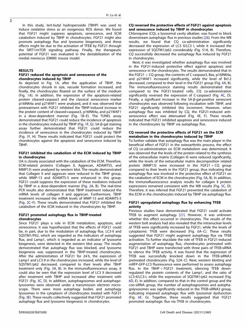
RESULTSFGF21 reduced the apoptosis and senescence of thechondrocytes induced by TBHPAs depicted in Fig. 1A, after the application of TBHP, thechondrocytes shrunk in size, vacuole formation increased, andfinally, the chondrocytes floated on the surface of the medium(Fig. 1A). In addition, the expression of the apoptosis-relatedprotein cleaved-caspase3 and the classical senescence markerp16INK4a and p21WAF1 were analyzed, and it was observed thatpretreatment with FGF21 inhibited the TBHP-induced increase inthe protein content of cleaved-caspase3, p16INK4a, and p21WAF1in a dose-dependent manner (Fig. 1B–E). The TUNEL assaydemonstrated that FGF21 could reduce the incidence of apoptosisin the chondrocytes induced by TBHP (Fig. 1F, G). SA-β-gal stainingassay further demonstrated that FGF21 could reduce theincidence of senescence in the chondrocytes induced by TBHP(Fig. 1F, H). These results indicated that FGF21 could protect thechondrocytes against the apoptosis and senescence induced byTBHP.
FGF21 inhibited the catabolism of the ECM induced by TBHPin chondrocytesOA is closely associated with the catabolism of the ECM. Therefore,ECM-related proteins (Collagen II, Aggrecan, ADAMTS5, andMMP13) were analyzed by western blot. The results demonstratedthat Collagen II and aggrecan were reduced in the TBHP group,while MMP-13 and ADAMTS-5 were enhanced in this group.FGF21 could suppress the expression of these mediators inducedby TBHP in a dose-dependent manner (Fig. 2A, B). The real-timePCR results also demonstrated that TBHP treatment reduced themRNA levels of collagen II and aggrecan. Furthermore, TBHPtreatment increased the mRNA levels of MMP-13 and ADAMTS-5(Fig. 2C–F). These results demonstrated that FGF21 inhibited thecatabolism of the ECM induced in the chondrocytes by TBHP.
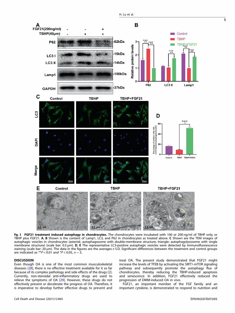
FGF21 promoted autophagy flux in TBHP-treatedchondrocytesSince FGF21 plays a role in ECM metabolism, apoptosis, andsenescence, it was hypothesized that the effects of FGF21 couldbe, in part, due to the modulation of autophagy flux. LC3-II andSQSTM1/P62, which are regarded as the indicators of autophagyflux, and Lamp1, which is regarded as an indicator of lysosomebiogenesis, were detected in the western blot assay. The resultsdemonstrated that autophagy flux was blocked, and lysosomebiogenesis was suppressed in the TBHP-treated chondrocytes.After the administration of FGF21 for 24 h, the expression ofLamp1 and LC3-II in the chondrocytes increased, while the level ofSQSTM1/p62 decreased, compared to their levels after TBHPtreatment only (Fig. 3A, B). In the immunofluorescence assay, itcould also be seen that the expression level of LC3 II decreasedafter treatment with TBHP and increased after treatment withFGF21 (Fig. 3C, D). In addition, autophagosomes and autophago-lysosomes were observed under a transmission electron micro-scope. There were more autophagy bodies and autophagylysosomes in the cytoplasm of chondrocytes treated with FGF21(Fig. 3E). These results collectively suggested that FGF21 promotedautophagy flux and lysosome biogenesis in chondrocytes.
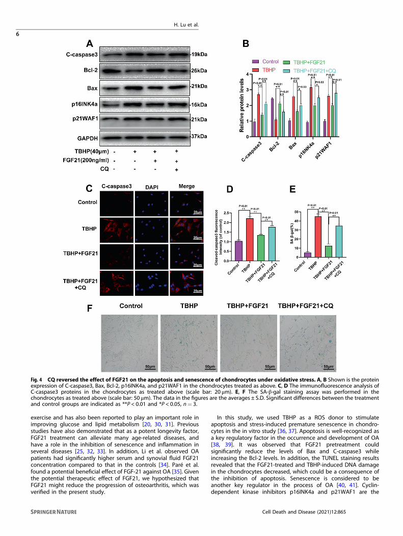
CQ reversed the protective effects of FGF21 against apoptosisand senescence induced by TBHP in chondrocytesChloroquine (CQ), a lysosomal cavity alkalizer, was found to blockdownstream autophagic flux in previous studies [26]. From the WBresults, we found that CQ co-administration significantlydecreased the expression of LC3 II/LC3 I, while it increased theexpression of SQSTM1/p62 considerably (Fig. S1A, B). Therefore,CQ successfully decreased the autophagy flux induced by FGF21in chondrocytes.Next, it was investigated whether autophagy flux was involved
in the FGF21-induced protective effect against apoptosis andsenescence in the chondrocytes. The results demonstrated that inthe FGF21+ CQ group, the contents of C-caspase3, Bax, p16INK4a,and p21WAF1 increased significantly, while the level of Bcl-2decreased, compared to their level in the FGF21 group (Fig. 4A, B).The immunofluorescence staining results demonstrated thatcompared to the FGF21-treated cells, CQ co-administrationsignificantly reversed the expression of C-caspase3 (Fig. 4C, D).Finally, a significant increase in the SA-β-gal-positive senescentchondrocytes was observed following incubation with TBHP, andFGF21 significantly inhibited this increment. However, whenautophagy flux was inhibited by CQ, the FGF21-induced anti-senescence effect was attenuated (Fig. 4E, F). These resultsindicated that FGF21 inhibited apoptosis and senescence inducedin the chondrocytes by TBHP via autophagy flux.
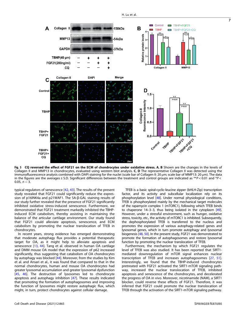
CQ reversed the protective effects of FGF21 on the ECMmetabolism in the chondrocytes induced by TBHPTo further validate that autophagy flux was the main player in thebeneficial effect of FGF21 in the osteoarthritis process, the effectof CQ co-administration on ECM metabolism was determined. Itwas observed that the levels of the protein related to the synthesisof the extracellular matrix (Collagen II) were reduced significantly,while the levels of the extracellular matrix decomposition relatedprotein (MMP13) were increased in the FGF21+ CQ groupcompared to their levels in the FGF21 group, thus indicating thatautophagy flux was involved in the protective effect of FGF21 onthe catabolism of ECM in the chondrocytes (Fig. 5A, B). In addition,the immunofluorescence analysis results for collagen II proteinexpressions remained consistent with the WB results (Fig. 5C, D).Therefore, it was inferred that FGF21 prevented the catabolism ofthe ECM in the chondrocytes induced by TBHP via autophagy flux.
FGF21 upregulated autophagy flux by enhancing TFEBactivityPrevious studies have demonstrated that FGF21 could activateTFEB to augment autophagy [21]. However, it was unknownwhether this effect occurred in chondrocytes. The results of thewestern bolt analysis had also revealed that the intranuclear levelsof TFEB were significantly increased by FGF21, while the levels ofcytoplasmic TFEB were decreased (Fig. 6A–C). These resultssuggested that FGF21 might augment autophagy flux via TFEBactivation. To further elucidate the role of TFEB in FGF21-inducedaugmentation of autophagy flux, chondrocytes pretreated withFGF21 and TBHP were transfected with three pairs of TFEB-siRNAto silence the TFEB activity. It was found that the expression ofTFEB was successfully knocked down in the TFEB-siRNA3pretreated chondrocytes (Fig. S2A–C). Next, western blotting andmRFP-GFP-LC3 fluorescence were performed to assess autophagyflux. In the TBHP+ FGF21 treatment, silencing TFEB down-regulated the protein contents of the Lamp1, and the ratio ofLC3-II/LC3-I, while the expression of SQSTM1/p62 increased (Fig.6D, E). In addition, compared to that in the control group and thecon-siRNA group, the number of autophagosomes and autopha-golysosomes was significantly reduced in the TFEB-siRNA3 group,indicating impaired autophagic flux with lysosomal dysfunction(Fig. 6F, G). Together, these results suggested that FGF21promoted autophagic flux via TFEB in chondrocytes.
H. Lu et al.
2
Cell Death and Disease (2021) 12:865
1234567890();,:
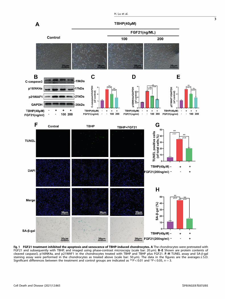
Fig. 1 FGF21 treatment inhibited the apoptosis and senescence of TBHP-induced chondrocytes. A The chondrocytes were pretreated withFGF21 and subsequently with TBHP, and imaged using phase-contrast microscopy (scale bar: 20 μm). B–E Shown are protein contents ofcleaved caspase3, p16INK4a, and p21WAF1 in the chondrocytes treated with TBHP and TBHP plus FGF21. F–H TUNEL assay and SA-β-galstaining assay were performed in the chondrocytes as treated above (scale bar: 50 μm). The data in the figures are the averages ± S.D.Significant differences between the treatment and control groups are indicated as **P < 0.01 and *P < 0.05, n= 3.
H. Lu et al.
3
Cell Death and Disease (2021) 12:865
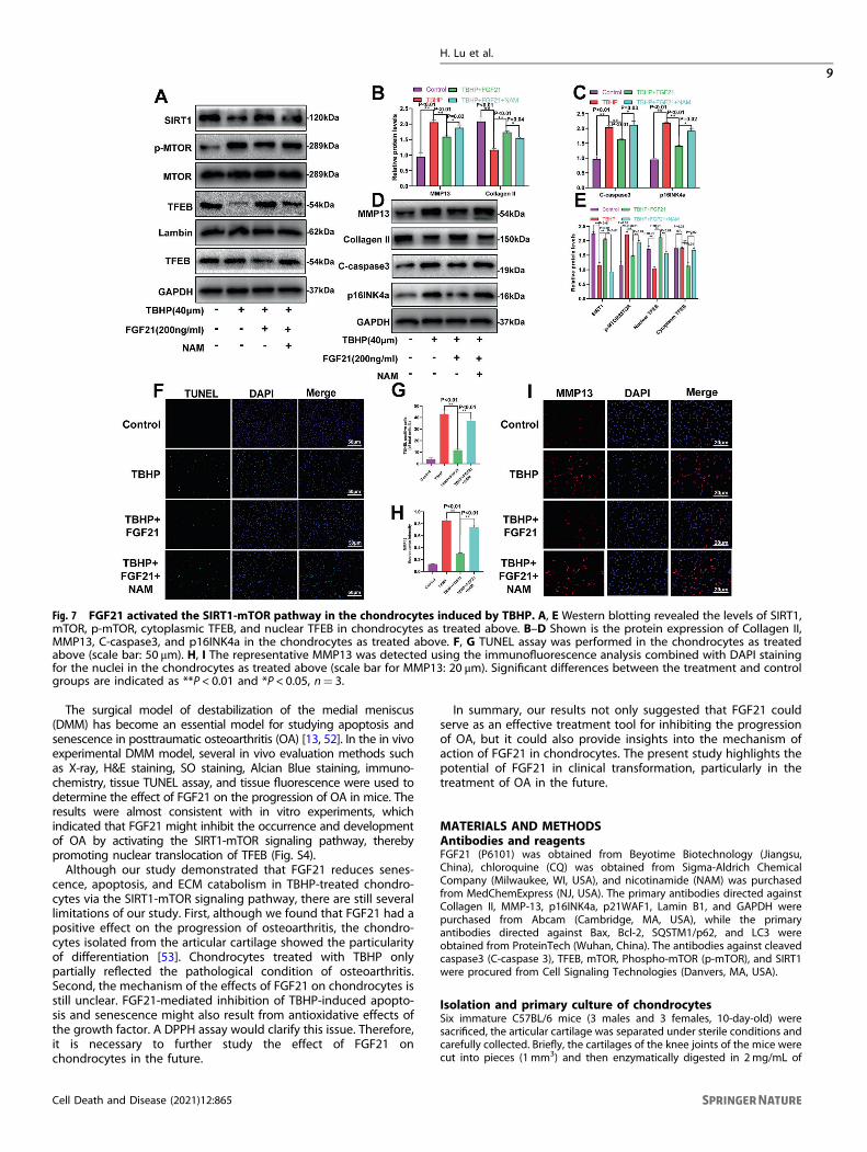
The SIRT1-mTOR pathway was activated by FGF21 and wasinvolved in the protective effect of FGF21 in the chondrocytesinduced by TBHPAccording to previously published reports, the SIRT-mTOR path-way has a crucial role in TFEB modulation [27]. Therefore, weassumed that the SIRT1-mTOR pathway would be activated in thechondrocytes treated with FGF21. Our results showed that FGF21increased the expression of SIRT1 and inhibited p-mTOR,indicating that FGF21 activated the SIRT1-mTOR pathway. Todetermine whether this FGF21-induced activation of TFEB wasmediated by the SIRT1-mTOR signaling pathway in chondrocytes,nicotinamide (NAM), a SIRT1 blocker, was used for the inhibition ofSIRT1 activation, and the results showed that FGF21 promoted theSIRT1-mTOR pathway-mediated TFEB nuclear translocation andthese effects were reversed after NAM application (Fig. 7A, E). Theresults of western blotting also indicated that NAM significantlyinhibited the FGF21-mediated protective effects on the ECM,apoptosis, and senescence (Fig. 7B–D). The TUNEL assay resultsand the immunofluorescence analysis further demonstrated theprotective effect of FGF21 on apoptosis and ECM catabolism (Fig.7F–I). Together, these results confirmed that FGF21 activated TFEBin the chondrocytes via the SIRT1-mTOR signaling pathway.
FGF21 reduced OA development through the SIRT1-mTORsignaling pathwayBased on the results of the in vitro experiment, we further studiedthe role of FGF21 in vivo, for which the DMM mouse model wasused. The X-ray images obtained eight weeks after surgeryrevealed that the DMM group had a wider joint space comparedto the DMM+ FGF21 group. However, the DMM+ FGF21+ NAM
group had a narrower joint space compared to the DMM+ FGF21group (Fig. 8A). The protective effect of FGF21 observed on thehistomorphology of knee joints in vivo was further confirmed fromthe results of the H&E and safranin O Fast Green staining andAlcian Blue staining, 8 weeks after the surgery. Erosion andhypocellularity of the superficial articular cartilage, along withproteoglycan loss, were observed in the DMM group. In contrast,the DMM+ FGF21 group presented a more complete cartilagesurface and richer proteoglycan (Fig. 8B). Consistent with thestaining results, the OARSI score declined in the FGF21-treatedgroup and increased in the DMM group. However, the combinedtreatment with NAM and FGF21 was observed to significantlyoffset the protective effect of FGF21 only on the articular cartilagestructure and the matrix (Fig. 8C).Furthermore, by immunohistochemistry, we found that the
FGF21 group had significantly higher LC3 and Lamp1 expressionand lower MMP13 and p16INK4a expressions in the chondrocytescompared to that in the DMM group (Fig. 8D–H). After theimmunofluorescence staining of joint tissues, it was found thatthe FGF21 group exhibited significantly higher levels of TFEB inthe nucleus of the chondrocytes, compared to that in the nucleusof the chondrocytes in the DMM group (Fig. S3A, B). The results ofthe TUNEL assay showed that the percentage of apoptosis in theFGF21 group was less than that in the DMM group (Fig. S3C, D).However, the NAM and FGF21 co-treatment groups exhibited agreater offset than the effect of FGF21 on the results of theabove-mentioned chondrocytes, which was consistent with theresults of the X-ray examination and histological staining. Tosummarize, these results demonstrated the therapeutic effect ofFGF21 in vivo.
Fig. 2 FGF21 treatment inhibited the TBHP-induced catabolism of the ECM. A, B Shown is the change in the levels of Aggrecan, Collagen II,ADAMTS5, and MMP13 in the chondrocytes, evaluated by the western blot analysis. C–F Shown is the mRNA expression of Aggrecan, CollagenII, ADAMTS5, and MMP13 in the chondrocytes treated with TBHP and TBHP plus FGF21, measured using real-time PCR. The data in the figuresare the averages ± S.D. Significant differences between the treatment and control groups are indicated as **P < 0.01 and *P < 0.05, n= 3.
H. Lu et al.
4
Cell Death and Disease (2021) 12:865
DISCUSSIONEven though OA is one of the most common musculoskeletaldiseases [28], there is no effective treatment available for it so farbecause of its complex pathology and side effects of the drugs [2].Currently, non-steroidal anti-inflammatory drugs are used torelieve the symptoms of OA [29]. However, these drugs do noteffectively prevent or decelerate the progress of OA. Therefore, itis imperative to develop further effective drugs to prevent and
treat OA. The present study demonstrated that FGF21 mightincrease the levels of TFEB by activating the SIRT1-mTOR signalingpathway and subsequently promote the autophagy flux ofchondrocytes, thereby reducing the TBHP-induced apoptosisand senescence. In addition, FGF21 effectively reduced theprogression of DMM-induced OA in vivo.FGF21, an important member of the FGF family and an
important cytokine, is demonstrated to respond to nutrition and
Fig. 3 FGF21 treatment induced autophagy in chondrocytes. The chondrocytes were incubated with 100 or 200 ng/ml of TBHP only, orTBHP plus FGF21. A, B Shown is the content of Lamp1, LC3, and P62 in chondrocytes as treated above. C Shown are the TEM images ofautophagic vesicles in chondrocytes (asterisk: autophagosome with double-membrane structure; triangle: autophagolysosome with singlemembrane structure) (scale bar: 0.5 µm). D, E The representative LC3-positive autophagic vesicles were detected by immunofluorescencestaining (scale bar: 20 μm). The data in the figures are the averages ± S.D. Significant differences between the treatment and control groupsare indicated as **P < 0.01 and *P < 0.05, n= 3.
H. Lu et al.
5
Cell Death and Disease (2021) 12:865
exercise and has also been reported to play an important role inimproving glucose and lipid metabolism [20, 30, 31]. Previousstudies have also demonstrated that as a potent longevity factor,FGF21 treatment can alleviate many age-related diseases, andhave a role in the inhibition of senescence and inflammation inseveral diseases [25, 32, 33]. In addition, Li et al. observed OApatients had significantly higher serum and synovial fluid FGF21concentration compared to that in the controls [34]. Paré et al.found a potential beneficial effect of FGF-21 against OA [35]. Giventhe potential therapeutic effect of FGF21, we hypothesized thatFGF21 might reduce the progression of osteoarthritis, which wasverified in the present study.
In this study, we used TBHP as a ROS donor to stimulateapoptosis and stress-induced premature senescence in chondro-cytes in the in vitro study [36, 37]. Apoptosis is well-recognized asa key regulatory factor in the occurrence and development of OA[38, 39]. It was observed that FGF21 pretreatment couldsignificantly reduce the levels of Bax and C-caspase3 whileincreasing the Bcl-2 levels. In addition, the TUNEL staining resultsrevealed that the FGF21-treated and TBHP-induced DNA damagein the chondrocytes decreased, which could be a consequence ofthe inhibition of apoptosis. Senescence is considered to beanother key regulator in the process of OA [40, 41]. Cyclin-dependent kinase inhibitors p16INK4a and p21WAF1 are the
Fig. 4 CQ reversed the effect of FGF21 on the apoptosis and senescence of chondrocytes under oxidative stress. A, B Shown is the proteinexpression of C-caspase3, Bax, Bcl-2, p16INK4a, and p21WAF1 in the chondrocytes treated as above. C, D The immunofluorescence analysis ofC-caspase3 proteins in the chondrocytes as treated above (scale bar: 20 μm). E, F The SA-β-gal staining assay was performed in thechondrocytes as treated above (scale bar: 50 μm). The data in the figures are the averages ± S.D. Significant differences between the treatmentand control groups are indicated as **P < 0.01 and *P < 0.05, n= 3.
H. Lu et al.
6
Cell Death and Disease (2021) 12:865
typical regulators of senescence [42, 43]. The results of the presentstudy revealed that FGF21 could significantly reduce the expres-sion of p16INK4a and p21WAF1. The SA-β-GAL staining results ofour study further revealed that the presence of FGF21 significantlyinhibited oxidative stress-induced senescence. Furthermore, wedemonstrated that FGF21 treatment markedly inhibited the TBHP-induced ECM catabolism, thereby assisting in maintaining thebalance of the articular cartilage environment. Our study foundthat FGF21 could alleviate apoptosis, senescence, and ECMcatabolism by promoting the nuclear translocation of TFEB inchondrocytes.In recent years, strong evidence has emerged demonstrating
that moderate autophagy flux provides a potential therapeutictarget for OA, as it might help to alleviate apoptosis andsenescence [13, 44]. Tang et al. observed in human OA cartilageand DMM mouse OA model that the expression of p62 increasedsignificantly, thus suggesting that catabolism of OA chondrocytesby autophagy was blocked [44]. Moreover, from the studies by Kimet al. and Ansari et al., it was found that compared to that in thenormal chondrocytes, human and mouse OA chondrocytes hadgreater lysosomal accumulation and greater lysosomal dysfunction[45, 46]. The destruction of lysosomes led to chondrocyteapoptosis and autophagy inhibition [47]. These results indicatedthat promoting the formation of autophagosomes and improvingthe function of lysosomes might restore autophagic flux, whichmight, in turn, protect chondrocytes against cellular damage.
TFEB is a basic spiral-cyclic-leucine zipper (bHLH-Zip) transcriptionfactor, and its activity and subcellular localization rely on itsphosphorylation level [48]. Under normal physiological conditions,TFEB is phosphorylated mainly by the mechanical target moleculesof the rapamycin complex 1 (mTORC1), following which TFEB bindsto chaperone 14–3–3, thus being isolated in the cytoplasm [49].However, under a stressful environment, such as hunger, oxidativestress, toxicity, etc., the activity of mTORC1 is inhibited. Subsequently,the dephosphorylated TFEB is transferred to the nucleus andpromotes the expression of various autophagy-related genes andlysosomal genes, which in turn promote autophagy and lysosomalbiogenesis [48, 50]. In the present study, FGF21 was demonstrated topromote the formation of autophagosomes and restore lysosomalfunction by promoting the nuclear translocation of TFEB.Furthermore, the mechanism by which FGF21 regulates the
level of TFEB was also studied. It has been reported that SIRT1-mediated downregulation of mTOR signal enhances nucleartranscription of TFEB and increases autophagosomes [27, 51].Interestingly, we found that the TBHP-induced chondrocytespretreated with FGF21 activated the SIRT1-mTOR signaling path-way, increased the nuclear translocation of TFEB, inhibitedapoptosis and senescence of the chondrocytes, and deceleratedthe progress of OA in vivo. Moreover, nicotinamide (NAM), a SIRT1blocker, could reverse these effects of FGF21. Therefore, it wasinferred that FGF21 could promote the nuclear translocation ofTFEB through the activation of the SIRT1-mTOR signaling pathway.
Fig. 5 CQ reversed the effect of FGF21 on the ECM of chondrocytes under oxidative stress. A, B Shown are the changes in the levels ofCollagen II and MMP13 in chondrocytes, evaluated using western blot analysis. C, D The representative Collagen II was detected using theimmunofluorescence analysis combined with DAPI staining for the nuclei (scale bar of Collagen II: 20 μm; scale bar of MMP13: 20 μm). The datain the figures are the averages ± S.D. Significant differences between the treatment and control groups are indicated as **P < 0.01 and *P <0.05, n= 3.
H. Lu et al.
7
Cell Death and Disease (2021) 12:865
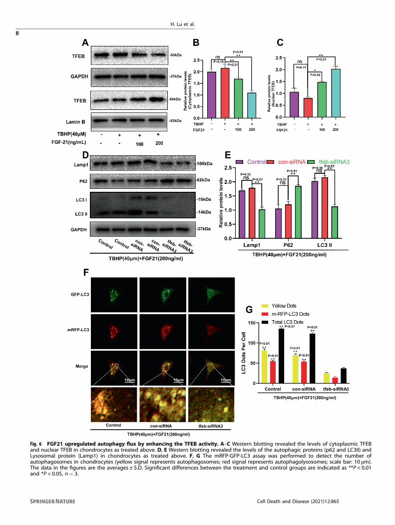
Fig. 6 FGF21 upregulated autophagy flux by enhancing the TFEB activity. A–C Western blotting revealed the levels of cytoplasmic TFEBand nuclear TFEB in chondrocytes as treated above. D, E Western blotting revealed the levels of the autophagic proteins (p62 and LC3II) andLysosomal protein (Lamp1) in chondrocytes as treated above. F, G The mRFP-GFP-LC3 assay was performed to detect the number ofautophagosomes in chondrocytes (yellow signal represents autophagosomes; red signal represents autophagolysosomes; scale bar: 10 μm).The data in the figures are the averages ± S.D. Significant differences between the treatment and control groups are indicated as **P < 0.01and *P < 0.05, n= 3.
H. Lu et al.
8
Cell Death and Disease (2021) 12:865
The surgical model of destabilization of the medial meniscus(DMM) has become an essential model for studying apoptosis andsenescence in posttraumatic osteoarthritis (OA) [13, 52]. In the in vivoexperimental DMM model, several in vivo evaluation methods suchas X-ray, H&E staining, SO staining, Alcian Blue staining, immuno-chemistry, tissue TUNEL assay, and tissue fluorescence were used todetermine the effect of FGF21 on the progression of OA in mice. Theresults were almost consistent with in vitro experiments, whichindicated that FGF21 might inhibit the occurrence and developmentof OA by activating the SIRT1-mTOR signaling pathway, therebypromoting nuclear translocation of TFEB (Fig. S4).Although our study demonstrated that FGF21 reduces senes-
cence, apoptosis, and ECM catabolism in TBHP-treated chondro-cytes via the SIRT1-mTOR signaling pathway, there are still severallimitations of our study. First, although we found that FGF21 had apositive effect on the progression of osteoarthritis, the chondro-cytes isolated from the articular cartilage showed the particularityof differentiation [53]. Chondrocytes treated with TBHP onlypartially reflected the pathological condition of osteoarthritis.Second, the mechanism of the effects of FGF21 on chondrocytes isstill unclear. FGF21-mediated inhibition of TBHP-induced apopto-sis and senescence might also result from antioxidative effects ofthe growth factor. A DPPH assay would clarify this issue. Therefore,it is necessary to further study the effect of FGF21 onchondrocytes in the future.
In summary, our results not only suggested that FGF21 couldserve as an effective treatment tool for inhibiting the progressionof OA, but it could also provide insights into the mechanism ofaction of FGF21 in chondrocytes. The present study highlights thepotential of FGF21 in clinical transformation, particularly in thetreatment of OA in the future.
MATERIALS AND METHODSAntibodies and reagentsFGF21 (P6101) was obtained from Beyotime Biotechnology (Jiangsu,China), chloroquine (CQ) was obtained from Sigma-Aldrich ChemicalCompany (Milwaukee, WI, USA), and nicotinamide (NAM) was purchasedfrom MedChemExpress (NJ, USA). The primary antibodies directed againstCollagen II, MMP-13, p16INK4a, p21WAF1, Lamin B1, and GAPDH werepurchased from Abcam (Cambridge, MA, USA), while the primaryantibodies directed against Bax, Bcl-2, SQSTM1/p62, and LC3 wereobtained from ProteinTech (Wuhan, China). The antibodies against cleavedcaspase3 (C-caspase 3), TFEB, mTOR, Phospho-mTOR (p-mTOR), and SIRT1were procured from Cell Signaling Technologies (Danvers, MA, USA).
Isolation and primary culture of chondrocytesSix immature C57BL/6 mice (3 males and 3 females, 10-day-old) weresacrificed, the articular cartilage was separated under sterile conditions andcarefully collected. Briefly, the cartilages of the knee joints of the mice werecut into pieces (1 mm3) and then enzymatically digested in 2mg/mL of
Fig. 7 FGF21 activated the SIRT1-mTOR pathway in the chondrocytes induced by TBHP. A, E Western blotting revealed the levels of SIRT1,mTOR, p-mTOR, cytoplasmic TFEB, and nuclear TFEB in chondrocytes as treated above. B–D Shown is the protein expression of Collagen II,MMP13, C-caspase3, and p16INK4a in the chondrocytes as treated above. F, G TUNEL assay was performed in the chondrocytes as treatedabove (scale bar: 50 μm). H, I The representative MMP13 was detected using the immunofluorescence analysis combined with DAPI stainingfor the nuclei in the chondrocytes as treated above (scale bar for MMP13: 20 μm). Significant differences between the treatment and controlgroups are indicated as **P < 0.01 and *P < 0.05, n= 3.
H. Lu et al.
9
Cell Death and Disease (2021) 12:865
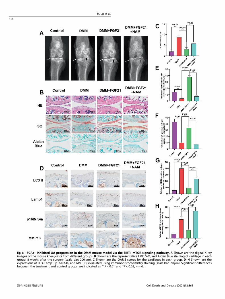
Fig. 8 FGF21 inhibited OA progression in the DMM mouse model via the SIRT1-mTOR signaling pathway. A Shown are the digital X-rayimages of the mouse knee joints from different groups. B Shown are the representative H&E, S-O, and Alcian Blue staining of cartilage in eachgroup, 8 weeks after the surgery (scale bar: 200 μm). C Shown are the OARIS scores for the cartilages in each group. D–H Shown are theexpressions of LC3, Lamp1, p16INK4a, and MMP13, evaluated using immunohistochemistry staining (scale bar: 20 μm). Significant differencesbetween the treatment and control groups are indicated as **P < 0.01 and *P < 0.05, n= 6.
H. Lu et al.
10
Cell Death and Disease (2021) 12:865
0.1% collagenase II at 37 °C for 4 h. The digested cartilage tissue pieceswere suspended in DMEM/F12 (Gibco, Invitrogen, Grand Island, NY)medium supplemented with 10% fetal bovine serum (FBS; HyClone,Thermo Scientific, Logan, UT, USA) and 1% penicillin/streptomycin (Gibco,Invitrogen, Grand Island, NY), and seeded into tissue culture flasks. Thechondrocytes were grown in an incubator maintained at 37 °C under 5%CO2 conditions. After 24 h of incubation, the culture medium was changed,and the second- or third-generation cells were retrieved for thesubsequent experiments.
Animal modelA total of 60 ten-week-old C57BL/6 male wild-type (WT) mice wereprocured from the Animal Center of the Chinese Academy of SciencesShanghai, China. The mice were randomly divided into four groups (n= 15in each) as the Control group (sham-operated), the DMM group (OA), theDMM+ FGF-21 group (OA treated with FGF-21), and the OA+ FGF-21+NAM (OA treated with FGF-21 and NAM) group. After anesthetization with2% (w/v) pentobarbital (40 mg/kg), as mentioned earlier [52], the mousemodel of osteoarthritis was developed by surgical destabilization of themedial meniscus (DMM). Eight weeks after the DMM surgery, the mice ineach group were sacrificed, and their joints were subjected to histologicalevaluation.
Western blot assayThe total protein in the chondrocytes was extracted using the RIPA lysisbuffer with 1 mM PMSF (phenylmethanesulfonyl fluoride). The proteinconcentration was determined using the BCA protein assay kit (Beyotime).The protein (40 ng) was separated on sodium dodecyl sulfate-polyacrylamide gel electrophoresis (SDS-PAGE) gels and transferred to apolyvinylidene difluoride (PVDF) membrane. After blocking the membraneby incubation with 5% non-fat milk for 2 h, the membrane was incubatedovernight at 4 °C with the following primary antibodies: aggrecan (1:1000),collagen II (1:1000), ADAMTS-5 (1:1000), MMP-13 (1:1000), p16INK4a(1:1000), p21WAF1 (1:1000), Lamin B1 (1:1000), GAPDH (1:5000), Bax(1:1000), Bcl-2 (1:1000), P62 (1:1000), Lc3 (1:1000), C-caspase3 (1:1000),TFEB (1:1000), mTOR (1:1000), Phospho-mTOR (p-mTOR) (1:1000), andSIRT1 (1:1000). Next, the bands were incubated with the respectivesecondary antibodies for 2 h at room temperature, followed by threewashes with TBST, and then visualized using an electrochemiluminescencereagent (Invitrogen). Finally, the intensity of these blots was determinedusing Image Lab 3.0 software (Bio-Rad).
TUNEL stainingThe apoptosis of chondrocytes was evaluated using an in-situ Cell DeathDetection Kit (Roche, South San Francisco, CA). The chondrocytes werefixed in 4% paraformaldehyde for ~1 h and then incubated for 10min eachwith 3% H2O2 and 0.2% Triton X-100. After three washes with PBS, the cellswere stained using the TUNEL staining solution and DAPI. Finally, theapoptosis of the chondrocytes was observed under an Olympusfluorescence microscope (Olympus Inc., Tokyo, Japan).
Transmission electron microscopyThe chondrocytes were fixed overnight in 2.5% glutaraldehyde, post-fixed in2% osmium tetroxide for 1 h, and finally stained with 2% uranyl acetate for1 h. After dehydration in a series of acetone solutions, the samples wereembedded in Araldite and excised into semi-thin sections, which were stainedwith toluidine blue to locate the cell position and were finally observed undera transmission electron microscope (Hitachi, Tokyo, Japan). In each section, 30cells were selected randomly, and their image was captured.
Transduction with mRFP-GFP-LC3 and analysisBefore co-culturing, the chondrocytes were cultured in a lower compartment,and (GeneChem, Shanghai, China) when 50–70% confluence was reached, thecells were transduced with mRFP-GFP-LC3, according to the instructions of themanufacturer. After the incubation, the autophagosomes in the chondrocyteswere observed under a confocal microscope (Leica TCS SP8, Germany), andthe total number of puncta (1mm) per cell was counted.
ImmunofluorescenceChondrocytes were seeded in six-well plates on glass coverslips, followedby washing with PBS, fixing in 4% paraformaldehyde, and permeation in0.1% Triton X-100 for 15min. After blocking with 5% bovine serum
albumin for 30min, the chondrocytes were incubated overnight at 4 °Cwith the primary antibody against LC3 (1:200), cleaved-caspase3 (1:400),MMP13 (1:100), or collagen II (1:100). The following day, the cells werewashed and incubated with Alexa Fluor 488or Alexa Fluor 594 for 1 h atroom temperature for labeling. The slides were observed under afluorescence microscope (Olympus Inc., Tokyo, Japan), and the fluores-cence intensity was measured using the Image J software 2.1 (Bethesda,MD, USA).
X-ray imaging methodEight weeks after the surgery, with or without treatment, the mice weresubjected to X-ray examination using a digital X-ray machine (KubtecModel PERT.8; KUB Technologies Inc.) to evaluate the articular surface,osteophyte formation, and calcification changes on the cartilage surface.Appropriate images were obtained at 50 kV and 160 µA.
Statistical analysisThe results were expressed as mean ± S.D. The data were analyzed usingthe SPSS statistical software program 18.0. The differences among thegroups were determined by performing a one-way analysis of variance(ANOVA) or t-test. Statistical differences among and/or between groupswere considered at P < 0.05. All the experiments were performedindependently three times and were consistently repeatable.
DATA AVAILABILITYThe datasets used and/or analyzed during the current study are available from thecorresponding author on reasonable request.
REFERENCES1. Xie SH, Wang Q, Wang LQ, Wang L, Song KP, He CQ. Effect of internet-based
rehabilitation programs on improvement of pain and physical function inpatients with knee osteoarthritis: systematic review and meta-analysis of ran-domized controlled trials. J Med Internet Res. 2021;23:e21542.
2. Roman-Blas JA, Bizzi E, Largo R, Migliore A, Herrero-Beaumont G. An update onthe up and coming therapies to treat osteoarthritis, a multifaceted disease. ExpertOpin Pharmacother. 2016;17:1745–56.
3. Liu F, Song DY, Huang J, Yang HQ, You D, Ni JD. Long non-coding RNA CIRinhibits chondrogenic differentiation of mesenchymal stem cells by epigeneti-cally suppressing ATOH8 via methyltransferase EZH2. Mol Med. 2021;27:12.
4. Wu D, Wong P, Guo C, Tam LS, Gu J. Pattern and trend of five major muscu-loskeletal disorders in China from 1990 to 2017: findings from the Global Burdenof Disease Study 2017. BMC Med. 2021;19:34.
5. Li H, Chen J, Li B, Fang X. The protective effects of dulaglutide against advancedglycation end products (AGEs)-induced degradation of type collagen andaggrecan in human SW1353 chondrocytes. Chem Biol Interact. 2020;322:108968.
6. Ansari MY, Ahmad N, Haqqi TM. Oxidative stress and inflammation in osteoarthritispathogenesis: Role of polyphenols. Biomed Pharmacother. 2020;129:110452.
7. Bolduc JA, Collins JA, Loeser RF. Reactive oxygen species, aging and articularcartilage homeostasis. Free Radic Biol Med. 2019;132:73–82.
8. Kim EN, Lee HS, Jeong GS. Cudratricusxanthone O inhibits H2O2-induced celldamage by activating Nrf2/HO-1 pathway in human chondrocytes. Antioxidants.2020;9788.
9. Morleo M, Brillante S, Formisano U, Ferrante L, Carbone F, Iaconis D, et al. Reg-ulation of autophagosome biogenesis by OFD1-mediated selective autophagy.EMBO J. 2021;40:e105120.
10. Barth S, Glick D, Macleod KF. Autophagy: assays and artifacts. J Pathol.2010;221:117–24.
11. Chao X, Ni HM, Ding WX. Insufficient autophagy: a novel autophagic flux scenariouncovered by impaired liver TFEB-mediated lysosomal biogenesis from chronicalcohol-drinking mice. Autophagy. 2018;14:1646–8.
12. Wang S, Ni HM, Chao X, Wang H, Bridges B, Kumer S, et al. Impaired TFEB-mediated lysosomal biogenesis promotes the development of pancreatitis inmice and is associated with human pancreatitis. Autophagy. 2019;15:1954–69.
13. Zheng G, Zhan Y, Li X, Pan Z, Zheng F, Zhang Z, et al. TFEB, a potential ther-apeutic target for osteoarthritis via autophagy regulation. Cell Death Dis.2018;9:858.
14. Nishimura T, Nakatake Y, Konishi M, Itoh N. Identification of a novel FGF, FGF-21,preferentially expressed in the liver. Biochim Biophys Acta. 2000;1492:203–6.
15. Tabari FS, Karimian A, Parsian H, Rameshknia V, Mahmoodpour A, Majidinia M,et al. The roles of FGF21 in atherosclerosis pathogenesis. Rev Endocr MetabDisord. 2019;20:103–14.
H. Lu et al.
11
Cell Death and Disease (2021) 12:865
16. Desai BN, Singhal G, Watanabe M, Stevanovic D, Lundasen T, Fisher FM, et al.Fibroblast growth factor 21 (FGF21) is robustly induced by ethanol and has aprotective role in ethanol associated liver injury. Mol Metab. 2017;6:1395–406.
17. Kharitonenkov A, Wroblewski VJ, Koester A, Chen YF, Clutinger CK, Tigno XT, et al.The metabolic state of diabetic monkeys is regulated by fibroblast growth factor-21. Endocrinology. 2007;148:774–81.
18. Salminen A, Kaarniranta K, Kauppinen A. Regulation of longevity by FGF21:interaction between energy metabolism and stress responses. Ageing Res Rev.2017;37:79–93.
19. Kakoty V, Sarathlal KC, Tang RD, Yang CH, Dubey SK, Taliyan R. Fibroblast growthfactor 21 and autophagy: a complex interplay in Parkinson disease. BiomedPharmacother. 2020;127:110145
20. Byun S, Seok S, Kim YC, Zhang Y, Yau P, Iwamori N, et al. Fasting-inducedFGF21 signaling activates hepatic autophagy and lipid degradation via JMJD3histone demethylase. Nat Commun. 2020;11:807.
21. Zhou K, Chen H, Lin J, Xu H, Wu H, Bao G, et al. FGF21 augments autophagy inrandom-pattern skin flaps via AMPK signaling pathways and improves tissuesurvival. Cell Death Dis. 2019;10:872.
22. Wei W, An XR, Jin SJ, Li XX, Xu M. Inhibition of insulin resistance by PGE1 viaautophagy-dependent FGF21 pathway in diabetic nephropathy. Sci Rep. 2018;8:9.
23. Zhang J, Cheng Y, Gu J, Wang S, Zhou S, Wang Y, et al. Fenofibrate increasescardiac autophagy via FGF21/SIRT1 and prevents fibrosis and inflammation in thehearts of Type 1 diabetic mice. Clin Sci. 2016;130:625–41.
24. Chen S, Chen ST, Sun Y, Xu Z, Wang Y, Yao SY, et al. Fibroblast growth factor 21ameliorates neurodegeneration in rat and cellular models of Alzheimer’s disease.Redox Biol. 2019;22:101133.
25. Ren B, Wang L, Shi L, Jin X, Liu Y, Liu RH, et al. Methionine restriction alleviatesage-associated cognitive decline via fibroblast growth factor 21. Redox Biol.2021;41:101940.
26. Mauthe M, Orhon I, Rocchi C, Zhou X, Luhr M, Hijlkema KJ, et al. Chloroquineinhibits autophagic flux by decreasing autophagosome-lysosome fusion.Autophagy. 2018;14:1435–55.
27. Mao K, Chen J, Yu H, Li H, Ren Y, Wu X, et al. Poly (ADP-ribose) polymerase 1inhibition prevents neurodegeneration and promotes alpha-synuclein degrada-tion via transcription factor EB-dependent autophagy in mutant alpha-synucleinA53T model of Parkinson’s disease. Aging Cell. 2020;19:e13163.
28. Fang J, Wang X, Jiang W, Zhu Y, Hu Y, Zhao Y, et al. Platelet-rich plasma therapyin the treatment of diseases associated with orthopedic injuries. Tissue Eng Part BRev. 2020;26:571–85.
29. Fraenkel L, Buta E, Suter L, Dubreuil M, Levy C, Najem C, et al. Nonsteroidal anti-inflammatory drugs vs cognitive behavioral therapy for arthritis pain: a rando-mized withdrawal trial. JAMA Intern Med. 2020;180:1194–202.
30. Staiger H, Keuper M, Berti L, Hrabe de Angelis M, Häring HU. Fibroblast growthfactor 21-metabolic role in mice and men. Endocr Rev. 2017;38:468–88.
31. Lewis JE, Ebling FJP, Samms RJ, Tsintzas K. Going back to the biology of FGF21:new insights. Trends Endocrinol Metab. 2019;30:491–504.
32. Kang K, Xu P, Wang M, Chunyu J, Sun X, Ren G, et al. FGF21 attenuates neuro-degeneration through modulating neuroinflammation and oxidant-stress.Biomed Pharmacother. 2020;129:110439.
33. Hua X, Sun DY, Zhang WJ, Fu JT, Tong J, Sun SJ, et al. P7C3-A20 alleviates fattyliver by shaping gut microbiota and inducing FGF21/FGF1, via the AMP-activatedprotein kinase/CREB regulated transcription coactivator 2 pathway. Br J Pharm.2021;178:2111–30.
34. Li ZC, Xiao J, Wang G, Li MQ, Hu KZ, Ma T, et al. Fibroblast growth factor-21concentration in serum and synovial fluid is associated with radiographic boneloss of knee osteoarthritis. Scand J Clin Lab Invest. 2015;75:121–5.
35. Paré F, Tardif G, Fahmi H, Ouhaddi Y, Pelletier JP, Martel-Pelletier J. In vivoprotective effect of adipsin-deficiency on spontaneous knee osteoarthritis inaging mice. Aging. 2020;12:2880–96.
36. Dierick JF, Eliaers F, Remacle J, Raes M, Fey SJ, Larsen PM, et al. Stress-inducedpremature senescence and replicative senescence are different phenotypes,proteomic evidence. Biochem Pharm. 2002;64:1011–7.
37. Tan X, Yu L, Yang R, Tao Q, Xiang L, Xiao J, et al. Fibroblast growth factor 10attenuates renal damage by regulating endoplasmic reticulum stress afterischemia-reperfusion injury. Front Pharm. 2020;11:39.
38. Hosseinzadeh A, Kamrava SK, Joghataei MT, Darabi R, Shakeri-Zadeh A, ShahriariM, et al. Apoptosis signaling pathways in osteoarthritis and possible protectiverole of melatonin. J Pineal Res. 2016;61:411–25.
39. An S, Hu H, Li Y, Hu Y. Pyroptosis plays a role in osteoarthritis. Aging Dis.2020;11:1146–57.
40. Coryell PR, Diekman BO, Loeser RF. Mechanisms and therapeutic implications ofcellular senescence in osteoarthritis. Nat Rev Rheumatol. 2021;17:47–57.
41. Xie J, Lin J, Wei M, Teng Y, He Q, Yang G, et al. Sustained Akt signaling in articularchondrocytes causes osteoarthritis via oxidative stress-induced senescence inmice. Bone Res. 2019;7:23.
42. Thounaojam MC, Jadeja RN, Warren M, Powell FL, Raju R, Gutsaeva D, et al.MicroRNA-34a (miR-34a) mediates retinal endothelial cell premature senescencethrough mitochondrial dysfunction and loss of antioxidant activities. Anti-oxidants. 2019;8328.
43. Li M, Yang X, Lu X, Dai N, Zhang S, Cheng Y, et al. APE1 deficiency promotescellular senescence and premature aging features. Nucleic Acids Res.2018;46:5664–77.
44. Tang Q, Zheng G, Feng Z, Chen Y, Lou Y, Wang C, et al. Trehalose amelioratesoxidative stress-mediated mitochondrial dysfunction and ER stress via selectiveautophagy stimulation and autophagic flux restoration in osteoarthritis devel-opment. Cell Death Dis. 2017;8:e3081.
45. Kim D, Song J, Kang Y, Park S, Kim YI, Kwak S, et al. Fis1 depletion in osteoarthritisimpairs chondrocyte survival and peroxisomal and lysosomal function. J MolMed. 2016;94:1373–84.
46. Ansari MY, Ball HC, Wase SJ, Novak K, Haqqi TM. Lysosomal dysfunction inosteoarthritis and aged cartilage triggers apoptosis in chondrocytes through BAXmediated release of Cytochrome c. Osteoarthr Cartil. 2021;29:100–12.
47. Gu M, Jin J, Ren C, Chen X, Pan Z, Wu Y, et al. 20-Deoxyingenol alleviatesosteoarthritis by activating TFEB in chondrocytes. Pharm Res. 2021;165:105361.
48. Settembre C, Di Malta C, Polito VA, Garcia Arencibia M, Vetrini F, Erdin S, et al.TFEB links autophagy to lysosomal biogenesis. Science. 2011;332:1429–33.
49. Young NP, Kamireddy A, Van Nostrand JL, Eichner LJ, Shokhirev MN, Dayn Y, et al.AMPK governs lineage specification through Tfeb-dependent regulation oflysosomes. Genes Dev. 2016;30:535–52.
50. Herzig S, Shaw RJ. AMPK: guardian of metabolism and mitochondrial home-ostasis. Nat Rev Mol Cell Biol. 2018;19:121–35.
51. Yau WW, Singh BK, Lesmana R, Zhou J, Sinha RA, Wong KA, et al. Thyroid hor-mone (T3) stimulates brown adipose tissue activation via mitochondrial bio-genesis and MTOR-mediated mitophagy. Autophagy. 2019;15:131–50.
52. Au M, Liu Z, Rong L, Zheng Y, Wen C. Endothelin-1 induces chondrocytesenescence and cartilage damage via endothelin receptor type B in a post-traumatic osteoarthritis mouse model. Osteoarthr Cartil. 2020;28:1559–71.
53. Charlier E, Deroyer C, Ciregia F, Malaise O, Neuville S, Plener Z, et al. Chondrocytededifferentiation and osteoarthritis (OA). Biochem Pharm. 2019;165:49–65.
ACKNOWLEDGEMENTSWe thank all the colleagues for their invaluable assistance during the execution ofthis study.
AUTHOR CONTRIBUTIONSWW and HW Lu conceived and designed the experiments. HW Lu, CJ, and DY Wucarried out the experiments. HDJ, ZL, and XC Li analyzed data and wrote themanuscript. WW and JP co-supervised the study and revised the paper. All authorsread and approved the final manuscript.
FUNDINGThis study is supported by the National Natural Science Foundation of China(81401772).
COMPETING INTERESTSThe authors declare no competing interests.
ETHICS STATEMENTAll surgical interventions, treatments, and postoperative animal care procedures arestrictly implemented following the “Guidelines for the Care and Use of LaboratoryAnimals of the National Institutes of Health” and are approved by the Animal Careand Use Committee of Wenzhou Medical University (wydw2020-0873).
ADDITIONAL INFORMATIONSupplementary information The online version contains supplementary materialavailable at https://doi.org/10.1038/s41419-021-04157-x.
Correspondence and requests for materials should be addressed to Jun Pan, XiucuiLi or Wei Wang.
Reprints and permission information is available at http://www.nature.com/reprints
H. Lu et al.
12
Cell Death and Disease (2021) 12:865

Publisher’s note Springer Nature remains neutral with regard to jurisdictional claimsin published maps and institutional affiliations.
Open Access This article is licensed under a Creative CommonsAttribution 4.0 International License, which permits use, sharing,
adaptation, distribution and reproduction in anymedium or format, as long as you giveappropriate credit to the original author(s) and the source, provide a link to the Creative
Commons license, and indicate if changes were made. The images or other third partymaterial in this article are included in the article’s Creative Commons license, unlessindicated otherwise in a credit line to the material. If material is not included in thearticle’s Creative Commons license and your intended use is not permitted by statutoryregulation or exceeds the permitted use, you will need to obtain permission directlyfrom the copyright holder. To view a copy of this license, visit http://creativecommons.org/licenses/by/4.0/.
© The Author(s) 2021
H. Lu et al.
13
Cell Death and Disease (2021) 12:865