ferrotoxicity and its amelioration by calcitriol in cultured
TRANSCRIPT

Research ArticleFerrotoxicity and Its Amelioration by Calcitriol in CulturedRenal Cells
Chandrashekar Annamalai,1 Rohit Seth,2 and Pragasam Viswanathan 1
1Renal Research Lab, Centre for Biomedical Research, School of Biosciences and Technology, Vellore Institute of Technology (VIT),Vellore, 632 014 Tamil Nadu, India2Department of Zoology, Guru Ghasidas Vishwavidyalaya, Bilaspur, 495009 Chhattisgarh, India
Correspondence should be addressed to Pragasam Viswanathan; [email protected]
Received 9 November 2020; Revised 22 January 2021; Accepted 15 February 2021; Published 23 February 2021
Academic Editor: Md. Atiar Rahman
Copyright © 2021 Chandrashekar Annamalai et al. This is an open access article distributed under the Creative CommonsAttribution License, which permits unrestricted use, distribution, and reproduction in any medium, provided the original workis properly cited.
Globally, acute kidney injury (AKI) is associated with significant mortality and an enormous economic burden. Whereas iron isessential for metabolically active renal cells, it has the potential to cause renal cytotoxicity by promoting Fenton chemistry-basedoxidative stress involving lipid peroxidation. In addition, 1,25-dihydroxyvitamin D3 (calcitriol), the active form of vitamin D, isreported to have an antioxidative role. In this study, we intended to demonstrate the impact of vitamin D on iron-mediatedoxidant stress and cytotoxicity of Vero cells exposed to iohexol, a low osmolar iodine-containing contrast media in vitro.Cultured Vero cells were pretreated with 1,25-dihydroxyvitamin D3 dissolved in absolute ethanol (0.05%, 2.0mM) at a dose of1mM for 6 hours. Subsequently, iohexol was added at a concentration of 100mg iodine per mL and incubated for 3 hours.Total cellular iron content was analysed by a flame atomic absorption spectrophotometer at 372 nm. Lipid peroxidation wasdetermined by TBARS (thiobarbituric acid reactive species) assay. Antioxidants including total thiol content were assessed byEllman’s method, catalase by colorimetric method, and superoxide dismutase (SOD) by nitroblue tetrazolium assay. The cellswere stained with DAPI (4′,6-diamidino-2-phenylindole), and the cytotoxicity was evaluated by viability assay (MTT assay).The results indicated that iohexol exposure caused a significant increase of the total iron content in Vero cells. A concomitantincrease of lipid peroxidation and decrease of total thiol protein levels, catalase, and superoxide dismutase activity were observedalong with decreased cell viability in comparison with the controls. Furthermore, these changes were significantly reversed whenthe cells were pretreated with vitamin D prior to incubation with iohexol. Our findings of this in vitro model of iohexol-inducedrenotoxicity lend further support to the nephrotoxic potential of iron and underpin the possible clinical utility of vitamin D forthe treatment and prevention of AKI.
1. Introduction
The prevalence of acute kidney injury (AKI) is increasing,and besides causing significant morbidity and mortality, itincreases the risk of progression to irreversible kidney dis-ease, thereby imposing a tremendous financial, societal, andpersonal burden [1, 2]. The pathogenesis of AKI is multifac-torial and involves a complex [3] interaction between vascu-lar, tubular, and inflammatory factors. This is followed eitherby repair and restoration of glomerular and tubular functionsor culminates in fibrosis and progression to chronic kidneydisease [4]. Currently, there is no specific and effective thera-
peutic modality and the treatment is mostly supportive innature [5].
Furthermore, despite the incidence being overestimated,contrast-induced acute kidney injury (CI-AKI) still poses amajor threat to patients undergoing contrast-associated trans-catheter diagnostic and interventional procedures by worsen-ing short- and long-term outcomes and prolonging hospitalstay [6, 7]. Iohexol, a water-soluble, low osmolar, and nonioniciodine-containing monomeric radiocontrast agent inducesrenal damage by several mechanisms which are quite complexand yet to be completely understood [8, 9]. Renal hypoperfu-sion and renal medullary hypoxia [8, 10], autocrine and
HindawiAnalytical Cellular PathologyVolume 2021, Article ID 6634429, 13 pageshttps://doi.org/10.1155/2021/6634429

paracrine impairment including increased endothelin andadenosine release, lower nitric oxide metabolite concentrationand enhanced oxidative stress [11] [12], and direct cellulardamage [13] resulting in endothelial dysfunction followed byvasoconstriction of vasa recta [14] [15], and consequent alter-ations in renal blood flow lead to the development of contrast-induced renal injury. High viscosity can further adverselyaffect renal perfusion and urine formation [8].
Besides, the metallobiology of iron has been in focus inrecent times due to its crucial pathophysiological role in kid-ney diseases. In particular, iron has been increasingly impli-cated in the induction of AKI and worse outcomes [16–20].In this context, the cytotoxic potential of iron in contrast-induced AKI has been put forth as an important mechanism[18]. Iron is an indispensable component of oxygen-bindingmolecules (for example, hemoglobin and myoglobin), cyto-chromes in the electron transport chain, and as a cofactor inmany enzymes and is involved in many fundamental biologi-cal processes [21, 22]. The kidneys play a vital role in prevent-ing iron loss in urine by reabsorbing the filtered iron.Furthermore, they express several proteins necessary for irontransport and metabolic activity and are thus actively involvedin systemic iron homeostasis [23, 24]. Although iron is essen-tial for the metabolically active renal cells [25], it has thepotential to induce renal cytotoxicity [26, 27]. This is attrib-uted to the unique property of iron to mediate electron trans-fer and participate in the oxidation-reduction (redox)reactions. While this is crucial for the functioning of severalbiological systems, it can render iron lethal by catalysing theFenton and Haber-Weiss reactions and promoting the pro-duction of reactive oxygen species (ROS) such as hydroxylradicals. This further overrides the cellular antioxidant defencemechanisms and induces oxidative injury of cell structures inaddition to causing local inflammation and vasoconstriction[26, 27]. It is worth noting that knowledge on the complexmechanisms of renal iron handling and iron-induced cellularinjury (ferroptosis) is limited and its understanding couldprovide novel therapeutic avenues [22, 28].
Concurrently, the kidneys also possess endogenous andexogenous protective agents to counteract cellular injury[28]. In this context, vitamin D is known to have cytoprotec-tive action because of its antioxidant potential [29–31]. Vita-min D is a prohormone with no intrinsic biological activityand is derived endogenously from the skin and exogenouslyfrom diet and supplements [32]. Both vitamin D2 (ergocalci-ferol) and vitamin D3 (cholecalciferol) differ in their sidechains thereby affecting the capacity to bind to vitamin D-binding protein (DBP) as well as efficacy. Vitamin D3 is con-siderably more effective than vitamin D2 [33, 34]. Duringexposure to the sun, ultraviolet rays (270–300 nm) photolyt-ically converts 7-dehydrocholesterol by breaking its B ring toform previtamin D3 which undergoes thermal isomerizationto vitamin D3 [35, 36]. Vitamin D3 bound to DBP is sequen-tially hydroxylated first in the liver by the cytochrome P450s(microsomal CYP2R1 and mitochondrial CYP27A1) [37] toform 25-hydroxyvitamin D3 and subsequently in theproximal tubules of the kidney [38, 39] by 1 α-hydroxylase(CYP27B1) to the bioactive 1,25-dihydroxyvitamin D3, alsoknown as calcitriol [40–42].
1,25-Dihydroxyvitamin D3 is known to possess severalpleiotropic effects [43, 44]. Apart from being a key regulatorof calcium homeostasis by modulating parathyroid hormonesecretion and increasing gut calcium absorption [45], it hasbeen proven to exert antioxidant [30, 31], anti-inflammatory,antiproliferative, and antineoplastic effects [46–49] as well.By virtue of these properties, calcitriol is cytoprotective bynature, whereas iron, especially the free form, is potentiallycytotoxic. Furthermore, the role of vitamin D in AKI is notclearly elucidated as in chronic kidney disease [50] and itneeds to be ascertained if vitamin D could mitigate ferrotoxi-city induced by radiocontrast media in vitro [28].
Therefore, we sought to investigate the combined roles ofiron and vitamin D in relation to oxidative stress and neph-rotoxicity in an in vitro model of iohexol-induced AKI usingVero cells. The main purpose of this study was to determinethe effect of iohexol on total cellular iron concentration andits influence on oxidative stress and cytotoxicity of Vero cellsand to study the impact of 1,25-dihydroxyvitamin D3 oniron-mediated oxidant stress and cellular injury.
2. Materials and Methods
2.1. Cell Culture. Vero cells (ATCC® CLL-81™) were pro-cured from Cell Repository, National Centre for Cell Science(NCCS), India. Dulbecco’s Modified Eagle’s Medium (HiMe-dia Laboratories, India) containing 10% fetal bovine serum(FBS) and 1% penicillin and streptomycin combination wasused. The cells were grown on a cover glass in 10 cm plasticdishes placed inside an incubator at 37°C containing a humidatmosphere consisting of 5% oxygen, 5% carbon dioxide, and90% nitrogen until a monolayer was formed. Periodic assess-ment of these cells was carried out to ensure freedom frommycoplasma contamination. Upon reaching 80-95% con-fluency, the cells were digested with trypsin, resuspended inserum-free medium, and passaged in a 1 : 3 proportion. Theculture media was replaced every 2 to 3 days to ensurecontinuous nutritional support for cell growth.
The optimal dose of iohexol was determined and the cellviability (MTT (3-(4,5-dimethylthiazol-2-yl)-2,5-diphenyl-tetrazolium bromide)) assay was studied by plating 1 × 10⁴viable cells per cm2 using 96-well plates.
Control cells were incubated similarly with sterile 0.9%normal saline (sodium chloride injection, B.P. 0.9% w/v,Schwitz Biotech, India) except that they were not pretreatedwith vitamin D3 and iohexol. Microscopy was performedusing a Leica DM IL inverted fluorescent microscopeequipped with appropriate fluorescent filters (Leica Micro-systems, India). Images were captured at 100x magnificationusing an attached Leica DFC 450C camera and processedwith LAS X software and exported.
2.2. Preparation of 1,25-Dihydroxyvitamin D3. 1,25-Dihy-droxyvitamin D3 (Cat: sc-202877, Santa Cruz BiotechnologyInc., USA) dissolved in absolute ethanol (0.05%, 2.0mM,HiMedia Laboratories, India) was used for our cell culturestudies. Vitamin D is light-sensitive and is stored away fromdirect light in a brown bottle at -20°C. On the day of use,further dilutions were made directly in the culture medium
2 Analytical Cellular Pathology
at the concentration of 1μM, a dose at which no significantchange in cell viability was observed to occur.
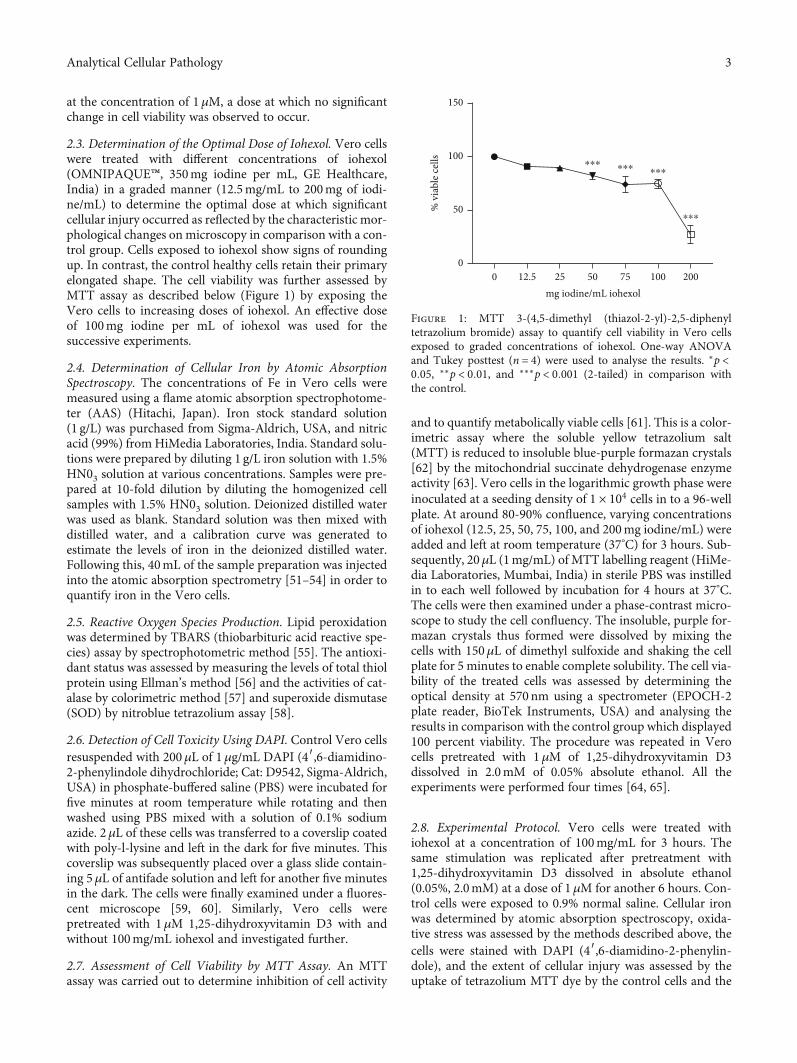
2.3. Determination of the Optimal Dose of Iohexol. Vero cellswere treated with different concentrations of iohexol(OMNIPAQUE™, 350mg iodine per mL, GE Healthcare,India) in a graded manner (12.5mg/mL to 200mg of iodi-ne/mL) to determine the optimal dose at which significantcellular injury occurred as reflected by the characteristic mor-phological changes on microscopy in comparison with a con-trol group. Cells exposed to iohexol show signs of roundingup. In contrast, the control healthy cells retain their primaryelongated shape. The cell viability was further assessed byMTT assay as described below (Figure 1) by exposing theVero cells to increasing doses of iohexol. An effective doseof 100mg iodine per mL of iohexol was used for thesuccessive experiments.
2.4. Determination of Cellular Iron by Atomic AbsorptionSpectroscopy. The concentrations of Fe in Vero cells weremeasured using a flame atomic absorption spectrophotome-ter (AAS) (Hitachi, Japan). Iron stock standard solution(1 g/L) was purchased from Sigma-Aldrich, USA, and nitricacid (99%) fromHiMedia Laboratories, India. Standard solu-tions were prepared by diluting 1 g/L iron solution with 1.5%HN03 solution at various concentrations. Samples were pre-pared at 10-fold dilution by diluting the homogenized cellsamples with 1.5% HN03 solution. Deionized distilled waterwas used as blank. Standard solution was then mixed withdistilled water, and a calibration curve was generated toestimate the levels of iron in the deionized distilled water.Following this, 40mL of the sample preparation was injectedinto the atomic absorption spectrometry [51–54] in order toquantify iron in the Vero cells.
2.5. Reactive Oxygen Species Production. Lipid peroxidationwas determined by TBARS (thiobarbituric acid reactive spe-cies) assay by spectrophotometric method [55]. The antioxi-dant status was assessed by measuring the levels of total thiolprotein using Ellman’s method [56] and the activities of cat-alase by colorimetric method [57] and superoxide dismutase(SOD) by nitroblue tetrazolium assay [58].
2.6. Detection of Cell Toxicity Using DAPI. Control Vero cellsresuspended with 200μL of 1μg/mL DAPI (4′,6-diamidino-2-phenylindole dihydrochloride; Cat: D9542, Sigma-Aldrich,USA) in phosphate-buffered saline (PBS) were incubated forfive minutes at room temperature while rotating and thenwashed using PBS mixed with a solution of 0.1% sodiumazide. 2μL of these cells was transferred to a coverslip coatedwith poly-l-lysine and left in the dark for five minutes. Thiscoverslip was subsequently placed over a glass slide contain-ing 5μL of antifade solution and left for another five minutesin the dark. The cells were finally examined under a fluores-cent microscope [59, 60]. Similarly, Vero cells werepretreated with 1μM 1,25-dihydroxyvitamin D3 with andwithout 100mg/mL iohexol and investigated further.
2.7. Assessment of Cell Viability by MTT Assay. An MTTassay was carried out to determine inhibition of cell activity
and to quantify metabolically viable cells [61]. This is a color-imetric assay where the soluble yellow tetrazolium salt(MTT) is reduced to insoluble blue-purple formazan crystals[62] by the mitochondrial succinate dehydrogenase enzymeactivity [63]. Vero cells in the logarithmic growth phase wereinoculated at a seeding density of 1 × 10⁴ cells in to a 96-wellplate. At around 80-90% confluence, varying concentrationsof iohexol (12.5, 25, 50, 75, 100, and 200mg iodine/mL) wereadded and left at room temperature (37°C) for 3 hours. Sub-sequently, 20μL (1mg/mL) of MTT labelling reagent (HiMe-dia Laboratories, Mumbai, India) in sterile PBS was instilledin to each well followed by incubation for 4 hours at 37°C.The cells were then examined under a phase-contrast micro-scope to study the cell confluency. The insoluble, purple for-mazan crystals thus formed were dissolved by mixing thecells with 150μL of dimethyl sulfoxide and shaking the cellplate for 5 minutes to enable complete solubility. The cell via-bility of the treated cells was assessed by determining theoptical density at 570 nm using a spectrometer (EPOCH-2plate reader, BioTek Instruments, USA) and analysing theresults in comparison with the control group which displayed100 percent viability. The procedure was repeated in Verocells pretreated with 1μM of 1,25-dihydroxyvitamin D3dissolved in 2.0mM of 0.05% absolute ethanol. All theexperiments were performed four times [64, 65].
2.8. Experimental Protocol. Vero cells were treated withiohexol at a concentration of 100mg/mL for 3 hours. Thesame stimulation was replicated after pretreatment with1,25-dihydroxyvitamin D3 dissolved in absolute ethanol(0.05%, 2.0mM) at a dose of 1μM for another 6 hours. Con-trol cells were exposed to 0.9% normal saline. Cellular ironwas determined by atomic absorption spectroscopy, oxida-tive stress was assessed by the methods described above, thecells were stained with DAPI (4′,6-diamidino-2-phenylin-dole), and the extent of cellular injury was assessed by theuptake of tetrazolium MTT dye by the control cells and the
2001007550mg iodine/mL iohexol
2512.500
50
100 ⁎⁎⁎ ⁎⁎⁎ ⁎⁎⁎
⁎⁎⁎
% v
iabl
e cel
ls
150
Figure 1: MTT 3-(4,5-dimethyl (thiazol-2-yl)-2,5-diphenyltetrazolium bromide) assay to quantify cell viability in Vero cellsexposed to graded concentrations of iohexol. One-way ANOVAand Tukey posttest (n = 4) were used to analyse the results. ∗p <0:05, ∗∗p < 0:01, and ∗∗∗p < 0:001 (2-tailed) in comparison withthe control.
3Analytical Cellular Pathology

Vero cells exposed to iohexol before and after pretreatmentwith 1,25-dihydroxyvitamin D3.
2.9. Statistical Analysis. The experimental results from fourindependent experiments (technical replicates) were repre-sented asmeans ± standard deviation (SD). One-way analysisof variance (ANOVA) with Tukey multiple comparison post-test was used to study the individual variances in relation tothe control group. Pearson correlation was used to estimatethe association between different study parameters. Statisticalsignificance was defined at p < 0:05 (2-tailed). SPSS (IBMCorp. Released 2017, IBM SPSS Statistics for Windows,Version 25.0. Armonk, NY: IBM Corp.) was used to analysethe results and GraphPad Prism Version 8.3.0 to create theartwork and illustrations.
3. Results
3.1. Determination of the Optimal Dose of Iohexol. Vero cellsincubated with different concentrations of iohexol were
examined under a microscope. 100% confluency wasobserved in the control cells which retained their primaryelongated morphology. With a progressive increase in theconcentration of iohexol, there was a concomitant increasein cell death. At a concentration of 50mg iodine per mLof iohexol, nearly 50% cell lysis was evident with the max-imum lysis occurring at 200mg iodine per mL of iohexolconcentration (Figure 2).
Vero cells were further exposed to increasing doses ofiohexol to determine cell viability using MTT assay. As illus-trated in Figure 1, in the dose-response study, the effect ofiohexol was noted to be concentration-dependent. A signifi-cant decrease in cell viability was conspicuous starting froma concentration of 50mg iodine per mL of iohexol withrespect to the control (100% viable cells). Cell survival wasthe lowest at the maximum iohexol concentration of200mg iodine per mL (Figure 1). An effective dose of100mg iodine per mL of iohexol was used for the successiveexperiments.
Control 12.5 mg I/mL 25 mg I/mL 50 mg I/mL
75 mg I/mL 100 mg I/mL 200 mg I/mL
100× 100× 100× 100×
100× 100× 100×
Figure 2: Vero cells under the microscope after subjecting to different doses of iohexol containing increasing concentration of iodine (I)(n = 4). Magnification 100x. The control group displays healthy, growing cells with elongated morphology (black arrows), whereas thecells incubated with iohexol show signs of rounding up.
Table 1: Descriptive statistics.
Control(n = 4)
Vitamin D(n = 4)
Iohexol(n = 4)
Iohexol+vitamin D(n = 4)
Total cellular iron (nmol/mg protein) 10:4 ± 1:2 10:8 ± 1:0 52:4 ± 9:1∗∗∗ 28:0 ± 4:7∗∗$$$
Lipid peroxidation (mg/mg protein) 0:2 ± 0:0 0:3 ± 0:1 0:6 ± 0:1∗∗∗ 0:4 ± 0:0∗$$$
Thiol protein (units/mg protein) 0:8 ± 0:1 0:7 ± 0:1 0:1 ± 0:0∗∗∗ 0:5 ± 0:2∗∗$$
Catalase (units/mg protein) 13:1 ± 0:2 12:2 ± 0:6∗ 1:0 ± 0:1∗∗∗ 6:4 ± 0:4∗∗∗$$$
Superoxide dismutase (units/mg protein) 100:0 ± 0:0 97:4 ± 0:8 72:4 ± 2:7∗∗∗ 91:43 ± 1:7∗∗∗$$$
Cell viability (%) 100:0 ± 0:0 96:7 ± 9:6 66:6 ± 7:5∗∗ 87:6 ± 13:6$
Results were represented asmean ± SD. One-way ANOVA and Tukey posttest (n = 4) were used to analyse the results. p < 0:05, ∗∗p < 0:01, and ∗∗∗p < 0:001 (2-tailed) versus control; $p < 0:05; $$p < 0:01, and $$$p < 0:001 (2-tailed) iohexol versus iohexol+vitamin D groups.
4 Analytical Cellular Pathology

3.2. Total Cellular Iron. Iohexol exposure caused a significantincrease in the total iron content in the Vero cells(52:4 ± 9:1 nmol/mg protein, p < 0:001) compared with thecontrol cells (10:4 ± 1:2 nmol/mg protein) and the cells incu-bated with vitamin D alone (10:8 ± 1:0 nmol/mg protein).The iron concentration was, however, noted to be signifi-cantly reduced in the cells pretreated with vitamin D(28:0 ± 4:7 nmol/mg protein) (Table 1, Figure 3).
3.3. Reactive Oxygen Species Production. Similar conditionswere reproduced to assess the oxidative stress markers inVero cells (Figure 4). There was a considerable increase inlipid peroxidation in cells exposed to iohexol alone(0:6 ± 0:1mg/mg protein, p < 0:001) compared with the con-trol cells (0:2 ± 0:0mg/mg protein). Furthermore, a concom-itant reduction in the antioxidant levels, namely, total thiolprotein (0:5 ± 0:2 units/mg protein, p < 0:01), catalase(6:4 ± 0:4 units/mg protein, p < 0:001), and superoxidedismutase (91:4 ± 1:7 units/mg protein, p < 0:001), wasobserved in those cells. On the other hand, these changeswere significantly reversed when the cells were treated withvitamin D prior to incubation with iohexol (Figure 4,Table 1). The cells treated with both vitamin D and iohexoldisplayed a relatively decreased oxidative stress than whentreated with iohexol alone. Similarly, not much differencein these levels existed between the control cells and the cellsexposed to calcitriol alone (Table 1, Figure 4).
3.4. Cell Toxicity Test Using DAPI Staining. Vero cells treatedwith iohexol alone and after pretreatment with vitamin D3were analysed after staining with DAPI. The morphology ofthe cells was investigated under a fluorescent microscope.The dead cells displayed a distorted appearance withfragmented or condensed nuclei (Figure 5).
3.5. Detection of Cell Viability by MTT Assay. Compared withthe control group, cell viability was noted to decrease
dramatically in iohexol-treated cells (p < 0:001) in aconcentration-dependent manner. This was indicated bythe lower absorbance rates of the experimental samples com-pared with negative control on MTT assay. However, priorexposure to vitamin D appeared to counteract the reductionin cell viability (p < 0:05) (Figure 6).
3.6. Correlation of Iron with Oxidative Stress and CellViability. Total cellular iron showed a positive correlationwith lipid peroxidation (Figure 7(a)) and a strong negativelinear relationship with the antioxidants, namely, thiol pro-tein, catalase, and superoxide dismutase (Figures 7(b)–7(d))and with cell viability (Figure 7(e)).
4. Discussion
Although iron is an essential element, its ability to switchbetween ferrous and ferric states can be deleterious to renalcells. Renal cells possess highly regulated mechanisms to con-trol cellular iron levels. Dysregulation of these mechanismsleads to disrupted cellular iron trafficking and consequentiron accumulation and altered iron homeostasis. Accord-ingly, suitable adaptive mechanisms are activated to enablethe renal cells to remain viable. Here, we showed that calci-triol exerted a protective role against ferrotoxicity inducedby iohexol in Vero cells. In the iohexol-treated cells, iron-mediated oxidative stress and a decrease in cell viability werereversed in the cells exposed to calcitriol.
This study exhibited a significant elevation in the total ironcontent in the Vero cells following exposure to the optimaldose of iohexol (Table 1, Figures 1–3). This was accompaniedby increased oxidative stress and a significant decline in theantioxidant concentrations including those of the thiol pro-tein, catalase, and superoxide dismutase (Table 1, Figure 4).In addition, significant cell death was visible on DAPI staining(Figure 5) along with a remarkable decrease in cell viability ina concentration-dependent manner (Figure 6). Strikingly,there existed a meaningful correlation between iron, oxidativestress, and cell viability (Figure 7). These findings are consis-tent with those of other studies in which iron and iron-containing proteins were revealed to cause direct injury torenal tubular cells in vivo using the whole kidney [66–68]and isolated proximal renal tubules [69, 70] and in otherin vitro studies [71–74]. Similarly, García-Alfonso et al. provedferrous [Fe(II)] and ferric [Fe(III)] forms of iron to produce adose-dependent toxicity in Vero cells [75]. Iron-induced oxi-dative stress and cell death (ferroptosis) were also demon-strated in some rat and murine experimental AKI [20, 76–81]. Further, there occurred an increase in the kidney and uri-nary iron content in both animal models of AKI [82–85] andin humans [86–89]. Interestingly, the use of iron chelators wasshown to reduce lipid peroxidation and improve renalfunctions [74, 90–93].
Most mammalian cells are protected from the detrimen-tal effects of oxidant stress by tight control of iron homeosta-sis involving the processes of iron uptake, utilization, andstorage. For instance, iron is transported in the circulationbound to transferrin [94] and sequestered inside the cell byferritin [95] while hepcidin regulates the intake of iron and
Control Vit D I I + Vit D0
20
40
60
80To
tal c
ellu
lar i
ron
(nm
ol/m
g pr
otei
n)
$$$
ns
⁎⁎⁎
⁎⁎
Figure 3: Relative concentrations of total cellular iron determinedby atomic absorption spectroscopy in Vero cells treated withiohexol at a concentration of 100mg iodine/mL or pretreated with1μM vitamin D3. Results are represented as mean ± SD. One-wayANOVA and Tukey posttest (n = 4) were used to analyse theresults. ns: not significant; p < 0:05, ∗∗p < 0:01, and ∗∗∗p < 0:001(2-tailed) in comparison with the control; $p < 0:05, $$p < 0:01,and $$$p < 0:001 (2-tailed) iohexol (I) versus iohexol+vitamin D (I+Vit D) groups.
5Analytical Cellular Pathology

its distribution by binding to the basolateral membrane fer-roportin, an iron exporter and facilitating its internalizationand degradation [96–98]. As a result, very minute quantities
0.0
0.2
0.4
0.6
0.8Li
pid
pero
xida
tion
(mg/
mg
prot
ein)
⁎⁎⁎
⁎$$$
ns
Control Vit D I I + Vit D
(a)
0.0
0.2
0.4
0.6
0.8
1.0
Thio
l pro
tein
(uni
ts/m
g pr
otei
n)
⁎⁎⁎
$$ns
Control Vit D I I + Vit D
⁎⁎
(b)
⁎
Control Vit D I I + Vit D0
5
10
15
Cata
lase
(uni
ts/m
g pr
otei
n)
$$$
⁎⁎⁎
⁎⁎⁎
(c)
Control Vit D I I + Vit D0
50
100
150
Supe
roxi
de d
ismut
ase
(% N
BT re
duct
ion) ns $$$
⁎⁎⁎
⁎⁎⁎
(d)
Figure 4: Levels of (a) lipid peroxidation, (b) thiol protein, (c) catalase, and (d) superoxide dismutase activity in Vero cells treated withiohexol at a concentration of 100mg iodine/mL or pretreated with 1μM vitamin D3. Results are represented as mean ± SD. One-wayANOVA and Tukey posttest (n = 4) were used to analyse the results. ns: not significant; ∗p < 0:05, ∗∗p < 0:01, and ∗∗∗p < 0:001 (2-tailed)versus control; $p < 0:05, $$p < 0:01, and $$$p < 0:001 (2-tailed) iohexol (I) versus iohexol+vitamin D (I+Vit D) groups.
Control Vit D
I 100 Vit D + I 100
Figure 5: Fluorescent micrographs of Vero cells stained with DAPI(4′,6-diamidino-2-phenylindole) demonstrating more prominentnuclear condensation (shown in blue) in the iohexol-treated (I100)group (white arrows) compared to the iohexol+vitamin D-treated(Vit D+I100) group. Magnification 100x.
Control Vit D I I +Vit D40
60
80
100
120
Cell
viab
ility
(%)
nsns$
⁎⁎
Figure 6: MTT 3-(4,5-dimethyl (thiazol-2-yl)-2,5-diphenyltetrazolium bromide) assay to quantify cell viability in Vero cellsexposed to iohexol and vitamin D3. Results are represented asmean ± SD. One-way ANOVA and Tukey posttest (n = 4) wereused to analyse the results. ns: not significant; ∗p < 0:05, ∗∗p < 0:01, and ∗∗∗p < 0:001 (2-tailed) in comparison with the control;$p < 0:05, $$p < 0:01, and $$$p < 0:001 (2-tailed) iohexol (I) versusiohexol+vitamin D (I+Vit D) groups.
6 Analytical Cellular Pathology

of highly reactive and toxic free or labile iron are present inthe circulation [21, 26, 99]. On the other hand, antioxidantrenoprotective mechanisms also exist inside the kidney cellsincluding superoxide dismutase and catalase to prevent oxi-dative injury [28]. Noteworthily, owing to its antioxidativeproperty, vitamin D has been shown to provide cytoprotec-tion [29–31].
This study noticed that cells treated with both calcitrioland iohexol tended to have decreased levels of cellular ironas well as a reduction in oxidative stress together with a con-comitant elevation in the concentrations of antioxidants,namely, the thiol protein, catalase, and superoxide dismutase,in comparison with those cells incubated with iohexol alone.As vitamin D is known to be cytoprotective, one can specu-
late that the Vero cells incubated without 1,25-dihydroxyvi-tamin D3 were protected from the oxidative damageinduced by iron.
This was supported by compelling evidence of renopro-tection exerted by calcitriol and its analogues in severalexperimental cell cultures and animal models. While somestudies have reported a reduction in malondialdehyde levelsby vitamin D [100] [101], others have illustrated an increasein the antioxidants like glutathione [102]. In an in vitro studyby Weih et al., calcitriol was noted to inhibit the proliferationof opossum kidney (OK) cells [103]. Paricalcitol, the syn-thetic vitamin D analogue, was elucidated by Ari et al. [100]to circumvent contrast-induced nephropathy by reducingoxidative stress. By virtue of its antioxidant effects, vitamin
0 10 20 30 40 50 60
y = 0.0066x + 0.175R2 = 0.866
700
0.1
0.2
0.3
0.4
0.5
0.6
0.7
Total cellular iron (nmol/mg protein)
Lipi
d pe
roxi
datio
n(m
g/m
g pr
otei
n)
(a)
y = –0.015x + 0.897R2 = 0.8983
0 10 20 30 40 50 60 700
0.10.20.30.40.50.60.70.80.9
1
Total cellular iron (nmol/mg protein)
Thio
l pro
tein
(uni
ts/m
g pr
otei
n)
(b)
y = –0.25x + 14.812R2 = 0.9394
0 10 20 30 40 50 60 70
Total cellular iron (nmol/mg protein)
0
2
4
6
8
10
12
14
16
Cata
lase
(uni
ts/m
g pr
otei
n)
(c)
y = –0.5486x + 104.85R2 = 0.9228
0 10 20 30 40 50 60 70
Total cellular iron (nmol/mg protein)
Supe
roxi
de d
ismut
ase
(% N
BT re
duct
ion)
0
20
40
60
80
100
120
(d)
y = –0.6482x + 101.9R2 = 0.8388
0 10 20 30 40 50 60 70
Total cellular iron (nmol/mg protein)
Cel
l via
bilit
y (%
)
0
20
40
60
80
100
120
(e)
Figure 7: Scatter plot depicting the correlation between total cellular iron, oxidative stress, and cell viability in cells treated with iohexol: (a)lipid peroxidation, (b) thiol protein, (c) catalase, (d) superoxide dismutase activity, and (e) cell viability. NBT: nitroblue tetrazolium.
7Analytical Cellular Pathology
D was found to protect against AKI induced by aminoglyco-side [102, 104], cyclosporine [105], rhabdomyolysis [106],and lipopolysaccharide [107]. Moreover, deficient calcitriollevels appeared to exacerbate inflammatory responses in ratswith ischemia reperfusion injury [108]. Low vitamin D levelswere also found to increase the risk of AKI and to predictmortality in critically ill patients [109, 110], denoting the sig-nificance of maintaining adequate vitamin D levels.
As described earlier, although contrast-induced acutekidney injury results from numerous pathophysiological pro-cesses such as ischemia due to changes in renal blood flow,direct renal tubular toxicity, and intratubular obstructionby the contrast material [8, 9], it would be helpful to analyseindividual mechanisms independently in vitro utilizing ahighly controlled cell culture system without the confound-ing factors encountered in vivo. Investigating the role ofiron-mediated cytotoxicity without the influence of tubularand prerenal factors would therefore be very informative.This study accomplishes this objective and is the first to high-light such a reduction in iron-induced oxidative stress andinjury by vitamin D in Vero cells exposed to iohexol. Of note,this study is an extension of a similar work carried out inWistar rats by us [111], thereby providing comprehensive
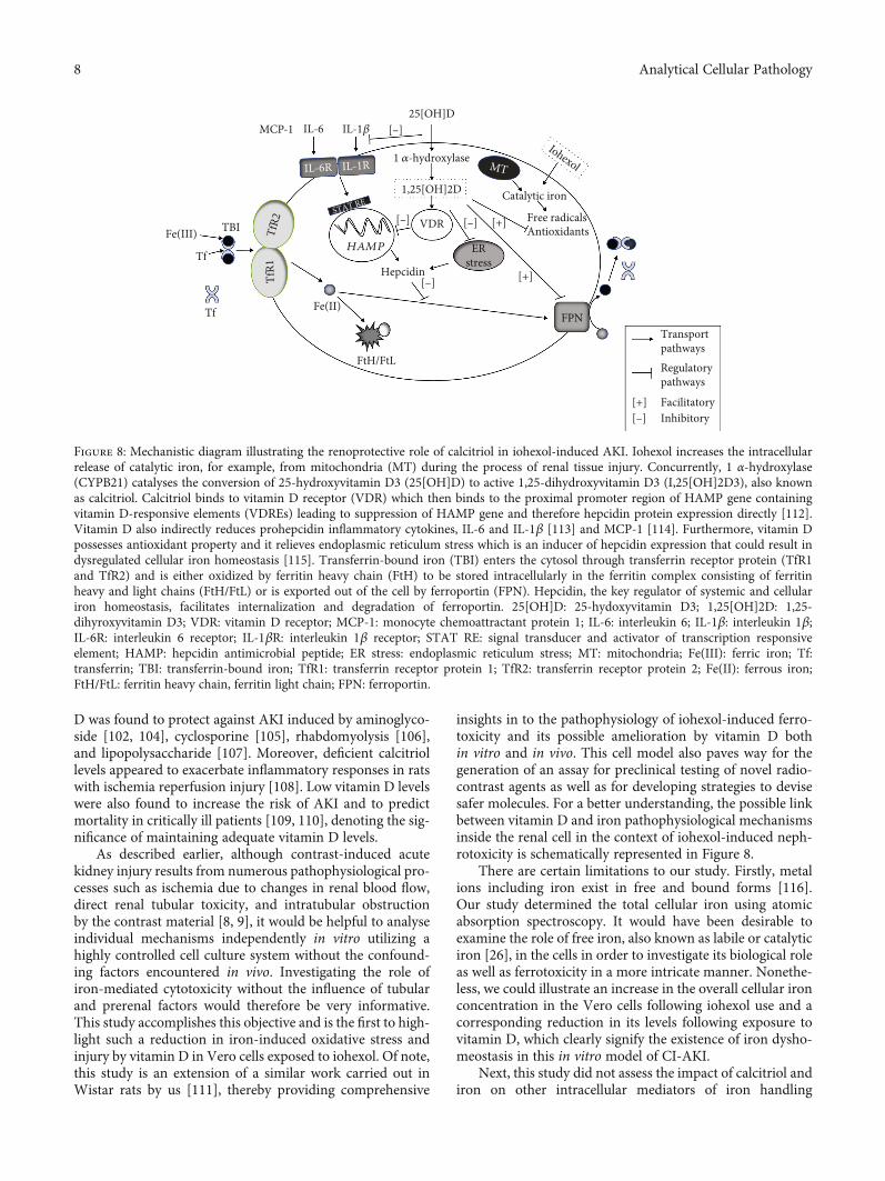
insights in to the pathophysiology of iohexol-induced ferro-toxicity and its possible amelioration by vitamin D bothin vitro and in vivo. This cell model also paves way for thegeneration of an assay for preclinical testing of novel radio-contrast agents as well as for developing strategies to devisesafer molecules. For a better understanding, the possible linkbetween vitamin D and iron pathophysiological mechanismsinside the renal cell in the context of iohexol-induced neph-rotoxicity is schematically represented in Figure 8.
There are certain limitations to our study. Firstly, metalions including iron exist in free and bound forms [116].Our study determined the total cellular iron using atomicabsorption spectroscopy. It would have been desirable toexamine the role of free iron, also known as labile or catalyticiron [26], in the cells in order to investigate its biological roleas well as ferrotoxicity in a more intricate manner. Nonethe-less, we could illustrate an increase in the overall cellular ironconcentration in the Vero cells following iohexol use and acorresponding reduction in its levels following exposure tovitamin D, which clearly signify the existence of iron dysho-meostasis in this in vitro model of CI-AKI.
Next, this study did not assess the impact of calcitriol andiron on other intracellular mediators of iron handling
VDR
HAMP
FtH/FtL
FPNTf
TfR1
TBIFe(III)
Tf
1,25[OH]2D
25[OH]D
1 𝛼-hydroxylase
IL-6 IL-1𝛽MCP-1
ERstress
Hepcidin
Catalytic iron
[+]
[–]
[–]
[–]
[–]
Free radicalsAntioxidants
[+]
MT
Fe(II)
Regulatorypathways
Transportpathways
[+] Facilitatory[–] Inhibitory
TfR2
IL-1RIL-6R
STAT RE
Iohexol
FPN
Figure 8: Mechanistic diagram illustrating the renoprotective role of calcitriol in iohexol-induced AKI. Iohexol increases the intracellularrelease of catalytic iron, for example, from mitochondria (MT) during the process of renal tissue injury. Concurrently, 1 α-hydroxylase(CYPB21) catalyses the conversion of 25-hydroxyvitamin D3 (25[OH]D) to active 1,25-dihydroxyvitamin D3 (I,25[OH]2D3), also knownas calcitriol. Calcitriol binds to vitamin D receptor (VDR) which then binds to the proximal promoter region of HAMP gene containingvitamin D-responsive elements (VDREs) leading to suppression of HAMP gene and therefore hepcidin protein expression directly [112].Vitamin D also indirectly reduces prohepcidin inflammatory cytokines, IL-6 and IL-1β [113] and MCP-1 [114]. Furthermore, vitamin Dpossesses antioxidant property and it relieves endoplasmic reticulum stress which is an inducer of hepcidin expression that could result indysregulated cellular iron homeostasis [115]. Transferrin-bound iron (TBI) enters the cytosol through transferrin receptor protein (TfR1and TfR2) and is either oxidized by ferritin heavy chain (FtH) to be stored intracellularly in the ferritin complex consisting of ferritinheavy and light chains (FtH/FtL) or is exported out of the cell by ferroportin (FPN). Hepcidin, the key regulator of systemic and cellulariron homeostasis, facilitates internalization and degradation of ferroportin. 25[OH]D: 25-hydoxyvitamin D3; 1,25[OH]2D: 1,25-dihyroxyvitamin D3; VDR: vitamin D receptor; MCP-1: monocyte chemoattractant protein 1; IL-6: interleukin 6; IL-1β: interleukin 1β;IL-6R: interleukin 6 receptor; IL-1βR: interleukin 1β receptor; STAT RE: signal transducer and activator of transcription responsiveelement; HAMP: hepcidin antimicrobial peptide; ER stress: endoplasmic reticulum stress; MT: mitochondria; Fe(III): ferric iron; Tf:transferrin; TBI: transferrin-bound iron; TfR1: transferrin receptor protein 1; TfR2: transferrin receptor protein 2; Fe(II): ferrous iron;FtH/FtL: ferritin heavy chain, ferritin light chain; FPN: ferroportin.
8 Analytical Cellular Pathology
including ferritin, hepcidin, and ferroportin. Lastly, iron che-lators have been proven efficacious in animals inflicted withAKI by decreasing the luminal or intracellular iron levels[90]. These were, however, not used in this study. Perhaps,their use to diminish iron concentration and the resultantoxidative stress would have seemed more informative wheninterpreted in correlation with the cytoprotective effect ofvitamin D. Importantly, it has to be understood that the mereassociation between iron and vitamin D noted in thisresearch work is not a compelling evidence per se to infer thatvitamin D annihilates ferrotoxicity. Notwithstanding thisshortcoming, the fact that calcitriol has several pleiotropiceffects and supported by the ample evidence demonstratingthe cytoprotection it offers in several models of AKI and alsoin an in vivo experiment performed by us using Wistar rats[111], it seems rational to corroborate the role of vitamin Din overcoming iron-induced renal cytotoxicity. Yet, it isimperative to have more conclusive proof in this respect bymeans of effective translational research.
There are few key recommendations for future investiga-tions depending on the present study. It will be important tofurther characterize and better understand the regulatorylinks between calcitriol and iron, especially at the cellularlevel involving the ferritin and the hepcidin-ferroportin axisand the associated downstream pathways by way of in-depth exploration using in vitro and in vivo models. Treat-ment of AKI is challenging owing to the complex nature ofits pathogenicity and currently is largely supportive withthere being no panacea for its optimal treatment. Conse-quently, multitarget therapeutic strategies are likely to bemore effective. In the light of this as well as based on ourstudy results, it would be worthwhile to evaluate the desirableeffects of calcitriol in relation to iron chelating agents to over-come iron-induced cellular injury, ideally in subjects withAKI due to varied aetiology and severity. Also, it is of para-mount importance to translate the findings of this cell culturestudy to patients by formulating treatment protocols follow-ing further validation through well-designed, randomizedcontrolled clinical trials.
5. Conclusions
Our study findings further substantiate the crucial roleplayed by iron in inducing renal cytotoxicity via oxidantstress in this model of iohexol-induced nephrotoxicity. Pre-treatment with calcitriol protects the cells by significantlydiminishing oxidative stress and ferrotoxicity and therebyenhancing cell viability. Overall, this work attempts to dem-onstrate an association between iron and vitamin D in thepathophysiology of AKI and provides some useful insightson iron being a vital target in the amelioration of nephrotox-icity, specifically by employing strategies to eliminate luminalor intracellular iron in addition to timely administration ofvitamin D.
Data Availability
Data shall be available from the corresponding author uponrequest.
Conflicts of Interest
The authors declare that they have no conflicts of interest.
Acknowledgments
The authors express their gratitude to Dr. Renu Bhatt and Dr.Rupal Purena, Guru Ghasidas Vishwavidyalaya, Bilaspur,Chhattisgarh, India, for their assistance in carrying out thecell culture work. Last but not least, they are indebted toVIT, Vellore, and Guru Ghasidas Vishwavidyalaya for theresources.
References
[1] P. K. T. Li, E. A. Burdmann, R. L. Mehta, and S. Martin,“Acute kidney injury: global health alert,” Journal of Nephro-pathology, vol. 2, no. 2, pp. 90–97, 2013.
[2] S. Lakhmir, Chawla, on behalf of the Acute Disease QualityInitiativeWorkgroup 16, R. Bellomo et al., “Acute kidney dis-ease and renal recovery: consensus report of the acute diseasequality initiative (ADQI) 16 workgroup,” Nature ReviewsNephrology, vol. 13, no. 4, pp. 241–257, 2017.
[3] G. R. Kinsey and M. D. Okusa, “Pathogenesis of acute kidneyinjury: foundation for clinical practice,” American Journal ofKidney Diseases: The Official Journal of the National KidneyFoundation, vol. 58, no. 2, pp. 291–301, 2011.
[4] J. V. Bonventre, “Pathophysiology of AKI: injury and normaland abnormal repair,” Contributions to Nephrology, vol. 165,pp. 9–17, 2010.
[5] S. M. Bagshaw and R. Wald, “Acute kidney injury: timing ofrenal replacement therapy in AKI,” Nature Reviews Nephrol-ogy, vol. 12, no. 8, pp. 445-446, 2016.
[6] R. Morcos, M. Kucharik, P. Bansal et al., “Contrast-inducedacute kidney injury: review and practical update,” ClinicalMedicine Insights: Cardiology, vol. 13, 2019.
[7] R. Rear, R. M. Bell, and D. J. Hausenloy, “Contrast-inducednephropathy following angiography and cardiac interven-tions,” Heart (British Cardiac Society), vol. 102, no. 8,pp. 638–648, 2016.
[8] E. Seeliger, M. Sendeski, C. S. Rihal, and P. B. Persson, “Con-trast-induced kidney injury: mechanisms, risk factors, andprevention,” European Heart Journal, vol. 33, no. 16,pp. 2007–2015, 2012.
[9] P. A. McCullough, F. Stacul, and C. Davidson, “Contrast-induced nephropathy: clinical insights and practical guidance- a report from the CIN consensus working panel-overview,”American Journal of Cardiology, vol. 98, pp. 2K–4K, 2006.
[10] S. N. Heyman, S. Rosen, and C. Rosenberger, “Renal paren-chymal hypoxia, hypoxia adaptation, and the pathogenesisof radiocontrast nephropathy,” Clinical Journal of the Amer-ican Society of Nephrology, vol. 3, no. 1, pp. 288–296, 2008.
[11] S. N. Heyman, S. Rosen, M. Khamaisi, J. M. Idée, andC. Rosenberger, “Reactive oxygen species and the pathogene-sis of radiocontrast-induced nephropathy,” InvestigativeRadiology, vol. 45, no. 4, pp. 188–195, 2010.
[12] J. Tumlin, F. Stacul, A. Adam et al., “Pathophysiology ofcontrast-induced nephropathy,” The American Journal ofCardiology, vol. 98, no. 6, pp. 14–20, 2006.
9Analytical Cellular Pathology

[13] M. M. Sendeski, “Pathophysiology of renal tissue damage byiodinated contrast media,” Clinical and Experimental Phar-macology & Physiology, vol. 38, no. 5, pp. 292–299, 2011.
[14] M. Sendeski, A. Patzak, and P. B. Persson, “Constriction ofthe vasa recta, the vessels supplying the area at risk for acutekidney injury, by four different iodinated contrast media,evaluating ionic, nonionic, monomeric and dimeric agents,”Investigative Radiology, vol. 45, no. 8, pp. 453–457, 2010.
[15] M. M. Sendeski, A. B. Persson, Z. Z. Liu et al., “Iodinated con-trast media cause endothelial damage leading to vasocon-striction of human and rat vasa recta,” American Journal ofPhysiology-Renal Physiology, vol. 303, no. 12, pp. F1592–F1598, 2012.
[16] V. J. Walker and A. Agarwal, “Targeting iron homeostasis inacute kidney injury,” Seminars in Nephrology, vol. 36, no. 1,pp. 62–70, 2016.
[17] D. E. Leaf, M. Rajapurkar, S. S. Lele et al., “Increased plasmacatalytic iron in patients maymediate acute kidney injury anddeath following cardiac surgery,” Kidney International,vol. 87, no. 5, pp. 1046–1054, 2015.
[18] S. S. Lele, B. N. Mukhopadhyay, M. M. Mardikar et al.,“Impact of catalytic iron on mortality in patients with acutecoronary syndrome exposed to iodinated radiocontrast–TheIscom Study,” American Heart Journal, vol. 165, no. 5,pp. 744–751, 2013.
[19] S. V. Shah, M. M. Rajapurkar, and R. Baliga, “The role of cat-alytic iron in acute kidney injury,” Clinical journal of theAmerican Society of Nephrology : CJASN, vol. 6, no. 10,pp. 2329–2331, 2011.
[20] R. Baliga, N. Ueda, and S. V. Shah, “Increase in bleomycin-detectable iron in ischaemia/reperfusion injury to rat kid-neys,” The Biochemical Journal, vol. 291, no. 3, pp. 901–905,1993.
[21] N. Zhao and C. A. Enns, “Iron transport machinery of humancells,” Current Topics in Membranes, vol. 69, pp. 67–93, 2012.
[22] S. Swaminathan, “Iron homeostasis pathways as therapeutictargets in acute kidney injury,” Nephron, vol. 140, no. 2,pp. 156–159, 2018.
[23] R. P. L. van Swelm and W. JFM, “The multifaceted role ofiron in renal health and disease,” Nature Reviews Nephrology,vol. 16, no. 2, pp. 77–98, 2020.
[24] A. M. F. Martines, R. Masereeuw, H. Tjalsma, J. G. Hoen-derop, J. F. M. Wetzels, and D. W. Swinkels, “Iron metabo-lism in the pathogenesis of iron-induced kidney injury,”Nature Reviews Nephrology, vol. 9, no. 7, pp. 385–398, 2013.
[25] P. Ponka, “Cellular iron metabolism,” Kidney internationalSupplement, vol. 69, pp. S2–11, 1999.
[26] B. Halliwell and J. M. Gutteridge, “[1] Role of free radicalsand catalytic metal ions in human disease: An overview,”Methods in Enzymology, vol. 186, pp. 1–85, 1990.
[27] D. P. Basile, M. D. Anderson, and T. A. Sutton, “Pathophys-iology of acute kidney injury,” Comprehensive Physiology,vol. 2, no. 2, pp. 1303–1353, 2012.
[28] P. Perco and G. Mayer, “Endogenous factors and mecha-nisms of renoprotection and renal repair,” European Journalof Clinical Investigation, vol. 48, no. 5, article e12914, 2018.
[29] P. Lips, “Vitamin D physiology,” Progress in Biophysics andMolecular Biology, vol. 92, no. 1, pp. 4–8, 2006.
[30] W. Zhong, B. Gu, Y. Gu, L. J. Groome, J. Sun, and Y. Wang,“Activation of vitamin D receptor promotes VEGF andCuZn-SOD expression in endothelial cells,” The Journal of
Steroid Biochemistry and Molecular Biology, vol. 140,pp. 56–62, 2014.
[31] S. Sardar, A. Chakraborty, and M. Chatterjee, “Comparativeeffectiveness of vitamin D3 and dietary vitamin E on peroxi-dation of lipids and enzymes of the hepatic antioxidant sys-tem in Sprague–Dawley rats,” International journal forvitamin and nutrition research Internationale Zeitschrift furVitamin- und Ernahrungsforschung Journal international devitaminologie et de nutrition, vol. 66, pp. 39–45, 1996.
[32] I. Medicine, F. N. Board, and Calcium CRDRIVD, Dietaryreference intakes for calcium and vitamin D, National Acade-mies Press, 2011.
[33] H. M. Trang, D. E. Cole, L. A. Rubin, A. Pierratos, S. Siu, andR. Vieth, “Evidence that vitamin D3 increases serum 25-hydroxyvitamin D more efficiently than does vitamin D2,”The American Journal of Clinical Nutrition, vol. 68, no. 4,pp. 854–858, 1998.
[34] L. A. Armas, B. W. Hollis, and R. P. Heaney, “Vitamin D2 ismuch less effective than vitamin D3 in humans,” The Journalof Clinical Endocrinology and Metabolism, vol. 89, no. 11,pp. 5387–5391, 2004.
[35] M. F. Holick, J. E. Frommer, S. C. McNeill, N. M. Richtand,J. W. Henley, and Potts JT Jr, “Photometabolism of 7-dehydrocholesterol to previtamin D3 in skin,” Biochemicaland Biophysical Research Communications, vol. 76, no. 1,pp. 107–114, 1977.
[36] T. OKANO, M. YASUMURA, K. MIZUNO, andT. KOBAYASHI, “Photochemical conversion of 7-dehydrocholesterol into vitamin D3 in rat skins,” Journal ofNutritional Science and Vitaminology, vol. 23, no. 2,pp. 165–168, 1977.
[37] J. B. Cheng, M. A. Levine, N. H. Bell, D. J. Mangelsdorf, andD. W. Russell, “Genetic evidence that the human CYP2R1enzyme is a key vitamin D 25-hydroxylase,” Proceedings ofthe National Academy of Sciences of the United States ofAmerica, vol. 101, no. 20, pp. 7711–7715, 2004.
[38] A. Nykjaer, D. Dragun, D. Walther et al., “An endocytic path-way essential for renal uptake and activation of the steroid25-(OH) vitamin D3,” Cell, vol. 96, no. 4, pp. 507–515, 1999.
[39] F. Takemoto, T. Shinki, K. Yokoyama et al., “Gene expressionof vitamin D hydroxylase and megalin in the remnant kidneyof nephrectomized rats,” Kidney International, vol. 64, no. 2,pp. 414–420, 2003.
[40] D. R. Fraser and E. Kodicek, “Unique biosynthesis by kidneyof a biological active vitamin D metabolite,” Nature, vol. 228,no. 5273, pp. 764–766, 1970.
[41] K. Takeyama and S. Kato, “The vitamin D3 1alpha-hydroxylase gene and its regulation by active vitamin D3,”Bioscience, Biotechnology, and Biochemistry, vol. 75,pp. 208–213, 2014.
[42] D. Zehnder and M. Hewison, “The renal function of 25-hydroxyvitamin D3-1alpha-hydroxylase,” Molecular andCellular Endocrinology, vol. 151, no. 1-2, pp. 213–220, 1999.
[43] R. Bouillon, W. H. Okamura, and A. W. Norman, “Structure-function relationships in the vitamin D endocrine system,”Endocrine Reviews, vol. 16, no. 2, pp. 200–257, 1995.
[44] R. Bland, D. Zehnder, and M. Hewison, “Expression of 25-hydroxyvitamin D3-1alpha-hydroxylase along the nephron:new insights into renal vitamin D metabolism,” CurrentOpinion in Nephrology and Hypertension, vol. 9, no. 1,pp. 17–22, 2000.
10 Analytical Cellular Pathology

[45] M. R. Haussler, G. K. Whitfield, C. A. Haussler et al., “Thenuclear vitamin D receptor: biological and molecular regu-latory properties revealed,” Journal of Bone and MineralResearch: the Official Journal of the American Society forBone and Mineral Research, vol. 13, no. 3, pp. 325–349,1998.
[46] M. R. Walters, “Newly identified actions of the vitamin Dendocrine system,” Endocrine Reviews, vol. 13, no. 4,pp. 719–764, 1992.
[47] R. Bouillon, H. Bischoff-Ferrari, and W. Willett, “Vitamin Dand health: perspectives frommice andman,” Journal of Boneand Mineral Research: the Official Journal of the AmericanSociety for Bone and Mineral Research, vol. 23, no. 7,pp. 974–979, 2008.
[48] S. Nagpal, S. Na, and R. Rathnachalam, “Noncalcemic actionsof vitamin D receptor ligands,” Endocrine Reviews, vol. 26,no. 5, pp. 662–687, 2005.
[49] D. Feldman, A. V. Krishnan, S. Swami, E. Giovannucci, andB. J. Feldman, “The role of vitamin D in reducing cancer riskand progression,” Nature Reviews Cancer, vol. 14, no. 5,pp. 342–357, 2014.
[50] W.-C. Liu, C.-C.Wu, Y.-M. Hung et al., “Pleiotropic effects ofvitamin D in chronic kidney disease,” Clinica chimica acta,vol. 453, pp. 1–12, 2016.
[51] S. J. Hill and A. S. Fisher, “Atomic absorption, methods andinstrumentation,” in Encyclopedia of Spectroscopy and Spec-trometry, J. C. Lindon, G. E. Tranter, and D. W. Koppenaal,Eds., pp. 37–43, Oxford, Academic Press, Third Edition edi-tion, 2017.
[52] D. T. Sawyer, W. R. Heineman, and J. M. Beebe, ChemistryExperiments for Instrumental Methods, Wiley, 1984.
[53] D. A. Skoog, D. M. West, and F. J. Holler, Fundamentals ofAnalytical Chemistry, Cengage Learning, 2013.
[54] M. Torre, M. C. González, O. Jiménez, and A. R. Rodríguez,“Study of analytical methods for iron determination in com-plex organic liquids by atomic absorption spectrometry,”Analytical Letters, vol. 23, no. 8, pp. 1519–1536, 1990.
[55] H. Ohkawa, N. Ohishi, and K. Yagi, “Assay for lipid peroxidesin animal tissues by thiobarbituric acid reaction,” AnalyticalBiochemistry, vol. 95, no. 2, pp. 351–358, 1979.
[56] G. L. Ellman, “Tissue sulfhydryl groups,” Archives of Bio-chemistry and Biophysics, vol. 82, no. 1, pp. 70–77, 1959.
[57] A. K. Sinha, “Colorimetric assay of catalase,” Analytical Bio-chemistry, vol. 47, no. 2, pp. 389–394, 1972.
[58] C. C. Winterbourn, R. E. Hawkins, M. Brian, and R. W. Car-rell, “The estimation of red cell superoxide dismutase activ-ity,” The Journal of Laboratory and Clinical Medicine,vol. 85, no. 2, pp. 337–341, 1975.
[59] J. Kapuscinski, “DAPI: a DNA-specific fluorescent probe,”Biotechnic & histochemistry: official publication of the Biolog-ical Stain Commission, vol. 70, pp. 220–233, 2009.
[60] K. D. Kim, O. Iwasaki, and K. Noma, “Chapter eight-an IF–FISH approach for covisualization of gene loci and nucleararchitecture in fission yeast,” in Methods in Enzymology, R.Marmorstein, Ed., vol. 574, pp. 167–180, Academic Press,2016.
[61] F. Denizot and R. Lang, “Rapid colorimetric assay for cellgrowth and survival: modifications to the tetrazolium dyeprocedure giving improved sensitivity and reliability,” Jour-nal of Immunological Methods, vol. 89, no. 2, pp. 271–277,1986.
[62] T. Mosmann, “Rapid colorimetric assay for cellular growthand survival: application to proliferation and cytotoxicityassays,” Journal of Immunological Methods, vol. 65, no. 1-2,pp. 55–63, 1983.
[63] T. F. Slater, B. Sawyer, and U. Straeuli, “STUDIES ONSUCCINATE-TETRAZOLIUM REDUCTASE SYSTEMSIIIStudies on succinate-tetrazolium reductase systems,” Bio-chimica et Biophysica Acta, vol. 77, pp. 383–393, 1963.
[64] P. Price and T. J. McMillan, “Use of the tetrazolium assay inmeasuring the response of human tumor cells to ionizingradiation,” Cancer Research, vol. 50, no. 5, pp. 1392–1396,1990.
[65] R. Seth, C. Yang, V. Kaushal, S. V. Shah, and G. P. Kaushal,“p53-dependent caspase-2 activation in mitochondrialrelease of apoptosis-inducing factor and its role in renal tubu-lar epithelial cell injury,” The Journal of Biological Chemistry,vol. 280, no. 35, pp. 31230–31239, 2005.
[66] S. Hamazaki, S. Okada, Y. Ebina, M. Fujioka, andO. Midorikawa, “Nephrotoxicity of ferric nitrilotriacetate.An electron-microscopic and metabolic study,” The Ameri-can Journal of Pathology, vol. 123, no. 2, pp. 343–350, 1986.
[67] K. A. Nath, “Heme oxygenase-1: a provenance for cytopro-tective pathways in the kidney and other tissues,” KidneyInternational, vol. 70, no. 3, pp. 432–443, 2006.
[68] G. Kovtunovych, M. A. Eckhaus, M. C. Ghosh, H. Ollivierre-Wilson, and T. A. Rouault, “Dysfunction of the heme recy-cling system in heme oxygenase 1-deficient mice: effects onmacrophage viability and tissue iron distribution,” Blood,vol. 116, no. 26, pp. 6054–6062, 2010.
[69] R. A. Zager and C. A. Foerder, “Effects of inorganic iron andmyoglobin on in vitro proximal tubular lipid peroxidationand cytotoxicity,” The Journal of Clinical Investigation,vol. 89, no. 3, pp. 989–995, 1992.
[70] L. Chen, B. H. Zhang, and D. C. Harris, “Evidence suggestingthat nitric oxide mediates iron-induced toxicity in culturedproximal tubule cells,” The American Journal of Physiology,vol. 274, no. 1, pp. F18–F25, 1998.
[71] H. T. Sponsel, A. C. Alfrey, W. S. Hammond, J. A. Durr,C. Ray, and R. J. Anderson, “Effect of iron on renal tubularepithelial cells,” Kidney International, vol. 50, no. 2,pp. 436–444, 1996.
[72] N. S. Sheerin, S. H. Sacks, and G. B. Fogazzi, “In vitro erythro-phagocytosis by renal tubular cells and tubular toxicity byhaemoglobin and iron,” Nephrology, dialysis, transplantation:official publication of the European Dialysis and TransplantAssociation-European Renal Association, vol. 14, no. 6,pp. 1391–1397, 1999.
[73] R. A. Zager and K. Burkhart, “Myoglobin toxicity in proximalhuman kidney cells: roles of Fe, Ca2+, H2O2, and terminalmitochondrial electron transport,” Kidney International,vol. 51, no. 3, pp. 728–738, 1997.
[74] M. S. Paller and B. E. Hedlund, “Extracellular iron chelatorsprotect kidney cells from hypoxia/reoxygenation,” Free Rad-ical Biology & Medicine, vol. 17, no. 6, pp. 597–603, 1994.
[75] C. García-Alfonso, J. López-Barea, P. Sanz, G. Repetto, andM. Repetto, “Changes in antioxidative activities induced byFe (II) and Fe (III) in cultured Vero cells,” Archives of Envi-ronmental Contamination and Toxicology, vol. 30, no. 4,pp. 431–436, 1996.
[76] A. Linkermann, R. Skouta, N. Himmerkus et al., “Synchro-nized renal tubular cell death involves ferroptosis,”
11Analytical Cellular Pathology
Proceedings of the National Academy of Sciences of the UnitedStates of America, vol. 111, no. 47, pp. 16836–16841, 2014.
[77] R. Baliga, Z. Zhang, M. Baliga, and S. V. Shah, “Evidence forcytochrome P-450 as a source of catalytic iron in myoglobi-nuric acute renal failure,” Kidney International, vol. 49,no. 2, pp. 362–369, 1996.
[78] R. Baliga, Z. Zhang, M. Baliga, N. Ueda, and S. V. Shah, “Invitro and in vivo evidence suggesting a role for iron incisplatin-induced nephrotoxicity,” Kidney International,vol. 53, no. 2, pp. 394–401, 1998.
[79] M. S. Paller, “Hemoglobin- and myoglobin-induced acuterenal failure in rats: role of iron in nephrotoxicity,” TheAmerican Journal of Physiology, vol. 255, no. 3, pp. F539–F544, 1988.
[80] R. E. Kirschner and G. A. Fantini, “Role of iron and oxygen-derived free radicals in ischemia-reperfusion injury,” Journalof the American College of Surgeons, vol. 179, no. 1, pp. 103–117, 1994.
[81] S. V. Shah and P. D. Walker, “Evidence suggesting a role forhydroxyl radical in glycerol-induced acute renal failure,” TheAmerican Journal of Physiology, vol. 255, pp. F438–F443,1988.
[82] T. Veuthey, M. C. D'Anna, andM. E. Roque, “Role of the kid-ney in iron homeostasis: renal expression of prohepcidin, fer-roportin, and DMT1 in anemic mice,” American Journal ofPhysiology. Renal Physiology, vol. 295, no. 4, pp. F1213–F1221, 2008.
[83] N. Ueda, R. Baliga, and S. V. Shah, “Role of ‘catalytic’ iron inan animal model of minimal change nephrotic syndrome,”Kidney International, vol. 49, no. 2, pp. 370–373, 1996.
[84] R. A. Zager, C. Foerder, and C. Bredl, “The influence of man-nitol on myoglobinuric acute renal failure: functional, bio-chemical, and morphological assessments,” J Am SocNephrol, vol. 2, no. 4, pp. 848–855, 1991.
[85] D. C. Harris, C. Tay, and B. J. Nankivell, “Lysosomal ironaccumulation and tubular damage in rat puromycin nephro-sis and ageing,” Clinical and Experimental Pharmacology &Physiology, vol. 21, no. 2, pp. 73–81, 1994.
[86] J. A. Moreno, C. Martín-Cleary, E. Gutiérrez et al., “AKI asso-ciated with macroscopic glomerular hematuria: clinical andpathophysiologic consequences,” Clinical journal of theAmerican Society of Nephrology: CJASN, vol. 7, no. 1,pp. 175–184, 2012.
[87] D. A. V. I. D. A. SEARS, P. E. A. R. L. R. ANDERSON, A. R.T. H. U. R. L. FOY, H. A. R. O. L. D. L. WILLIAMS, andW. I.L. L. I. A. M. H. CROSBY, “Urinary iron excretion and renalmetabolism of hemoglobin in hemolytic diseases,” Blood,vol. 28, no. 5, pp. 708–725, 1966.
[88] J. Ballarin, Y. Arce, R. T. Balcells et al., “Acute renal failureassociated to paroxysmal nocturnal haemoglobinuria leadsto intratubular haemosiderin accumulation and CD163expression,” Nephrology Dialysis Transplantation, vol. 26,no. 10, pp. 3408–3411, 2011.
[89] H. Wang, K. Nishiya, H. Ito, T. Hosokawa, K. Hashimoto,and T. Moriki, “Iron deposition in renal biopsy specimensfrom patients with kidney diseases,” American Journal of Kid-ney Diseases: The Official Journal of the National KidneyFoundation, vol. 38, no. 5, pp. 1038–1044, 2001.
[90] M. S. Paller and B. E. Hedlund, “Role of iron in postischemicrenal injury in the rat,” Kidney International, vol. 34, no. 4,pp. 474–480, 1988.
[91] K. Mori, H. T. Lee, D. Rapoport et al., “Endocytic delivery oflipocalin-siderophore-iron complex rescues the kidney fromischemia-reperfusion injury,” The Journal of Clinical Investi-gation, vol. 115, no. 3, pp. 610–621, 2005.
[92] B. de Vries, S. J. Walter, L. von Bonsdorff et al., “Reduction ofcirculating redox-active iron by apotransferrin protectsagainst renal ischemia-reperfusion injury,” Transplantation,vol. 77, no. 5, pp. 669–675, 2004.
[93] J. Mishra, K. Mori, Q. Ma et al., “Amelioration of ischemicacute renal injury by neutrophil gelatinase-associated lipoca-lin,” J Am Soc Nephrol, vol. 15, no. 12, pp. 3073–3082, 2004.
[94] M. Cazzola, H. A. Huebers, M. H. Sayers, A. P. MacPhail,M. Eng, and C. A. Finch, “Transferrin saturation, plasma ironturnover, and transferrin uptake in normal humans,” Blood,vol. 66, no. 4, pp. 935–939, 1985.
[95] E. C. Theil, “Ferritin: the protein nanocage and iron biomin-eral in health and in disease,” Inorganic Chemistry, vol. 52,no. 21, pp. 12223–12233, 2013.
[96] H. Drakesmith, E. Nemeth, and T. Ganz, “Ironing out ferro-portin,” Cell Metabolism, vol. 22, no. 5, pp. 777–787, 2015.
[97] E. Nemeth and M. S. Tuttle, “Hepcidin regulates cellular ironefflux by binding to ferroportin and inducing its internaliza-tion,” Hepcidin regulates cellular iron efflux by binding to fer-roportin and inducing its internalization. science, vol. 306,no. 5704, pp. 2090–2093, 2004.
[98] B. Qiao, P. Sugianto, E. Fung et al., “Hepcidin-induced endo-cytosis of ferroportin is dependent on ferroportin ubiquitina-tion,” Cell Metabolism, vol. 15, no. 6, pp. 918–924, 2012.
[99] M. W. Hentze, M. U. Muckenthaler, B. Galy, andC. Camaschella, “Two to tango: regulation of mammalianiron metabolism,” Cell, vol. 142, no. 1, pp. 24–38, 2010.
[100] E. Ari, A. E. Kedrah, Y. Alahdab et al., “Antioxidant andrenoprotective effects of paricalcitol on experimentalcontrast-induced nephropathy model,” The British Journalof Radiology, vol. 85, no. 1016, pp. 1038–1043, 2012.
[101] K. Husain, E. Suarez, A. Isidro, and L. Ferder, “Effects of par-icalcitol and enalapril on atherosclerotic injury in mouse aor-tas,” American Journal of Nephrology, vol. 32, no. 4, pp. 296–304, 2010.
[102] E. Hur, A. Garip, A. Camyar et al., “The effects of vitamin don gentamicin-induced acute kidney injury in experimentalrat model,” International Journal of Endocrinology,vol. 2013, Article ID 313528, 7 pages, 2013.
[103] M. Weih, S. Orth, T. Weinreich, H. Reichel, and E. Ritz,“Inhibition of growth by calcitriol in a proximal tubular cellline (OK),” Nephrology Dialysis Transplantation, vol. 9,pp. 1390–1394, 1994.
[104] G. Bulut, Y. Basbugan, E. Ari et al., “Paricalcitol may improveoxidative DNA damage on experimental amikacin-inducednephrotoxicity model,” Renal Failure, vol. 38, no. 5,pp. 751–758, 2016.
[105] J. W. Park, E. H. Bae, I. J. Kim et al., “Paricalcitol attenuatescyclosporine-induced kidney injury in rats,” Kidney Interna-tional, vol. 77, no. 12, pp. 1076–1085, 2010.
[106] N. G. Reis, H. D. C. Francescato, L. F. de Almeida, C. G. A. daSilva, R. S. Costa, and T. M. Coimbra, “Protective effect of cal-citriol on rhabdomyolysis-induced acute kidney injury inrats,” Scientific Reports, vol. 9, no. 1, p. 7090, 2019.
[107] J. Du, S. Jiang, Z. Hu et al., “Vitamin D receptor activationprotects against lipopolysaccharide-induced acute kidneyinjury through suppression of tubular cell apoptosis,”
12 Analytical Cellular Pathology

American Journal of Physiology-Renal Physiology, vol. 316,no. 5, pp. F1068–f1077, 2019.
[108] A. C. de Bragança, R. A. Volpini, D. Canale et al., “Vitamin Ddeficiency aggravates ischemic acute kidney injury in rats,”Physiological Reports, vol. 3, no. 3, p. e12331, 2015.
[109] P. Lee, J. A. Eisman, and J. R. Center, “Vitamin D deficiencyin critically ill patients,” The New England Journal of Medi-cine, vol. 360, no. 18, pp. 1912–1914, 2009.
[110] A. B. Braun, A. A. Litonjua, T. Moromizato, F. K. Gibbons,E. Giovannucci, and K. B. Christopher, “Association of lowserum 25-hydroxyvitamin D levels and acute kidney injuryin the critically ill,” Critical Care Medicine, vol. 40, no. 12,pp. 3170–3179, 2012.
[111] C. Annamalai, R. N. Ganesh, and P. Viswanathan, “Ferro-toxicity and its amelioration by endogenous vitamin D inexperimental acute kidney injury,” Experimental Biologyand Medicine, vol. 245, no. 16, pp. 1474–1489, 2020.
[112] J. Bacchetta, J. J. Zaritsky, J. L. Sea et al., “Suppression of iron-regulatory hepcidin by vitamin D,” J Am Soc Nephrol, vol. 25,no. 3, pp. 564–572, 2014.
[113] S. M. Zughaier, J. A. Alvarez, J. H. Sloan, R. J. Konrad, andV. Tangpricha, “The role of vitamin D in regulating theiron-hepcidin-ferroportin axis in monocytes,” Journal ofClinical & Translational Endocrinology, vol. 1, no. 1,pp. e19–e25, 2014.
[114] J. A. Alvarez, S. M. Zughaier, J. Law et al., “Effects of high-dose cholecalciferol on serum markers of inflammation andimmunity in patients with early chronic kidney disease,”European Journal of Clinical Nutrition, vol. 67, no. 3,pp. 264–269, 2013.
[115] A. E. Riek, J. Oh, J. E. Sprague et al., “Vitamin D suppressionof endoplasmic reticulum stress promotes an antiatherogenicmonocyte/macrophage phenotype in type 2 diabeticpatients,” The Journal of Biological Chemistry, vol. 287,no. 46, pp. 38482–38494, 2012.
[116] K. M. Dean, Y. Qin, and A. E. Palmer, “Visualizing metal ionsin cells: an overview of analytical techniques, approaches, andprobes,” Biochimica et Biophysica Acta (BBA) - MolecularCell Research, vol. 1823, no. 9, pp. 1406–1415, 2012.
13Analytical Cellular Pathology