fcrγ activation regulates inflammation-associated squamous carcinogenesis
TRANSCRIPT

Cancer Cell
Article
FcRg Activation Regulates Inflammation-AssociatedSquamous CarcinogenesisPauline Andreu,1,5 Magnus Johansson,1,5 Nesrine I. Affara,1,5 Ferdinando Pucci,3 Tingting Tan,1 Simon Junankar,1
Lidiya Korets,1 Julia Lam,1 David Tawfik,1 David G. DeNardo,1 Luigi Naldini,3 Karin E. de Visser,4 Michele De Palma,3
and Lisa M. Coussens1,2,*1Department of Pathology2Helen Diller Family Comprehensive Cancer CenterUniversity of California, San Francisco, San Francisco, CA 94143, USA3Angiogenesis and Tumor Targeting Research Unit, San Raffaele-Telethon Institute for Gene Therapy and Vita-Salute SanRaffaele University,San Raffaele Institute, Milan, 20132, Italy4Division of Molecular Biology, The Netherlands Cancer Institute, Amsterdam, 1066 CX, the Netherlands5These authors contributed equally to this work*Correspondence: [email protected] 10.1016/j.ccr.2009.12.019
SUMMARY
Chronically activated leukocytes recruited to premalignant tissues functionally contribute to cancer develop-ment; however, mechanisms underlying pro- versus anti-tumor programming of neoplastic tissues byimmune cells remain obscure. Using the K14-HPV16 mouse model of squamous carcinogenesis, we reportthat B cells and humoral immunity foster cancer development by activating Fcg receptors (FcgRs) on residentand recruitedmyeloid cells. Stromal accumulation of autoantibodies in premalignant skin, through their inter-action with activating FcgRs, regulate recruitment, composition, and bioeffector functions of leukocytes inneoplastic tissue, which in turn promote neoplastic progression and subsequent carcinoma development.These findings support a model in which B cells, humoral immunity, and activating FcgRs are required forestablishing chronic inflammatory programs that promote de novo carcinogenesis.
INTRODUCTION
Clinical, epidemiological, and experimental studies have estab-lished that chronic inflammation contributes to various aspectsof solid tumor development (de Visser et al., 2006; Mantovaniet al., 2008). In particular, chronic inflammatory diseases,including several autoimmune disorders, are associated withincreased risk of cancer development (Brandtzaeg et al., 2006;Dalgleish and O’Byrne, 2002), revealing that B cell hyperactivitycombined with altered cellular immunity cooperate to initiateand/or sustain persistent inflammation that enhances overallcancer risk in afflicted tissues.Deposition of B lymphocyte-derived immunoglobulins (Igs) is
a common occurrence in premalignant and malignant stromaof human cancers (de Visser et al., 2006; Tan and Coussens,2007). In addition, high levels of circulating immune complexes(CIC) are associated with increased tumor burden and poor
prognosis in patients with breast, genitourinary, and head andneck malignancies (Tan and Coussens, 2007). While little isknown about the function of CICs in tumor development, therole of CICs in inflammatory and autoimmune diseases is undis-puted. CIC deposition in stroma has been implicated as aninitiator of inflammatory cascades by mechanisms that includeactivation of complement pathways and engagement of thereceptors for the crystallizable region (Fc) of IgG (FcgRs) onthe surface of leukocytes (Takai, 2005). As such, FcgRs repre-sent a functional link between adaptive and innate immunity bycoupling interactions between circulating (auto)antibodies andinnate immune cells (Nimmerjahn and Ravetch, 2008).Four classes of IgG receptor FcgRs have been identified,
FcgRI/CD64, FcgRII/CD32, FcgRIII/CD16, and FcgRIV, differingby their distinct affinity for IgG isotypes, cellular distributions,and effector functions (Nimmerjahn and Ravetch, 2008). Acti-vating types of FcgRs form multimeric complexes including the
Significance
Andreu and colleagues demonstrate that peripheral activation of humoral immunity and subsequent activation of FcgRs onmyeloid cells regulate critical features of carcinogenesis that foster solid tumor development. This work reveals previouslyunrecognized targets for therapeutic intervention in solid tumors, namely, B cells and FcRg-signaling pathways that workin concert to differentially regulate not only the composition of leukocytes recruited to premalignant tissues but also theirbioeffector functions once present.
Cancer Cell 17, 121–134, February 17, 2010 ª2010 Elsevier Inc. 121

0
10
20
30
40
50
60
(-)LM
1m 4m 6m
*
*
A%
CD
45
+ c
ell
sC
% B
rd
U+ k
era
tin
oc
yte
sC
D3
1+ s
tru
ctu
re
s /
FO
V
F
0
10
20
30
40
50
60
# G
r1
+ C
ell
s /
FO
V
B
0
10
20
30
40
50
60
# M
as
t c
ell
s /
FO
V
G
H
MM
P9
(n
g/µ
g)
0
50
100
150
200
VE
GF
(p
g/m
g)
4m
HPV16
**
*
*
e
d
e
d
HPV16/JH+/-
HPV16/JH-/-
e
d
e
d
HPV16/JH+/-
HPV16/JH-/-
HPV16/JH+/-
HPV16/JH-/-
e
e
d
d
e
HPV16/JH+/-
HPV16/JH-/-
e
d
HPV16/JH-/-
(FVB/n, N5)
27.1 ± 2.7%% Dys (4 mo)
% Dys (6 mo)
6.2 ± 1.8%
29.7 ± 3.5% 8.4 ± 2.8%
HPV16/JH+/-
(FVB/n, N5)
Phenotype*
(age)
% Hyp (1 mo) 100% 100%
I
p value
d0.008
0.005
(-)LM
1m 4m 6m4m
HPV16 (-)LM
1m 4m 6m4m
HPV16
(-)LM
1m 4m 6m4m
HPV16
(-)LM
1m 4m 6m4m
HPV16 * % ear epidermis with hyperplastic or dysplastic
histopathology at age indicated
6m
HPV16(-)LM
4m
30
20
25
15
10
5
0
25
20
15
10
5
0
*
*
*
*
*0.3
0.2
0.1
0
D
% o
f C
D4
5+ c
ell
s Other CD45+
CD11b- Gr1
+
CD11b+ Gr1
-
CD11b+ Gr1
+
CD3+ CD8
+
CD3+ CD4
+
CD19+ B220
+
0
20
40
60
80
100
HPV16HPV16
1m 4m 6m
JH+/-
JH-/-
4m 1m 4m 6m
*
*
*
HPV16
4m6m4m1m
(-)LM
(-)LM (-)LM
*
*
*
4m
RAG1-/-
*
*
4m
E
% C
D11
b+ G
r1
+ c
ell
s
% C
D11
b+ G
r1
- c
ell
s
CD11c+ F4/80
-
CD11c- F4/80
+
CD11c- F4/80
-
(-)LM HPV16
JH-/-
0
20
40
60
80
100
(-)LM HPV16
JH-/-
0
20
40
60
80
100
JH+/-
JH-/-
JH+/-
JH-/-
JH+/-
JH-/-
JH+/-
JH-/-
JH+/-
JH-/-
JH+/-
JH-/-
Figure 1. B Cells Are Critical Regulators of Premalignant Progression in HPV16 Mice(A) Percentage of CD45+ cells in skin single cell suspensions isolated from negative littermates (!LM), HPV16/JH+/!, and HPV16/JH!/!mice at 1, 4, and 6months
of age assessed by flow cytometry.
(B and C) Mast cells (B, blue staining) and Gr1+ myeloid cells (C, brown staining) in skin of HPV16/JH+/! and HPV16/JH!/! mice at 1, 4, and 6 months of age
assessed quantitatively after chloroacetate esterase histochemistry or Gr1 immunohistochemistry (IHC), respectively.
(D) Flow cytometric analysis of immune cell lineages expressed as percentages of total CD45+ leukocyte infiltrates in ear tissue of negative littermates (!LM),
HPV16, HPV16/JH+/!, HPV16/JH!/!, and HPV16/RAG1!/! mice at 1, 4, and 6 months of age.
(E) Dendritic (CD11c+) and macrophage (F4/80+) lineage cell composition of CD11b+Gr1+ (left) and CD11b+Gr1! (right) myeloid populations evaluated by flow
cytometry in skin of negative littermates (!LM), HPV16, and HPV16/JH!/! mice at 4 months of age.
(F) Angiogenic vasculature in skin tissue sections from negative littermates (!LM), HPV16/JH+/!, and HPV16/JH!/! mice at 1, 4, and 6 months of evaluation by
CD31/PECAM-1 IHC revealing endothelial cells (brown staining).
(G) Reduced VEGF-A and active MMP-9 protein levels in skin extracts from HPV16/JH!/! versus HPV16/JH+/! mice (4 and 6 months) as assessed by ELISA.
Cancer Cell
FcRg Activation Potentiates Squamous Carcinogenesis
122 Cancer Cell 17, 121–134, February 17, 2010 ª2010 Elsevier Inc.

Fc receptor common g chain (FcRg) that contains an intracellulartyrosine-based activating motif (ITAM), whose activation triggersoxidative bursts, cytokine release, phagocytosis, antibody-dependent cell-mediated cytotoxicity, and degranulation (Takai,2005). In contrast, engagement of FcgRIIB (or FcgRII in mice),which contains an immune tyrosine-based inhibitory motif, abro-gates ITAM-mediated inflammatory responses and instead regu-lates alternative signaling cascades (Takai, 2005). FcRg expres-sion is necessary for assembly and cell-surface localization ofFcgRI, FcgRIII, and FcgRIV; as such, FcRg!/! mice (Takaiet al., 1994) are deficient for all activating FcgRs, whereas FcgRIIexpression is unaltered. Given that developing solid tumorsdisplay similar characteristics to tissues damaged by autoim-mune dysfunction, e.g., chronic immune cell infiltration, tissueremodeling, angiogenesis, and altered cell survival pathways,we speculated that similar humoral immune-mediated regulatorypathways may be involved in solid tumor development.Using a transgenic mousemodel of multistage epithelial carci-
nogenesis, i.e., K14-HPV16 mice (Coussens et al., 1996), wepreviously revealed that adaptive immunity is an important regu-lator of inflammation-associated cancer development (de Visseret al., 2005). Combined B and T lymphocyte deficiency in HPV16mice, e.g., HPV16/RAG1!/! mice, resulted in a failure to initiateand/or sustain leukocyte infiltration during premalignancy (deVisser et al., 2005). As a consequence, tissue remodeling, angio-genesis, and epithelial hyperproliferation were significantlyreduced, culminating in attenuated premalignant progressionand a 43% reduction in carcinoma incidence (de Visser et al.,2005). Importantly, adoptive transfer of B lymphocytes or serumfrom HPV16 mice into HPV16/RAG1!/! mice reinstated chronicinflammation in premalignant tissues, indicating that B cell-derived soluble mediators were necessary to potentiate malig-nant progression. In the present study, we investigated whetherB cell-derived IgGs regulate neoplastic progression and sub-sequent carcinoma development by engagement of FcgRsexpressed on resident and recruited immune cells.
RESULTS
Humoral Immunity-Mediated Promotion of SquamousCarcinogenesis in HPV16 MiceHPV16mice express the early region genes of human papilloma-virus type 16 (HPV16) under control of the human keratin 14 pro-motor/enhancer (Arbeit et al., 1994). By 1 month of age, HPV16mice develop epidermal hyperplasias with 100% penetrancecharacterized by a terminally differentiated hyperproliferativeepidermis. Between 3 and 6 months of age, hyperplastic lesionsadvance focally into angiogenic dysplasias with prominenthyperproliferative epidermis that fails to undergo terminal differ-entiation and a dermis containing significant CD45+ leukocyteinfiltration encompassing CD117+ mast cells and CD11b+Gr1+
immature myeloid cells (IMCs; Coussens et al., 1999; de Visseret al., 2005; Junankar et al., 2006) (Figures 1A–1H). By 1 yearof age, 60% of HPV16 mice (FVB/n, N25) develop malignantskin carcinomas, 50% of which are squamous cell carcinomas(SCCs) that metastasize to regional lymph nodes with a "30%frequency (Coussens et al., 1996). HPV16 mice lacking mastcells (Coussens et al., 1999), leukocyte-derived matrix metallo-proteinase (MMP)-9 (Coussens et al., 2000), or B and T lympho-cytes, e.g., HPV16/RAG1!/! mice (de Visser et al., 2005), exhibitattenuated parameters of premalignant progression culminatingin reduced SCC development.Given that adoptive transfer of B cells or serum (isolated from
HPV16 mice) into HPV16/RAG1!/! mice restored hallmarks ofpremalignant progression (de Visser et al., 2005), we hypothe-sized that humoral immunity represented the critical featureof premalignant progression regulating chronic inflammation.To investigate this, we generated HPV16 mice deficient forB220+CD19+ mature B cells (Chen et al., 1993), e.g., HPV16/JH!/! mice (Figure S1A, available online). Similar to HPV16/RAG1!/! mice, HPV16/JH!/! mice exhibited reduced infiltrationof premalignant skin by CD45+ leukocytes, including mastcells and Gr1+ myeloid cells (Figures 1A–1C). Whereas CD11b+
Gr1+F4/80!CD11c! IMCs constitute the most abundant CD45+
leukocyte subtype in premalignant HPV16 skin (Figures 1Dand 1E), immune cell infiltrates in HPV16/JH!/! mice insteadrevealed an increased relative proportion of CD11b+Gr1! cells(Figure 1D) that contained F4/80+ and CD11c+ cells (Figure 1E),as well as expanded populations of CD3+CD4+ and CD3+CD8+
T cells (Figure 1D). In addition, HPV16/JH!/! mice exhibitedreduced presence of CD31+ blood vessels (Figure 1F), vascularendothelial growth factor (VEGF), and MMP-9 protein levels(Figure 1G and Figure S1B); reduced keratinocyte hyperprolifer-ation (Figure 1H); and diminished presence of focal dysplasticlesions (Figure 1I). Together, these data indicate that B cellsare critical components of adaptive immunity regulating prema-lignant progression and characteristics of early squamous carci-nogenesis in HPV16 mice.
HPV16-Induced Autoantibody Complexes Induce AcuteInflammationBecause parameters of premalignant progression were rein-stated in HPV16/RAG1!/! mice by adoptive transfer of serumfrom HPV16 animals, and because dysplastic skin of HPV16mice is characterized by stromal depositions of IgG and IgM(de Visser et al., 2005), we hypothesized that Igs were themediators by which B cells promote premalignant progression.To assess this, we first evaluated CIC presence in serum ofHPV16 mice and found increased concentrations parallelingpremalignant progression (Figure 2A). Using direct immunofluo-rescence with biotinylated IgGs isolated from HPV16 serum(IgGHPV16) as detector antibodies, we revealed that IgGHPV16
(H) Keratinocyte proliferation in skin of negative littermates (!LM), HPV16/JH+/!, andHPV16/JH!/!mice at 1, 4, and 6months of age, as evaluated by quantitation
of bromodeoxyuridine (BrdU)-positive keratinocytes (red staining).
(I) Percentage of ear skin in HPV16/JH!/! and HPV16/JH+/! mice developing hyperplastic lesions by 1 month of age (Hyp) or dysplasia by 4 and 6 months of age
(Dys).
(A–I) Results shown represent mean ± SEM (n = 5–8 mice) and asterisks (*) indicate statistically significant differences (p < 0.05, Mann-Whitney). Representative
images of HPV16/JH+/! and HPV16/JH!/! mouse skin at 4 months of age are shown. Values represent average of five high-power fields of view per mouse and
five mice per category. FOV, field of view; solid red line, epidermal-dermal interface; e, epidermis; d, dermis. Scale bars represent 50 mm. See also Figure S1.
Cancer Cell
FcRg Activation Potentiates Squamous Carcinogenesis
Cancer Cell 17, 121–134, February 17, 2010 ª2010 Elsevier Inc. 123

cognate antigens were localized to both epithelial and dermalcompartments of neoplastic skin (Figure 2B, inset). In additionto HPV16 E7 oncoprotein-specific autoantibodies (Figure S2)(Daniel et al., 2005), we identified high titer IgGs specific fortype I, II, and IV collagens, but not laminins 111 and 332(Figure 2B).
To determine whether stromal deposition of autoantibodieswas sufficient to induce an acute inflammatory responsein vivo, we injected IgGHPV16 versus IgG isolated from negativelittermate mice (IgGwt) intradermally into syngeneic FVB/n mice.While IgGHPV16 and IgGwt antibodies were similarly detected indermis (Figure 2C), only mice injected with IgGHPV16 exhibitedan acute inflammatory response characterized by a significant
increase in CD45+ and Gr1+ cell recruitment (Figure 2D). Sincesimilar concentrations of IgGHPV16 versus IgGwt differentiallyinduced leukocyte recruitment in vivo, we hypothesized thathigher proportions of IgGHPV16 as opposed to IgGwt were presentin their active form, e.g., in immune complexes (ICs), and indeed,we found that IgGHPV16 contains significantly higher levels of bothIgG/C3 and IgG/C1q ICs as compared to IgGwt (Figure 2E).
Differential Expression of FcgR Ig Receptorson Leukocytes in HPV16 Neoplastic SkinPremalignant progression in HPV16 mice is independent ofcomplement cascade activation via complement factor C3(de Visser et al., 2004); thus, we hypothesized that CICs
A
C D
IgG
WT
IgG
HP
V16
CD45 Gr1WT, FVB/n
PB
S ID
IgG
HP
V1
6 ID
e
d
IgG
WT ID
e
d
20
40
60
80
# C
D45
+ / F
OV
0
10
20
30
40
# G
r1
+ / F
OV
*
0
IgGWT
IgGHPV16
PBS
*
0
0.05
0.10
0.15
0.20
0.25
C1q
/!-Ig
G (
OD
) C1q-IC/d
JH+/+
JH-/-
(-)LM
6m4m
HPV16
*
* *
0
0.3
0.6
0.9
!-C
3/!
-Ig
G (
OD
)JH
+/+
JH-/-
(-)LM
6m4m
HPV16
*
* *
HPV16 6m
IgG
HP
V16
e
d
0
1
2
3
101
103
102
IgG
Tit
ers (
OD
)
Lm-111 Lm-332 Col-I Col-II Col-IV
101
103
102
101
103
102
101
103
102 10
110
310
2
B
(-)LM
HPV16
HPV16/JH-/-
*
*
*
* *
*
*
Serum dilution (fold)
0
0.1
0.2
0.3
0.4
C1q
/!-Ig
G (
OD
)
*
IgGWT
IgGHPV16
E
0
0.4
!-C
3/!
-Ig
G (
OD
)
0.6
*
0.2
Figure 2. CICs from HPV16 Mice Induce Acute Inflammation(A) CICs titers in serum collected from negative littermate (!LM), HPV16/JH+/+, and HPV16/JH!/! mice assessed by ELISA with anti-mouse C3 Ig (left) and C1q
(right). The specificity of C1q/IgG binding to ICs was assessed by disrupting C1q-CIC binding using a high-salt solution (C1q/IC/d; gray bars).
(B) Antibody titers for laminin (Lm)-111, Lm-332, collagen (Col)-I, Col-II, and Col-IV in serum from littermate (!LM), HPV16, and HPV16/JH!/! (4 months) were
evaluated by indirect ELISA. Antigens recognized by biotinylated IgGHPV16 were localized by immunofluorescence on skin sections from 4-month-old HPV16
mice (inset; IgGs: red staining).
(C) Immunofluorescence (green staining) staining on syngeneic skin tissue sections reveals presence of intradermally injected purified IgGs isolated from negative
littermate (IgGwt) or HPV16 (IgGHPV16) 24 hr after intradermal injections.
(D) Number of infiltrating CD45+ leukocytes andGr1+ cells in skin tissue sections 24 hr after intradermal injections of PBS, IgGwt, or IgGHPV16 into ears of syngeneic
wild-type FVB/n mice. Values reflect averages from five high-power fields per mouse.
(E) Normalized CIC titers of IgG purified from negative littermates (IgGwt) or HPV16 (IgGHPV16) serum assessed by ELISA with anti-mouse C3 Ig (left) and C1q
(right).
(B and C) Blue staining, DAPI; white line, epidermal-dermal interface; e, epidermis; d, dermis. Scale bars represent 50 mm.
(B–D) Results shown are mean percentages ± SEM (n = 5–8mice) and asterisks (*) indicate statistically significant differences (p < 0.05, Mann-Whitney). See also
Figure S2.
Cancer Cell
FcRg Activation Potentiates Squamous Carcinogenesis
124 Cancer Cell 17, 121–134, February 17, 2010 ª2010 Elsevier Inc.

accumulating in young HPV16 mice promoted neoplasticprogression through activation of FcRg signaling. FcgRs arebroadly expressed on immune cells and encompass both acti-vating, e.g., FcgRI, III, and IV (including the FcRg subunit), andinhibitory, e.g., FcgRII, subtype complexes (Nimmerjahn andRavetch, 2008). Immunodetection of FcRg, FcgRI, and FcgRII/III revealed increased presence of CD45+FcgR+ cells in dermalregions of premalignant HPV16 skin (Figure 3A). Using flowcytometry, we revealed that while CD11b+Gr1+ IMCs andCD45+CD117+ mast cells expressed FcgRIII, CD11b+Gr1! cells,including F4/80+ macrophages and CD11c+ dendritic cells(DCs), expressed FcgRI and FcgRIII, and no expression of anyFcgR was detected on CD3+ T cells or CD45! nonimmune cellsin neoplastic skin (Figure 3B). B cells were not evaluated giventheir undetectable levels in HPV16 skin (Figure 1D) (de Visseret al., 2005).
Leukocyte FcRg Is Necessary for Tumor Developmentand Squamous CarcinogenesisBecause IgGHPV16 induced acute inflammatory responses insyngeneic nontransgenic mice, and proinflammatory-typeFcgRs were expressed on infiltrating leukocytes in premalignantHPV16 skin, we evaluated whether carcinoma growth or de novosquamous carcinogenesis were FcgR dependent. First weassessed transplantable tumor growth with syngeneic FcRg!/!
(Takai et al., 1994) versus FcRg+/!mice injected subcutaneouslywith PDSC5 cells, a carcinoma cell line derived from a poorlydifferentiated SCC (Arbeit et al., 1996). Mice lacking FcRg failedto mount a robust angiogenic response (Figure 3C) as well as tosupport transplantable tumor growth (Figure 3D), which wasindependent of humoral immune responsiveness as serum Igtiters increased in PDSC5-injected mice irrespective of FcRgexpression (Figure 3D).To evaluate whether de novo tumorigenesis was similarly
FcRg dependent, we generated a cohort of HPV16/FcRg!/!
mice that retained expression of inhibitory FcgRII (Figure S3A).Quantitative evaluation of CD45+ immune cell infiltrates inneoplastic skin by flow cytometry revealed reduced leukocyteinfiltration in HPV16/FcRg!/!mice as compared to age-matchedHPV16/FcRg+/! tissue (Figure 4A). Similar to HPV16/JH!/!mice,mast cells and IMCs were significantly reduced in HPV16/FcRg!/! mice, concomitant with an increased relative influx ofCD11b+Gr1! macrophages and DCs, as well as CD3+CD4+
and CD3+CD8+ lymphocytes (Figures 4B–4D). F4/80 andCD11c lineage marker expression on both CD11b+Gr1+ andCD11b+Gr1! cells were unperturbed by absence of FcRg (Fig-ure 4E and Figure S3D). FcgRIII-expressing CD45+CD49b+CD3!
NK cells represented a minor population in control HPV16 skin,recruitment of which was not modified by FcRg deficiency(Figures S3B and S3C).To determinewhether the activating types of FcgRs alsomedi-
ated other parameters of de novo squamous carcinogenesis, weanalyzed age-matched HPV16/FcRg!/! mice at canonical timepoints (1, 4, and 6 months of age) and found reduced develop-ment of angiogenic vasculature (Figure 4F), VEGF, and MMP-9protein levels (Figure 4G and Figure S3E); reduced keratinocytehyperproliferation and appearance of focal dysplastic regions(Figures 4H and 4I); and significantly reduced incidence ofSCC development (Figure 4I). Diminished de novo carcinogen-
esis in HPV16/FcRg!/! mice was independent of B cellresponses and humoral immunity as shown by Ig isotype switch-ing and accumulation of IgG1 and IgG2a in HPV16/FcRg!/! mice(Figure S3F). Our interpretation of these findings was that periph-eral activation of humoral immunity in young HPV16 micepromoted squamous carcinogenesis by locally activatingFcRg-mediated signaling on resident and recruited immune cellsin neoplastic skin. These in turn activate angiogenic programs invascular cells, thus enabling tissue expansion via keratinocytehyperproliferation, culminating in increased SCC development.
Mast Cell FcRg Activation Regulates Parametersof Premalignant Progression and Enhances TumorDevelopmentGiven that we found that combinations of activating FcgRs wereexpressed on infiltrating leukocytes in HPV16 skin, and giventhat absence of either humoral immunity or FcRg altered leuko-cyte composition during premalignant composition, we soughtto delineate which distinct FcRg+ leukocyte population exhibitedprotumorigenic properties.FcgRIII+ mast cells regulate tissue remodeling, angiogenesis,
and keratinocyte hyperproliferation in HPV16 mice (Coussenset al., 1999; Coussens et al., 2000), thus we evaluated whetherFcRg-dependent activation of mast cells was in part necessaryfor neoplastic progression. Using conditioned medium gener-ated from FcRg-deficient versus FcRg-proficient bone marrow-derived mast cells (BMMCs) previously stimulated with either ratIgG, IgGHPV16, or IgGwt, we found that Ig-activated mast cellsinduced human umbilical vein endothelial cell (HUVEC) migration(Figure 5A), VEGF expression (Figure 5B), and CD45+ peripheralblood leukocyte (PBL) recruitment (Figure 5C), which weredependent on FcRg expression. The diminished capabilities ofFcRg-deficient mast cells were conserved in vivo as PDSC5tumor growth was significantly diminished in mast cell-deficient(kitsh/sh) mice, whereas presence of FcRg-proficient BMMCssignificantly enhanced tumor growth, development of angio-genic vasculature, and infiltration by Gr1+ leukocytes (Figures5D and 5E). These features were not significantly altered inPDSC5 tumors grown in the presence of FcRg!/! BMMCs ascompared to PDSC5 cells alone (Figure 5E). Thus, these findingssupport a model in which mast cells respond to CIC depositionin early neoplastic stroma by activating FcgR-mediated path-ways, leading to PBL recruitment and angiogenesis, whichtogether establish a microenvironment permissive for tumordevelopment.
FcRg-Independent and -Dependent Propertiesof CD11b+ Myeloid Cells in HPV16 MiceAs described both in cancer patients and mice harboring sometransplantable tumors (Ostrand-Rosenberg, 2008), CD11b+Gr1+
cells accumulate in spleen and peripheral blood of HPV16mice (Figure S4A). Morphological analysis of CD11b+Gr1+ cellsisolated from neoplastic skin and spleen of HPV16mice revealedcharacteristics of immature granulocytes, including presenceof elongated band-shaped, nonfragmented nuclei (Figure 6A)in cells encompassing a mixed population expressing CD45,7/4, CD14, CD44, IL4Ra, CD80, and CD86, but not CD34(Figure S4B). In contrast, CD11b+Gr1! cells (from skin) accumu-late only in draining lymph nodes of HPV16 mice (Figure S4A),
Cancer Cell
FcRg Activation Potentiates Squamous Carcinogenesis
Cancer Cell 17, 121–134, February 17, 2010 ª2010 Elsevier Inc. 125

B CD45+
CD45-
CD45+ CD3
+CD11b
+ Gr1
+CD11b
+ Gr1
-CD45
+ CD117
+
Fc"R
III
Fc"R
I
100 101 102 103 104100 101 102 103 104
100 101 102 103 104100 101 102 103 104100 101 102 103 104
100 101 102 103 104
100 101 102 103 104
HPV16/JH-/- Ctrl IgHPV16
6m 6m
e
d
c
HPV16
e
d
c
e
d
c
(-)LM
Fc"R
II/I
IIA
DC
FcR"-/-
FcR"+/-
0
10
20
30
40
CD
31
+ s
tru
ctu
re
s /
FO
V
*
1m
100 101 102 103 1040
20
40
60
80
100
%M
AX
100 101 102 103 104
100 101 102 103 104100 101 102 103 1040
20
40
60
80
100
%M
AX
100 101 102 103 104
Fc"R
I
Fc"R
II/I
II
CD45
HPV16
CD45 Fc"RII/III
e
d
e
d
e
d
e
d
e
d
e
d
e
d
e
d
DAPI
DAPI
Fc
R"
Fc
R"
FcR"
0
2
1
4
3
PDSC5; FcR"-/-
PDSC5; FcR"+/-
WT control
**
**
IgG
Tit
ers (
mg
.ml-
1)FcR"+/-
(n=7)
44403632282420
400
350
300
250
200
150
100
50
0
450
FcR"-/- (n=10)
days postinjection
Tu
mo
r v
olu
me
(m
m3)
**
*
* * **
**
**
*
FcR"-/-
FcR"+/-
Figure 3. Infiltration of FcgR+ Leukocytes during Neoplastic Progression in HPV16 Mice(A) Infiltration of leukocytes expressing FcRg (red staining, top), FcgRI (red staining, middle), and FcgRII/III (brown staining, bottom) in premalignant skin of HPV16
mice evaluated in tissue sections by immunofluorescence. Shown in the right panels is double immunofluorescence staining revealing expression of FcRg and
FcgRII/III on CD45+ leukocytes. Solid line, epidermal-dermal interface; e, epidermis; d, dermis; blue staining, DAPI. Scale bars represent 50 mm.
(B) Differential expression of FcgRI and FcgRIII by individual leukocyte populations in premalignant skin of HPV16 (red) and HPV16/JH!/! (blue) mice at 4 months
of age. Live cells were gated as CD45+ leukocytes, CD45+CD3+ T lymphocytes, CD11b+Gr1+ IMCs, CD11b+Gr1! mixed macrophages/DCs, and CD45+CD117+
mast cells. Grey line, Ig control.
(C) PDSC5 tumor cells were injected as matrigel plugs into FcRg+/! and FcRg!/! mice (FVB/n). Neovascularization was evaluated by CD31 IHC (brown staining).
Values reflect number of CD31+ vessels averaged from five high-power fields per mouse (n = 5–8 mice). Scale bars represent 25 mm.
(D) Deficient tumor growth in mice lacking FcRg. PDSC5 tumor cells were injected s.c. into FcRg+/! and FcRg!/! syngeneic FVB/n mice. Titers of IgG in serum of
wild-type (WT) FVB/n versus transplanted FVB/n mice evaluated by ELISA.
(C and D) Results shown are mean percentages ± SEM. Asterisks (*) indicate statistically significant differences (p < 0.05, unpaired t test).
Cancer Cell
FcRg Activation Potentiates Squamous Carcinogenesis
126 Cancer Cell 17, 121–134, February 17, 2010 ª2010 Elsevier Inc.

exhibited a more mature phenotype with larger cellulardiameters, dense granules, vacuole-rich cytoplasm, and roundnuclei (Figure 6A), and reflected subpopulations orientatedtoward dendritic (CD11c+) and macrophage lineages (F4/80+)(Figure S4B).To delineate which distinct FcRg+ myeloid population present
in HPV16 neoplastic skin exerted FcRg-dependent protumorproperties, and considering the fact that differential activationof FcgRs regulates DC maturation (Takai, 2005), we evaluatedexpression of CD86, CD80, and MHC-II by flow cytometry inCD11b+Gr1!CD11c+F4/80! DCs from cervical lymph nodesand premalignant skin of HPV16/FcRg+/! and HPV16/FcRg!/!
mice and found no significant differences (Figure S5A). Similarly,when we audited gene expression of CD11b+Gr1!CD11c+F4/80! skin DCs isolated from age-matched HPV16/FcRg+/! andHPV16/FcRg!/! mice by low-density qPCR arrays, we foundno change in expression of genes reflecting DC maturation.However, HPV16/FcRg!/! DCs reflected myeloid populationpolarized toward a TH1 state as shown by enhanced expres-sion of Nos2, IL1a, IFNg, IL12a, Ptgs2, and IL6 (Figure S5B andTable S1).CD11b+Gr1+ myeloid-derived suppressor cells (MDSCs) have
been reported to promote tumor development by enhancingangiogenesis and by inhibiting T lymphocyte-mediated anti-tumor immunity (Ostrand-Rosenberg, 2008). Since CD11b+Gr1+
accumulate in HPV16 neoplastic tissue and peripheral sites andexhibit an immature morphology (Figure 6A and Figure S4B), weassessed their immune-suppressive capabilities as comparedto CD11b+Gr1+ cells isolated from tumors and spleens of 4T1mammary tumor-bearing mice, a model where immune-sup-pressive properties of MDSCs have been previously described(Figure S5C) (Ostrand-Rosenberg, 2008). Neither CD11b+Gr1+
cells isolated from premalignant skin nor from spleen of HPV16mice demonstrated in vitro inhibition of polyclonal activationof either CD4+ or CD8+ T cells (Figure S5C). In addition, CD11b+
Gr1+ cells failed to produce reactive oxygen species, majormediators implicated in myeloid-mediated immune suppression(Figure S5D) (Ostrand-Rosenberg, 2008). Moreover, usinglow-density qPCR arrays, gene expression analysis of HPV16/FcRg!/! versus HPV16/FcRg+/! skin CD11b+Gr1+ cells revealeddownregulation of IL1a, Ptgs2, and TNFa, indicative of a dimin-ished proinflammatory state (Figure S5E and Table S1). More-over, when co-injected with PDSC5, CD11b+Gr1+ cells isolatedfrom skin of HPV16 mice failed to alter either tumor growth ordevelopment of angiogenic vessels (Figures 6B and 6C) anddid not exhibit proangiogenic activity in vitro (Figure 6D). Thus,although present in significant numbers, CD11b+Gr1+ cells infil-trating neoplastic skin are likely to represent a population of bonafide immature cells.
FcRg Activation Mediates the Protumor and AngiogenicBioactivities of CD11b+Gr1!F4/80+ MacrophagesIn contrast to IMCs, both spleenic and skin CD11b+Gr1!CD11c!
F4/80+ macrophages induced HUVEC migration in vitro bya FcRg-dependent mechanism (Figure 6D) and significantlyenhanced PDSC5 tumor growth in vivo (Figures 6B and 6C).Moreover, macrophage-enhanced tumorgenicity of PDSC5 cellswas also FcRg dependent (Figures 7A and 7B). Interestingly,bone marrow-derived FcRg!/! macrophages, when admixed
with PDSC5 cells, not only failed to promote transplantabletumor development but also impeded tumor growth (Figure 7B).Given that DC gene expression analyses revealed altered
programming toward a TH1-type state, we reasoned thatperhaps, similarly, in the absence of FcRg signaling, macro-phages were also reprogrammed. Indeed, using low-densityqPCR arrays and RT-PCR, gene expression analyses of macro-phages isolated from HPV16/FcRg+/! versus HPV16/FcRg!/!
skin revealed significant upregulation of genes reflecting clas-sical ‘‘M1’’ activation, including Il1b, Il12a, Cxcl10, Nos2,Cxcl11, and IL1a, whereas genes reflecting alternative ‘‘M2’’(Il13, Cd163, Ccl17, Il4, and Ym1) or ‘‘M2-like’’ (Ccl1) activationwere significantly downregulated in HPV16/FcRg!/! as com-pared to HPV16/FcRg+/! macrophages (Figure 7C).Among the differentially expressed genes, the angiostatic
chemokines Cxcl10 and Cxcl11 were significantly elevated inHPV16/FcRg!/! macrophages (Figure 7C). We confirmed thatmRNA expression of Cxcl10, as well as its receptor Cxcr3,were significantly upregulated in whole neoplastic skin of4-month-old HPV16/FcRg!/! and HPV16/JH!/!, as comparedto age-matched HPV16/FcRg+/! skin by qRT-PCR (Figure 7D).Given that VEGF-induced HUVEC migration was significantlyinhibited by CXCL10 in a CXCR3-dependent manner (Figure 7E),we evaluated FcRg-deficient versus FcRg-proficient macro-phages isolated from the respective HPV16 cohorts andrevealed CXCR3-dependent angiostatic activity of FcRg!/!
macrophages (Figure 7F).
DISCUSSION
We revealed a provocative and functional role for B cells and acti-vating type FcgRs as potentiators of squamous carcinogenesis.Using a transgenic mouse model of epithelial carcinogenesisand mice lacking either B cells or activating FcgRs, we foundthat IC stimulation of leukocyte FcRg is critical for establishinga protumor microenvironment in premalignant tissue that directsnot only recruitment of leukocytes from peripheral blood but alsoleukocyte composition, phenotype, and bioeffector functionsonce within neoplastic tissue (Figure 8). As such, proangiogenicand protumorigenic functions of mast cells and macrophagesare differentially regulated by humoral immunity and functionallycontribute to squamouscarcinogenesis (Figure8). Thesefindingshave broad clinical implications as they reveal critical signalingpathways regulated by humoral immunity and FcRg to targettherapeutically in patients at risk for cancer development, e.g.,patients suffering from chronic inflammatory diseases, as wellas individuals harboring premalignant lesions where chronicinflammation compromises tissue integrity and enhances risk ofmalignancy.
Regulation of Protumor Immunity by B Cells, HumoralImmunity, and Activating FcgRsWhile early and persistent inflammatory-type reactions in oraround developing neoplasms are thought to regulate tumordevelopment (de Visser et al., 2005; Mantovani et al., 2008),tumor-promoting properties of adaptive leukocytes have notbeen fully elucidated.As thecentral componentof humoral immu-nity, B lymphocytes function in antibody production, antigenpresentation, and secretion of proinflammatory cytokines. In the
Cancer Cell
FcRg Activation Potentiates Squamous Carcinogenesis
Cancer Cell 17, 121–134, February 17, 2010 ª2010 Elsevier Inc. 127

A%
CD
45
+ c
ells
0
10
20
30
40
50
B
0
10
20
30
40
50
60
# M
as
t c
ells
/ F
OV
# G
r1
+ c
ells / F
OV
D
% B
rd
U+
ke
ra
tin
oc
yte
sC
D3
1+
stru
ctu
re
s /
FO
V
5
10
15
20
25
C
H
FcR"-/-
FcR"+/-
*FcR"-/-
FcR"+/-
FcR"-/-
FcR"+/-
FcR"-/-
FcR"+/-
FcR"-/-
FcR"+/-
FcR"-/-
FcR"+/-
e
d
e
d
HPV16/FcR"+/-
HPV16/FcR"-/-
e
d
e
d
HPV16/FcR"+/-
HPV16/FcR"-/-
HPV16/FcR"+/-
e
d
e
d
HPV16/FcR"-/-
HPV16/FcR"-/-
HPV16/FcR"+/-
e
d
e
d
HPV16/FcR"-/-
(FVB/n, N5)
% Dys (by 4 mo)*
% Dys (by 6 mo)*
HPV16/FcR"+/-
(FVB/n, N5)
Phenotype
(age)
% Hyp (by 1 mo)* 100% 100%
SCC Incidence 20.4% 12.5%
0
p value
0.045
18.3 ± 4.2% 3.6±1.6%
27.1 ± 4.3% 4.5±2.2%
0.008
0.009
(-)LM
1m 4m 6m4m
HPV16 (-)LM
1m 4m 6m4m
HPV16 (-)LM
1m 4m 6m4m
HPV16
(-)LM
1m 4m 6m4m
HPV16
(-)LM
1m 4m 6m4m
HPV16
SCC Hazard Ratio 1.0 0.401
(0.180-0.758)
0.0067
* % ear epidermis evidencing hyperplastic or dysplastic
histopathology at age indicated
100
0
20
40
60
80
40
30
20
10
0
*
**
*
*
*
*
*
*
*
*
4m 6m
(-)LMHPV16
F
% C
D45
+ c
ells
HPV16 HPV16
Other CD45+
CD11b- Gr1
+
CD11b+ Gr1
-
CD11b+ Gr1
+
CD3+ CD8
+
CD3+ CD4
+
CD19+ B220
+
0
20
40
60
80
100
FcR"+/-FcR"-/-
4m 1m 4m 6m 4m 1m 4m 6m
*
*
*
*
*
G
MM
P9
(n
g/µ
g)
VE
GF
(p
g/m
g)
0
50
100
150
200
0.3
0.2
0.1
0
I
E
% C
D11
b+ G
r1
+ c
ells
% C
D11
b+ G
r1
- c
ells
CD11c+ F4/80
-
CD11c- F4/80
+
CD11c- F4/80
-
(-)LM HPV16
FcR"-/-
0
20
40
60
80
100
(-)LM HPV16
FcR"-/-
0
20
40
60
80
100
Figure 4. FcRg Expression Is a Critical Determinant of Squamous Carcinogenesis in HPV16 Mice(A) Percentage of CD45+ cells in skin of HPV16/FcRg+/! and HPV16/FcRg!/! mice at 1, 4, and 6 months assessed by flow cytometry.
(B and C) Mast cells (B, blue staining) and Gr1+ myeloid cells (C, brown staining) in skin of HPV16/FcRg+/! and HPV16/FcRg!/!mice at 1, 4, and 6 months of age
assessed quantitatively after chloroacetate esterase histochemistry or Gr1 IHC, respectively.
(D) Flow cytometric analysis of immune cell lineages as percentages of total CD45+ leukocytes in single-cell suspensions from nontransgenic, HPV16/FcRg+/!,
and HPV16/FcRg!/! transgenic skin at 1, 4, and 6 months of age.
(E) Dendritic (CD11c+) andmacrophage (F4/80+) cell composition in CD11b+Gr1+ andCD11b+Gr1!myeloid populations evaluated by flow cytometry of single cell
suspensions derived from skin of negative littermate (!LM), HPV16, and HPV16/FcRg!/! mice at 4 months of age.
(F) Attenuated angiogenesis in premalignant skin of HPV16/FcRg!/!mice. Density of angiogenic vasculature evaluated quantitatively by CD31 IHC (brown stain-
ing) on age-matched tissue sections.
(G) Reduction in total VEGF-A and active MMP-9 protein levels in skin tissue extracts from age-matched HPV16/FcRg!/! and HPV16/FcRg+/! mice as deter-
mined by ELISA.
(H) Keratinocyte proliferation evaluated as percentage of bromodeoxyuridine (BrdU)-positive keratinocytes (red staining) in ear skin tissue sections representing
age-matched negative littermate (!LM), HPV16/FcRg+/!, and HPV16/FcRg!/! premalignant skin.
(I) Percentages of ear skin (area) exhibiting hyperplasia by 1 month of age (Hyp) or dysplasia by 4 or 6 months of age (Dys). Values represent percentages of
mice with specific neoplastic phenotypes. Statistical significance was determined using the Mann-Whitney test. Lifetime incidence of SCC was determined
Cancer Cell
FcRg Activation Potentiates Squamous Carcinogenesis
128 Cancer Cell 17, 121–134, February 17, 2010 ª2010 Elsevier Inc.

context of cancer, in addition to altering local and circulatinglevels of cytokines, B cells also inhibit TH1-mediated anti-tumorimmunity. In a transplantable model of colorectal cancer, partialB cell depletion resulted in significantly reduced tumor burden(Barbera-Guillem et al., 2000), while B cell-deficientmice showedresistance to syngeneic tumors (Shah et al., 2005). In addition,overexpression of tumor necrosis factor receptor-associatedfactor 3 in lymphocytes activates humoral immune responsesthat result in chronic inflammation and enhanced incidence ofcancer, particularly SCCs (Zapata et al., 2009).
Using HPV16/B cell-deficient mice, we found that pre-malignant progression was significantly attenuated and essen-tially stalled at an early hyperplastic stage. In the absence ofB cells, leukocyte recruitment from peripheral blood wasreduced, vasculature failed to mount an angiogenic response,and hyperproliferation of keratinocytes failed to support tissueexpansion to a carcinoma in situ state. Thus, peripheral activa-tion of B cells represents an early event in premalignant pro-gression that promotes subsequent neoplastic programmingof tissue.
C
D E
A B
WT; PDSC5 (n=13)
kitsh/sh
; PDSC5 (n=13)
WT; PDSC5:FcR"+/- BMMC (n=10)
WT; PDSC5:FcR"-/- BMMC (n=10)
900
800
700
600
500
400
300
200
100
0
days postinjection
23 28 33 38 43 4818
Tu
mo
r v
olu
me
(m
m3)
*
###
**
*
**
*
**
*
*
0
50
100
150
200
250
# H
UV
EC
/ F
OV
+-Fc"R-stim. - ++
- -- --
*
--
DC101
- --
- -- -+
IgGWT
IgGHPV16
-
+
-
-
-
+
-
-
-
+
-
+
-
+
-
+
-
-
+
-
-
-
+
+
-
-
+
-
-
-
+
+
*
FcR"-/-FcR"+/+
*
*
Fc"R-stim.
BMMC
- + - + - +
FcR"-/-FcR"+/+
CD
45
+ P
BL
s/ F
OV
*
80
60
40
20
0
WT; PDSC5
kitsh/sh
; PDSC5
WT; PDSC5:FcR"+/- BMMC
WT; PDSC5:FcR"-/- BMMC
CD
31
+ s
tru
ctu
re
s /
FO
V
* **
*
0
10
20
30
Gr1
+ c
ell
s /
FO
V
0
10
20
30
40 **
** **
0
40
80
120
160
VE
GF
(n
g/m
l) *
FcR"-/-FcR"+/+
- -
Fc"R-stim.
IgGWT
IgGHPV16
- +
Cromolyn
- - - +
- - + -- - + -
- + - -+ + - -
- - - -+ - - -
**
Figure 5. Activating FcgRs Regulate Protumor Functions of Mast Cells(A) FcgR-dependent chemotaxis of HUVECs in response to FcgR-stimulated mast cells isolated from FcRg+/! or FcRg!/! mice. BMMCs were stimulated with
2.4G2 and anti-rat IgG (25 mg/ml; FcgR-stim.), IgGHPV16, or IgGwt (30 mg/ml). HUVECmigration in response to conditioned mediumwas assessed using a Boyden
chamber assay. A specific VEGF-R2 inhibitor (DC101; 100 mg/ml) was used. HUVEC migration was quantitated by enumerating the number of migrating cells in
four random fields per membrane (1253 magnification). Samples were assayed in quadruplet for each assessed condition.
(B) BMMCs from FcRg+/! or FcRg!/! mice were FcgR stimulated or activated with IgGHPV16 or IgGwt in the presence or absence of the mast cell stabilizer
cromolyn (10 mM). Levels of VEGF-A in conditioned medium were assessed by ELISA. Representative analysis from three independent experiments is shown.
(C) PBLmigration in response to conditionedmedium fromFcRg+/! versus FcRg!/!BMMCs after FcgR stimulation was evaluated with a Boyden chamber assay.
PBLs migrating to the lower chamber were visualized by H&E staining in five to eight random fields per well. Samples were assayed in triplicate for each tested
condition.
(D) FcRg-proficient mast cells enhance tumorigenicity. PDSC5 tumor cells (blue) alone or admixed with FcRg+/! (red) or FcRg!/! (green) BMMCs were injected
s.c. into FVB/n (WT) or kitsh/sh (gray) mice at a 1:1 ratio. The asterisk (*) indicates statistically significant differences between PDSC5 cells in combination with
FcRg+/! versus FcRg!/! BMMCs. The number sign (#) indicates statistically significant differences between tumor growth in syngeneic FVB/n or kitsh/sh mice.
(E) FcRg-proficient mast cells induce angiogenesis and leukocyte infiltration of transplantable tumors. Blood vessels and leukocyte infiltration were evaluated by
CD31 and Gr1 IHC. Values represent average of five high-power fields of view per tumor and six to ten tumors per category.
(A–E) Data are represented as means ± SEM; *p < 0.05; **p < 0.01 (unpaired t test).
in HPV16/FcRg+/! and HPV16/FcRg!/! mice (97 and 132 mice/group, respectively). Tumor incidence was analyzed with the generalized Wilcoxon test. Hazard
ratio was determined by Kaplan Meyer analysis of tumor incidence. All mice reflect similarly backcrossed groups at FVB/n, N5.
(A–H) Results shown representmean ± SEM (n = 5–8mice) and asterisks (*) indicate statistically significant differences (p < 0.05,Mann-Whitney). Values represent
average of five high-power fields of view per mouse. Representative images of HPV16/FcRg+/! and HPV16/FcRg!/! skin tissue sections at 4 months of age are
shown. Red line, epidermal-dermal interface; e, epidermis; d, dermis. Scale bars represent 50 mm. See also Figure S3.
Cancer Cell
FcRg Activation Potentiates Squamous Carcinogenesis
Cancer Cell 17, 121–134, February 17, 2010 ª2010 Elsevier Inc. 129
How do B cells ‘‘promote’’ solid tumor formation? It is wellknown that cancer patients develop antibodies to tumor-associ-ated antigens—evidence exist for c-myc, HER-2/neu, and p53(Lu et al., 2008). In addition, high circulating levels of ICs areassociated with increased tumor burden and poor prognosis inpatients with breast, genitourinary, and head and neck malig-nancies (Tan and Coussens, 2007). In a chemical carcinogenesismouse model of papilloma growth, immunization against aninherited oncoprotein failed to eradicate tumor cells butrather induced tumor growth (Siegel et al., 2000). Increasedlevels of Ig in neoplastic microenvironments result in accumula-tion of ICs that favor tumor-promoting inflammatory responsesincluding recruitment and activation of several myeloid cell types(Barbera-Guillem et al., 1999). When injected into syngeneicnontransgenic mice, CICs (from HPV16 mice) alone were suffi-cient to trigger an acute inflammatory response. While largelynot described in the context of cancer, the significance of inflam-matory responses to autoantibodies has been studied in mousemodels of autoimmune disease (Nimmerjahn andRavetch, 2008)where FcgRs have been recognized as effectors that inducerecruitment of CD11b-expressingmyeloid cells into tissue (Berg-told et al., 2006). Thus, mice deficient in activating-type FcgRsare resistant to IC-mediated hypersensitive reactions, such asalveolitis, glomerulonephritis, and skin Arthus reaction, whilemice deficient in FcgRII exhibit enhanced IC-mediated inflam-matory responses (Takai, 2005). Herein, we found differentialpresence of both activating and inhibitory types of FcgRs oninfiltrating myeloid cells, and using FcRg-deficient mice, we
demonstrated a functional role for the Fc activating receptorsin cancer promotion. Therefore IgG-mediated activation of FcRgon resident and recruited leukocytes, including mast cells andmacrophages, regulated not only PBL recruitment but alsoleukocyte activation and bioeffector function within the neo-plastic microenvironment.Activation of complement pathways is an alternative mecha-
nism by which IgG could induce leukocyte activation andrecruitment, further leading to chronic inflammation. Markiewskiet al. (2008) reported that transplanted renal tumor growth wasregulated by complement activation and that C5a depositionwas associated with MDSC recruitment and subsequent CTLsuppression. Complement factor C3 does not alter parametersof neoplastic progression in HPV16 mice (de Visser et al.,2005); however, C3-independent mechanisms do exist (Bau-mann et al., 2001). As such, we cannot exclude a role for comple-ment factors as sensors of IC deposition in HPV16 mice, poten-tially regulating C3-independent FcgR activation.It is intriguing to speculate that targeting B lymphocyte/Ig/
FcgR pathway by inactivating either B lymphocytes or FcRgsignaling may be tractable for anti-cancer therapy. In supportof this approach, patients with rheumatoid arthritis, systemiclupus erythematosus, and others, have benefited following Bcell-depletion therapy using a chimeric monoclonal antibodyspecific to human CD20, e.g., Rituximab (Gurcan et al., 2009).Rituximab has also shown clinical efficacy in adult acute lympho-blastic leukemia (Gokbuget and Hoelzer, 2006). Support forextending Rituximab into solid tumor therapy comes from
A
CD
11
b+ G
r1
+
CD
11
b+ G
r1
-
Skin Spleen
D
# H
UV
EC
/ F
OV
120
140
160
100
80
60
40
20
0
IMC
Skin
MØ IMC
Spleen
MØ
**
**
Fc"R-stim. +- +- +- +-
*
##
**
48
B
days postinjection
days postinjection
Tu
mo
r v
olu
me
(m
m3)
Tu
mo
r v
olu
me
(m
m3)
PDSC5 (n=10)
PDSC5:IMCskin
(n=5)
PDSC5:MØskin
(n=5)
44403632282420
44403632282420
0
200
400
600
800
1000
1200
0
200
400
600
800
1000
1200
*
*
*
*
*
#
#
#
*
*
*
*
PDSC5 (n=10)
PDSC5:IMCspleen
(n=12)
PDSC5:MØspleen
(n=10)
48
C
SpleenSkinCD
31
+ s
tru
ctu
re
s /
FO
V
0
5
30
**
**
10
15
20
25
**
PDSC5
PDSC5:IMC
PDSC5:MØ
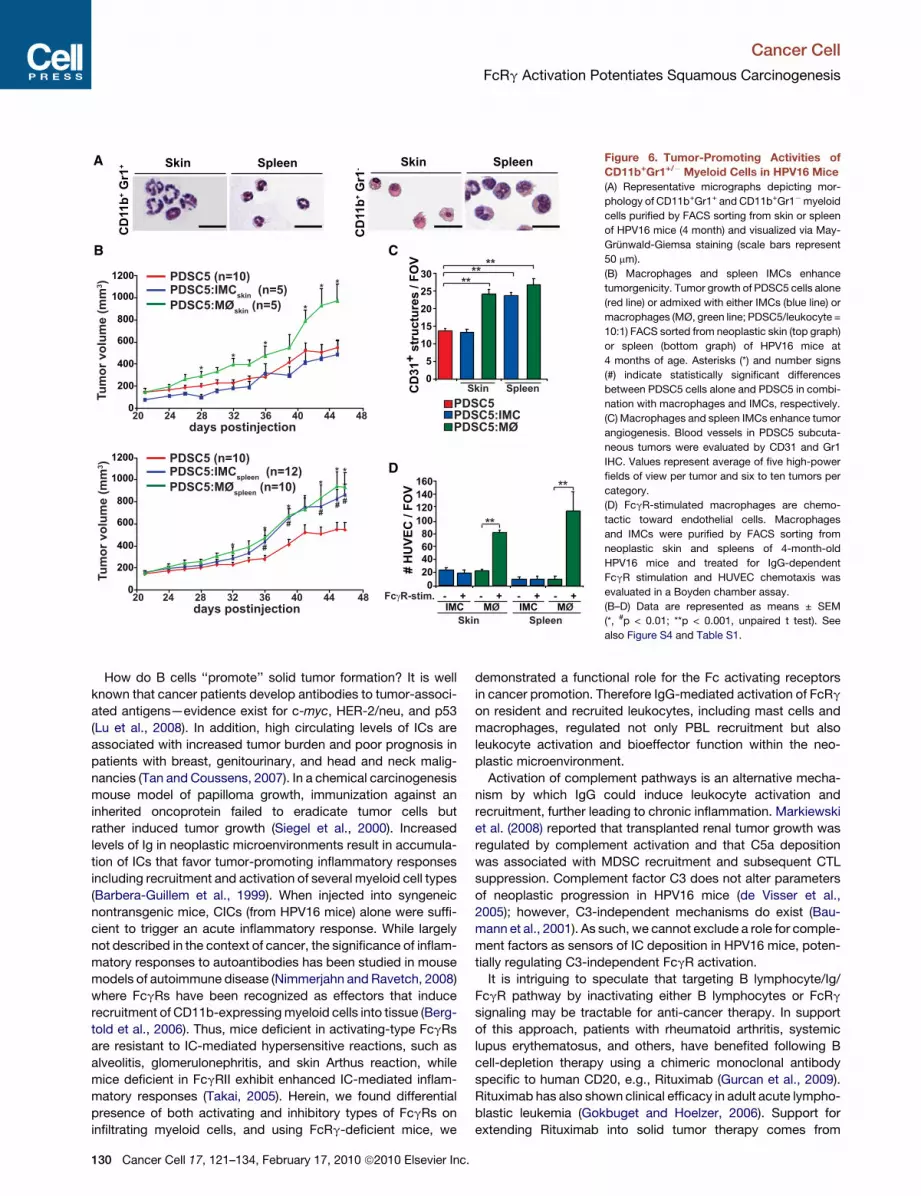
Skin SpleenFigure 6. Tumor-Promoting Activities ofCD11b+Gr1+/! Myeloid Cells in HPV16 Mice(A) Representative micrographs depicting mor-
phology of CD11b+Gr1+ and CD11b+Gr1!myeloid
cells purified by FACS sorting from skin or spleen
of HPV16 mice (4 month) and visualized via May-
Grunwald-Giemsa staining (scale bars represent
50 mm).
(B) Macrophages and spleen IMCs enhance
tumorgenicity. Tumor growth of PDSC5 cells alone
(red line) or admixed with either IMCs (blue line) or
macrophages (MØ, green line; PDSC5/leukocyte =
10:1) FACS sorted from neoplastic skin (top graph)
or spleen (bottom graph) of HPV16 mice at
4 months of age. Asterisks (*) and number signs
(#) indicate statistically significant differences
between PDSC5 cells alone and PDSC5 in combi-
nation with macrophages and IMCs, respectively.
(C) Macrophages and spleen IMCs enhance tumor
angiogenesis. Blood vessels in PDSC5 subcuta-
neous tumors were evaluated by CD31 and Gr1
IHC. Values represent average of five high-power
fields of view per tumor and six to ten tumors per
category.
(D) FcgR-stimulated macrophages are chemo-
tactic toward endothelial cells. Macrophages
and IMCs were purified by FACS sorting from
neoplastic skin and spleens of 4-month-old
HPV16 mice and treated for IgG-dependent
FcgR stimulation and HUVEC chemotaxis was
evaluated in a Boyden chamber assay.
(B–D) Data are represented as means ± SEM
(*, #p < 0.01; **p < 0.001, unpaired t test). See
also Figure S4 and Table S1.
Cancer Cell
FcRg Activation Potentiates Squamous Carcinogenesis
130 Cancer Cell 17, 121–134, February 17, 2010 ª2010 Elsevier Inc.

a clinical study with colon cancer patients in which numbersof CD21-hyperpositive lymphocytes were reduced in parallelwith reduction in tumor burden (Barbera-Guillem et al., 2000).Although Rituximab effectively deletes circulating B cells, noincreased susceptibility to infection has been observed inpatients with rheumatoid arthritis or non-Hodgkin’s lymphoma(Gokbuget and Hoelzer, 2006), thus supporting the approachand manipulation of humoral immunity and/or its downstreameffector pathways as a therapeutic possibility.
Programming Recruited Leukocytes and InhibitingProtumor ImmunityClinical and experimental data indicate that chronic presenceand activation of immune cells, e.g., mast cells, macrophages,
Tie2-expressing monocytes, neutrophils, DCs, IMCs, and CD4+
T cells, promote tumor development by activating angiogenicprograms, suppressing antitumor immunity (Mantovani et al.,2008), and enhancing tumor cell migration and metastasis(DeNardo et al., 2009; Pollard, 2008). When chronically activatedin tumor microenvironments, some myeloid cells are pro-grammed such that they deliver a diversity of bioactive media-tors to neoplastic tissues, including chemokines, cytokines,matrix remodeling enzymes, and cytotoxic proteins (Mantovaniet al., 2008). Identification of the critical programs that inducethese protumor pathways would reveal important mediators topotentially target with anticancer therapeutics.Mast cells exert duality as cancer mediators with some clinical
studies indicating their presence in human cancers correlates
D
F
# H
UV
EC
/ F
OV
aCXCR3
VEGF + + + + + +
- - + + - -
-- -- + +
-- + + - +
0
40
80
120
160
200
FcRg+/- MØ
FcRg-/- MØ
E60
# H
UV
EC
/ F
OV
VEGF
-
-
-
-
-
+
-
+
+
+
+
+
CXCL10
0
20
40
aCXCR3
JH-/-
HPV16
FcRg-/-(-)LM
0
0.4
0.8
1.2
1.6
1.8
0
4
8
12 *
*
**
Cxcl10
Cxcr3
A
49 54
0
100
200
300
400
500
600
700
800
19 24 29 34 39 44
*
*
*
*
*
**
***
***
***
49
0
100
200
300
400
500
600
700
800
19 24 29 34 39 44
**
**
** * **
PDSC5 (n=14)
PDSC5:FcRg+/- MØ
BM (n=11)
PDSC5:FcRg-/- MØ
BM (n=11)
PDSC5 (n=9)
PDSC5:FcRg+/- MØ
skin (n=9)
PDSC5:FcRg-/- MØ
skin (n=9)
days postinjection
Tu
mo
r v
olu
me
(m
m3)
days postinjection
Tu
mo
r v
olu
me
(m
m3)
*
**
*
**
*
**
**
**
#
##
###
##
B
C
CD11b+Gr1
-CD11c
-
skin
(MØ)
Polarization
M1M1M1M1
M1M1M1M1M1M1
M1M1
M2
M1
M2M2b
M2
M2
M2
Gene Fold DCt p value
Il13 -14.6 11.8 ***Cd4 -13.0 9.1 ***Ctla4 -12.5 9.6 ***Cd3e -11.5 10.6 ***Icos -11.0 9.6 ***Cd163
Ccl1
-10.5-10.2
5.215.2
****
Il5 -10.3 15.7 **Cd8a -9.7 11.0 **Il2ra -9.1 9.7 **Il6 -7.2 8.8 **Cd28 -7.0 10.4 **Ccl17 -6.8 13.9 *Gzmb -6.8 10.7 **Il4 -6.7 11.8 *Cd40lg -6.0 14.2 *Csf2 -5.7 12.2 *Il17 -5.5 12.0 *Ym1 -4.9 5.9 *Ccr4 -4.9 11.7 *Cxcr3 -4.9 9.5 *H2-Eb1 -4.7 3.4 *Gusb -4.5 6.2 *Il1b 4.3 0.7 *
* p<0.05 ** p<0.01 *** p<0.001
Ptgs2 5.0 4.3 *Il12a 6.7 8.4 **Cxcl10 7.5 3.9 **Nos2 9.0 4.8 **Csf3 9.5 7.6 **Cxcl11 9.7 5.6 **Il1a 16.8 4.0 ***
Figure 7. FcRg Expression RegulatesProangiogenic and Protumorigenic Proper-ties of Macrophages(A and B) FcRg expression regulated macrophage
protumor activity. PDSC5 tumor cells were
injectedalone (blue) or admixedwithmacrophages
derived from neoplastic skin (MØskin) of either
HPV16/FcRg+/! (red) or HPV16/FcRg!/! (green)
mice (A) or instead from bone marrow-derived
macrophages (MØBM) isolated from FcRg+/! (red)
or FcRg!/! (green) mice (B). The asterisk (*) indi-
cates statistically significant differences between
PDSC5 cells admixed with FcRg+/! versus
FcRg!/! macrophages. The number sign (#) indi-
cates statistically significant differences between
PDSC5 alone and PDSC5 admixed with FcRg!/!
macrophages. Data are represented as means ±
SEM; p < 0.05, unpaired t test.
(C) Genes differentially expressed in macro-
phages isolated from neoplastic skin of HPV16/
FcRg!/! versus HPV16/FcRg+/! mice. mRNA
expression levels in macrophages from HPV16/
FcRg!/! neoplastic skin (4 month old) are indi-
cated as fold change as compared to HPV16/
FcRg+/! macrophages with DCT of each gene
calculated with b2-microglobulin as endogenous
control. Expression was considered as statisti-
cally significantly deregulated when p < 0.05
(t test, FcRg!/! versus FcRg+/!).
(D) Quantitative real-time PCR analysis of Cxcl10
and Cxcr3 mRNA expression in ear tissue from
4-month-old negative littermate (!LM), HPV16,
HPV16/JH!/!, and HPV16/FcRg!/! mice (n =
3–5 mice/cohort). *p < 0.05; **p < 0.01, unpaired
t test.
(E) VEGF165-induced (100 ng/ml) HUVEC chemo-
taxis was evaluated in a Boyden chamber assay
after pretreatment with recombinant CXCL10
(100 ng/ml) in the presence or absence of
CXCR3 blocking antibody (10 mg/ml). *p < 0.05;
**p < 0.01, unpaired t test.
(F) Macrophages isolated from HPV16/FcRg!/!
mice exhibit angiostatic activity. HUVEC chemo-
taxis in a Boyden chamber assay was evaluated
after pretreatment with conditioned medium iso-
lated from macrophages (purified from skin of HPV16/FcRg+/! or HPV16/FcRg!/! mice) following activation with LPS (10 ng/ml) and aCXCR3 blocking Ig
(10 mg/ml). *, p < 0.05; **, p < 0.01, unpaired t test.
(E and F) Pretreated HUVECs were evaluated for chemotactic migration in response to VEGF (100 ng/ml) using a Boyden chamber assay. Quantitative values
reflect the number of migrating HUVECs averaged from four to five high-power fields per insert and four inserts per treatment ± SEM. At least three independent
analyses were performed and one representative experiment is shown. See also Figure S5 and Table S1.
Cancer Cell
FcRg Activation Potentiates Squamous Carcinogenesis
Cancer Cell 17, 121–134, February 17, 2010 ª2010 Elsevier Inc. 131

with a favorable clinical outcome, whereas others clearlyimplicate them as protumor mediators (Theoharides and Conti,2004). In HPV16 mice (Coussens et al., 1999), and in othermodels of cancer development (Nakayama et al., 2004;Soucek et al., 2007), mast cell-derived factors foster tumor cellsurvival and angiogenesis. With IgG-mediated stimulation ofmast cells enhancing tumor development and angiogenesis inHPV16 mice, these data indicate a functional requirement forFcRg engagement for activating protumor cascades, leadingto PBL recruitment into neoplastic skin, angiogenesis, and tumordevelopment.
Pharmacologic inhibition of macrophages minimizes cervicalcarcinogenesis in HPV16 mice (Giraudo et al., 2004), whereaselimination of macrophages during mammary carcinogenesislimits cancer progression and metastasis (Pollard, 2008) byreducing angiogenesis in premalignant tissues. Interestingly,pro- versus anti-metastatic properties of infiltrating macro-phages in MMTV-PyMT mice are programmed by CD4+ Tlymphocytes via an IL-4-dependent mechanism (DeNardoet al., 2009). In contrast, in ovarian cancer, macrophage pheno-type is regulated by IL-1R and MyD88, which together maintaina macrophage immunosuppressive M2 phenotype (Hagemannet al., 2008). In the present study, we report that B cells andFcRg are key parameters regulating pro- versus antitumorprogramming of macrophages during squamous carcinogenesis
through a mechanism that not only involves modified M1 versusM2 or M2-like polarization but also the differential expression ofan angiostatic chemokine Cxcl10 and its receptor Cxcr3. Assuch, activation of angiogenic vasculature in developing tumorsis regulated by multiple subsets of myeloid cells, each exhibitinga distinct signature of pro- versus antiangiogenic molecules, aswell as likely being programmed by distinct effector pathwaysdependent on the tissue microenvironment.These experimental findings imply that reprogramming
myeloid cell phenotypes and/or altering the immune microenvi-ronment to foster antitumor versus protumor activity couldimprove survival of patients with cancer by limiting cancer devel-opment and perhaps stabilizing premalignant or premetastaticdisease. Proof-of-concept studies supporting this notion wererecently reported by De Palma et al. (2008) demonstrating thatmyeloid cells could be used as vehicles to deliver immune medi-ators to tumor microenvironments and essentially reprogramthem.
ConclusionsResults from this studydemonstrate a key role forB lymphocytes,humoral immunity, and activation of FcRg signaling pathwaysin myeloid cells as promoting forces for squamous carcinogen-esis. With regards to therapy, our data indicate that anti-cancerstrategies targeting B cells, Ig, or FcRg may harbor therapeuticefficacy in limiting risk of malignant conversion in patientssuffering from chronic inflammatory diseases or in patientsharboring premalignant lesions whose molecular and/or immu-nologic characteristics favor tumor development. The efficacyand safety of Rituximab for various autoimmune disorders andsome hematological cancers could be extended to potentiallycombat squamous neoplasms in which IC deposition is promi-nent and/or activation of FcRg-mediated signaling is evident.
EXPERIMENTAL PROCEDURES
Animal HusbandryGeneration and characterization of HPV16, JH!/!, FcRg!/!, and c-kitsh/sh
mice have previously been described (Arbeit et al., 1994; Coussens et al.,
1996; Lyon and Glenister, 1982; Takai et al., 1994). To generate HPV16
mice in the JH!/! and FcRg!/! backgrounds, JH+/! and FcRg+/! mice were
backcrossed into the FVB/n strain to N5 and intercrossed with HPV16 mice.
c-kitsh/+ mice were backcrossed five generations into FVB/n. All mouse
experiments complied with National Institutes of Health guidelines and were
approved by the University of California San Francisco Institutional Animal
Care and Use Committee. Characterization of neoplastic stages has been
reported previously (Coussens et al., 1996).
In Vivo AssaysFor evaluation of proinflammatory properties of IgGs, serum-purified IgGs
(20 ml; 12 mg/ml) were injected intradermally into ears of (!)littermates. For
performing matrigel plug assays, PDSC5 cells (Arbeit et al., 1996) (1.5 3
106/100 ml) were suspended in 300 ml of Matrigel and injected s.c. in the groin
area of 7-week-old mice. For tumorgenicity assays, PDSC5 cells (0.5 3
106 cells) were suspended in 100 ml of Matrigel in PBS (1:1) and inoculated
s.c. into flanks of 7-week-old mice. Tumors were measured at 2 day intervals
with a digital caliper, and tumor volume was calculated with the equation
V (mm3) = a 3 b2/2 (a is the largest diameter and b the smallest diameter).
Low-Density qPCR ArraysImmune Panel TaqMan arrays (Applied Biosystems) were used to measure the
expression of 96 genes in three biological replicates. Analysis of raw data
Figure 8. Activation of the FcRg Pathway in Mast Cells and Macro-phages by Humoral Immunity Regulates Squamous Carcinogenesisof HPV16 MiceHPV16 oncogene expression initiates keratinocytes and triggers early
neoplastic progression accompanied by neo- and self-antigen presentation,
peripheral B cell activation/maturation, and secretion of Igs. Autoantibodies
subsequently accumulate in dermal stroma of neoplastic tissue as vasculature
becomes initially angiogenic and ‘‘leaky.’’ IgGs interact with Fcg receptors on
resident and recruited myeloid cells where they induce differential recruitment
of leukocytes from peripheral blood and regulate mast cell and macrophage
bioeffector functions once present in neoplastic tissue. FcRg deficiency not
only impairs the protumorigenic properties of mast cells and macrophages
but also reprogram macrophages leading to enhanced angiostatic and M1
bioactivity.
Cancer Cell
FcRg Activation Potentiates Squamous Carcinogenesis
132 Cancer Cell 17, 121–134, February 17, 2010 ª2010 Elsevier Inc.

obtained with Immune Panel TaqMan arrays have been performed using an
implemented covariance model, as previously described (Pucci et al., 2009).
Statistical AnalysesStatistical analyses were performed using GraphPad Prism version 4 and/or
InStat version 3.0a forMacintosh (GraphPadSoftware). Specific tests included
Mann-Whitney (unpaired, nonparametric, two-tailed), unpaired t test, Fisher’s
exact test, chi-square test, and log rank analysis. p values < 0.05 were consid-
ered statistically significant.
SUPPLEMENTAL INFORMATION
Supplemental Information includes five figures, Supplemental Experimental
Procedures, and one table and can be found with this article online at
doi:10.1016/j.ccr.2009.12.019.
ACKNOWLEDGMENTS
The authors thank theUniversity of California San Francisco Helen Diller Family
Comprehensive Cancer Center Laboratory for Cell Analysis and Mouse
Pathology shared resource core facilities and members of the Coussens
laboratory for critical discussion. The authors acknowledge support from the
Cancer Research Institute (P.A.), the Swedish Research Council (M.J.), the
American Association for Cancer Research (N.I.A.), the American Cancer
Society and National Cancer Institute postdoctoral training grants (T32-
CA09043 and T32-CA108462; D.D.), the National Institutes of Health/National
Cancer Institute grants (R01CA130980, R01CA13256, R01CA098075, and
P01CA72006), and the Department of Defense Breast Cancer Research
Program Era of Hope Scholar Award (W81XWH-06-1-0416; L.M.C.). L.N.
and M.D.P. were supported by the Associazione Italiana per la Ricerca sul
Cancro, the European Union (FP6 Tumor-Host Genomics), and the Italian
Ministry of Health (Challenge in Oncology).
Received: August 19, 2009
Revised: November 14, 2009
Accepted: December 9, 2009
Published online: February 4, 2010
REFERENCES
Arbeit, J.M., Munger, K., Howley, P.M., and Hanahan, D. (1994). Progressive
squamous epithelial neoplasia in K14-human papillomavirus type 16 trans-
genic mice. J. Virol. 68, 4358–4368.
Arbeit, J.M., Olson, D.C., and Hanahan, D. (1996). Upregulation of fibroblast
growth factors and their receptors during multi-stage epidermal carcinogen-
esis in K14-HPV16 transgenic mice. Oncogene 13, 1847–1857.
Barbera-Guillem, E., May, K.F., Jr., Nyhus, J.K., and Nelson, M.B. (1999).
Promotion of tumor invasion by cooperation of granulocytes and macro-
phages activated by anti-tumor antibodies. Neoplasia 1, 453–460.
Barbera-Guillem, E., Nelson, M.B., Barr, B., Nyhus, J.K., May, K.F., Jr., Feng,
L., and Sampsel, J.W. (2000). B lymphocyte pathology in human colorectal
cancer. Experimental and clinical therapeutic effects of partial B cell depletion.
Cancer Immunol. Immunother. 48, 541–549.
Baumann, U., Chouchakova, N., Gewecke, B., Kohl, J., Carroll, M.C., Schmidt,
R.E., and Gessner, J.E. (2001). Distinct tissue site-specific requirements of
mast cells and complement components C3/C5a receptor in IgG immune
complex-induced injury of skin and lung. J. Immunol. 167, 1022–1027.
Bergtold, A., Gavhane, A., D’Agati, V., Madaio, M., and Clynes, R. (2006).
FcR-bearing myeloid cells are responsible for triggering murine lupus
nephritis. J. Immunol. 177, 7287–7295.
Brandtzaeg, P., Carlsen, H.S., and Halstensen, T.S. (2006). The B-cell system
in inflammatory bowel disease. Adv. Exp. Med. Biol. 579, 149–167.
Chen, J., Trounstine, M., Alt, F.W., Young, F., Kurahara, C., Loring, J.F., and
Huszar, D. (1993). Immunoglobulin gene rearrangement in B cell deficient
mice generated by targeted deletion of the JH locus. Int. Immunol. 5, 647–656.
Coussens, L.M., Hanahan, D., and Arbeit, J.M. (1996). Genetic predisposition
and parameters of malignant progression in K14-HPV16 transgenic mice. Am.
J. Pathol. 149, 1899–1917.
Coussens, L.M., Raymond, W.W., Bergers, G., Laig-Webster, M., Behrendt-
sen, O., Werb, Z., Caughey, G.H., and Hanahan, D. (1999). Inflammatory
mast cells up-regulate angiogenesis during squamous epithelial carcinogen-
esis. Genes Dev. 13, 1382–1397.
Coussens, L.M., Tinkle, C.L., Hanahan, D., and Werb, Z. (2000). MMP-9
supplied by bone marrow-derived cells contributes to skin carcinogenesis.
Cell 103, 481–490.
Dalgleish, A.G., and O’Byrne, K.J. (2002). Chronic immune activation and
inflammation in the pathogenesis of AIDS and cancer. Adv. Cancer Res. 84,
231–276.
Daniel, D., Chiu, C., Giraud, E., Inoue, M., Mizzen, L.A., Chu, N.R., and
Hanahan, D. (2005). CD4+ T cell-mediated antigen-specific immunotherapy
in a mouse model of cervical cancer. Cancer Res. 65, 2018–2025.
De Palma, M., Mazzieri, R., Politi, L.S., Pucci, F., Zonari, E., Sitia, G.,
Mazzoleni, S., Moi, D., Venneri, M.A., Indraccolo, S., et al. (2008). Tumor-
targeted interferon-alpha delivery by Tie2-expressing monocytes inhibits
tumor growth and metastasis. Cancer Cell 14, 299–311.
de Visser, K.E., Korets, L.V., and Coussens, L.M. (2004). Early neoplastic
progression is complement independent. Neoplasia 6, 768–776.
de Visser, K.E., Korets, L.V., and Coussens, L.M. (2005). De novo carcinogen-
esis promoted by chronic inflammation is B lymphocyte dependent. Cancer
Cell 7, 411–423.
de Visser, K.E., Eichten, A., and Coussens, L.M. (2006). Paradoxical roles of
the immune system during cancer development. Nat. Rev. Cancer 6, 24–37.
DeNardo, D.G., Baretto, J.B., Andreu, P., Vasquez, L., Kolhatkar, N., Tawfik,
D., and Coussens, L.M. (2009). CD4+ T cells regulate pulmonary metastasis
of mammary carcinomas by enhancing protumor properties of macrophages.
Cancer Cell 16, 91–102.
Giraudo, E., Inoue, M., and Hanahan, D. (2004). An amino-bisphosphonate
targets MMP-9-expressing macrophages and angiogenesis to impair cervical
carcinogenesis. J. Clin. Invest. 114, 623–633.
Gokbuget, N., and Hoelzer, D. (2006). Novel antibody-based therapy for acute
lymphoblastic leukaemia. Best Pract. Res. Clin. Haematol. 19, 701–713.
Gurcan, H.M., Keskin, D.B., Stern, J.N., Nitzberg, M.A., Shekhani, H., and
Ahmed, A.R. (2009). A review of the current use of rituximab in autoimmune
diseases. Int. Immunopharmacol. 9, 10–25.
Hagemann, T., Lawrence, T., McNeish, I., Charles, K.A., Kulbe, H., Thompson,
R.G., Robinson, S.C., and Balkwill, F.R. (2008). ‘‘Re-educating’’ tumor-associ-
ated macrophages by targeting NF-kappaB. J. Exp. Med. 205, 1261–1268.
Junankar, S.R., Eichten, A., Kramer, A., de Visser, K.E., and Coussens, L.M.
(2006). Analysis of immune cell infiltrates during squamous carcinoma
development. J. Investig. Dermatol. Symp. Proc. 11, 36–43.
Lu, H., Goodell, V., and Disis, M.L. (2008). Humoral immunity directed against
tumor-associated antigens as potential biomarkers for the early diagnosis of
cancer. J. Proteome Res. 7, 1388–1394.
Lyon, M.F., and Glenister, P.H. (1982). A new allele sash (Wsh) at the W-locus
and a spontaneous recessive lethal in mice. Genet. Res. 39, 315–322.
Mantovani, A., Allavena, P., Sica, A., and Balkwill, F. (2008). Cancer-related
inflammation. Nature 454, 436–444.
Markiewski, M.M., DeAngelis, R.A., Benencia, F., Ricklin-Lichtsteiner, S.K.,
Koutoulaki, A., Gerard, C., Coukos, G., and Lambris, J.D. (2008). Modulation
of the antitumor immune response by complement. Nat. Immunol. 9,
1225–1235.
Nakayama, T., Yao, L., and Tosato, G. (2004). Mast cell-derived angiopoietin-1
plays a critical role in the growth of plasma cell tumors. J. Clin. Invest. 114,
1317–1325.
Nimmerjahn, F., and Ravetch, J.V. (2008). Fcgamma receptors as regulators of
immune responses. Nat. Rev. Immunol. 8, 34–47.
Ostrand-Rosenberg, S. (2008). Immune surveillance: a balance between
protumor and antitumor immunity. Curr. Opin. Genet. Dev. 18, 11–18.
Cancer Cell
FcRg Activation Potentiates Squamous Carcinogenesis
Cancer Cell 17, 121–134, February 17, 2010 ª2010 Elsevier Inc. 133

Pollard, J.W. (2008). Macrophages define the invasive microenvironment in
breast cancer. J. Leukoc. Biol. 84, 623–630.
Pucci, F., Venneri, M.A., Biziato, D., Nonis, A., Moi, D., Sica, A., Di Serio, C.,
Naldini, L., and De Palma, M. (2009). A distinguishing gene signature shared
by tumor-infiltrating Tie2-expressing monocytes (TEMs), blood ‘‘resident’’
monocytes and embryonic macrophages suggests common functions and
developmental relationships. Blood 114, 901–914.
Shah, S., Divekar, A.A., Hilchey, S.P., Cho, H.M., Newman, C.L., Shin, S.U.,
Nechustan, H., Challita-Eid, P.M., Segal, B.M., Yi, K.H., and Rosenblatt, J.D.
(2005). Increased rejection of primary tumors in mice lacking B cells: inhibition
of anti-tumor CTL and TH1 cytokine responses by B cells. Int. J. Cancer 117,
574–586.
Siegel, C.T., Schreiber, K., Meredith, S.C., Beck-Engeser, G.B., Lancki, D.W.,
Lazarski, C.A., Fu, Y.X., Rowley, D.A., and Schreiber, H. (2000). Enhanced
growth of primary tumors in cancer-prone mice after immunization against
the mutant region of an inherited oncoprotein. J. Exp. Med. 191, 1945–1956.
Soucek, L., Lawlor, E.R., Soto, D., Shchors, K., Swigart, L.B., and Evan, G.I.
(2007). Mast cells are required for angiogenesis and macroscopic expansion
of Myc-induced pancreatic islet tumors. Nat. Med. 13, 1211–1218.
Takai, T. (2005). Fc receptors and their role in immune regulation and autoim-
munity. J. Clin. Immunol. 25, 1–18.
Takai, T., Li, M., Sylvestre, D., Clynes, R., and Ravetch, J.V. (1994). FcR
gamma chain deletion results in pleiotrophic effector cell defects. Cell 76,
519–529.
Tan, T.T., and Coussens, L.M. (2007). Humoral immunity, inflammation and
cancer. Curr. Opin. Immunol. 19, 209–216.
Theoharides, T.C., and Conti, P. (2004). Mast cells: the Jekyll and Hyde of
tumor growth. Trends Immunol. 25, 235–241.
Zapata, J.M., Llobet, D., Krajewska, M., Lefebvre, S., Kress, C.L., and Reed,
J.C. (2009). Lymphocyte-specific TRAF3 transgenic mice have enhanced
humoral responses and develop plasmacytosis, autoimmunity, inflammation,
and cancer. Blood 113, 4595–4603.
Cancer Cell
FcRg Activation Potentiates Squamous Carcinogenesis
134 Cancer Cell 17, 121–134, February 17, 2010 ª2010 Elsevier Inc.

1
Cancer Cell, Volume 17
Supplemental Information
FcR Activation Regulates Inflammation-Associated
Squamous Carcinogenesis Pauline Andreu, Magnus Johansson, Nesrine I. Affara, Ferdinando Pucci, Tingting Tan, Simon Junankar, Lidiya Korets, Julia Lam, David Tawfik, David G. DeNardo, Luigi Naldini, Karin E. de Visser, Michele De Palma, and Lisa M. Coussens

2
SUPPLEMENTAL DATA
Figure S1. Generation of B cell-deficient HPV16 mice (accompanies Figure 1)
A) Analysis of B220+CD19+ B cells by flow cytometry in lymph node, spleen and peripheral
blood from HPV16 and HPV16/JH-/- mice (4-mo). Right panel: Western blot for IgG, IgA and
IgM in serum from negative litterate (lane 1), HPV16 (lane 2 and 6) and HPV16/JH-/- (lane 3-5)
mice. Ig heavy and light chains are indicated (arrows).
B) Protein levels of pro-MMP9 in skin extracts from age-matched (-)LM, HPV16/JH+/- and
HPV16/JH-/- mice (4-mo) as assessed by ELISA. Error bars represent SEM, asterisk (*)

3
indicates statistically significant differences between HPV16/JH+/- and HPV16/JH-/- mice (p <
0.05, Mann-Whitney).
Figure S2. Anti-E7 IgG quantification (accompanies Figure 2)
Serum titers of anti-E7 IgG evaluated by indirect ELISA in serum from (-)LM and HPV16 (4-
and 6-mo). Error bars represent SEM; asterisk (*) indicates statistically significant differences
with (-)LM (p < 0.05, Mann-Whitney).

4
Figure S3. Deficient carcinogenesis in HPV16/FcR -deficient mice (accompanies Figure 4)
A) Immunolocalization of FcR left panel), Fc RI (middle panel) and Fc RII/III (right panel)
expression in HPV16/Fc R-/- mice at 4-mo of age. Solid line, epidermal-dermal interface;
epidermis, e; dermis, d; scale bar, 50 m.

5
B) Representative dot-plot graphs showing CD45+CD49b+CD3- NK cells in premalignant skin of
HPV16, HPV16/JH-/-, and HPV16/FcR mice (4-mo).
C) Differential expression of Fc RI and Fc RIII by NK cells in premalignant skin of HPV16
(red), HPV16/JH-/- (blue) and HPV16/FcR -/- (green) mice at 4-mo of age. NK cells were gated
as live CD45+CD49b+CD3- leukocytes. Grey line: Ig control.
D) Immunolocalization of CD11c and F4/80-expressing cells in premalignant skin of
HPV16/Fc R+/- and HPV16/Fc R-/- mice at 4-mo of age (red staining). White line depicts
epidermal-dermal interface; e: epidermis; d: dermis; scale bar, 50 m.
E) Protein levels of pro-MMP9 in skin extracts from (-)LM, HPV16/FcR +/- and HPV16/FcR -/-
mice assessed by ELISA. Error bars represent SEM, asterisk (*) indicates statistically significant
differences between age-matched mice (p < 0.05, Mann-Whitney).
F) Circulating Ig levels were evaluated by ELISA in serum from (-)LM, HPV16 (4- and 6-mo)
and HPV16/FcR -/- (6-mo). Error bars represent SEM, asterisk (*) indicates statistically
significant differences with (-)LM (p < 0.05, Mann-Whitney).

6
Figure S4. Lineage characteristics of CD11b+Gr1+/- myeloid cell subpopulations in
premalignant skin, secondary lymphoid organs and blood of HPV16 mice (accompanies
Figure 6)
A) Percentages of CD11b+Gr1+/- myeloid cells in spleen, cervical lymph nodes (cLN), and
peripheral blood assessed by flow cytometry of single-cell suspensions derived from negative

7
age-matched negative littermates (-LM), HPV16/FcR +/+ and HPV16/FcR -/- mice. Results
shown represent mean percentages (n=5-8 mice) SEM. (*) and (**) indicate statistically
significant differences; (*) p < 0.05 and (**) p < 0.01 (Mann-Whitney).
B) Phenotype of CD11b+/-Gr1+/- myeloid cells evaluated by flow cytometry to assess leukocyte
lineage and maturation markers. Single-cell suspensions of neoplastic skin or spleen from mice
(4-mo) were gated as live CD11b+Gr1+ (blue line) and CD11b+Gr1- (green line), respectively,
and each population was evaluated for expression of the indicated cell surface markers in
comparison with isotypic Ig fluorescent levels (red line). At least 3 independent analyses were
performed and one representative overlay plot is shown.

8
Figure S5. Functional characterization of myeloid lineage cells infiltrating premalignant skin
of HPV16 mice (accompanies Figure 7)

9
A) FcR -deficiency does not modify expression of co-stimulatory molecules on DCs. Single-cell
suspensions derived from skin and cLN of HPV16/FcR +/- and HPV16/FcR -/- mice (4-mo) were
gated as CD45+CD11c+ dendritic cells and evaluated for MHCII, CD80 and CD86 expression
levels, as compared to background fluorescent levels (Isotype Ig control, dark grey line). For
each antigen, at least 3 independent analyses were performed and overlay plots for one
representative experiment are shown.
B) Genes differentially expressed in dendritic cells from HPV16/FcR +/- and HPV16/FcR -/-
mice. mRNA expression levels in DCs from HPV16/FcR +/- neoplastic skin (4-mo) are indicated
as fold-change as compared to HPV16/FcR +/- DCs with CT of each gene calculated using 2-
microglobulin as an endogenous control. Genes are considered as statistically significantly de-
regulated when p (t test, FcR +/- versus FcR -/- group) <0.05.
C) CD11b+Gr1+ IMCs purified from either skin or spleen of HPV16 mice do not inhibit T cell
polyclonal activation. IMCs were purified by flow cell sorting from premalignant tissue or
spleen of HPV16 mice (4-mo) or 4T1 tumor-bearing mice. CD4+ and CD8+ T cells, respectively,
were magnetically purified from (-)LM mouse spleen and co-incubated with isolated IMCs at
various ratio (1:16 to 1:1, CD11b+Gr1+:T cell) for 72-hr in plates coated with anti-CD3/CD28
agonist antibodies. BrdU was added to culture medium for the last 18-hr of co-culture.
Incorporation of BrdU was evaluated by ELISA.
D) IMCs from skin and spleen of HPV16 mice do not produce reactive-oxygen species. Single-
cell suspensions derived from skin and spleen of HPV16 mice (4-mo) were incubated with an
oxidation-sensitive probe (DCFDA) in the absence (green line) or presence of H2O2 (5.0 M,
blue line). Live cells were gated as CD11b+Gr1+and evaluated for DCFDA fluorescent levels as
compared to background fluorescent levels (No treatment, dark grey line). 3 independent
analyses were performed and overlay plot for one representative experiment is shown.
E) Genes differentially expressed in IMCs from HPV16/FcR +/- and HPV16/FcR -/- mouse skin.
Gene expression levels in IMCs from HPV16/FcR -/- neoplastic skin (4-mo) are indicated as
fold-change as compared to HPV16/FcR +/- IMCs with CT of each gene calculated using 2-
microglobulin as an endogenous control. Gene are considered as statistically significantly de-
regulated when p (t test, FcR +/- versus FcR -/- group) <0.05.

10
SUPPLEMENTAL EXPERIMENTAL PROCEDURES
Serum collection, immunoglobulin purification and biotinylation
Blood was collected by cardiac puncture and left at room temperature for 2 hr for coagulation,
then centrifuged at 2000 rpm for 15 min. Serum was separated and stored at -80°C. Ig
purification was performed as described by the manufacturer�’s recommendations using the
Pierce Thiophilic Adsorption Kit (Pierce Biotechnology, Rockford, IL). Biotinylation of purified
IgGs was performed as described by the manufacturer�’s recommendations using the EZ-Link
NHS-PEO Solid Phase Biotinylation Kit (Pierce).
Intradermal injection of concentrated immunoglobulins
Purified IgGs were loaded into the filter unit of Amicon Ultra-4 centrifugal devices (Millipore,
Bedford, MA). Devices were spun in a centrifuge with swinging bucket rotor carriers set at 3000
x g for 45 min. Concentrated IgGs were collected immediately after centrifugation, diluted to a
final concentration of 12 µg/µl and injected intradermally using 28-gauge insulin syringes to wt
mice anesthetized with 2.0% isoflurane/98% O2 mixture.
IF-detection of antibody deposition
Tissue samples were frozen directly in glycerol-based freezing medium (Tissue-Tek® OCT,
Sakura Finetek, CA). Tissue sections (10-µm) were cut using a Leica CM1900 cryostat (Leica
Microsystem, Bannockburn, IL), air-dried, and fixed in cold acetone. To prevent non-specific
binding, tissue sections were incubated for 30 min with rat anti-mouse CD16/CD32 mAb (1:50;
2.4G2, BD Biosciences, San Jose, CA) in blocking buffer (5% goat serum and 2.5% bovine
serum albumin (BSA) in PBS). Sections were then incubated with FITC-conjugated goat anti-
mouse IgG ( -chain specific; 1:100; Sigma-Aldrich, St. Louis, MO), anti-mouse IgG1 (1:100;
Invitrogen, Carlsbad, CA), anti-mouse IgG2a (1:50; Invitrogen), and IgM (1:50; -chain
specific, Sigma) in 0.5X blocking buffer for 1 hr at room temperature. Sections were mounted
using Vectashield containing DAPI (Vector Laboratories, Burlingame, CA). All IF experiments
included negative controls to assess for background staining, which was negligible
Immunohistochemistry
Age-matched tissue samples from transgenic and control animals were immersion-fixed in 10%

11
neutral-buffered formalin followed by dehydration through graded series of alcohols and xylene
and embedded in paraffin. 5- m thick paraffin sections were cut using a Leica 2135 microtome
(Leica Microsystem) and mounted onto SuperFrost/Plus slides (Thermo Fisher Scientific,
Waltham, MA). Tissue sections were deparaffinized using xylene and rehydrated through a
graded series of alcohol, and subjected to enzyme- and immuno-histochemical (IHC) detection
as previously described (Coussens et al., 1996).
To visualize serine esterase activity in mast cells, chloroacetate esterase (CAE)
histochemistry was performed as previously described (Coussens et al., 1999). Briefly, 1.0 mg
of naphthol AS-D chloroacetate (Sigma) was dissolved in 20 l of N, N-dimethyl formamide
(DMF) and 1.0 ml buffer (8% DMF, 20% ethylene glycol monoethylether in 80mM Tris-maleate
pH 7.5). The resulting solution was subsequently added to 1.0 mg of Fast Blue BB salt (Sigma)
and applied to tissue sections. Sections were then dehydrated and mounted in glycerol.
To detect endothelial cells and neutrophils, endothelium specific marker PECAM1/CD31
and Gr1 were visualized on paraffin-embedded tissue sections. For antigen retrieval, proteinase
K was used (Dako, Glostrup, Denmark). Non-specific binding was blocked using PBS
containing 5% goat serum (Thermo Fisher) and 2.5% BSA (Blocking buffer). Sections were
incubated with rat anti-mouse CD31 (1:50, BD Biosciences) and rat anti-mouse 7/4, (1:500,
Cedarlane Labs, Burlington, NC) in 0.5X blocking buffer for 2 hr at room temperature. Sections
were then incubated with biotinylated rabbit anti-rat IgG secondary antibodies (1:200, Vector
Laboratories) for 45 min at room temperature. Slides were subsequently incubated with
horseradish peroxidase conjugated avidin complex (ABC Elite, Vector Laboratories) for 30 min,
followed by incubation with Fast 3,3 diaminobenzidine (DAB, Vector Laboratories). Sections
were counterstained with methyl green, dehydrated, and mounted with Cytoseal 60 (Thermo
Fisher). To detect Fc R-expressing immune cells, tissue sections were incubated with rat anti-
mouse CD45 (1:500, BD Pharmingen) and Fc RI-III (1:500, R&D Systems, Minneapolis, MN)
and processed as described above. Hamster anti-mouse CD11c (1:50, BD Pharmingen) and rat
anti-mouse F4/80 (1:50, R&D Systems) were used to detect dendritic cells and macrophages
respectively. All immunolocalization experiments were repeated on multiple tissue sections and
included negative controls for determination of background staining, which was negligible.
Photographs were captured at high-magnification (40X) on a Leica DM-RXA microscope
attached to a Leica digital camera (Leica Microsystem) operated by OpenLab software�™

12
(Improvision, Waltham, MA). Quantitative analysis of innate immune cells was performed by
counting cells in five high-power fields (40X) per age-matched tissue section from five mice per
group. Data presented reflect the mean total cell count per field from the ventral ear leaflet.
Determination of keratinocyte proliferation index
Mice received intraperitoneal injections of bromodeoxyuridine (BrdU; Roche Diagnostics,
Indianapolis, IN) dissolved in PBS (50 g per g of mouse body weight) 90 min prior to sacrifice.
5- m thick paraffin sections were deparaffinized in xylene, rehydrated in graded ethanol, and
subjected to antigen retrieval by steam heating in Citra�™ antigen retrieval solution (BioGenex,
San Ramon, CA). BrdU-positive cells were detected according to manufacturers
recommendations using the BrdU Labeling Kit II (Roche). Briefly, BrdU staining was revealed
using the chromogen Vector Red alkaline phosphatase substrate (Vector Laboratories). Slides
were then counterstained with methyl green. Photographs were captured at high-magnification
(40X) on a Leica DM-RXA microscope as described above. The proliferative index (percentage
of BrdU-positive nuclei over the total number of keratinocytes) was quantified in five high-
power fields per tissue section and included five mice per group.
Flow cytometry
Ear skin from PBS-perfused mice was manually minced using scissors, followed by a 30 min
enzymatic digestion with 2.0 mg/ml collagenase A (Roche) and 1.0 mg/ml Hyaluronidase
(Worthington, Lakewood, NJ) in serum-free Dulbecco�’s modified eagles medium (DMEM)
(Invitrogen) at 37oC using continuous stirring conditions. The digest was quenched by adding
DMEM containing 10% FBS (Invitrogen) and was subsequently filtered through a 70-µm nylon
filter (Falcon). To prevent non-specific binding, cells were incubated for 10 min at 4oC with rat
anti-mouse CD16/CD32 mAb (1:200, BD Bioscience) in PBS containing 1.0 % BSA.
Subsequently, cells were washed and incubated for 20 min with 50 l of fluorophore-conjugated
anti-mouse antibodies; B220 (RA3-6B2), CD3e (145-2C11), CD4 (6K1.5), CD8a (53-6.7),
CD11b (M1/70), CD11c (N418), CD14 (Sa2-8), CD19 (MB19-1), CD31 (MEC 13.3), CD44
(IM7), CD45 (30-F11), CD80 (16-10A1), CD86 (GL1), CD115 (AFS98), CD117 (2B8), F4/80
(BM8), Fc RI alpha (MAR-1), Fc RII/III (93), FcgammaR III (275003), Gr-1 (RB6-8C5),
MHCII (M5/114.15.2) (all from eBioscience, San Diego, CA) CD64 a and b (Fc RI; PE; X54-

13
5/7.1), CD124 (M1), MHCI (KH114) (from BD-Pharmingen), and polyclonal anti-Fc RIII/CD16
(PE; R&D Systems). Antibodies were used at 1:200 dilution in PBS containing 1% BSA. To
delineate between viable and dead cells, 7-amino-actinomycin D (7-AAD; 1:10; BD
Biosciences) was used. Data acquisition was performed on a FACSCalibur using CellQuestPro
software (BD Biosciences) and analysis was performed using FlowJo software program (Tree
Star Inc, Ashland, OR).
Leukocyte isolation
Immune cells were isolated from neoplastic skin, lymph node, and spleen by either flow sorting
or magnetic sorting. Preparation of single cell suspensions from skin has been described above.
Single cell suspensions from lymph nodes or spleens were prepared by passing tissue through
70- m nylon strainers (Falcon). Cells were then incubated for 10 min at 4°C with 2.4G2 (1:200)
in PBS containing 1% of BSA to prevent nonspecific antibody binding. Subsequently, cells were
incubated for 20 min with appropriate fluorescent primary antibodies that included anti-Gr-1
(RB6-8G5), -CD11b (93), -CD11c (N418) and/or -F4/80 (BM8, eBioscience) at 1:200 dilution,
depending on the population to be isolated. To exclude non-viable cells, single-cell suspensions
were labeled with 7-AAD. Selected cells were then flow sorted using a FACSAria using
Vantage DiVa software (BD Biosciences). CD4+ and CD8+ splenocytes were isolated using
magnetic bead selection according to manufactures specifications (Miltenyi Biotec, Auburn,
CA).
Indirect ELISA
ELISA plates were coated with recombinant protein (1.0 µg/well) and diluted in sodium
carbonate buffer (pH 9.3). The following proteins were reconstituted according to manufacturer
recommendations: purified HPV16 E7 (Zymed, South San Francisco, CA), laminin-I, laminin-
IV, collagen-I (Sigma-Aldrich), collagen-II (Axxora, San Diego, CA), and collagen-IV (BD
Biosciences). After overnight incubation at 4°C, plates were washed and then blocked using 1.0
% BSA for 1 hr. Serial 10 fold successive dilutions of sera in PBS-Tween/1.0 % BSA were
added and incubated at room temperature for 2 hr. Plates were incubated with biotinylated goat
anti-mouse IgG for 1 hr, followed by streptavidin-HRP (Amersham Biosciences,
Buckinghamshire, England) for 30 min, and developed with Sigma FAST OPD kit (Sigma) for

14
10-min. Enzymatic activity was stopped using sulfuric acid (3M). Optical density (OD) was
measured at 450 nm with wavelength correction set to 540 nm on a SpectraMax 340
spectrophotomoter (Molecular Devices, Sunnyvale, CA). Antibody concentrations were
calculated using SoftMax Pro 4.1 (Molecular Devices).
Immunoassays for MMP9, cytokines, CIC and Ig ELISA
Conditioned medium and/or tissue lysates (50 g of protein) were analyzed for levels of pro-
MMP9, MMP9 and VEGF-A (R&D systems) using immunoassay commercially available
antibody-pairs, as described by the manufacturer. OD was measured and analyzed as described
above.
To analyze CIC, ELISA was performed on purified serum using the CIC Mouse ELISA Kit
according to manufacturer�’s instructions (Alpha Diagnostic, San Antonio, TX). The method is
based on the specific binding of C1q to immune complexes, followed by a secondary step where
application of anti-IgG antibodies confirms the presence of CIC. To verify presence of CIC in
serum, a high salt �“confirmation solution�” was added to dissociate C1q-CIC binding. Serum
samples displaying 30% decrease in absorbance following addition of �“confirmation solution�”
were considered positive for presence of CIC. CIC levels were also evaluated using C3-IgG
ELISA where plates were coated with goat anti-mouse C3 (MP Biomedicals, Solon, OH) and
analyzed as described above.
To analyze Ig levels, serum ELISA was performed as described by the manufacturer�’s
recommendations using the clonotyping system-HRP kit (SouthernBiotech, Birmingham AL).
Preparation of bone marrow-derived mast cells (BMMC) and bone marrow-derived
macrophages (BMM)
Bone marrow cells were flushed out of femurs of 4-week old mice using a 23-gauge needle.
Cells were then cultured in RPMI 1640 supplemented with 10% FBS, 2.0 mM L-glutamine,
100U/ml penicillin, 100 µg/ml streptomycin, non-essential amino acids, 14.2 mM -
mercaptoethanol (cRPMI), to which was added either murine recombinant IL3 (for BMMC, 30
ng/ml; PeproTech, Rocky Hill, NJ) or murine recombinant M-CSF (for BMM, 20 ng/ml;
PeproTech, Rocky Hill, NJ). Differentiating mast cells were cultured once per week by
transferring non-adherent cells and replenishing half of the medium with a fresh one in the

15
presence of IL3 (10 ng/ml). To verify differentiation of BMMC, flow cytometric analysis was
performed 4 weeks later to assess expression of mast cell markers CD117/c-kit and Fc R-1
(eBiosciences). BMMC differentiation was further assessed with toluidine blue for
metachromatic staining. Differentiating macrophages were cultured by transferring non-adherent
cells 24 hr after bone-marrow cells isolation and replenishing the medium with a fresh one every
48h. BMM differentiation was further assessed by flow cytometry evaluation of F4/80 and CD45
expression. Purity was usually >97%. The resulting populations were used between weeks 4 and
12. All cell cultures were maintained at 37°C in a humidified atmosphere with 5% CO2.
Stimulation of leukocytes with IgG
BMMC were starved for 4 hr in cRPMI medium in the absence of IL3, followed by culturing at
high density (5x10⁶ cells/ ml) in cRPMI medium containing IL3 (5.0 ng/ml) in combination with
IL4 (20 ng/ml; PeproTech) for 4 days. BMMC or FACS-sorted myeloid population were then
washed, resuspended at 5 x 10⁵ cells/ml in DMEM supplemented with 1.0 % BSA, and incubated
with 2.4G2 (10 g/ml) for 10 min at 4°C. To induce cross-linking, cells were then washed and
incubated with 25 g/ml goat F(ab 2) anti-rat IgG (Jackson Immunoresearch, West Grove, PA) in
the presence or absence of the mast cell stabilizer cromolyn (10 M; Sigma). Cells were plated
at 105 cells/200 l in 96-well flat-bottom plates for 24 hr at 37°C. Conditioned medium was
collected, centrifuged to remove cellular debris, and stored at -70°C for subsequent analysis.
T cell activation assay
Magnetic bead (Miltenyi Biotech) sorted CD4+ or CD8+ T lymphocytes were added to CD3
coated plates (BD Biosciences) at a concentration of 500,000 cells/ml in presence of CD28
antibody (5.0 mg/ml, eBioscience) in RPMI 1640 supplemented with 10% FBS, 2.0 mM L-
glutamine, 100 U/ml penicillin, 100 µg/ml streptomycin, non-essential amino acids, and 14.2
mM 2-mercaptoethanol. T cells were co-cultured for 72 hr with increasing ratio of flow-sorted
CD11b+Gr1+ and 18 hr before termination of the assay BrdU (1.0 M) was added to the culture
medium. BrdU incorporation was evaluated using BrdU Cell Proliferation Assay according to
manufactures specifications (Millipore).

16
Endothelial cell chemotaxis assays
Confluent human umbilical vein endothelial cells (HUVEC, ATCC) monolayers were harvested
and re-suspended in F-12K medium (ATCC) supplemented with 1.0 % BSA. HUVECs were
then seeded at 105 cells (100 l) onto the top chamber of transwell filters (8- m, Corning,
Corning, NY). The filters were then placed in a 24-well plate that contains conditioned medium
collected from cell sorted CD11b+Gr1+/- subpopulations stimulated with LPS (10 ng/ml; Sigma),
IFN /IL13 (10 ng/ml), or IgG (25 g/ml), as well as control and IgG-stimulated BMMC (600
l). Addition of chemotactic factors, including rVEGF165 (100 ng/ml, R&D Systems) or 10%
FBS to lower chambers served as positive controls. To verify that HUVEC-induced migration
was VEGF-dependent, a rat monoclonal function-blocking antibody against VEGFR2 (DC101;
100 g/ml; UCSF Hybridoma Core) was added to lower chambers when indicated. Chambers
were incubated for 8 hr at 37°C in a CO2 incubator. Non-migrating cells were gently removed
from the filter surface using cotton swabs. Inserts were fixed in cold methanol, followed by
incubation with Diff-Quick stain (IMEB Inc, San Marcos, CA). Inserts were then mounted with
Cytoseal 60 (Thermo Fisher). HUVEC migration to the underside of the transwell membrane
was quantitated by enumerating the number of migrated cells in four random fields (125x total
magnification) per insert.
To evaluate CXCL10 and 11-dependent anti-angiogenic activities, HUVEC were cultured
for 48 hr in complete F-12K medium supplemented with CXCL10 or CXCL11 (100 nM;
Peprotech). HUVEC pre-treated with CXCL10 and 11 were then seeded at 105 cells onto the top
chamber of transwell filters (8- m) in serum free DMEM medium containing 0.1% BSA.
Addition of VEGF165 (100 ng/ml) or 10% FBS to the lower chamber compartment served as
chemoattractants for HUVEC migration. The chambers were then incubated for 4 hr at 37oC in
5% CO2.
Leukocyte chemotaxis assay
For cell migration, PBLs were collected from peripheral blood of HPV16 mice following cardiac
puncture, and seeded (105 cells/ 100 l DMEM containing 0.1% BSA) onto the top chamber of
transwell filters (3- m; Corning). Filters were placed in a 24-well plate that contains
conditioned medium isolated from control or IgG-stimulated BMMC. 8 hr following incubation,
the lower chamber compartments were fixed in cold methanol, followed by incubation with Diff-

17
Quick stain (IMEB Inc.). Leukocyte migration to the lower chamber was quantitated by
enumeration of the number of the number of cells in 5-8 random fields of view per well using a
Lumar microscope (100x total magnification). Samples were run in triplicates for each
experimental group.
Angiogenesis matrigel plug assay
Matrigel matrix with reduced growth factor composition (BD Biosciences) was diluted 1:1 in
cold PBS. PDSC5 cells (Clone 6) were re-suspended at a density of 1.5x10⁶/100 l and pre-
mixed with diluted Matrigel in a total volume of 300 µl. Matrigel was injected s.c. in the ventral
side of 7 weeks old mice in the groin area. At day 26 post-injection, Matrigel plugs appeared as
lumps on the ventral side of mice. Mice bearing plugs (n=5 per group) were sacrificed, plugs
were recovered and fixed in 10% neutral-buffered formalin and paraffin embedded. The extent
of neovascularization was evaluated by staining for rat anti-mouse CD31 (BD biosciences).
In vivo tumorgenicity assays
PDSC5 cells (Clone 6) suspended in 100 l of diluted cold Matrigel in PBS (1:1) (0.5x10⁶ cells)
were inoculated s.c. in the flanks of 7 week-old mice. Tumor dimensions were measured at 2-
day interval using a digital caliper, and tumor volume was calculated using the equation: V
(mm3) = a x b2/2, where a is the largest diameter and b is the smallest diameter. In some
experiments, PDSC5 cells were co-transplanted with CD11b+Gr1+/- sorted from ears and spleens
of HPV16/Fc R-/- versus HPV16/Fc R+/- mice or IgG-stimulated BMMC at a ratio of 1:1, 3:1 or
10:1 (PDSC5:cells).
Individual qPCR assays
Whole skin. Skin pieces from PBS-perfused mice were snap-frozen and ground to a powder in
liquid nitrogen. Total mRNA was purified following RNeasy Micro/Mini kit guidelines
(Qiagen). RNA was quantified with a NanoDrop ND-1000 (Thermo Fisher Scientific) and retro-
transcribed with SuperScript III. Primers specific for Cxcl10, Cxcr3, -actin (Superarray) were
used and relative gene expression determined using RT2 Real-Timetm SYBR Green/ROX PCR
master mix (Superarray) on an ABI 7900HT quantitative PCR machine (ABI biosystems). The
comparative threshold cycle method was used to calculate fold change in gene expression, which

18
was normalized to -actin as reference gene.
Flow-sorted leukocytes from ear tissue. Single TaqMan gene expression assays (ABI
biosystems) were used to quantify transcripts for the following genes: Arg1, Ccl1, Cd163, Mrc1
and Ym1. SYBER Green chemistry (ABI biosystems) was used to quantify transcripts for Ccl17
and Ccl22. The comparative threshold cycle method was used to calculate fold change in gene
expression, which was normalized to -2m as reference gene.
Low-density qPCR arrays
For each cell sorting session, ear tissue pooled from 4-6 PBS-perfused mice was excised and
minced into single-cell suspensions and selected lineages were flow-sorted to obtain 50-200,000
cells per cell population. Total mRNA was purified following RNeasy Micro/Mini kit guidelines
(Qiagen). RNA was quantified with a NanoDrop ND-1000 (Thermo Fisher Scientific) and retro-
transcribed with SuperScript III. qPCR analyses were performed with TaqMan assays using
Immune Panel TaqMan array (Applied Biosystems, for flow-sorted leukocytes populations),
measuring the expression of 96 genes in 2-3 technical replicates (Pucci et al., 2009). 100 ng-1.0
g of cDNA was loaded on each array. qPCR was run for 40 cycles (low-density arrays) in
standard mode using an ABI7900HT apparatus (Applied Biosystems).
Collection of raw data and determination of gene expression
The SDS 2.2.1 software was used to extract raw data (CT and raw fluorescence). The difference
( CT) between the threshold cycle (CT) of each gene and that of the reference genes (B2m or -
actin) was used to determine gene expression. A threshold of 0.1 was used. The lower the CT,
the higher the gene expression level.
Statistical analysis of gene expression data
To calculate the fold-change of gene expression between HPV16/FcR -/- and HPV16/ FcR +/- cell
populations, we used an implemented covariance model (ANCOVA), as previously describe
(Pucci et al., 2009). This multiple regression approach is aimed at modelling jointly the impact
of different explicative covariates on the outcome of interest. In this case, the outcome variable is
CT, and the covariates are the Experiment (i.e. each biological replicate; XExp), the Gene (XGene),
and the genetic background (i.e. HPV16/FcR +/-, HPV16/FcR -/-; XBG). The multiple regression

19
formula reads as follows:
CT = 0 + 1 · XExp + 2 · XBG + 3 · XBG�•Gene +
where CT is the threshold cycle, i are the coefficients calculated by the model that represents the
impact of the respective qualitative variable Xi, is the residual error. Xi is set to zero when Exp
= �“first replicate�”, Gene = �“B2m�”, and BG = �“HPV16�”.
The implemented model leads to a procedure equivalent to test the CT (equal to the
interaction BG�•Gene) (Yuan et al., 2006). The advantage of this procedure with respect to two-
by-two t-test comparisons lies on the joint nature of the modeling of all covariates included.
This allows avoiding the high frequency of type I errors (false positive results) when multiple
comparisons are performed. Estimation technique is based on Likelihood Ratio Test. The model
is implemented in R-statistical software (version 2.6.1; see http://www.R-project.org).
Significance level is chosen at = 0.05.

20
SUPPLEMENTAL REFERENCES
Coussens, L. M., Hanahan, D., and Arbeit, J. M. (1996). Genetic predisposition and parameters of malignant progression in K14- HPV16 transgenic mice. Am J Path 149, 1899-1917.
Coussens, L. M., Raymond, W. W., Bergers, G., Laig-Webster, M., Behrendtsen, O., Werb, Z., Caughey, G. H., and Hanahan, D. (1999). Inflammatory mast cells up-regulate angiogenesis during squamous epithelial carcinogenesis. Genes Dev 13, 1382-1397.
Pucci, F., Venneri, M. A., Biziato, D., Nonis, A., Moi, D., Sica, A., Di Serio, C., Naldini, L., and De Palma, M. (2009). A distinguishing gene signature shared by tumor-infiltrating Tie2-expressing monocytes (TEMs), blood "resident" monocytes and embryonic macrophages suggests common functions and developmental relationships. Blood 114, 901-914.
Yuan, J. S., Reed, A., Chen, F., and Stewart, C. N., Jr. (2006). Statistical analysis of real-time PCR data. BMC Bioinformatics 7, 85.