familial adenomatous polyposis-associated thyroid cancer
TRANSCRIPT
Familial Adenomatous Polyposis-AssociatedThyroid Cancer
A Clinical, Pathological, and Molecular Genetics Study
Claudio Soravia,*‡ Sonia L. Sugg,*† Terri Berk,‡
Angela Mitri,† Hong Cheng,† Steven Gallinger,*‡
Zane Cohen,*‡ Sylvia L. Asa,† andBharati V. Bapat†‡
From the Departments of Surgery * and Pathology and Laboratory
Medicine† and the Familial Gastrointestinal Cancer Registry,‡
Mount Sinai Hospital, University of Toronto, Toronto, Canada
We report two familial adenomatous polyposis (FAP)kindreds with thyroid cancer, harboring two appar-ently novel germline APC mutations. The clinical phe-notype in the first kindred was typical of classicaladenomatous polyposis, whereas the second kindredexhibited an attenuated adenomatous polyposis phe-notype. There was a female predominance with amean age of 34 years (range, 23–49) at cancer diag-nosis. Multiple sections of four thyroid tumors fromthree FAP patients were analyzed in detail. Histologi-cal examination of thyroid tumors showed a range ofmorphological features. Some tumors exhibited typi-cal papillary architecture and were associated withmultifocal carcinoma; in others, there were unusualareas of cribriform morphology, and spindle-cellcomponents with whorled architecture. Immunoreac-tivity for thyroglobulin and high molecular weightkeratins was strong. Somatic APC mutation analysisrevealed an insertion of a novel long interspersednuclear element-1-like sequence in one tumor sam-ple, suggesting disruption of APC. In three FAP pa-tients, ret/PTC-1 and ret/PTC-3 were expressed in thy-roid cancers. No positivity was observed for ret/PTC-2. p53 immunohistochemistry was positive inonly one section of a recurrent thyroid tumor sample.Our data suggest that genetic alterations in FAP-associated thyroid cancer involve loss of functionof APC along with the gain of function of ret/PTC,while alterations of p53 do not appear to be an earlyevent in thyroid tumorigenesis. (Am J Pathol 1999,154:127–135)
Familial adenomatous polyposis (FAP) is an inherited,autosomal dominant syndrome typically characterized bythe development of hundreds of colorectal adenomas.1
FAP is caused by germline mutations of the adenomatouspolyposis coli (APC) gene.2,3 Many FAP kindreds alsomanifest various extracolonic features and this entity waspreviously described as the Gardner syndrome.4,5 Be-nign FAP-associated extracolonic lesions include uppergastrointestinal adenomas, congenital hypertrophic reti-nal pigment epithelial (CHRPE) lesions, desmoid tumors,osteomas, epidermoid cysts, and dental abnormalities. Inaddition to duodenal and periampullary neoplasms, ex-traintestinal cancers have been reported that includecancers of the thyroid gland,6 adrenal gland,7 and brain.8
The majority of germline mutations occur in the first half ofthe APC gene. Specific genotype-phenotype correlationshave been established with regard to an attenuated ad-enomatous polyposis coli (AAPC) phenotype (,100 pol-yps),9 sparse polyposis phenotype (1000–2000 pol-yps),10 diffuse polyposis phenotype (.5000 polyps),11
CHRPE,12 and desmoid tumors.13
The Leeds Castle Polyposis Group has reported anincidence of 1.2% of thyroid carcinoma in FAP patients.14
Although the relative risk of thyroid cancer has beenestimated to be 7.6% (95% CL 2.5–17.7) in FAP,15 theabsolute risk of developing thyroid cancer is only approx-imately 2%16 and hence routine screening is not recom-mended. FAP-associated thyroid cancer is typically char-acterized by female predominance (94%), age at tumordiagnosis , 30 years (78%), papillary differentiation(89%), and multifocal development.17 Most reports onFAP-associated thyroid cancer are case studies withoutmolecular genetic investigations and/or specific geno-type-phenotype correlations.6,17–21 However, three re-cent reports have described APC germline mutations atcodons 848 and 1061.18–20
The molecular pathogenesis of sporadic thyroid carci-noma is still a subject of investigation. Rearrangementsinvolving the ret proto-oncogene have been specificallyimplicated in the development of papillary thyroid can-cer.22 ret/PTC-1 and ret/PTC-3 are formed by paracentricinversions of the long arm of chromosome 10 fusing thetyrosine kinase domain of ret to H4 and ele-1, respec-tively. ret/PTC-2 is formed by the fusion of the tyrosine
Accepted for publication September 24, 1998.
Address correspondence to Dr. Bharati V. Bapat, Department of Pa-thology and Laboratory Medicine, Mount Sinai Hospital, University ofToronto, Toronto, M5G 1X5, Canada. E-mail: [email protected].
American Journal of Pathology, Vol. 154, No. 1, January 1999
Copyright © American Society for Investigative Pathology
127

kinase domain of ret to 59-terminal sequences derivedfrom the regulatory subunit RIa of cAMP-dependent pro-tein kinase A.22 These ret/PTC rearrangements appear tobe an early event in thyroid tumorigenesis.23 Alterationsof p53 have been implicated as a late event that corre-lates with dedifferentiation. However, the factors that de-termine progression of intermediate forms of differentia-tion in thyroid cancer are not clear. Tumor suppressorgenes like APC do not appear to be involved in theprogression of sporadic thyroid cancer.24–27 However,interactions between ret/PTC1 activation and APC muta-tions have been postulated in the development of thyroidcancer in FAP patients.28
In the present study, we report two FAP kindreds har-boring two distinct germline APC mutations with a vari-able expression of adenomatous polyposis and thyroid
cancer. Further, we have investigated the respectiveroles of APC, ret/PTC, and p53 genes in the developmentof thyroid cancer in these FAP patients.
Patients and Methods
Selection of Patients
Two FAP kindreds with associated thyroid cancer wereidentified at the Familial Gastrointestinal Cancer Registryat Mount Sinai Hospital, Toronto, Canada. Clinical, endo-scopic, operative, histological, and follow-up data aresummarized in Tables 1 and 2. Molecular genetic testingwas offered with pretest and posttest genetic counsel-ing.29 Patient accrual, blood samples, tissue specimen
Table 1. Clinical Data of Kindred #1
Patient SexAge at Diagnosis
(Death) CRCLocation of
CRCAdenomas#, Location
Surgery (2ndsurgery) ECM/other (age)
I-1 F 35 (63) Y nr 2 colostomy 2II-2 F 42 (51) Y R 2 R colectomy 2
Y rectum 2 2 2IL-5 M 48 Y rectum 2 PC osteomasIL-6 M 37 (39) Y R, L, rectum 2 PC 2II-8 M 25 (28) Y L 2 IRA cancer recurrence deathII-9 M 50 (50) Y rectum 2 APR cancer recurrence deathIII-1 F 38 (55) N 2 multiple, R5L IRA (IPAA) thyroid cancer (38)
endometrial cancer (54)thyroid cancer recurrence (55)
UGI polyposisIII-2 M 23 Y rectum multiple, R5L IRA (APR) dental abnormalities
UGI polyposisepidermoid cysts
III-3 M 31 Y L 60, R5L IRA (APR) 2III-4 M 23 N 2 multiple, R5L IRA mandible osteoma
UGI polyposisIII-5 M 35 N 2 multiple, R5L IRA (APR) desmoid tumorIII-6 F 31 N 2 multiple, R5L IRA desmoid tumorIII-7 M 26 (46) N 2 multiple, R5L IRA (APR) accidental deathIII-9 M 32 (35) Y rectum multiple, R5L PC cancer deathIII-11 F 24 N 2 multiple, R5L IRA (IPAA) thyroid cancer (24)
thyroid cancer recurrence (41)R maxilla osteoma
III-12 M 24 N 2 multiple, R5L IRA thyroid cancer (23)III-16 M 21 N 2 multiple, R.L IRA skull osteoma
UGI polyposisceliac disease
III-17 M 24 N 2 .100, R5L IRA 2III-26 F 22 N 2 multiple, R5L IRA (APR) 2III-29 F 27 Y rectum 1, L LAR (IRA) 2III-30 M 25 N 2 multiple, R5L IRA 2IV-3 M 27 N 2 multiple, R5L IRA 2IV-6 M 17 N 2 multiple, R5L IRA 2IV-8 F 20 N 2 multiple, R5L IRA 2IV-9 M 20 N 2 multiple, R5L IRA 2IV-10 M 17 N 2 multiple, R5L none 2IV-12 M 22 N 2 multiple, R5L IRA 2IV-13 M 23 N 2 multiple, R5L IRA desmoid tumorIV-23 F N 2 4, L none celiac disease
epidermoid cystsIV-28 F 13 N 2 several, R5L IRA 2IV-29 F 9 N 2 none none 2 normal colonoscopiesIV-30 M 9 N 2 several, R.L none 2IV-44 F 10 N 2 several, R.L none 2IV-46 M 13 N 2 several, R.L none 2
CRC, colorectal carcinoma; ECM, extracolonic manifestations; IPAA, restorative proctocolectomy with ileal pouch-anal anastomosis; APR,abdomino-perineal resection; PC, proctoclectomy with terminal end ileostomy; UGI, upper gastrointestinal; LAR, low anterior resection.
128 Soravia et alAJP January 1999, Vol. 154, No. 1

accrual, and predictive genetic testing were carried outaccording to protocols approved by the Human EthicsCommittee of the University of Toronto.
Molecular Genetic Analysis of GermlineAPC Mutations
APC molecular screening was done using the proteintruncation test (PTT) as previously reported.30 RNA andDNA extraction was performed using TRIzol and DNAzolaccording to the manufacturer’s protocol (Life Technolo-gies, Burlington, Ontario). Reverse-transcriptase poly-merase chain reaction (RT-PCR) was carried out usingstandard techniques. Briefly, cDNA was generated fromtotal RNA (2–5 mg) using random hexamers, 13 first-strand buffer, 0.5 mmol/L deoxynucleotide triphosphates,10 mmol/L DTT, and 200 U Superscript II reverse tran-scriptase (Bethesda Research Laboratories, Burlington,Ontario). cDNA and genomic DNA were amplified underthe following conditions: initial denaturation at 95°C for 2minutes; 35–40 cycles, each consisting of denaturation(95°C, 30 seconds), annealing (63–65°C, 1 minute 30seconds), and extension (70°C, 2 minutes), and a finalextension (70°C, 5 minutes). Five-milliliter aliquots of PCRproducts were electrophoresed on a 1.5% agarose gel toconfirm amplification.
The in vitro-synthesized PTT assay was performed us-ing a commercial kit TNT T7 Quick Coupled Transcrip-tion/Translation System (Promega, Madison, WI) accord-ing to the manufacturer’s protocol. Briefly, APC exon 15was amplified in four overlapping segments as previouslydescribed.30 Exons 1 to 14 were also amplified in twooverlapping segments, 1A (exons 1–9) and 1B (exons8–14).9 The 59 end of each forward primer had a T7promoter sequence and a translation initiation site forcoupled in vitro transcription and translation and theseproducts were separated on 12.5% polyacrylamide gels.Positive PTT assays were confirmed by two independentPCR reactions.
For sequence determination, PCR products generatedfrom the putative positive PTT samples were purifiedusing the QIAquick Gel Extraction Kit (Qiagen, Chats-worth, CA). The dideoxy-mediated chain-terminationmethod was used for DNA sequencing.31 For this pur-pose, we used a-33P dideoxynucleotide and the Thermo-Sequenase radiolabeled terminator sequencing kit (Am-
ersham Life Science, Oakville, Ontario). Sequencing wascarried out according to the manufacturer’s protocol.Finally, the gel was autoradiographed using a BioMax MRfilm (Kodak) for 24–48 hours at room temperature.
Molecular Genetic Analysis of ret/PTC andSomatic APC Mutations in Thyroid Cancers
Tissue sections of 20 mm thickness were deparaffinizedfrom archival specimens in 1 ml xylene at room temper-ature for 20 minutes and washed once with 100% etha-nol. After centrifugation, the tissue pellet was air-driedand resuspended in 200 ml of solution containing 6mg/ml proteinase K (Sigma Canada Ltd., Oakville, Ontar-io), 1 mol/L guanidinium isothiocyanate, 25 mmol/Lb-mercaptoethanol, 0.5% Sarcosyl, and 20 mmol/L Tris(pH 7.5) and incubated at 45°C for 6 hours. One equiv-alent volume of 70% phenol/30% chloroform was addedand phase separation was carried out at 4°C for 20minutes followed by centrifugation at 14,000 3 g. Over-night precipitation at 220°C followed the addition of onevolume of isopropanol and 2 mg of glycogen to the aque-ous supernatant. The pellet formed after centrifugation at14,000 3 g was washed with 70% ethanol, air-dried, andresuspended in 10 ml of DEPC water containing RNaseinhibitor.
RT was performed on one-fifth of the paraffin-extractedRNA samples. The reaction mixture contained 5 mmol/LMgCl2, 1 mmol/L dNTP, 2.5 mmol/L respective antisenseprimer, 1 U/ml ribonuclease inhibitor, and 2.5 U/ml Molo-ney leukemia virus reverse transcriptase (Perkin-Elmer,Branchburg, NJ). RT was performed under following con-ditions: 15 minutes at 42°C, followed by 5 minutes ofdenaturation at 99°C and cooled for 5 minutes at 5°C. Theintegrity of the RNA and efficiency of the RT reaction ineach sample was confirmed by PCR for the housekeep-ing gene PGK-1.22 Each reaction mixture contained 1mmol/L sense and 0.5 mmol/L antisense primers, 0.3mmol/L dNTPs, 2 mmol/L MgCl2, and 5 U Taq polymerase(Perkin-Elmer). After an initial denaturation at 94°C for 2minutes, amplification was performed over 35 cyclesconsisting of 94°C for 30 seconds, 57°C (PGK-1) or 55°Cfor 2 minutes (ret/PTC-1, -2, and -3), 72°C for 2 minutes,and a final extension at 72°C for 4 minutes. The productswere resolved on a 1.2% agarose gel containing ethidiumbromide.
Table 2. Clinical Data of Kindred #2
Patient SexAge at
Diagnosis CRCLocationof CRC
Adenomas#, Location Surgery ECM/other (age)
II-2 F 68 Y nr nr IRA 2II-10 F nr Y R nr nr 2III-1 F 51 no 2 20, R5L none thyroid cancer (49)III-2 F 47 no 2 34, R.L IRA thyroid cancer (36)
cancer in situ endometrium (35)III-3 M 42 no 2 4, L none 2III-5 M 37 no 2 55, R.L none 2IV-3 M 27 no 2 9, R.L none 2IV-4 M 25 no 2 multiple, R.L none 2
CRC, colorectal cancer; ECM, extracolonic manifestations; IRA, total colectomy with ileo-rectal anastomosis; nr, not reported.
FAP-Associated Thyroid Cancer 129AJP January 1999, Vol. 154, No. 1
DNA extracted from sections of archival thyroid tumorspecimens were screened for somatic APC mutations inthe mutation cluster region (MCR, APC codons 1286–1513) of the gene.32 The MCR was divided into 8 over-lapping segments and screened for altered conformantsby SSCP analysis.33 PCR conditions were as follows:initial denaturation at 94°C for 4 minutes, followed by 30cycles, each consisting of denaturation (94°C, 1 minute),annealing (55–57°C, 1 minute), and extension (72°C, 1minute) as previously reported.33
Southern Hybridization
PCR products were transferred to nylon membranes(Boehringer Mannheim, Laval, PQ) by upward capillaryaction and fixed by UV cross-linking. Digoxigenin labeledprobes for ret/PTC-1 and -3 were generated by RT-PCRof thyroid tumors known to harbor ret/PTC rearrange-ments.22 A cDNA probe for ret/PTC-2 was kindly pro-vided by Dr. Jhiang (Columbus, Ohio). The primers foreach probe were identical to those used for RT-PCR(Boehringer Mannheim). Labeling, hybridization, anddetection were performed according to manufacturer’sprotocol.
Histology and Immunohistochemistry
Paraffin blocks of thyroid tumor specimens were sec-tioned at 5 mm thickness for histological and immunohis-tochemical evaluation of ret/PTC.23 Immunostaining wasperformed using a rabbit polyclonal IgG antibody to thecarboxyl terminus of ret (Santa Cruz Biotechnology,Santa Cruz, CA). Tissue sections were mounted on sialin-ized slides and pretreated with 45% formic acid for 15minutes at room temperature. Endogenous peroxidasewas blocked with 3% hydrogen peroxide and nonspecificbinding was prevented by incubation with a proteinblocker reagent (Signet, Dedham, MA). The primary an-tibody (1:1000 dilution) was incubated at room tempera-ture overnight, followed by detection with the ultrastrepta-vidin system (Signet). Immunohistochemical stains forp53 protein were performed on paraffin sections (5 mm)of thyroid tumor specimens, using a streptavidin-biotintechnique and a monoclonal antibody (DO-7, dilution1/100, Novocastra Laboratories Ltd., Newcastle-On-Tyne, UK).26
Results
Molecular Genetic Testing and Characterizationof Germline APC Mutations
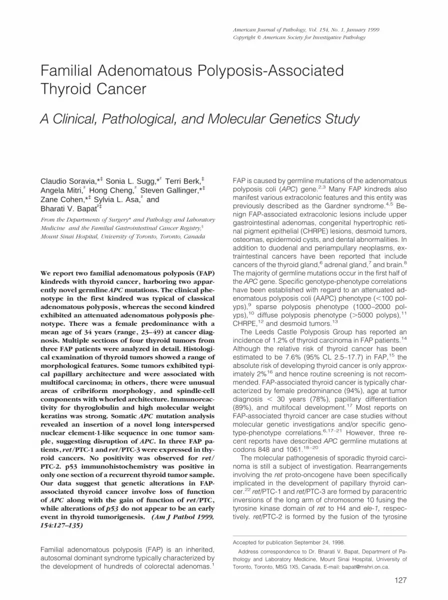
Two apparently novel APC germline mutations werefound in these two FAP kindreds.34,35 In kindred #1, PTTassay demonstrated a truncated mutant protein band of;40 kd in APC segment 1B (Figure 1A). In kindred #2, atruncated band of ;50 kd in size was detected in APCsegment 1A (Figure 1B). DNA sequence analysis re-vealed the truncation in kindred #1 to be caused by a
transversion T 3 G, resulting in a substitution of leucineby a stop codon (TTA3 TGA) at nucleotide 2092 (codon698 in exon 15). In kindred #2, the APC mutation oc-curred at nucleotides 937–938 (deletion GA) within exon9 (codon 313), resulting in a frameshift leading to a stopcodon (TGA) at nucleotides 975–977.
Histopathology of Thyroid Tumors
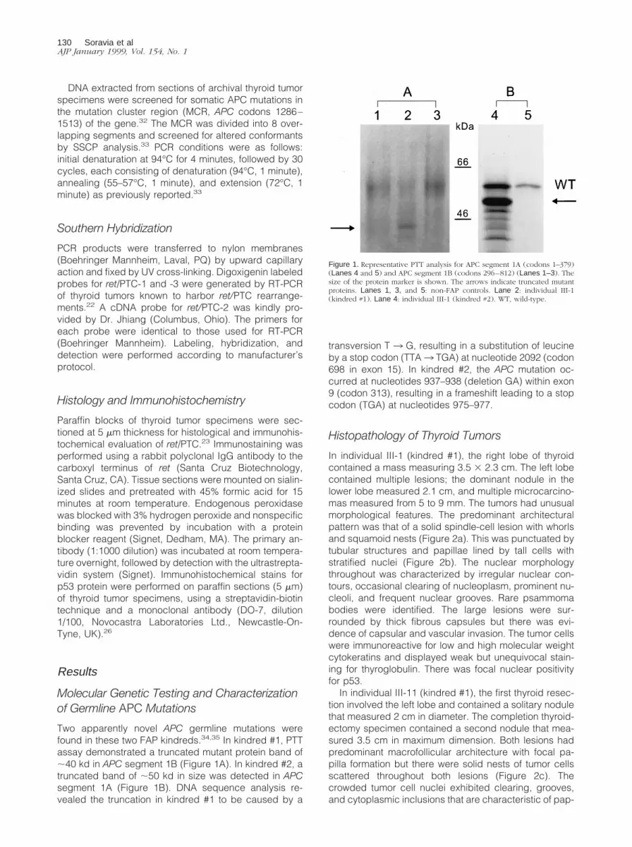
In individual III-1 (kindred #1), the right lobe of thyroidcontained a mass measuring 3.5 3 2.3 cm. The left lobecontained multiple lesions; the dominant nodule in thelower lobe measured 2.1 cm, and multiple microcarcino-mas measured from 5 to 9 mm. The tumors had unusualmorphological features. The predominant architecturalpattern was that of a solid spindle-cell lesion with whorlsand squamoid nests (Figure 2a). This was punctuated bytubular structures and papillae lined by tall cells withstratified nuclei (Figure 2b). The nuclear morphologythroughout was characterized by irregular nuclear con-tours, occasional clearing of nucleoplasm, prominent nu-cleoli, and frequent nuclear grooves. Rare psammomabodies were identified. The large lesions were sur-rounded by thick fibrous capsules but there was evi-dence of capsular and vascular invasion. The tumor cellswere immunoreactive for low and high molecular weightcytokeratins and displayed weak but unequivocal stain-ing for thyroglobulin. There was focal nuclear positivityfor p53.
In individual III-11 (kindred #1), the first thyroid resec-tion involved the left lobe and contained a solitary nodulethat measured 2 cm in diameter. The completion thyroid-ectomy specimen contained a second nodule that mea-sured 3.5 cm in maximum dimension. Both lesions hadpredominant macrofollicular architecture with focal pa-pilla formation but there were solid nests of tumor cellsscattered throughout both lesions (Figure 2c). Thecrowded tumor cell nuclei exhibited clearing, grooves,and cytoplasmic inclusions that are characteristic of pap-
Figure 1. Representative PTT analysis for APC segment 1A (codons 1–379)(Lanes 4 and 5) and APC segment 1B (codons 296–812) (Lanes 1–3). Thesize of the protein marker is shown. The arrows indicate truncated mutantproteins. Lanes 1, 3, and 5: non-FAP controls. Lane 2: individual III-1(kindred #1). Lane 4: individual III-1 (kindred #2). WT, wild-type.
130 Soravia et alAJP January 1999, Vol. 154, No. 1
illary carcinoma. Capsular invasion was seen in bothspecimens. Immunoreactivity for thyroglobulin and kera-tins was strong. The p53 antibodies have a short half-lifefor normal protein, resulting in only focal faint or negativestaining, whereas mutant p53 usually accumulates in thenucleus, producing intense positivity. In our tumor spec-imens, p53 was identified only in nuclei of cells in solidnests (Figure 2f).
In individual III-1 (kindred#2), the thyroid gland con-tained multiple nodules in both lobes varying from 0.6 to2.3 cm in diameter. Three nodules, including the largest,
had cytologic features of papillary carcinoma; the re-mainder were considered hyperplastic nodules. The car-cinomas had the architecture of follicular variant typepapillary carcinomas with follicles storing hypereosino-philic colloid and occasional papillae. They also con-tained areas of cribriform architecture (Figure 2d) or for-mation of solid nests. The lesions were unencapsulatedand infiltrated surrounding parenchyma but vascular in-volvement was not seen. The tumors contained high mo-lecular weight cytokeratins and thyroglobulin but immu-noreactive p53 was not identified.
Figure 2. a, b: Histological features of the thyroid tumor of individual III-1 (kindred # 1). The tumor has a predominant solid spindle cell architectural pattern withwhorls and squamoid nests (a) and focal papillary structures with more typical papillary morphology (b). H&E, Magnification, 390. c: Histology of the thyroidtumor of individual III-11 (kindred #1). This tumor is predominantly a follicular variant papillary carcinoma but contains scattered small papillary structures andfocal solid areas. H&E, magnification, 335. d: Histology of the thyroid tumor of individual III-1 (kindred # 2). This tumor has papillary and follicular architectureand focally there is an unusual cribiform pattern (top left). H&E, magnification, 335. e: ret immunostaining localizes cytoplasmic immunoreactivity in tumor cells.Immunoperoxidase technique with hematoxylin counterstain, Magnification, 390. f: Immunostains for p53 show nuclear reactivity in scattered tumor cells withina solid area of the tumor of the patient III-11 (kindred #1). Immunoperoxidase technique with hematoxylin counterstain. Magnification, 3120.
FAP-Associated Thyroid Cancer 131AJP January 1999, Vol. 154, No. 1
ret/PTC and Somatic APC Gene Analyses inThyroid Cancers
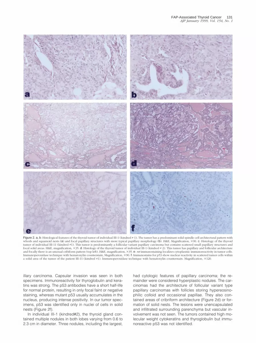
Thyroid tumor blocks from 3 FAP patients were screenedby RT-PCR for ret/PTC gene rearrangements. In patientIII-1 (kindred #1) the dominant tumor and multifocal mi-crocarcinomas were positive for ret/PTC-1. In patientIII-11 (kindred #1) the dominant tumor was positive forret/PTC-1 and three of four blocks showed focal positivityfor ret/PTC-3 (Figure 3). In patient III-1 (kindred #2) mul-tifocal papillary carcinoma was found. The three largesttumors were all positive for ret/PTC-1. No positivity forret/PTC-2 was observed in any tumor specimen.
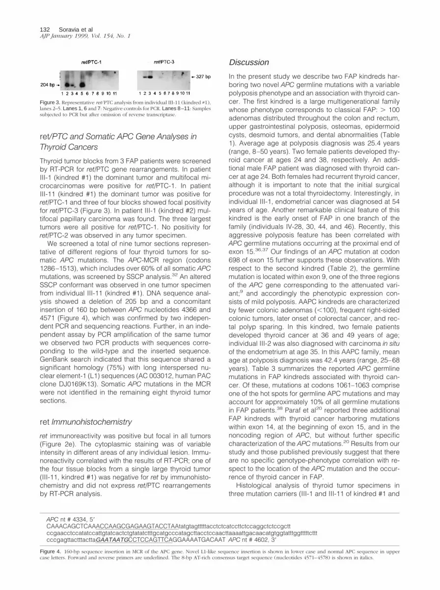
We screened a total of nine tumor sections represen-tative of different regions of four thyroid tumors for so-matic APC mutations. The APC-MCR region (codons1286–1513), which includes over 60% of all somatic APCmutations, was screened by SSCP analysis.32 An alteredSSCP conformant was observed in one tumor specimenfrom individual III-11 (kindred #1). DNA sequence anal-ysis showed a deletion of 205 bp and a concomitantinsertion of 160 bp between APC nucleotides 4366 and4571 (Figure 4), which was confirmed by two indepen-dent PCR and sequencing reactions. Further, in an inde-pendent assay by PCR amplification of the same tumorwe observed two PCR products with sequences corre-ponding to the wild-type and the inserted sequence.GenBank search indicated that this sequence shared asignificant homology (75%) with long interspersed nu-clear element-1 (L1) sequences (AC 003012, human PACclone DJ0169K13). Somatic APC mutations in the MCRwere not identified in the remaining eight thyroid tumorsections.
ret Immunohistochemistry
ret immunoreactivity was positive but focal in all tumors(Figure 2e). The cytoplasmic staining was of variableintensity in different areas of any individual lesion. Immu-noreactivity correlated with the results of RT-PCR; one ofthe four tissue blocks from a single large thyroid tumor(III-11, kindred #1) was negative for ret by immunohisto-chemistry and did not express ret/PTC rearrangementsby RT-PCR analysis.
Discussion
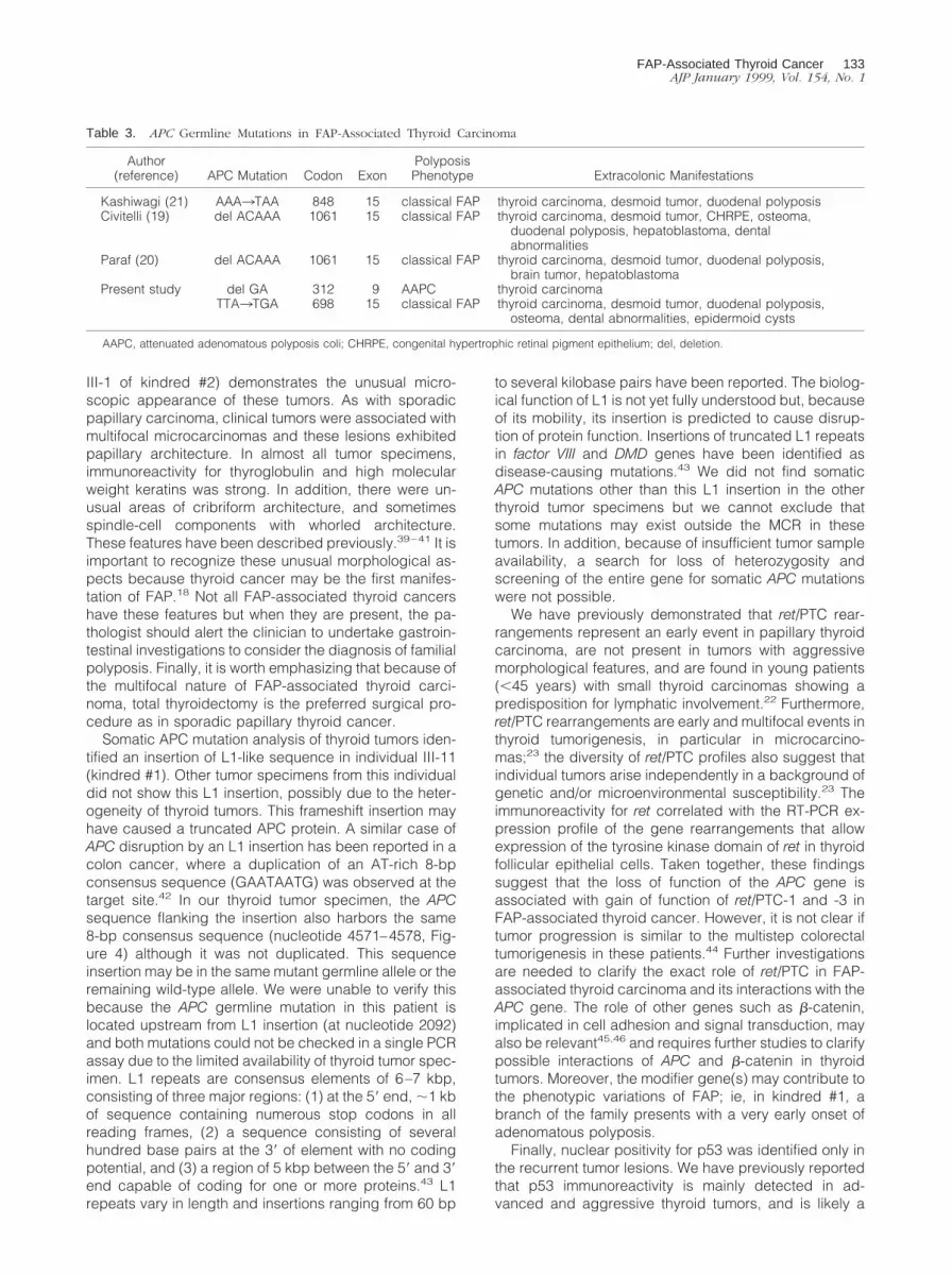
In the present study we describe two FAP kindreds har-boring two novel APC germline mutations with a variablepolyposis phenotype and an association with thyroid can-cer. The first kindred is a large multigenerational familywhose phenotype corresponds to classical FAP: . 100adenomas distributed throughout the colon and rectum,upper gastrointestinal polyposis, osteomas, epidermoidcysts, desmoid tumors, and dental abnormalities (Table1). Average age at polyposis diagnosis was 25.4 years(range, 8–50 years). Two female patients developed thy-roid cancer at ages 24 and 38, respectively. An addi-tional male FAP patient was diagnosed with thyroid can-cer at age 24. Both females had recurrent thyroid cancer,although it is important to note that the initial surgicalprocedure was not a total thyroidectomy. Interestingly, inindividual III-1, endometrial cancer was diagnosed at 54years of age. Another remarkable clinical feature of thiskindred is the early onset of FAP in one branch of thefamily (individuals IV-28, 30, 44, and 46). Recently, thisaggressive polyposis feature has been correlated withAPC germline mutations occurring at the proximal end ofexon 15.36,37 Our findings of an APC mutation at codon698 of exon 15 further supports these observations. Withrespect to the second kindred (Table 2), the germlinemutation is located within exon 9, one of the three regionsof the APC gene corresponding to the attenuated vari-ant;9 and accordingly the phenotypic expression con-sists of mild polyposis. AAPC kindreds are characterizedby fewer colonic adenomas (,100), frequent right-sidedcolonic tumors, later onset of colorectal cancer, and rec-tal polyp sparing. In this kindred, two female patientsdeveloped thyroid cancer at 36 and 49 years of age;individual III-2 was also diagnosed with carcinoma in situof the endometrium at age 35. In this AAPC family, meanage at polyposis diagnosis was 42.4 years (range, 25–68years). Table 3 summarizes the reported APC germlinemutations in FAP kindreds associated with thyroid can-cer. Of these, mutations at codons 1061–1063 compriseone of the hot spots for germline APC mutations and mayaccount for approximately 10% of all germline mutationsin FAP patients.38 Paraf et al20 reported three additionalFAP kindreds with thyroid cancer harboring mutationswithin exon 14, at the beginning of exon 15, and in thenoncoding region of APC, but without further specificcharacterization of the APC mutations.20 Results from ourstudy and those published previously suggest that thereare no specific genotype-phenotype correlation with re-spect to the location of the APC mutation and the occur-rence of thyroid cancer in FAP.
Histological analysis of thyroid tumor specimens inthree mutation carriers (III-1 and III-11 of kindred #1 and
Figure 3. Representative ret/PTC analysis from individual III-11 (kindred #1),lanes 2–5. Lanes 1, 6 and 7: Negative controls for PCR. Lanes 8–11: Samplessubjected to PCR but after omission of reverse transcriptase.
APC nt # 4334, 59CAAACAGCTCAAACCAAGCGAGAAGTACCTAAtatgtagtttttacctctcatccttctccaggctctccgcttccgaacctccatatccattgtatcactctgtatatctttgcatgcccatagcttacctccaacttaaaattgacaacatgtggtatttggtttttctttcccgagttactttacttaGAATAATGCCTCCAGTTCAGGAAAATGACAAT APC nt # 4602, 39
Figure 4. 160-bp sequence insertion in MCR of the APC gene. Novel L1-like sequence insertion is shown in lower case and normal APC sequence in uppercase letters. Forward and reverse primers are underlined. The 8-bp AT-rich consensus target sequence (nucleotides 4571–4578) is shown in italics.
132 Soravia et alAJP January 1999, Vol. 154, No. 1
III-1 of kindred #2) demonstrates the unusual micro-scopic appearance of these tumors. As with sporadicpapillary carcinoma, clinical tumors were associated withmultifocal microcarcinomas and these lesions exhibitedpapillary architecture. In almost all tumor specimens,immunoreactivity for thyroglobulin and high molecularweight keratins was strong. In addition, there were un-usual areas of cribriform architecture, and sometimesspindle-cell components with whorled architecture.These features have been described previously.39–41 It isimportant to recognize these unusual morphological as-pects because thyroid cancer may be the first manifes-tation of FAP.18 Not all FAP-associated thyroid cancershave these features but when they are present, the pa-thologist should alert the clinician to undertake gastroin-testinal investigations to consider the diagnosis of familialpolyposis. Finally, it is worth emphasizing that because ofthe multifocal nature of FAP-associated thyroid carci-noma, total thyroidectomy is the preferred surgical pro-cedure as in sporadic papillary thyroid cancer.
Somatic APC mutation analysis of thyroid tumors iden-tified an insertion of L1-like sequence in individual III-11(kindred #1). Other tumor specimens from this individualdid not show this L1 insertion, possibly due to the heter-ogeneity of thyroid tumors. This frameshift insertion mayhave caused a truncated APC protein. A similar case ofAPC disruption by an L1 insertion has been reported in acolon cancer, where a duplication of an AT-rich 8-bpconsensus sequence (GAATAATG) was observed at thetarget site.42 In our thyroid tumor specimen, the APCsequence flanking the insertion also harbors the same8-bp consensus sequence (nucleotide 4571–4578, Fig-ure 4) although it was not duplicated. This sequenceinsertion may be in the same mutant germline allele or theremaining wild-type allele. We were unable to verify thisbecause the APC germline mutation in this patient islocated upstream from L1 insertion (at nucleotide 2092)and both mutations could not be checked in a single PCRassay due to the limited availability of thyroid tumor spec-imen. L1 repeats are consensus elements of 6–7 kbp,consisting of three major regions: (1) at the 59 end, ;1 kbof sequence containing numerous stop codons in allreading frames, (2) a sequence consisting of severalhundred base pairs at the 39 of element with no codingpotential, and (3) a region of 5 kbp between the 59 and 39end capable of coding for one or more proteins.43 L1repeats vary in length and insertions ranging from 60 bp
to several kilobase pairs have been reported. The biolog-ical function of L1 is not yet fully understood but, becauseof its mobility, its insertion is predicted to cause disrup-tion of protein function. Insertions of truncated L1 repeatsin factor VIII and DMD genes have been identified asdisease-causing mutations.43 We did not find somaticAPC mutations other than this L1 insertion in the otherthyroid tumor specimens but we cannot exclude thatsome mutations may exist outside the MCR in thesetumors. In addition, because of insufficient tumor sampleavailability, a search for loss of heterozygosity andscreening of the entire gene for somatic APC mutationswere not possible.
We have previously demonstrated that ret/PTC rear-rangements represent an early event in papillary thyroidcarcinoma, are not present in tumors with aggressivemorphological features, and are found in young patients(,45 years) with small thyroid carcinomas showing apredisposition for lymphatic involvement.22 Furthermore,ret/PTC rearrangements are early and multifocal events inthyroid tumorigenesis, in particular in microcarcino-mas;23 the diversity of ret/PTC profiles also suggest thatindividual tumors arise independently in a background ofgenetic and/or microenvironmental susceptibility.23 Theimmunoreactivity for ret correlated with the RT-PCR ex-pression profile of the gene rearrangements that allowexpression of the tyrosine kinase domain of ret in thyroidfollicular epithelial cells. Taken together, these findingssuggest that the loss of function of the APC gene isassociated with gain of function of ret/PTC-1 and -3 inFAP-associated thyroid cancer. However, it is not clear iftumor progression is similar to the multistep colorectaltumorigenesis in these patients.44 Further investigationsare needed to clarify the exact role of ret/PTC in FAP-associated thyroid carcinoma and its interactions with theAPC gene. The role of other genes such as b-catenin,implicated in cell adhesion and signal transduction, mayalso be relevant45,46 and requires further studies to clarifypossible interactions of APC and b-catenin in thyroidtumors. Moreover, the modifier gene(s) may contribute tothe phenotypic variations of FAP; ie, in kindred #1, abranch of the family presents with a very early onset ofadenomatous polyposis.
Finally, nuclear positivity for p53 was identified only inthe recurrent tumor lesions. We have previously reportedthat p53 immunoreactivity is mainly detected in ad-vanced and aggressive thyroid tumors, and is likely a
Table 3. APC Germline Mutations in FAP-Associated Thyroid Carcinoma
Author(reference) APC Mutation Codon Exon
PolyposisPhenotype Extracolonic Manifestations
Kashiwagi (21) AAA3TAA 848 15 classical FAP thyroid carcinoma, desmoid tumor, duodenal polyposisCivitelli (19) del ACAAA 1061 15 classical FAP thyroid carcinoma, desmoid tumor, CHRPE, osteoma,
duodenal polyposis, hepatoblastoma, dentalabnormalities
Paraf (20) del ACAAA 1061 15 classical FAP thyroid carcinoma, desmoid tumor, duodenal polyposis,brain tumor, hepatoblastoma
Present study del GA 312 9 AAPC thyroid carcinomaTTA3TGA 698 15 classical FAP thyroid carcinoma, desmoid tumor, duodenal polyposis,
osteoma, dental abnormalities, epidermoid cysts
AAPC, attenuated adenomatous polyposis coli; CHRPE, congenital hypertrophic retinal pigment epithelium; del, deletion.
FAP-Associated Thyroid Cancer 133AJP January 1999, Vol. 154, No. 1
useful prognostic index of clinical behavior.26 Takingthese findings together, we suggest that p53 is a ratherlate event in thyroid tumorigenesis.
In summary, although specific APC mutation genotypemay not correlate with manifestations of thyroid tumors inFAP kindreds, rearrangements in ret/PTC-1 and -3 have amore relevant effect in FAP-associated thyroid tumori-genesis.
Acknowledgments
We thank Mrs. Julie Precious, Mrs. Colette Devlin, andMs. Lily Ramyar for technical assistance. We acknowl-edge the FAP patients who participated in the study.C.S. is the recipient of a Postdoctoral Research Fellow-ship from the Geneva University Hospital, Geneva,Switzerland.
References
1. Bussey HJR: Familial Polyposis Coli. Families Studies, Histopathol-ogy, Differential Diagnosis, and Results of Treatment. Baltimore, TheJohn Hopkins University Press, 1975
2. Groden J, Thliveris A, Samowitz W, Carlson M, Gelbert L, Albertsen H,Joslyn G, Stevens J, Spirio L, Robertson M, Sargeant L, Krapchno K,Wolff E, Burt R, Hughes JP, Warrington J, McPherson J, Wasmuth J,Le Paslier D, Abderrahim H, Cohen D, Leppert M, White R: Identifi-cation and characterization of the familial adenomatous polyposis coligene. Cell 1991, 66:589–600
3. Kinzler KW, Nilbert MC, Su L-K, Vogelstein B, Bryan TM, Levy DB,Smith KJ, Preisinger AC, Hedge P, McKechnie D, Finniear R,Markham A, Groffen J, Boguski MS, Altschul SF, Horii A, Ando H,Miyoshi Y, Miki Y, Nishisho I, Nakamura Y: Identification of FAP locusgenes from chromosome 5q21. Science 1991, 253:661–665
4. Gardner EJ: A genetic and clinical study of intestinal polyposis, apredisposing factor for carcinoma of the colon and rectum. Am J HumGenet 1951, 3:167–176
5. Gardner EJ, Richards RC: Multiple cutaneous and subcutaneouslesions occurring simultaneously with hereditary polyposis and osteo-matosis. Am J Hum Genet 1953, 5:139–147
6. Plail RO, Bussey HJR, Glazer G, Thomson JPS: Adenomatouspolyposis: an association with carcinoma of the thyroid. Br J Surg1987, 74:377–380
7. Schneider NR, Cubilla AL, Chaganti RSK: Association of endocrineneoplasia with multiple polyposis of the colon. Cancer 1983, 51:1171–1175
8. Turcot J, Depres JP, Pierre FS: Malignant tumours of the centralnervous system associated with familial polyposis of the colon: reportof two cases. Dis Colon Rectum 1959, 2:465–468
9. Soravia C, Berk T, Madlensky L, Mitri A, Cheng H, Gallinger S, CohenZ, Bapat B: Genotype-phenotype correlations in attenuated adeno-matous polyposis coli. Am J Hum Genet 1998, 62:1290–1301
10. Nagase H, Miyoshi Y, Horii A, Aoki T, Petersen GM, Vogelstein B,Maher E, Ogawa M, Maruyama M, Utsunomiya J, Baba S, NakamuraY: Screening for germ-line mutations in familial adenomatous polyp-osis patients: 61 new patients and a summary of 150 unrelatedpatients. Hum Mut 1992, 1:467–473
11. Nagase H, Miyoshi Y, Horii A, Ogawa M, Utsunomiya J, Baba S,Sasazuki T, Nakamura Y: Correlation between the location of germ-line mutations in the APC gene and the number of colorectal polypsin familial adenomatous polyposis patients. Cancer Res 1992, 52:4055–4057
12. Olschwang S, Tiret A, Laurent-Puig P, Muleris M, Parc R, Thomas G:Restriction of ocular fundus lesions to a specific subgroup of APCmutations in adenomatous polyposis coli patients. Cell 1993, 75:959–968
13. Caspari R, Olschwang S, Friedl W, Mandl M, Boisson C, Boker T,Augustin A, Kadmon M, Moslein G, Thomas G, Propping P: Familial
adenomatous polyposis: desmoid tumours and lack of ophthalmiclesions (CHRPE) associated with APC mutations beyond codon 1444.Hum Mol Genet 1995, 4:337–340
14. Bulow C, Bulow S, Group LCP: Is screening for thyroid carcinomaindicated in familial adenomatous polyposis? Int J Colorect Dis 1997,12:240–242
15. Giardiello FM, Offerhaus GJA, Lee DH, Krush AJ, Tersmette AC,Booker SV, Kelley NC, Hamilton SR: Increased risk of thyroid andpancreatic carcinoma in familial adenomatous polyposis. Gut 1993,34:1394–1396
16. Houlston R, Stratton M: Genetics of non-medullary thyroid cancer. QJ Med 1995, 88:685–693
17. Bell B, Mazzaferi EL: Familial adenomatous polyposis (Gardner’ssyndrome) and thyroid carcinoma. Dig Dis Sciences 1993, 38:185–190
18. Stigt J, Vasen HFA, van der Linde K, van Vliet A: Thyroid carcinomaas first manifestation of familial adenomatous polyposis. NetherJ Med 1996, 49:116–118
19. Civitelli S, Tanzini G, Cetta F, Petracci M, Pacchiarotti M, Civitelli B:Papillary thyroid carcinoma in three siblings with familial adenoma-tous polyposis. Int J Colorect Dis 1996, 11:34–37
20. Paraf F, Olschwang S, Nihoul-Fekete C, Kazandjian V, Brousse N,Schmitz J: Polypose adenomateuse familiale et cancer de la thyroıde.Gastroenterol Clin Biol 1997, 21:74–77
21. Kashiwagi H, Konishi F, Kanazawa K, Miyaki M: Sisters with familialadenomatous polyposis affected with thyroid carcinoma, desmoidtumour and duodenal polyposis. Br J Surg 1996, 83:228
22. Sugg SL, Zheng L, Rosen IB, Freeman JL, Ezzat S, Asa SL: ret/PTC-1,-2, and -3 oncogene rearrangements in human thyroid carcinomas:implications for metastatic potential? J Clin Endocrinol Metab 1996,81:3360–3365
23. Sugg SL, Ezzat S, Rosen IB, Freeman JL, Asa SL: Distinct multipleret/PTC gene reaarrangements in multifocal papillary thyroid neopla-sia. J Clin Endocrinol Metab 1998, 83:4116–4122
24. Colletta G, Sciacchitano S, Palmirotta R, Ranieri A, Zanella E, CamaA, Mariani Costantini R, Battista P, Pontecorvi A: Analysis of adeno-matous polyposis coli in thyroid tumours. Br J Cancer 1994, 70:1085–1088
25. Curtis L, Wyllie AH, Shaw JJ, Williams GT, Radulescu A, DeMicco C,Haugen DR, Varhaug JE, Lillehaug JR, Wynford-Thomas D: Evidenceagainst involvment of APC mutations in papillary thyroid carcinoma.Eur J Cancer 1994, 30A:984–987
26. Hosal SA, Apel RL, Freeman JL, Azadian A, Rosen IB, LiVolsi VA, AsaSL: Immunohistochemical localization of p53 in human thyroidneoplasms: correlation with biological behavior. Endocr Pathol 1997,8:21–28
27. Zeki K, Spambalg D, Sharifi N, Gonsky R, Fagin JA: Mutations of theadenomatous polyposis coli gene in sporadic thyroid neoplasms.J Clin Endocrinol Metab 1994, 79:1317–1321
28. Cetta F, Chiappetta G, Melillo R, Petracci M, Montalto G, Santoro M,Fusco A: The ret/PTC-1 oncogene is activated in familial adenoma-tous polyposis-associated thyroid papillary carcinomas. J Clin Endo-crinol Metab 1998, 83:1003–1006
29. Soravia C, Bapat B, Cohen Z: Familial adenomatous polyposis (FAP)and Hereditary nonpolyposis colorectal cancer (HNPCC): a review ofclinical, genetic and therapeutic aspects. Schweiz Med Wochenschr1997, 127:682–690
30. Powell SM, Petersen GM, Krush AJ, Booker S, Jen J, Giardello FM,Hamilton SR, Vogelstein B, Kinzler K: Molecular diagnosis of familialadenomatous polyposis. N Engl J Med 1993, 329:1982–1987
31. Sanger F, Nicklen S, Coulson A: DNA sequencing with chain-termi-nating inhibitors. Proc Natl Acad Sci USA 1977, 74:5463–5467
32. Miyoshi Y, Nagase H, Horii A, Ichii S, Nakatsuru S, Aoki T, Mori T,Nakamura Y: Somatic mutations of the APC gene in colorectaltumours: mutation cluster region in the APC gene. Hum Mol Genet1992, 1:229–233
33. Bapat B, Odze R, Mitri A, Ward M, Gallinger S: Identification ofsomatic APC gene mutations in periampullary adenomas in a patientwith familial adenomatous polyposis (FAP). Hum Mol Genet 1993,2:1957–1959
34. Beroud C, Soussi T: APC gene: database of germline and somaticmutations in human tumours and cell lines. Nucleic Acids Res 1996,24:121–124
35. De Vries E, Ricke D, De Vries T, Hartmann A, Blaszyk H, Liao D,
134 Soravia et alAJP January 1999, Vol. 154, No. 1
Soussi T, Kovach J, Sommer S: Database of mutations in the p53 andAPC tumor suppressor genes designed to facilitate molecular epide-miological analyses. Hum Mutat 1996, 7:202–213
36. Bunyan D, Shea-Simonds J, Reck A, Finnis D, Eccles D: Genotype-phenotype correlations of new causative APC gene mutations inpatients with familial adenomatous polyposis. J Med Genet 1995,32:728–731
37. Distante S, Nasioulas S, Somers GR, Cameron DJ, Young MA, ForrestSM, Gardner RJ: Familial adenomatous polyposis in a 5 year oldchild: clinical, pathological, and molecular genetic study. J MedGenet 1996, 33:157–160
38. Nagase H, Nakamura Y: Mutations of the APC (adenomatous polyp-osis coli) gene. Hum Mutat 1993, 3:425–434
39. Cetta F, Toti P, Petracci M, Montalto G, Disanto A, Lore F, Fusco A:Thyroid carcinoma associated with familial adenomatous polyposis.Histopathology 1997, 31:231–236
40. Harach HR, Williams GT, Williams ED: Familial adenomatous polyp-osis associated thyroid carcinoma: a distinct type of follicular cellneoplasm. Histopathology 1994, 25:549–561
41. Hizawa K, Iida M, Aoyagi K, Oohata Y, Mibu R, Yamasaki K, Hirata T,Fujishima M: Association between thyroid cancer of cribriform variantand familial adenomatous polyposis. J Clin Pathol 1996, 49:611–613
42. Miki Y, Nishisho I, Horii A, Miyoshi Y, Utsunomiya J, Kinzler KW,Vogelstein BV, Nakamura Y: Disruption of the APC gene by a retro-transposal insertion of L1 sequence in a colon cancer. Cancer Res1992, 52:643–645
43. Kazazian HH, Moran JV: The impact of L1 retrotransposons on thehuman genome. Nat Genet 1998, 19:19–24
44. Kinzler K, Vogelstein B: Lessons from hereditary colorectal cancer.Cell 1996, 87:159–170
45. Korinek V, Barker N, Morin P, van Wichen D, de Weger R, Kinzler K,Vogelstein B, Clevers H: Constitutive transcriptional activation by ab-catenin-Tcf complex in APC2/2 colon carcinoma. Science 1997,275:1784–1787
46. Rubinfeld B, Robbins P, El-Gamil M, Albert I, Porfiri E, Polakis P:Stabilization of b-catenin by genetic defects in melanoma cell lines.Science 1997, 275:1790–1792
FAP-Associated Thyroid Cancer 135AJP January 1999, Vol. 154, No. 1