factors affecting brown adipose tissue activity in animals and man
TRANSCRIPT

4
Factors Affecting Brown Adipose TissueActivity in Animals and Man
DANIEL RICQUIERGERARD MORY
Brown adipose tissue (BAT) is the only known tissue whose main functionis heat production. BAT is an important site of non-shivering thermogenesis and perhaps of diet-induced thermogenesis. Thus. this tissue probablyplays a significant role in the regulation of energetic equilibrium in mostmammals. Before describing those factors which control BAT activity, wewill briefly describe its organization and function. The specialist reader isreferred to the great number of excellent reviews on BAT and itsmitochondria which have recently been produced (Cannon and Lindberg.1979; Nicholls. 1979; Lindberg, Cannon and Nedergaard, 1981; Nederguard and Lindberg. 1982; Girardier, 1983; Nicholls and Locke, 1983; Cannon and Ncdergaard, 1984a and b; Himms-Hagen, 1984; Nedergaard andCannon, 1984a; Nicholls and Locke, 1984).
Between 1961 and 1965. several groups of workers showed that heat production was the principal function of BAT. This conclusion was based onstudies of three different physiological states where a mammal's bodyrequires heat production: birth, acclimation to cold, and arousal fromhibernation (reviews: Smith and Horwitz, 1969; Hull and Hardman, 1970).
PHYLOGENETIC DISTRIBUTION, BODY LOCATION ANDl\IORPHOLOGY
BAT exists in most mammals but has never been identified in nonmammals. even in homoiothermic animals such as birds. In the human species, BAT has been well characterized in the newborn (reviews: Hull andHardman, 1970; Cannon and Johansson, 1980; Lean and James. 1983).Although islets or pieces of BAT have been observed in human adults(Heaton, 1972; Tanuma et ai, 1976; Hassi, 1977; Huttunen, Hirvonen andKinnula, 1981), BAT in the human adult has not been quantified, and thequestion of its physiological importance is presently a subject of controversy (see Astrup et al, 1984).
BAT can account for 0.5 to 5 per cent of the body weight of most rnam-
Clinics ill Endocrinology and Metabolism-Vol. 13. No.3. November 1984 501

502 DANIEL RICQUIER AND GERARD MORY
mals . This tissue is generally more abundant in interscapular, cervical andthoracic regions in small mammals such as rodents and lagomorphs and inthe perirenal region of larger species such as the lamb (review: Afzelius ,1970).
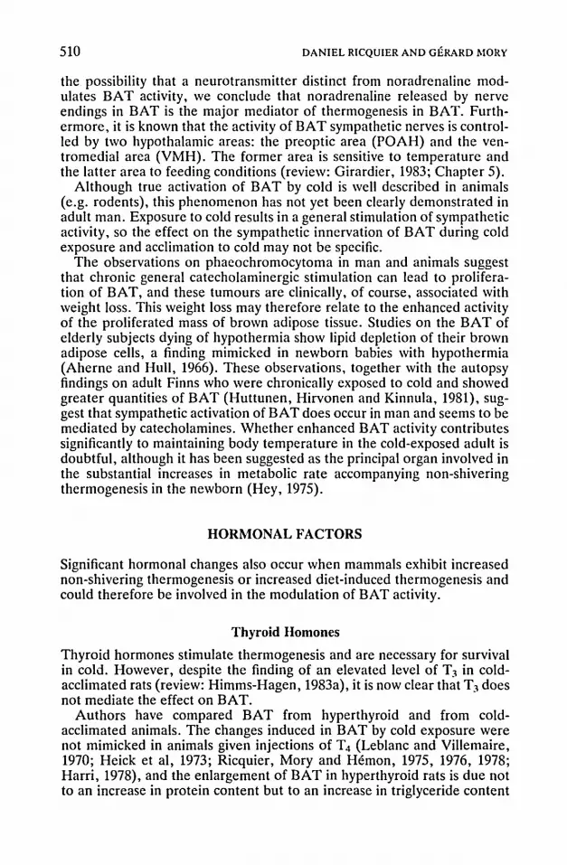
BAT is composed of brown adipocytes which have characteristic features (Figure 1), with many small multilocular triglyceride droplets and anexceptionally high content of mitochondria which are well filled with cristae. BAT is also characterized by numerous blood capillaries and by a richnetwork of sympathetic nerves . These sympathetic fibres innervate arterialblood vessels , and they also directly innervate brown adipocytes throughabundant synaptic varicosities which run between the adipocyte cells. Thetransmitter released by these varicosities is noradrenaline (reviews: Cottle,1970; Schonbaum, Steiner and Sellers, 1970; Barnard, Mory and Nechad,1980). Several authors have proposed that sympathetic relays exist insideBAT, but this has been repudiated by recent investigations. Further, noother aminergic fibres, such as serotoninergic or dopaminergic fibres, havebeen found in BAT (review: Mory, Combes-George and Nechad, 1983).
THE MECHANISMS OF THERMOGENESIS IN THE BROWNADIPOCYTE
It is now well established that thermogenesis in the brown fat cell resultsmainly from a physiological uncoupling of oxidative phosphorylation inmitochondria. Elevated amounts of free fatty acids can be generated in thecell through lipolysis, fatty acid synthesis and importation of fatty acids.These free fatty acids are quickly oxidized inside mitochondria. In contrastto most mitochondria, where respiration and AOP phosphorylation areobligatorily linked, free energy from the oxidation of substrates is directlydissipated as heat. This uncoupling mechanism involves a specific regulatedtransport of protons across the inner membrane (reviews: Nicholls andLocke , 1983, 1984). The proton pathway is specific to brown fat mitochondria and is inhibited by nucleotides (AOP, ATP, GOP and GTP) and acti vated by free fatty acids (reviews: Nicholls and Locke, 1983, 1984). Themolecular basis for this unique H + conductance pathway is a specific protein (M, 32 (00), named uncoupling protein or thermogenin, located in theinner mitochondrial membrane. Before its identification as the proteinresponsible for the uncoupling of mitochondria (Heaton et ai, 1978), thiscomponent had been shown to be present in large amounts in the membranes of mitochondria from active brown fat and in smaller amounts inmitochondria from weakly active BAT (Ricquier and Kader, 1976). Manystudies subsequently led to the conclusion that the total amount of uncoupling protein in BAT determines the capacity of the tissue for thermogenesis (reviews: Nicholls and Locke, 1984; Cannon and Nedergaard, 1984b).Thus, factors which are able to modulate the activity and the amount of theuncoupling protein are factors which control the activity of BAT.
When thermogenesis in BAT is activated at birth, during acclimation tocold, and during arousal from hibernation the heat produced by BAT rep-

tll:l:lo::;:z>a:;;otilm--IVitilcm;I-
::l<:~
Figure 1. Electron micrographs of interscapular brown adipose tissue taken from a cold-acclimated rat.Courtesy of Dr M. Nechad. Left (la): x 2000. Right (lb): x 30000. L = lipid droplets: N = nucleus:M = mitochondria; G = glycogen: C = capillary.
Ulow

504 DANIEL RICQUIER AND GERARD MaRY
resents a large part (30 to 80 per cent) of total non-shivering thermogenesis(Hull and Hardman, 1970; Foster and Frydrnan, 1979; Nedergaard andCannon, 1984b). In addition to its considerable role in non-shivering thermogenesis (at least in rodents: Foster and Frydman , 1979), recent work hasimplicated BAT in diet-induced thermogenesis and regulation of bodyweight (Rothwell and Stock, 1979) (see Chapters 1 and 2 of this volume).
NEURAL FACTORS
The factors which control non-shivering thermogenesis and diet-inducedthermogenesis in BAT are probably the same factors and can be dividedinto neural and hormonal factors. Non-shivering thermogenesis is essentially controlled by sympathetic nerves and noradrenaline (review: Jansky,1973), and BAT is a major site of non-shivering thermogenesis (Foster andFrydman, 1979). Thus, sympathetic neural factors could control BATactivity.
Noradrenaline released by the sympathetic neural afferents in BAT isbound to receptors on the surface of brown adipocytes. Three types ofcatecholamine receptors have been detected in brown adipocytes ofrodents. Classed according to number and physiological function, the mostimportant are B-adrenoreceptors which are associ ated with adenylatecyclase activity and cAMP production. They exhibit an affinity pattern foragonists, characteristic of the B.-subtype: isoprenaline (isoproterenol) »noradrenaline> adrenaline » phenylephrine (reviews: Girardier, 1983;Cannon and Nedergaard, 1984a) . However, Arch et al (1984) have synthesized and tested a new B-agon ist which specifically stimulates B-receptors ofBAT, and they concluded that these receptors are not true B.-receptors.
Brown adipocytes of rodents also contain aI-receptors, of which the pattern of affinity for a-antagonists is prazosin > phentolamine » yohimbine (Mohell, Nedergaard and Cannon, 1983; Mohell, Svartengren andCannon , 1983). As for a.-receptors in other cells, calcium and phosphatidylinositol are involved in the activation of aI-receptors of brown fat cells(Garcia-Sainz, Hasler and Fain, 1980; Mohell, Wallace and Fain, 1984;review: Nedergaard et al, 1984).
Rat brown adipocytes also possess az-adrenoreceptors, the stimulationof which inhibits B-effects (Sundin and Fain, 1983). Such receptors werenot found in hamster BAT (McMahon and Schimmel, 1982). These threetypes of receptors (B., a. and a2) were found in lamb brown adipocytes(Fain et al, 1984). The catecholamine receptors of BAT vasculature havenot been studied.
Although noradrenaline, which is the natural mediator of thermogenesisin BAT, can interact with both a-adrenergic and B-adrcnergic receptors,most heat produced by brown fat cells results from interaction with ~I
receptors (Mohell, Nedergaard and Cannon, 1983; review: Cannon andNedergaard,1984b).

BROWN ADIPOSE TISSUE ACfIVITY 505
Sympathetic Activation of Brown Adipose Tissue
Acute exposure to cold induces a rapid increase in noradrenaline release inBAT (Cottle, 1970; Young et aI, 1982). This sympathetic activation persists if exposure to cold is chronic (Cottle, 1970; Kennedy, Hammond andHamolsky, 1977). Such an elevated release of noradrenaline induces only amoderate 'down-regulation' of the number of B-receptors, but there is alsoreduced coupling between B-receptors and cyclase (Svartengren, Svobodaand Cannon, 1982).
Dietary intake also modulates the release of noradrenaline in BAT. A48-hour fast markedly decreases sympathetic activity in rat BAT (Young etaI, 1982), while chronic overfeeding using the 'cafeteria diet' enhancesnoradrenaline release in BAT (Young et aI, 1982). However, short-term oracute sucrose-overfeeding affects this release only slightly and contrastswith that observed during acute exposure to cold (Young et aI, 1982): seeChapter 3.
Acute cold exposure
The response of BAT to acute cold exposure is mainly metabolic. The lipidstores are rapidly mobilized to supply fuel to the mitochondrial machinerywhich produces heat through uncoupling of oxidative phosphorylation.Within one minute of the start of lipolysis in the brown adipocyte , the cellcan increase its respiration rate 10 to 40 times. Free fatty acids are not onlysubstrates for mitochondrial respiration but also immediately increase proton conductance of the inner mitochondrial membrane and hence uncouplerespiration (Rial, Poustie and Nicholls, 1983; review: Nicholls and Locke,1984). A marked increase in the ability of GDP to bind to isolated BATmitochondria also occurs within the first hour of exposure of rats to cold(Desautels, Zaror-Behrens and Himrns-Hagen, 1978). GDP binds to theuncoupling protein (Heaton et aI, 1978), and the binding is considered toreflect the activity of the proton conductance pathway of BAT mitochondria. During a short exposure to cold, there is also a rapid increase inlipoprotein lipase activity in BAT which allows the tissue to oxidize circulating lipids (Radomski and Orme, 1971; Cameheim, Nedergaard andCannon, 1984). Moreover, the metabolic activation of brown fat cells isaccompanied by a large increase in blood flow through the tissue(Kuroshima, Konno and Itoh, 1967; Foster and Frydrnan, 1979).
All these events are undoubtedly mediated by the activation of thesympathetic nerves in BAT and the subsequent release of noradrenaline.It has long been known that surgical denervation of BAT prevents lipidmobilization in the tissue during cold exposure (Hausberger, 1934; review:Girardier, 1983). Oxidative metabolism of isolated brown adipocytes canbe directly increased in vitro by adding noradrenaline and almost totallysuppressed by adding B-blockers such as propranolol (reviews: Lindberg,Bieber and Houstek, 1976; Nedergaard and Lindberg, 1982; Cannon andNedergaard, 1984a). The catabolic cascade starts with the binding ofnoradrenaline to B-receptors and continues with an increase in cAMP con-

506 DANIEL RICQUIER AND GERARD MaRY
centration and activation of hormone-sensitive lipase. According to recentdata, 80 per cent of the increase in oxygen consumption following noradrenaline binding to brown fat cells results from stimulation of the B-receptoradenyl ate cyclase system and 20 per cent corresponds to an a-receptormediated effect (Mohell, Nedergaard and Cannon, 1983). The rapidincrease in GDP binding to isolated mitochondria of rats during acute coldexposure can be induced by a l-hour intravenous infusion of noradrenaline(Desautels and Himms-Hagen, 1979), and this effect can be largelyimpaired by pretreating the animals with propranolol (Mory et aI, 1984b).The enhancement of lipoprotein lipase activity during short cold exposurecan be reproduced by injecting isoprenaline or noradrenaline and isblocked by propranolol (Radomski and Horme, 1971; Carneheim, Nedergaard and Cannon, 1984). The increase in blood-flow which is characteristic of the response of BAT to cold is also a consequence of stimulation ofthe tissue by noradrenaline (Hull and Hardman, 1970; Foster andDepocas, 1980). Acute cold exposure causes mobilization of serotoninfrom BAT mast cells (Mory, Combes-George and Nechad, 1983) which inturn can stimulate noradrenaline release from nerve endings (Steiner andEvans, 1976) and can thus amplify sympathetic activation.
Chronic cold exposure
When animals are chronically exposed to cold, the metabolic activation ofBAT persists and lipolysis, lipogenesis, oxygen consumption and lipoprotein lipase activity remain elevated (review: Nicholls and Locke, 1984).With prolonged cold exposure in adult animals, BAT shows further adaptive changes which are the opposite of the normal involution expected inthis tissue after the first month of life in most non-hibernating rodents. Thisadaptive response can be detected within three to five hours of exposure tocold and reaches a maximum after several weeks of cold exposure (Table1). Cold acclimation of rats also leads to increased blood flow throughBAT (Kuroshima, Konno and Itoh, 1967, Foster and Frydman, 1979). Theorgan also proliferates with endothelial precursor celis maturing and leading to a doubling or tripling of the organ's DNA content (review: Barnard,Mory and Nechad, 1980). Mitochondrial mass also increases markedly onan organ and cell basis; the proportion of the 32 OOO-M r mitochondrialuncoupling protein is also strikingly increased (reviews: Nicholls andLocke, 1984; Nedergaard and Cannon, 1984a). The selective enrichment inuncoupling protein can be detected within 3 h of cold exposure (Mory etaI, 1984b). These events are probably explained by the observation that thelevel of mRNA encoding the uncoupling protein increases during prolonged exposure to cold (Ricquier et ai, 1983b; Bouillaud et aI, 1984).Moreover, numerous enzymatic activities, such as cytochrome c oxidaseactivity, are enhanced and the fatty acid composition of phospholipids isaltered (review: Barnard, Mory and Nechad, 1980). All the modificationsobserved during prolonged cold exposure allow a striking increase in theheat production capacity of BAT.
Several observations clearly indicate that the integrity of BAT innerva-

Table 1. The trophic response of brownadipose tissue of rats acclimated to coldand itscontrol by the sympathetic innervation
ChemicalChronic cold sympathectomyand Daily injection Continuous delivery Phacochromocytoma
exposure" cold exposure" of catccholamines" of noradrenaline" tumour (PC 12cells)"
Weight of BAT + 0 + + +DNA content + 0 + + +Protein concentration + 0 + + +Phospholipidconcentration + 0 0 n.d, +Unsaturation degree of phospholipids + 0 0 n.d, +Mitochondrialprotein concentration + 0 n.d. + +Cytochrome c oxidase activityper cell + 0 + + 0Uncouplingprotein concentration in mitochondria + 0 0 + +GDP binding to isolated mitochondria + 0 0 + +
+ = Large increase.o= No or weak increase.n.d. =Not determined.• Data from Barnard, Mory and Nechad (1980)and Mory et al (1982).b Data from Desautels and Himms-Hagen (1979)and Mory, Ricquier and Hernon (1980).C Data from Mory et al (1984a).d Data from Ricquier ct al (1983a).
Ol::<lo~z>o:;;oVlrn
"""ViVlc:tTl>B-e~
V1
~

508 DANIEL RICQUIER AND GERARD I\IORY
tion is necessary for development of the tissue. After 5 h cold exposure,normal cell proliferation does not occur in surgically denervated BAT(Hunt and Hunt, 1967), and the increase in the uncoupling protein concentration is inhibited by pretreatment of animals with propranolol (Mory etai, 1984b). Likewise, the major features of the trophic response of BAT incold-acclimated animals (increases in DNA, total and mitochondrial protein, cytochrome c oxidase activity and uncoupling protein, alterations ofphospholipids) are suppressed or largely impaired in rats chemically sympathectomized using guanethidine treatment (Mory et ai, 1982). The roleof innervation has also been studied using BAT grafts (Nechad and Olson,1983).
Daily injections of noradrenaline or of isoprenaline in rats for severalweeks can induce growth of BAT, including hyperplasia and increasesin protein content and cytochrome c oxidase activity (Leblanc andVillemaire, 1970; Heick et al, 1973; Desautels and Himms-Hagen, 1979;Mory, Ricquier and Hernon, 1980). However, these rats did not exhibit thecharacteristic increase in uncoupling protein observed during cold exposure(Desautels and Himms-Hagen, 1979; Mory, Ricquier and Hernon, 1980).This observation implied that noradrenaline and sympathetic innervationwere necessary but not sufficient to induce the full adaptive response ofBAT. However, recent experiments with continuous infusion (rather thansingle daily injections) of noradrenaline, using implanted mini-osmoticpumps, showed that BAT of noradrenaline-infused rats developed in a waysimilar to that observed in cold-acclimated rats (Mory et ai, 1984a). Theproportion of mitochondrial uncoupling protein in these experimentsincreased as much as in those experiments in which animals were exposedto prolonged cold. The effect was observed only when the pump deliveringnoradrenaline was placed near interscapular BAT, and not when the pumpwas implanted 6 to 9 em away from the tissue (Mory et ai, 1984, unpublished data). These data favour the proposition that noradrenaline releasedby nerve endings in BAT is the agent responsible for the adaptive trophicresponse of BAT. This conclusion is in excellent agreement with theobservations made by Arch et al (1984) and by Young, Wilson and Arch(1984). These authors have shown development of BAT and a specificincrease in the uncoupling protein in rats chronically treated with longacting ~-adrenoreceptor agonists such as fenoterol or by a new compound(from Beecham Laboratories) which has a strong affinity for the ~
adrenoreceptors of BAT.
Phaeochromoeytoma in Animals and Humans
The development of BAT, and sometimes of BAT pseudotumours, hasbeen described in humans and animals bearing phaeochromocytomastumours which secrete large amounts of catecholamines (reviews: Feyrter,1973; Girardier, 1983). The implantation of phaeochrornocytoma tumours(PC 12 cell line) in rats induces the development of BAT and stimulates thesynthesis of the uncoupling protein (Ricquier et ai, 1983a; Bouillaud et ai,1984). An ultrastructural and biochemical study of perirenal fat from

BROWN ADIPOSE TISSUE ACTIVITY 509
humans bearing phaeochromocytoma tumour has revealed that this tissuewas typical of BAT, with mitochondria behaving biochemically like BATmitochondria (Ricquier, Nechad and Mory, 1982; Bouillaud, CombesGeorge and Ricquier, 1983). Conversely, perirenal fat in patients bearingno phaeochromocytoma resembles typical unilocular white adipose tissue.Interestingly, it has been reported that brown fat pseudotumours can beconfused with the tumour itself in patients with phaeochromocytoma(review: Girardier, 1983). Thus, sympathetic factors released by tumorouschromaffin cells (probably catecholamines) are able to stimulate BATdevelopment and to induce maturation of precursor cells and/or 'dedifferentiation' of unilocular fat cells. Furthermore, the apparent transformation of white fat cells into brown adipocytes has been described in severechronic hypoxaemia which induces sympathetic activation in man (Teplitzet al, 1974). The major effects induced by catecholamine infusions or in thepresence of phaeochromocytoma are indicated in Table 1.
The Role of Neural Factors in the Response to Diet
Rats overfed a cafeteria diet have more BAT than animals fed a stock diet(Rothwell and Stock, 1979; Chapter 1). This growth of BAT is due tohyperplasia (Himms-Hagen, Triandafillou and Gwilliam, 1981; Bukowiecki et al, 1982), and GOP binding to isolated BAT mitochondriaincreases (Brooks et ai, 1980; Himms-Hagen, Triandafillou and Gwilliam,1981). Moreover, blood-flow through BAT is largely increased (Rothwelland Stock, 1981a). Thus, chronic overeating and chronic cold exposureinduce almost similar modifications in BAT, leading to an enhancement ofthe thermogenic capacity of the tissue (see Chapter 1). Furthermore, asingle meal has been reported to activate BAT metabolism (Glick, Teagueand Bray, 1981). However, an increase in the proportion of uncouplingprotein has not been observed so far (Himms-Hagen, Triandafillou andGwilliam,1981).
As in the case of cold exposure, a great number of observations haveshown that sympathetic innervation of BAT and released noradrenalineplay key roles in the development of BAT during chronic overfeeding(review: Stock and Rothwell, 1981). Chronic cafeteria feeding increasesnoradrenaline release in BAT of rats (Young et al, 1982), and the trophicresponse of BAT in rats overfed with sucrose is impaired by chemicalsympathectomy (Sundin and Nechad, 1983). Conversely, BAT of obeseanimals has a low thermogenic capacity (see Chapter 2 of this issue)which is associated with a decrease in noradrenaline turnover in the tissue(Knehans and Romsos, 1982; Levin, Triscari and Sullivan, 1983) andwith a chronic lack of sympathetic activation of BAT (Seydoux et al, 1981).
Conclusion: Neural Factors
The evidence of the interaction between BAT and its sympathetic innervation clearly shows that the neural distribution within this tissue plays anessential role in the control of tissue activity. Although we cannot exclude
510 DANIEL RICQUIER AND GERARD MORY
the possibility that a neurotransmitter distinct from noradrenaline modulates BAT activity, we conclude that noradrenaline released by nerveendings in BAT is the major mediator of thermogenesis in BAT. Furthermore, it is known that the activity of BAT sympathetic nerves is controlled by two hypothalamic areas: the preoptic area (POAH) and the ventromedial area (VMH). The former area is sensitive to temperature andthe latter area to feeding conditions (review: Girardier, 1983; Chapter 5).
Although true activation of BAT by cold is well described in animals(e.g. rodents), this phenomenon has not yet been clearly demonstrated inadult man. Exposure to cold results in a general stimulation of sympatheticactivity, so the effect on the sympathetic innervation of BAT during coldexposure and acclimation to cold may not be specific.
The observations on phaeochromocytoma in man and animals suggestthat chronic general catecholaminergic stimulation can lead to proliferation of BAT, and these tumours are clinically, of course, associated withweight loss. This weight loss may therefore relate to the enhanced activityof the proliferated mass of brown adipose tissue. Studies on the BAT ofelderly subjects dying of hypothermia show lipid depletion of their brownadipose cells, a finding mimicked in newborn babies with hypothermia(Aherne and Hull, 1966). These observations, together with the autopsyfindings on adult Finns who were chronically exposed to cold and showedgreater quantities of BAT (Huttunen, Hirvonen and Kinnula, 1981), suggest that sympathetic activation of BAT does occur in man and seems to bemediated by catecholamines. Whether enhanced BAT activity contributessignificantly to maintaining body temperature in the cold-exposed adult isdoubtful, although it has been suggested as the principal organ involved inthe substantial increases in metabolic rate accompanying non-shiveringthermogenesis in the newborn (Hey, 1975).
HORMONAL FACTORS
Significant hormonal changes also occur when mammals exhibit increasednon-shivering thermogenesis or increased diet-induced thermogenesis andcould therefore be involved in the modulation of BAT activity.
Thyroid Homones
Thyroid hormones stimulate thermogenesis and are necessary for survivalin cold. However, despite the finding of an elevated level of T) in coldacclimated rats (review: Himms-Hagen, 1983a), it is now clear that T) doesnot mediate the effect on BAT.
Authors have compared BAT from hyperthyroid and from coldacclimated animals. The changes induced in BAT by cold exposure werenot mimicked in animals given injections of T4 (Leblanc and Villemaire,1970; Heick et al, 1973; Ricquier, Mory and Hernon, 1975, 1976, 1978;Harri, 1978), and the enlargement of BAT in hyperthyroid rats is due notto an increase in protein content but to an increase in triglyceride content

BROWN ADIPOSE TISSUE ACfIVITY 511
(Lachance, 1953; Heick et ai, 1973; Ricquier, Mory and Hernon, 1976;Triandafillou, Gwilliam and Himrns-Hagen, 1982). Recently, it has alsobeen reported that thyroxine injections in rats decrease the level ofmitochondrial uncoupling protein, measured by GDP binding to mitochondria (Sundin, 1981; Triandafillou, Gwilliam and Himms-Hagen, 1982), andprevent the normal cold-induced changes occurring in BAT (Ricquier,Mory and Hernon, 1976, 1978).
Other studies have been carried out in hypothyroid animals . Such animals have a BAT composition similar to that found in the cold-acclimatedanimal-e .g., increases in wet weight, DNA content, mitochondrial protein, cytochrome c oxidase activity (Mory et ai , 1981). However, BATfrom hypothyroid animals is refractory in its reaction to noradrenaline asshown by (1) a poor lipolytic response to added noradrenaline (review:Hernon, Ricquier and Mory, 1976) or to cold stress (Mory et ai, 1981); (2)a sharply decreased metabolic response to nerve stimulation (Seydoux,Giacobino and Girardier, 1982); and (3) no increase in blood-flow duringchronic cold exposure (Kuroshima, Konno and Itoh, 1967). Moreover,although the amount of mitochondrial uncoupling protein per cell was notincreased in hypothyroid rats, the total amount in the organ was doubled(Mory et al, 1981; Triandafillou, Gwilliam and Himms-Hagen, 1982). Sellers, Flattery and Steiner (1974) had previously observed that hypothyroidrats provided with the minimum dose of T, for survival in cold were able todevelop their BAT during prolonged cold exposure.
It is noteworthy that, in 1850, Curling described 'symmetrical swellingsof fat tissue at the side of the neck' in human babies with cretinism. Later itwas recognized that this tissue was BAT (Shattock, 1909). The development of BAT in young cretins is due mainly to accumulation of fat becauseof inhibited lipolysis and a high lipoprotein lipase activity (Hernon, Ricquier and Mory, 1975, 1976).
This complex involvement of the thyroid (also noted in Chapter 8) maybe simply explained as follows: in comparison with euthyroid animals,hyperthyroid animals have increased metabolism with a rise in bodytemperature and therefore a reduced drive for BAT thermogenesis fromthe hypothalamus via the sympathetic nervous system. The oppositemechanism could occur in the hypothyroid animal. Nevertheless,hypothyroidism does not completely mimic the effects of cold exposure,since noradrenaline sensitivity of BAT is abnormal.
The role of thyroid hormones in BAT may therefore be restricted to apermissive role (Triandafillou, Gwilliam and Himrns-Hagen, 1982;Hernon, Ricquier and Mory, 1974; Sellers, Flattery and Steiner, 1974).These hormones may neither stimulate the activity of BAT directly normediate the cold-induced development of the thermogenic capacity of thetissue. We can unequivocally conclude that BAT is not a target of thyroidstimulated thermogenesis.
Pancreatic Hormones
It has been reported that glucagon secretion is increased during cold exposure and that this hormone is able to directly stimulate lipolysis in BAT

512 DANIEL RICQUIER AND GERARD MaRY
(review: Kuroshirna et al, 1984). Glucagon has a marked calorigenic actionwhen it is added to isolated brown adipocytcs but such an effect is obtainedwith a concentration of glucagon which is 3000 times the glucagonaemiameasured by the same authors . Thus, the hypothesis of a physiological rolefor glucagon in the induction of BAT thermogenesis is doubtful.
Insulin has obvious metabolic effects on isolated brown adipocytes orfragments of tissue. These effects are in many ways similar to thoseobserved in white adipose tissue (reviews: Nedergaard and Lindberg, 1982;McCormack, 1982). These effects include stimulation of glucose uptake,glucose oxidation and oxygen consumption, and stimulation of fatty acidsynthesis via activation of pyruvate dehydrogenase and acetyl-CoA carboxylase. There is a decrease in adrenaline-stimulated glycerol and freefatty acid release, and a smaller rise in cyclic AMP induced by addednoradrenaline.
BAT is a major site of lipogenesis in the cold-adapted rat (McCormackand Denton, 1977; Trayhurn, 1979; review: McCormnck, 1982). Glucose isthe predominant substrate for fatty acid synthesis in BAT (review: McCormack, 1982) and this process is stimulated by insulin (McCormack andDenton, 1977; Agius and Williamson, 1980; Agius et al, 1981). It is alsoknown that glycolysis in the brown fat cell is necessary for thermogenesis inorder to increase the level of citric acid cycle intermediates (Nedergaardand Lindberg, 1982). McCormack (1982) has proposed that, underappropriate circumstances, glucose could be a substrate for oxidation inBAT, and that 'the function of insulin could be seen as a switch mechanismwhereby the fat fuel present in the tissue for thermogenesis can be conserved and augmented when glucose, as an alternative fuel is available inthe bloodstream'. Increased lipogenesis in BAT does not necessarily meanthat there is increased thermogenesis in the tissue.
An increase in BAT temperature is obtained in response to electricalstimulation of the ventromedial hypothalamic nucleus (Perkins et al,1981). Such stimulation of the YMH enhances lipogenesis preferentially inBAT, in normal and in diabetic animals (Shimazu and Takahashi, 1980).This effect is probably obtained via activation of the sympathetic innervation of BAT. Thus, although insulin undoubtedly stimulates lipogenesis inisolated brown fat cells, such a process can also be stimulated by othermediators. Perhaps the effects of insulin on BAT in vivo are indirect anddue to its action on glucose receptors in the YMH zone. This hypothesisallows for the reduction in BAT diet-induced thermogenesis in diabeticanimals (Goodbody and Trayhurn, 1981; Seydoux et al, 1983, 1984) and inrats with hypothalamic obesity (Seydoux et al, 1981, 1982; Himms-Hagen,1983b).
Seydoux et al (1983, 1984) have studied BAT of streptozotocin-diabeticrats and of rats chronically infused with insulin. The data obtained indicatethat insulin increases BAT mass, metabolic capacity of BAT, fatty acid Boxidation, and the concentration of uncoupling protein in mitochondria.At present, it is not known whether or not all or some of these insulineffects are due to secondary increased sympathetic activity in BAT, but ahigh capacity of BAT for non-shivering thermogenesis requires insulin. It

BROWN ADIPOSE TISSUE ACTIVITY 513
has also been suggested that insulin is necessary for the diet-induced thermogenic response in BAT (Rothwell and Stock, 1981b). However, thetrophic response of BAT to cafeteria feeding was not dependent on insulin,although the noradrenaline-mediated thermogenic response of BAT inthese animals required insulin (Rothwell and Stock, 1981b). The understanding of the role of insulin in BAT of cafeteria-fed animals is complicated by the fact that authors have reported opposite responses of insulinaernia in these animals (Rothwell and Stock, 1981b; Cunningham et al,1983). On the other hand, the BAT in obese infants of diabetic mothers(Aherne and Hull, 1966) has more lipid than normal so the higher insulinoutput of these infants may well have affected BAT metabolism.
In conclusion, insulin stimulates glucose metabolism, fatty acid synthesisand thermogenesis in BAT. These effects of insulin are probably induceddirectly and indirectly via activation of the YMH area. Recently, a markedincrease in the glucagon and insulin content of BAT has been found in thecold-acclimated rat , but the significance of these unconfirmed findings isunclear (Kuroshima et al, 1984).
Adrenal Hormones
Although adrenaline is able to stimulate thermogenesis in brown fat cells,it is unlikely that adrenaline released by adrenals plays a role in activationof BAT since the circulating level of adrenaline is normally too low for thispurpose (Girardier, 1983). The effects of phaeochromocytomas havealready been discussed.
The level of uncoupling protein (as estimated by GDP binding to isolated mitochondria) is lower in obese Zucker rats than in lean animals, butthis low level can be restored to normal if the fatty rats are adrenalectomized (Holt and York, 1982; Holt, York and Fitzsimons, 1983). Brown fatof Zucker rats is reactivated following adrenalectomy (Marchington et al,1983), and brown fat is suppressed in mice treated with corticosterone(Galpin et al, 1983). In clinical pathology, it has been claimed that patientswith Addison's disease show a growth in BAT (Afzelius , 1970). It is recognized that addisonian patients have a low rate of production of adrenocorticoid hormones, so these may normally exert a suppressor effect on BATin vivo in humans as well as in experimental animals. The basis for theinhibitoryeffects of glucocorticosteroids and the stimulatory effects ofadrenalectomy have yet to be explained. Speculatively, it can be proposedthat adrenalectomy triggers sympathetic innervation of BAT.
Pituitary Hormones
Repeated injections of rats with ACfH (Laury and Portet , 1980; Harri,1981) or acclimation of hypophysectomized animals to cold (Fellenz et al,1982; Goubern ct ai, 1984; Laury et al, 1984) has clearly demonstrated thatnone of the pituitary hormones is directly involved in the development ofthe thermogenic capacity of BAT. Interestingly, it has been observed thathypophysectomized rats kept at 28°C exhibited several alterations in their

514 DANIEL RICQUIER AND GERARD MORY
BAT which are characteristic of the cold-acclimated state (Laury et ai,1984). This may reflect the lack of inhibitory effects of T4 and glucorcorticoids.
Other Hormones
The stimulatory effects of serotonin on brown fat (Mory and Ricquier,1981) are due to the secondary release of noradrenaline (see above). Melatonin is also able to stimulate the growth of BAT in hibernating species(Heldmaier and Hoffman, 1974; Sinnamon and Pivorun, 1981). Nevertheless, the intact pineal is not a prerequisite for the cold-induced increase inBAT occurring in the cold-acclimated rat (Kott and Horwitz, 1983).
CONCLUSION AND PERSPECTIVES
We may conclude that the action of thyroid hormones can probably be restricted to a permissive role. Large doses of thyroxine or of glucocorticoidshave inhibitory effects on BAT activity. Among hormones, insulin seemsto be the only one which is able to stimulate (directly and/or indirectly) theactivity of BAT.
Factors which affect the activity of BAT regulate brown adipocyte proliferation and differentiation, the development of mitochondria, and thesynthesis of the uncoupling protein, and modulate the availability ofoxidizable substrates. We conclude that noradrenaline released by BATinnervation is the main factor controlling heat generation and developmentof the tissue. Moreover, BAT is centrally controlled by the hypothalamus,and while insulin activates BAT its role is of lesser importance.
Both in vivo and in vitro experiments emphasized the role of noradrenaline in BAT activation. Cultures of brown adipocytes have been recentlydeveloped (Nechad, 1983; Sugihara et ai, 1983; Forest et ai, 1984) and canbe used in the future to study the direct role of neurotransmitters and hormones in BAT development.
Most studies have been carried out in rodents rather than in man. Inorder to deal with the question of a possible role of BAT in the regulationof thermogenesis and body weight in man, it is necessary to developmethods for the unambiguous characterization of brown adipocytes inhealthy adult man. The very high thermogenic capacity of BAT is recognized, as is the identification of functional 'BAT in the human neonate andin adult patients. Throughout this chapter, clinical examples have beenincluded which demonstrate the involvement of BAT in the conditions ofhypothyroidism, cold exposure, Addison's disease, and phaeochromocytoma. The role of these changes in determining associated changes inenergy balance is, of course, unknown, but they may playa part. It doesnot seem unreasonable to think that, in the foreseeable future, there willbe appropriate pharmacological treatments of some obese patients toinduce BAT development and contribute to weight loss.

BROWN ADIPOSE TISSUE ACTIVITY
ACKI'OWLEDGEMENTS
515
We wish to thank Dr Myriam Nechad for the illustration (Figure 1) and Suzann McKay for theexcellent reading of the manuscript. We also thank the following colleagues for having sent usreprints and preprints of their work : Drs J. Arch, E. Ashwell, A. Astrup, B. Cannon, Z .Glick. J. Himrns-Hagen, M. C. Laury, J. McCormack. J. Nedergaard, D. Nicholls, J.Seydoux and P. Trayhurn. We appreci ated the secretarial assistance of Marlene Darde.
Experiments of the authors were financially supported by grants from the Centre Nationalde la Recherche Scientifique.
REFERENCES
Afzclius, B. A. (1970) Brown adipose tissue: its gross anatomy. histology and cytology. InBrown Adipose Tissue (Ed .) Lindberg, O. Pl'. 1-31. New York: Elsevier.
Agius, L. & Williamson, D. H. (1980) Lipogenesis in interscapular brown adipose tissue ofvirgin, pregnant and lactating rats . Biochemical Journal, 190,477-480.
Agius, L., Rolls, B. J., Rowe, E. A. & Williamson, D. M. (1981) Increased lipogenesis inbrown adipose of lactating rats fcd a cafeteria diet. The possible involvement of insulin inbrown adipose tissue hypertrophy. FEBS Leiters, 123,45-48.
Aherne, W. & Hull, D. (1966) Brown adipose tissue and heat production in the newborninfant. Journal of Pathology and Bacteriology, 91,223-234.
Arch, J. R. S., Ainsworth, A. T., Ellis, R. D. M. et al (1984) Treatment of obesity with thermogenic B-adrenoreceptor agonists : studies on BRL 26830 A in rodents . InternationalJournal of Obesity, in press.
Astrup, A., Bulow, J., Christensen, N. J. & Madsen, J. (1984) Ephedrine-induced thermogenesis in man: no role for interscapular brown adipose tissue . Clinical Science, 66, 179186.
Barnard, T., Mory, G. & Nechad, M. (1980) Biogenic amines and the trophic response ofbrown adipose tissue . In Biogenic Amines ill Development (Ed.) Parvcz, S. & Parvez, H.pp , 391-439. Amsterdam: Elsevier.
Bouillaud, F., Combes-George, M. & Ricquier, D. (1983) Mitochondria of adult humanbrown adipose tissue contain a 32000-M, uncoupling protein. Bioscience Reports, 3,775-780.
Bouillaud, F., Ricquier, D., Mory, G. & Thibault, J. (1984) Increased level of mRNA for theuncoupling protein in brown adipose tissue of rats during thermogenesis induced by coldexposure or norepinephrine infusion . Journal of Biological Chemistry, in press.
Brooks, S. L., Rothwell, N. 1., Stock, M. J. et al (1980) Increased proton conductance pathway in brown adipose tissue mitochondria of rats exhibiting diet-induced thermogenesis.Nature, 286, 274-276.
Bukowiecki, L., Collet, A. J., Follea, N. et al (1982) Brown adipose tissue hyperplasia: a fundamental mechanism of adaptation to cold and hyperphagia. American Journal of Physiology, 242, E353-E359.
Cannon, B. & Johansson, B. W. (1980) Nonshivering thermogenesis in the newborn. InMolecular Aspects of Medicine (Ed.) Baum, H. & Gergely, J. Vol 3, pp. 119-223.Oxford: Pergamon Press.
Cannon. B. & Lindberg, O. (1979) Mitochondria from brown adipose tissue: isolation andproperties. Methods ill Enzymology, 55F, 65-78.
Cannon, B. & Nedergaard, J. (1984a) Brown adipose tissue. The molecular mechanisms controlling activity and thermogenesis. In New Perspectives ill Adipose Tissue (Ed.) Van, R.& Cryer, A. Butterworth, in press.
Cannon, B. & Nedergaard, J. (1984b) The biochemistry of an inefficient tissue: brown adipose tissue. Essays in Biochemistry, Volume 20, in press.
Carneheim, C., Nedergaard, J. & Cannon, B. (1984) B-Adrenergic stimulati on of lipoproteinlipase activity in rat brown adipose tissue during cold acclimation. American Journal ofPhysiology, 246, E327-E333.

516 DANIEL RICQUIER AND GERARD MORY
Cottle, W. H. (1970) Th e innervat ion of brown adipose tissue. In Brown A dipose Tissue ( Ed .)Lindberg, O. pp. 155-17fl. New York: Elsevier.
Cunningh am. J .. Calles, J .• Eisikowitz. L. et al (1983) Increased efficiency of weight gain andalt er ed cellularity of bro wn adipose tissue in rats with impaired glucose toler ance duringdiet -induced thermogenesis. Diabet es , 32, 1023-1027.
De saut els. M. & Hirnms-Hagen. J . (1979 ) Rol es o f noradrenaline and protein synthesis in thecold-induced increase in purine nucleotide binding by rat brown adipose tissu e rnitochondria. Cana dian Journal of Biochemistry, 57, 968-976.
Desautels. M.. Zaror-Behrens, G. & Himms-H agcn, J. (1978) Increased purine ncclcotidebinding. alt er ed polypeptide composition. and thermogene sis in brown adipose tissuemito chondria of co rd-acclimated rats. Canadian Journal of Biochemistry, 56, 378-3S3 .
Fain, J . N.• Mohell , N., Wallace . 1\1. A. & Mitis, I. (19S-t) Met abolic effects of ~ . u . and u!adrcn orcccptor activation on brown ad ipocytes isolat ed from the perirenal tissue of fetallambs. Metabolism , 33,289-297.
Fellenz. M .• Triand afillou, J .• Gwilli arn, C. & Him rns-l la gcn, J . (1982) Growth o f interscapula r brown adipose tissue in cold-acc limated hypophysec tomized rats maint ained o n thyrox ine and corticosterone. Canadian Jo urnal of Bioch em istry, 60, fl38-S-t2.
Feyrtcr, F. (1973) Ein adrcnolipoides Syndrorn . Norm ologie und Pathologic des brauncnFcugewcbcs der Mcnschcn . In Normale un d Pathologische A natomic (Ed.) Doerr, B.Vol. 27. Stuttgart : Thieme.
Forest. C . , Ricq uier, D .• Vannicr, C. & Doglio, A . (\98-t) Etablissement d'une lignee ccllulairc clonalc issue de tissu adipcux brun de souris C 57 BU6J. Proceedingsof the FirstConference on Obesity, Paris . 26 March. Association Francaisc d'etude ct de recherchesur l'o besite .
Foster, D. O . & Dcpocas, F. (1980) Evidence agai nst noradrenerg ic regulation of vaso di latation in rat brown adipose tissue. Canadian Journal of Physiology and Pha rmacology . 58,1-t1 8-1-t 25.
Foster . D . O . & Frydrnaru M. L. (1979) Tissu e distribution of cold-induced thermogenesi s inconscious warm - or cold- acclim ated rat s reevaluated from chan ges in tissue blo od flow:the dominant role of brown adipose tissue in the replacement of shive ring by non shivcring th er mogene sis. Canadian Journal of Phys iology and Pharm acology , 57, 257-270.
Galpin, K. S., Hend erson, R. G . , James, W. P. T. & Trayhurn , 1'. (1983) GDI' bindin g tobrown-ad ipose-tissue mitoc hondria of mice tre ated chronically with cort icosterone.Bioch em ical Journal, 21-t, 265-268.
Garcia-Sainz, J. A .• Hasler, A . K. & Fain, J . N. (1980) Alpha-I adrenergic act ivation ofphosphat idylinosito11abeling in isolated brown fat cells. Biochemical Pharmacology, 29,333{}-3333.
Girardier, L. (1983) Brown fat : an energy dissipating tissue . In Mammalian Therm ogenesis(Ed.) G irard ier . L. & Sto ck . 1\1. J . pp. 50--98 . London : Chapman and Hall.
Glick. Z .• Teague. R. J . & Bra y, G. A. (1981) Brown adipose tissue: thermic responseincreased by a single low prot ein , high carboh ydrate meal. Science, 213, 1125-1 127.
G ood bod y. A. E. & Trayhurn , P. ( 1981) GDP binding to brown-adipose-tissue mitochond riaof d iabet ic-ob ese (db /db) mice. Biochemi cal Journal. 19-t, 1019-10 22.
Goubcm, 1\1. , Laury, M. C,; Ziz ine, L. & Portet , R. (19S-t) Effects of temperature in lipoprotein-l ipase and lipogenic en zyme activities in brown adipose tissue of hypophysectomizedra ts. H ormone and M etabolic Research, in pr ess.
Harri, M. (1978) Alprenolol fails to antagonize the metabolic changes followin g repeatedthyroxine inj ections in th e rat. Acta Physiologica Scandinavica, 103, 52-58.
Harri, M. (1981) Effects of ACTH and Alprenolol treatments on muscle and brown fatenzyme activities and weights in the rat. Acta Physiologica Scandinavica, 113, 213-216.
Hassi, J. (1977) The brown adipose tissue in man. Acta Universitatis Ouluensis , se ries DMed ica . 21, 1&-92.
Hausb crgcr , F. X. (193-t) Uber die Innervation dcr Fettorgane. Zeitschrift fi ir Mikrosk opieund Anatomic Forschun g, 36, 231-266.
Heaton, J . M. (1972) Th e distr ibut ion of brown ad ipose tissu e in the human. Journal of Allatomy, 112,35-39 .
Heaton, G . M.• Wagcnvoord , R. J ., Kemp , A . & Nicholls, D. G. (1978) Brown-adiposetissue mitochondria: photoaffinity labelling of the regul atory site o f energy dissipation.European Journal of Bioch emistry, 82,51 5-521.

BROWN ADIPOSE TISSUE ACfIVITY 517
Heick. H. M. C.• Vachon. C,; Kallal, M. A. ct al (1973) The effects of thyroxine and isopropylnorndrcnalinc on cytochrome oxidase activity in brown adipose tissue . CanadianJonrual of Physiology ami Pharmacology. 51, 751-75S.
I Icldrnaier, G. & Hoffmann, K. (1974) Melatonin stimulates growth of brown adipose tissue.Nature. 247, 22+-225.
Hernon. P.• Ricquicr, O. & Mory, G. (1975) The lipoprotein lipase activity of brown adiposetissue during early postnatal development of the normal and hypothyroid rat. Hormoneand Metabolic Research, 7, 4SI-lS4.
Hernon. P.. Ricquicr, O. & Mory, G. (1976) A role for thyroid hormones in the response ofbrown adipose tissue to chronic cold. In Regulation of Depressed Metabolism and Thermogenesis (Ed.) Jansky. L. & Musacchia, X. J . pp. 17+-195. Springfield: C. C. Thomas.
Hey. E. (1975) Thermal neutrality. British MedicalBllllerill. 31, 69-74.Himms-Hagen, J. (19S3a) Thyroid hormones and thermogenesis. In Mammalian Thermo
genesis (Ed .) Girardier, L. & Stock . M. 1. pp. 141-177. London: Chapman and Hall .Himms-Hagcn, J. (1983b) Brown adipose tissue thermogenesis in obese animals. Nutrition
Reviews, 41, 261-267.Hirnms-Hagen, J. (1984) Brown adipose tissue thermogenesis, energy balance and obesity .
Canadian Journal of Biochemistry and Molecular Biology, in press.Himms-Hagcn, J .• Triandafillou, J. & Gwilliarn, C. (1981) Brown adipose tissue of cafeteria
fed rats. American Journal of Physiology, 241. EII6-EI2U.Holt, S. & York, O. A. (1982) The effect of adrenalectomy on GOP binding to brown
adipose-tissue mitochondria of obese rats. Biochemical Journal, 208,819-822.Holt, S., York, O. A. & Fitzsimons, J. T. R. (1983) The effects of corticosterone. cold expo
sure and overfeeding with sucrose on brown adipose tissue of obese Zucker rats (fa/fa).Biochemical Journal, 214,215-223.
Hull, D. & Hardman,M. J. (1970) Brown adipose tissue in newborn mammals. In BrownAdipose Tissue (Ed.) Lindberg. O. pp. 97-115. New York: Elsevier.
Hunt, T. E. & Hunt, E. A. (1967) A radiographic study of proliferation in brown fat of the ratexposed to cold. Anatomical Record, 157,537-546.
Huttunen , P., Hirvoncn, J. & Kinnula, V. (1981) The occurrence of brown adipose tissue inoutdoor workers. European Journal ofApplied Physiology. 46, 339-346.
Jansky, L. (1973) Non-shivering thermogenesis and its thermoregulatory significance. Biological Review, 48,85-132.
Kennedy, O. R., Hammond, R. P. & Hamolsky, M. \Y. (1977) Thyroid cold acclimationinfluences on norepinephrine metabolism in brown fat. American Iournal of Physiology,232, E565-E569.
Knehans, A. W. & Romsos, D. R. (l9S2) Reduced norepinephrine turn over in brown adipose tissue of ob/ob mice. Americall Journal of Physiology, 242, E253-E261.
KOIt, K. S. & Horwitz, B. A. (1983) Photoperiod and pinealectomy do not affect cold-induceddeposition of brown adipose tissue in the Long Evans rat. Cryobiology, 20, 100-105.
Kuroshirna, A., Konno, N. & ltoh, S. (1967) Increase in the blood flow through brown adipose tissue in response to cold exposure and norepinephrine in the rat . Japanese Journalof Physiology, )7,523-537.
Kuroshirna, A., Yahata, T .• Habara, Y. S: Ohno, T. (1984) Hormonal regulation of brownadipose tissue with spceial reference to the participation of endocrine pancreas . Journalof Thermal Biology, in press.
Lachance. J. P. (1953) Quelques aspects de la biochimie du tissu adipeux brun interscapulairechez Ie rat blanc. Laval Medical, 18, 1258-1290, 1402-1441.
Laury, M. C. & Portet, R. (1980) Effects of chronic corticotrophin treatment on brown adipose tissue of cold-acclimated rats. Pfliigers Archiv, 3~, 159-166.
Laury, M. C.; Azma, F., Zizine, L. & Porter, R. (1984) Brown adipose tissue and thermogenesis in hypophysectomized rats in relation to temperature acclimation. PfliigersArchiv, 400, 171-177.
Lean, M. E. J. & James. W. P. T. (1983) Uncoupling protein in human brown adipose tissuemitochondria. Isolation and detection by specific antiserum. FEBS Letters, 163,235-240.
Leblanc, J. S: Villcmairc, A (1970) Thyroxine and noradrenaline on noradrenaline sensitivity. cold resistance. and brown fat. American Iournal of Physiology, 218, 17-t2-1745.
Levin . B. E., Triscari, J. & Sullivan, C. (1983) Studies on origin of abnormal sympatheticfunction in obese Zucker rats . American Journal of Physiology, 245, E87-E93.

518 DANIEL RICQUIER AND GERARD MORY
Lindberg, 0., Bieber, L. & Houstek , J. (1976) Brown adipose tissue metabolism: an attemptto apply results from in vitro experiments on tissue in vivo. In Regulation of DepressedMetabolism and Thermog enesis (Ed.) Jansky, L. & Muss achia, X. J. pp. 117-136. Springfield: C. C. Thomas.
Lindberg. 0., Cannon, B. & Nedergaard, J. (1981) Thermogenic mitochondria. In Mitocltondria and Microsomes (Ed .) Lee, C. P. pp. 93-119. Reading: Addison-Wesley.
Marchington, D .• Rothwell, N. J .. Stock, M. J. & York. D. A. (1983) Energy balance, dietinduced thermogenesis and brown adipose tissue in lean and obese (falfa) Zucker ratsafter adrenalectomy. Journal of Nutrition, 113, 1395-1402.
McCormack, J . G. (1982) The regulation of fatty acid synthesis in brown adipose tissue byinsulin. Progress in Lipid Research, 21, 195-223.
McCormack. J. G. & Denton. R . M. (1977) Evidence that fatty acid synthesis in the interscapular brown adipo se tissue of cold-adapted rats is increased in vivo by insulin bymechanisms involving parallel activation of pyruvate dehydrogenase and acetylcoenzyme A carboxylase. Biochemical Journal, 166, 627-630.
McMahon , K. K. & Schimmel , R. J . (1982) Apparent absence of alpha-2 adrenergic receptorsfrom hamster brown adipocytes. Life Sciences, 30, 1185-1192.
Mohell, N., Nedergaard, J . & Cannon, B. (1983) Quantitative differentiation of u- and ~,
adrenergic respiratory responses in isolated hamster brown fat cells: evidence for thepresence of an u.,adrenergic component. European Journal of Pharmacology. 93,183-193 .
Mohell, N., Svartengren, J. & Cannon, B. (1983) Identification of ("H]prazosin binding sitesin brown ad ipose tissue as ul,adrenergic receptors. European Journal of Pharmacology,92,5-25.
Mohell, N., Wallace, M. & Fain, J . N. (1984) Alphaj-adrenergic stimulation of phosph at idylinositol turnover and respirat ion of brown fat cells. Molecular Pharmacology, 25,64--69.
Mory, G . & Ricquier, D. (1981) The trophic effect of serotonin on the brown adipose tissueof the rat and its mediation by the symp athetic nervous system, Molecular Physiology, I,113-118.
Mory, G., Combes-Georges, M. & Nechad, M. (1983) Localization and physiological variations of serotonin and dop amine in thc brown adipose tissue of the rat. Biology of theCell, 48, 159-166.
Mory, G .• Ricquier, D. & Hernon, P. (1980) Effects of chronic treatments upon the brownadipose tissue of rats. II . Comparison between the effects of catecholamine injectionsand cold adaptation. Journal de Physiologie, 76, 859-864.
Mory, G., Ricquier, D., Pesquies, P. & Hernon , P. (1981) Effects of hypothyroidism on thebrown adipose tissue of adult rats: comparison with the effects of adaptation to cold.Journal of Endocrinology, 91,515-524.
Mory , G ., Ricquier, D. , Nechad, M. & Hernon. P. (1982) Impa irment of trophic response ofbrown fat to cold in guanethidine-treated rats . American Journal of Physiology, 2ol2,CI59-CI65.
Mory, G., Bouill aud, F. , Combes-Georges, M. & Ricquier, D. (1984a) Noradrenaline controis the concentration of the uncoupling protein in brown adipo se tissue . FEBS Letters,166, 393-396. -
Mory, G., Bouill aud, F., Combes-George, M. et al (l984b) La synth esc de la protcine decouplante des mitochondries du tissu adipeux brun dans differentes situations physiologiques. Proceedings of the First Conference on Obesity, Paris, 23 March. AssociationFrancaisc d'etude et de recherche sur l'obesite .
Nechad, M. (1983) Development of brown fat cells in monolayer culture. II. Ultrastructuralcharacterization of precursors, differentiating adipocytes and their mitochondria.Experimental Cell Research. 149, 119-127.
Nechad, M. & Olson, L. (1983) Development of interscapular brown adipose tissue in thehamster. II . Differentiation of transplants in the anterior chamber of the eye: role of thesympathetic innervation. Biology of the Cell. 48, 167-174.
Nedergaard, J . & Cannon, B. (1984a) Thermogen ic mitochondria. In New ComprehensiveBiochemistry (Ed .) Ernster, L. New York: Elsevier.
Nedergaard , J. & Cannon, B. (1984b) Preferential utilization of brown adipose tissue lipidsduring arousal from hibernation in the golden hamster. American Iournal of Physiology,in pre ss.

BROWN ADIPOSE TISSUE ACfIVITY 519
Nedergaard, J. & Lindberg, D. (1982) The brown fat cell. International Review of Cytology,74, 187-286.
Nedergaard, J., Connolly. E., Nanberg, E. & Mohcll, N. (1984) A possible physiological roleof the Na +ICa2 + exchange mechanism of brown fat mitochondria in the mediation of aladrenergic signals. Biochemical Society Transactions, 12, 393--396.
Nicholls, D. G. (1979) Brown adipose tissue mitochondria. Biochimica et Biophysica Acta,549,1-29.
Nicholls. D. & Locke. R. (1983) Cellular mechanisms of heat dissipation. In MammalianThermogenesis (Ed.) Girardier, L. & Stock, M. J. pp. 8-49. London: Chapman and Hall.
Nicholls. D. & Locke. R. (1984) Thermogenic mechanisms in brown fat. PhysiologicalReviews, 64, 1-64.
Perkins, N. M.. Rothwell. N. J .. Stock. M. J. & Stone, T. W. (1981) Activation of brown adipose tissue thermogenesis by the ventromedial hypothalamus. Nature, 289, 401~02.
Radomski, M. W. & Orrne, T. (1971) Response of lipoprotein lipase in various tissues to coldexposure. American Journal of Physiology, 220, 1852-1856.
Rial, E., Poustie. A. & Nicholls, D. G. (1983) Brown-adipose-tissue mitochondria: the regulation of the 32000-M, uncoupling protein by fatty acids and purine nucleotides. European Journal of Biochemistry, 137, 197-203.
Ricquier, D. & Kader, J.-C. (1976) Mitochondrial protein alteration in active brown fat. Asodium dodecyl sulfatepolyacrylamide gel electrophoretic study. Biochemical and Biophysical Research Communications, 73,577-583.
Ricquier, D., Mory, G. & Hernon, P. (1975) Alterations of mitochondrial phospholipids inthe rat brown adipose tissue after chronic treatment with cold or thyroxine. FEBS Letters, 53,342-346.
Ricquier, D., Mory, G. & Hernon, P. (1976) Effects of chronic treatments upon the brownadipose tissue of young rats. I. Cold exposure and hyperthyroidism. PfliigersArchiv, 362,241-246.
Ricquier, D., Nechad, M. & Mory, G. (1982) Ultrastructural and biochemical characterization of human brown adipose tissue in pheochromocytoma. Journal of Clinical Endocrinology and Metabolism, 54, 803-807.
Ricquier, D .• Mory, G., Nechad, M. & Hernon. P. (1978) Effects of cold adaptation and readaptation upon the mitochondrial phospholipids of brown adipose tissue. Journal dePhysiologic, 74,695-702.
Ricquier, D .• Mory, G., Nechad, M. et al (1983a) Development and activation of brown fat inrats with pheochromocytoma PC 12 tumors. American Iournal of Physiology, 245, C 172CI77.
Ricquier, D .• Thibault, J., Bouillaud, F. & Kuster. Y. (1983b) Molecular approach to thermogenesis in brown adipose tissue. Cell-free translation of mRNA and characterization of themitochondrial uncoupling protein. Journal of Biological Chemistry, 258,6675-6677.
Rothwell, N. J. & Stock, M. J. (1979) A role for brown adipose tissue in diet-induced thermogenesis. Nature, 281,31-35.
Rothwell, N. J. & Stock, M. J. (198Ia) Influence of noradrenaline on blood flow to brownadipose tissue in rats exhibiting diet-induced thermogenesis. Pfiiigers Archiv, 389, 237242.
Rothwell. N. J. & Stock. M. J. (198Ib) A role for insulin in the diet-induced thermogenesis ofcafeteria-fed rats. Metabolism, 30, 673-678.
Schonbaurn, E .• Steiner, G. & Sellers, E. A. (1970) Brown adipose tissue and norepinephine.In Brown Adipose Tissue (Ed.) Lindberg. O. pp. 179-196. New-York: Elsevier.
Sellers, E. A., Flattery. K. V. & Steiner; G: (1974) Cold acclimation of hypothyroid rats.American Journal of Physiology, 226, 290-294.
Seydoux, J., Giacobino, J. P. & Girardier, L. (1982) Impaired metabolic response to nervestimulation in brown adipose tissue of hypothyroid rats. Molecular and Cellular Endocrinology, 25,213--226.
Seydoux, J., Rohner-Jeanrenaud, F., Assirnacopoulos-Jeannet, F. et al (1981) Functional disconnection of brown adipose tissue in hypothalamic obesity in rats. Pfiugers Archiv, 390,1-4.
Seydoux, J .• Ricquier, D., Rohner-Jeanrenaud, F. et al (1982) Decreased guanine nucleotidebinding and reduced equivalent production by brown adipose tissue in hypothalamicobesity. FEBS Letters, 146, 161-164.

520 DANIEL RICQUIER AND GERARD MORY
Seydoux, J .. Chinct, A., Schneider-Picard, G. et al (1983) Brown adipose tissue in strcptozotocin-diabetic rats. Endocrinology. 113,60-1-610.
Scydoux, J ., Trimble, E. R., Bouillaud, F. et al (198-l) Modulation of [l-oxidation and protonconductance pathway of brown adipose tissue in hypo- and hyperinsulinemic states.FEBS Letters, 166, ui-i-s.
Shattock. S. G . (1909) On normal tumor-like formations of fat in man and the lower mammals. Proceedings of the Royal Society of Medicine, 2,207-270.
Shimazu, T . S: Takahashi, A. (1980) Stimulation of hypothalamic nuclei has differentialeffects on lipid synthesis in brown and white adipose tissue. Nature, 284,62-63.
Sinnamon, W. B. & Pivorun, E . B, (1981) Melatonin induces hypertrophy of brown adiposetissue in Spermophilus tricedemlineatus. Cryobiology. 18,603-607.
Smith, R . E. & Horwitz, B. A. (1969) Brown fat and thermogenesis. Physiological Reviews,49, 330-425.
Steiner, G. & Evans, S. (1976) Effect of serotonin on brown adipose tissue and on its sympathetic neurons. American Journal of Physiology, 231,3-1-39.
Stock, M. J . & Rothwell, N. J . (1981) Sympathetic control of brown adipose tissue in the regulation of body weight. Biochemical SocietyTransactions, 9, 525-527.
Sugihara, H .. Miyahara. S., Yoemitsu, N. & Ohta, K. (1983) Hormonal sensitivity of brownfat cells of fetal rats in monolayer culture. Experimental and Clinical Endocrinology, 82,309-319.
Sundin, U. (1981) GDP binding to rat brown fat mitochondria: effects of thyroxine at different ambient temperatures. American JOIITl/al o] Physiology. 241, CI3-1-CI39.
Sundin, U. & Fain. J. (1983) u2·Adrenergic inhibition of lipolysis and respiration in rat brownadipocytcs, Biochemical Pharmacology. 32, 3117-3120.
Sundin, U. & Nechad, M. (1983) Trophic response of rat brown fat by glucose feeding:involvement of sympathetic nervous system. American Journal oJ Physiology, 224, C142C149.
Svartengren, J., Svoboda, P. & Cannon, B. (1982) Desensitisation of [s-adrenergic responsiveness in vivo. Decreased coupling between receptors and adenylate cyclase in isolatedbrown-fat cells. European JOIITl/al of Biochemistry. 128,481--488.
Tanuma, Y., Ohata, M., Ito, T. & Yokochi, C. (1976) Possible function of human brown adipose tissue as suggested by observation on perirenal brown fats from necropsy cases ofvariable age groups. Archivum Histologicum Japonicum, 39, 117-145.
Tcplitz, C., Goss, G., Hammond. R. & Harnolsky, M. (1974) The ultrastructural morphogenesis in the direct transformation of periadrenal white fat into brown adipose tissue(BAT) in adult man-proof of white and brown adipocyte interchangeability. Laboratory Investigation, 30,405.
Trayhurn, P. (1979) Fatly acid synthesis in vivo in brown adipose tissue, liver and white adipose tissue of the cold-acclimated rat. FEBS Letters, I~, 13-16.
Triandafillou, J., Gwilliarn, C. & Hirnrns-Hagen, J . (1982) Role of thyroid hormone in coldinduced changes in rat brown adipose tissue mitochondria. Canadian Journal of Biochemistry 60, 530--537.
Young, J. B., Saville, E., Rothwell, N. J. et al (1982) Effect of diet and COld-exposure onnorepinephrine turnover in brown adipose tissue of the rat. Journal of Clinical Investigation, 69, 1061-1071.
Young, P., Wilson, S. & Arch, J. R. S. (1984) Prolonged ~-adrenoreceptor stimulationincreases the amount of GDP-binding protein in brown adipose tissue mitochondria. LifeSciences, 34,1111-1117.