eye tracking during a visual paired comparison task as a predictor of early dementia
TRANSCRIPT
Eye tracking during a visual paired comparison task as a predictorof early dementia
Michael D. Crutcher, Ph.D.,Yerkes National Primate Research Center, Emory University, Emory Alzheimer's Disease ResearchCenter, Atlanta, GA
Rose Calhoun-Haney, Ph.D.,Yerkes National Primate Research Center, Emory University, Emory Alzheimer's Disease ResearchCenter, Atlanta, GA
Cecelia M. Manzanares,Yerkes National Primate Research Center, Emory University, Emory Alzheimer's Disease ResearchCenter, Atlanta, GA
James J. Lah, M.D., Ph.D.,Emory University School of Medicine, Emory Alzheimer's Disease Research Center, Atlanta, GA
Allan I. Levey, M.D., Ph.D. [Director], andEmory Center for Neurodegenerative Disease, Emory University School of Medicine, EmoryAlzheimer's Disease Research Center, Atlanta, GA
Stuart M. Zola, Ph.D. [Director]Yerkes National Primate Research Center, Emory University, Emory Alzheimer's Disease ResearchCenter, Veterans Administration Medical Center, Atlanta, GA
AbstractThe authors present profiles of performance on a behavioral task (Visual Paired Comparison) usinginfrared eye tracking that could potentially be useful in predicting the onset of Alzheimer's Disease.Delay intervals of 2 sec and 2 min were used between the initial viewing of a picture and when thepicture was displayed alongside a novel picture. Eye-tracking revealed that at the 2 second delay, 6mild cognitively impaired patients (MCI), 15 matched control subjects (NC), and 4 neurologicalcontrol subject's with Parkinson's Disease (PD) performed comparably, i.e., viewed the novel picturegreater than 71% of the time. When the delay increased to 2 minutes, MCI patients viewed the novelpicture only 53% of the time (p < .05), while NC and PD remained above 70%. These findings areconsistent with the idea that the MCI patients did not remember well which picture was recentlyviewed. These findings demonstrate the usefulness of this task for assessing normal as well asimpaired memory function.
Keywordsmild cognitive impairment; Alzheimer's Disease; eye tracking; early diagnosis; visual pairedcomparison; preferential looking
Corresponding author: Stuart Zola, Ph.D. Yerkes National Primate Research Center, 954 N. Gatewood, Atlanta, GA 30329; e-mail:E-mail: [email protected]: The authors have reported no conflicts of interest.Portions of this article were presented at the 34th annual meeting of the Society for Neuroscience, San Diego, California, October 2004.
NIH Public AccessAuthor ManuscriptAm J Alzheimers Dis Other Demen. Author manuscript; available in PMC 2010 June 1.
Published in final edited form as:Am J Alzheimers Dis Other Demen. 2009 ; 24(3): 258–266. doi:10.1177/1533317509332093.
NIH
-PA Author Manuscript
NIH
-PA Author Manuscript
NIH
-PA Author Manuscript
IntroductionThe diagnosis of mild cognitive impairment (MCI) refers to individuals who have memoryloss but relatively preserved abilities in other cognitive areas (1). Unfortunately, this populationappears to be at high risk for developing dementia, especially Alzheimer's Disease (AD) (2).A review of the literature suggests that the progression rate from MCI to AD is between 6 –25% per year (3). Accordingly, patients with MCI are an important target for the developmentof research strategies that could lead to early diagnosis and possible prevention of dementia(4).
The memory impairment associated with MCI has been linked to structural changes beginningin the medial temporal lobe (5,6). In particular, structures in the medial temporal lobe, includingthe hippocampal region, together with the entorhinal, perirhinal, and parahippocampal corticeshave been found to make up what is now referred to as the medial temporal lobe memorysystem. Damage to components of this system produce impairments in declarative memory,i.e. the ability to consciously recollect facts and events (7,8). These impairments in declarativememory give rise to the hallmark memory complaints made by AD patients and observed bytheir family members. However, given the lengthy prodromal phase of AD, which can last upto 7 to 10 years (9,10), many of the early memory changes that take place can go undetecteduntil well into the course of the disease. Therefore, it will be critical to have available verysensitive memory tests in order to detect memory deficits as early in the disease process aspossible.
A task that is proving to be highly sensitive to memory impairment is the visual paired-comparison (VPC) task (11). The VPC task is a recognition memory task that assesses theproportion of time an individual spends viewing a new picture compared to a picture they havepreviously seen, i.e. novelty preference. An important characteristic of normal individuals isthat they tend to focus disproportionately more attention on those aspects of the environmentthat are the most novel (12-14). In regards to the VPC task, expected normal performancewould presumably be characterized by more time spent looking at the new picture than the oldone. By contrast, memory impaired performance might be characterized by looking times thatwere about equally distributed between the novel and familiar pictures, i.e. impaired declarativememory for what has already been viewed.
It has previously been demonstrated in three species, rats (15) humans (16,17) and monkeys(18,19), that lesions of the hippocampus produce impaired declarative memory and impairedperformance on the VPC task. In monkeys, performance on the task was impaired even when70 – 80% of the hippocampus was spared (19). Moreover, monkeys with hippocampal lesionsperformed relatively worse on the VPC task than on other tests of recognition memory whenthe same delay intervals were used. Therefore, the VPC task appears to be very sensitive tominimal damage to the hippocampus and ought to be especially useful in detecting impaireddeclarative memory in individuals with little detectable damage to the hippocampus, e.g.patients with MCI who are in the early stages of AD.
The VPC task also has many advantages over other memory measures. Unlike many declarativetasks that require extensive training, the VPC task requires little to no instruction. Additionally,the VPC task requires no language comprehension or production, as well as minimal motoroutput, hence its previous successful use with rodents (15), primates (19), infants (20) andadults (17,21). Therefore, the VPC task can be used with participants whose verbal and motorskills substantially vary. This is quite beneficial when assessing for cognitive deficits inindividuals with varying educational backgrounds and intellectual capabilities.
Performance on the VPC task can be analyzed in considerable detail when it is administeredin conjunction with the use of non-invasive infrared eye tracking. Recent studies have taken
Crutcher et al. Page 2
Am J Alzheimers Dis Other Demen. Author manuscript; available in PMC 2010 June 1.
NIH
-PA Author Manuscript
NIH
-PA Author Manuscript
NIH
-PA Author Manuscript
advantage of these new eye tracking techniques to examine eye movement patterns in patientpopulations with Alzheimer's Disease (13,22) and Parkinson's Disease (23). For example,Daffner and colleagues (13) demonstrated that patients with AD exhibited reduced curiosityfor novel and irregular features of the visual environment and spent significantly less timelooking at novel/provocative stimuli than normal controls. They suggested this behavior wasreflective of disruption in neural pathways. Accordingly, the use of infrared eye trackingequipment with the VPC task to investigate memory performance in normal and memoryimpaired individuals could be revealing.
In the present study we have used the VPC task, combined with non-invasive eye tracking, toinvestigate the following possibilities: 1. Can the VPC task detect mild cognitive impairmentin humans? 2. Is performance on the VPC task sensitive specifically to hippocampal damage?If true, neurologic patients without damage to the hippocampus (e.g. patients with Parkinson'sDisease) should perform comparably to age-matched controls, and perform differently thanMCI patients. 3. Can impaired performance exhibited by the MCI group on the VPC task beattributed to aspects of performance other than memory?
Methods and MaterialsParticipants
Three subject groups were assessed. Group MCI: Six subjects diagnosed with mild cognitiveimpairment (mean age = 70.0, SD = 8.1); Group PD: Four subjects with Parkinson's Disease(mean age = 63.8, SD = 6.4); Group NC: Fifteen normal elderly control subjects (mean age =67.5, SD = 5.6). All participants were recruited from the Alzheimer's Disease Research Centerat Emory University, Atlanta, Ga. Informed consent was obtained for each participant inaccordance with the regulations of the Institutional Review Board at Emory University.
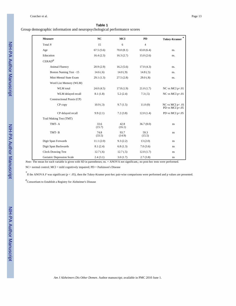
A detailed medical, social and family history was obtained from each subject. MCI and PDpatients had caregivers or informants who could corroborate their history. Participantscompleted the five subtests of the Consortium to Establish a Registry for Alzheimer's Disease(CERAD) neuropsychological battery that included the following subtests: Animal Fluency,Boston Naming Test – 15 item (BNT-15), Mini-Mental Status Exam (MMSE), Word ListMemory (WLM) and Constructional Praxis (CP). Additional neuropsychological testsincluded Trail-Making Tests Parts A and B (TMT-A, TMT-B), Digit Span subtest of theWechsler Adult Intelligence Scale – Revised (WAIS-R), and the Clock Drawing Test (24).The Geriatric Depression Scale (GDS) was administered to assess for the presence ofdepressive symptomatology. Group demographic information and neuropsychologicalperformance for the three groups are summarized in Table 1. MCI and PD patients also receiveda full neurological examination. Clinical diagnoses of MCI, PD, or NC were establishedfollowing a standardized assessment and review by three clinicians, expert in evaluation andmanagement of Geriatric Neurology patients. Clinical diagnosis of MCI required evidence ofa decline in baseline function in memory and possibly additional cognitive domains, with theseverity of symptoms or consequent functional limitations insufficient to meet DSM-III (R)criteria for Dementia. A diagnosis of PD was given if the participant fulfilled the criteria forPD according to the United Kingdom Parkinson's Disease Society Brain Bank clinicaldiagnostic criteria (25). Participants were classified as NC if they demonstrated no evidenceof cognitive decline from baseline functioning based on their clinical interview and assessment.Exclusion criteria included a history of substance abuse or learning disability, dementia,neurological (e.g. stroke, tumor) or psychiatric illness. Because the VPC task involves visualmemory, subjects were also excluded if: 1) the eye tracking equipment could not achieve properpupil and corneal reflection due to physiological constraints or visual problems (e.g. droopyeyelid, cataracts, detached retinas, glaucoma, pupils too small [7 subjects]); and/or 2) theycould not complete the calibration procedure (3 subjects).
Crutcher et al. Page 3
Am J Alzheimers Dis Other Demen. Author manuscript; available in PMC 2010 June 1.
NIH
-PA Author Manuscript
NIH
-PA Author Manuscript
NIH
-PA Author Manuscript
Equipment and StimuliDuring the task, participants' eye movements were continuously recorded using an AppliedScience Laboratories (ASL) Model 5000 remote pan/tilt camera system. A ring of filtered near-infrared LEDs illuminated the eye and a high-speed, near-infrared sensitive CCD cameracaptured the pupil and corneal reflection. The gaze angle was determined by the relativepositions of corneal and pupil centers with an accuracy of ±0.75°. The system sampled at 60Hz, with a temporal resolution of 16 ms and linearity less than 10%. The participants wereseated approximately 26 inches from a 19-inch flat panel computer screen that displayed thestimuli. No physical constraints other than a chinrest were used with the participants.Calibration for each subject was accomplished using a nine-point array. Eye fixation and eyemovement data were recorded with ASL EYEPOS software. All images were black and white,high contrast clipart images measuring 4.4 inches wide by 6.5 inches high. Unique pictureswere used for each trial.
ProcedureParticipants were brought into the testing room and seated comfortably in front of the monitorand their heads positioned within the chinrest to maintain their head/viewing position. Prior topresentation of the VPC task, a 9-point calibration procedure was completed. This wasaccomplished by having the subject fixate nine points at known locations on the computermonitor. The experimenter adjusted the calibration until the subject's fixations accuratelymapped onto the calibration points on the screen. This calibration procedure enabled the eyetracking system to accurately compute the subject's gaze position on the computer monitor.Next, participants were informed that images would begin to appear on the computer screen.They were simply instructed that they should look at the images “as if watching television.”During the calibration and the test phase, the subjects eye fixations and eye movements wererecorded and stored for later analyses.
The entire testing procedure lasted approximately 25 - 30 minutes, including the calibrationsession. For the VPC task, subjects were administered four blocks of five trials (delay order:2-minute delay, 2-second delay, 2-second delay, 2-minute delay) for a total of 20 trials. Eachtrial consisted of two phases; a familiarization phase followed by a test phase. During thefamiliarization phase, two identical pictures were presented side-by-side on the monitor forfive seconds. The monitor then went dark for a delay interval of either two seconds or twominutes. Then, in the test phase, two pictures were again presented side-by-side for fiveseconds. One of the images was identical to the image presented during the familiarizationphase and the other was a novel image. The side of presentation of the novel picture was selectedpseudorandomly and it was presented equally often on the left or right side of the monitorscreen. After the test phase of the trial, the monitor was darkened for 20 seconds until thebeginning of the next trial. In order to ensure subject attention for test trials that had two-minutedelays, the experimenter verbally alerted all subjects that there was “approximately ten secondsbefore the next pair of images.”
Data AnalysisEye fixation and eye movement data for each participant were extracted and analyzed off-lineusing ASL EYENAL software. A fixation was defined as a point of gaze continually remainingwithin 1° of visual angle for a period of 100 msec or more. For the data analysis in the currentstudy, the fixations analyzed occurred within two designated areas of interest (AOIs): the areaof the novel image, and the area of the familiar image. Fixations outside the two areas werenot included in the present analysis.
Eye tracking data were characterized using three measures: (1) total looking time (i.e. the totalsum of the duration for all fixations); (2) total number of fixations (i.e. the total number of
Crutcher et al. Page 4
Am J Alzheimers Dis Other Demen. Author manuscript; available in PMC 2010 June 1.
NIH
-PA Author Manuscript
NIH
-PA Author Manuscript
NIH
-PA Author Manuscript
fixations that met the ≥ 100 msec criterion); and (3) percent looking time on novel image. Foreach measure, we calculated the median of the ten trials at each delay interval (2-sec, 2-min)for each subject. Finally, each measure was analyzed using a separate 3 × 2 repeated measuresANOVA, with group (MCI, PD, NC) as the between-subjects factor and delay (2-seconds, 2-minutes) as the within-subjects factor. All post-hoc pairwise comparisons were performedusing the Tukey-Kramer test at α = .05 (two-tailed).
ResultsDemographics and global cognitive status
Analyses revealed there were no significant differences among the three subject groups in age,education, or global cognitive functioning as measured by several of the tests used by theCERAD, as well as the Trail Making Test, Digit Span, Clock Drawing, and the Geriatricdepression Scale (all p's > .05). ). However, the MCI group was impaired on both the WordList Memory Total and the Word List Memory Delayed Recall measures compared to the NCgroup (p's < .01). The MCI group was also impaired relative to both the NC (p < .01) and PD(p < .05) groups on a visuo-construction task as measured by the CP copy measure. On thedelayed recall version of this task the MCI group performed worse than the PD group (p < .05). No significant group differences in performance on any other neuropsychologicalmeasures were detected (all p's > .05). Results are summarized in Table 1.
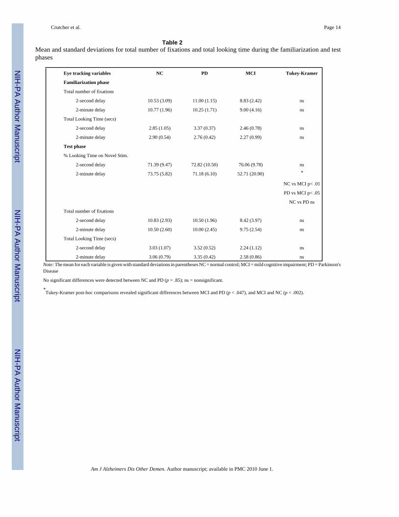
Familiarization phase: Total looking time and total number of fixationsDuring the familiarization phase, subjects were presented with two identical stimuli for 5seconds prior to a 2-second or 2-minute delay. For total looking time (Table 2), the effects ofgroup (F (2,22) = 1.55, p = .24), delay (F (1,22) = 1.73, p = .20) and group by delay interaction(F (2,22) = 1.09, p = .35) were nonsignificant, suggesting that the three groups did not differin the overall amount of time they spent looking at the familiarization images prior to eitherdelay. Similarly, for the total number of fixations i.e., looking at either of the two identicalstimuli, during the familiarization phase (Table 2), the effects of group (F (2,22) =1.26, p = .30), delay (F (1,22) = 0.04, p = .85), and group by delay interaction (F (2,22) = 0.22, p = .80)were nonsignificant. These results indicate that all three groups made a similar number offixations during the familiarization phase.
Test phase: Percent time looking at the novel imageDuring the test phase, subjects were presented with the original image from the familiarizationphase together with a novel image for 5 seconds. The percent time looking at the novel imagewas the main measure of interest. There was a significant group by delay interaction (F (2,22)= 5.39, p = .012). For the 2-second delay, all three groups spent similar amounts of time lookingat the novel image (71-76%; F (2,22) = 0.50, p = .61); Figure 1 and Table 2. However, for the2-minute delay, the groups differed in their percent time looking at the novel stimulus (F (2,22)= 7.69, p =.003). Specifically, the MCI group spent only 53% of their total looking time viewingthe novel image, compared to the PD group (71%) and the NC group (74%) (p's < .05). ThePD and NC groups did not differ from one another (p =.91).
Additional analyses revealed that impaired performance of the MCI group at the 2-minute delaycould not be accounted for by group differences in the overall time spent looking at the imagesor by group differences in the overall number of fixations on the images. For total number offixations (Table 2), the effects of group (F (2,22) = 0.81, p = .46), delay (F (1,22) = 0.09, p = .77) and group by delay interaction (F (2,22) =1.12, p = .34) were nonsignificant. Thisdemonstrates that all three groups made similar numbers of fixations during the test phase.Furthermore, for total looking time the effects of group (F (2,22) = 2.03, p = .16), delay (F(1,22) = 0.16, p = .69), and group by delay interaction (F (2,22) = 0.72, p = .50) were also
Crutcher et al. Page 5
Am J Alzheimers Dis Other Demen. Author manuscript; available in PMC 2010 June 1.
NIH
-PA Author Manuscript
NIH
-PA Author Manuscript
NIH
-PA Author Manuscript
nonsignificant. Therefore, groups did not differ in the amount of overall time spent looking atimages during the test phase.
DiscussionThe present study, to our knowledge, is the first to combine eye tracking together with a visualpaired comparison (VPC) task of recognition memory. In the following sections we addressthe questions we raised in the Introduction.
1) Can the VPC task detect mild cognitive impairment in humans?This work has demonstrated that patients diagnosed with MCI display impaired recognitionmemory performance compared to NC and PD groups. Specifically, all three groupsdemonstrated equivalent recognition memory performance (characterized by increasedviewing time of the novel image relative to the familiar image) at the 2-sec delay. However,the MCI group showed a significant reduction in the amount of time they spent looking at thenovel image when the delay interval was increased to two minutes (Figure 1). At the 2-mindelay, NC and PD groups spent 74% and 71% (respectively) of the total looking time viewingthe novel stimulus; obversely, they viewed the familiar image only 26 - 29 % of the total lookingtime. By contrast, the MCI group spent only 53% of the total looking time viewing the novelstimulus, and 47% of the total looking time viewing the familiar stimulus. Thus, the MCI groupspent about equal amounts of time looking at both the novel and familiar images. These resultssuggest that the delay interval of 2 minutes sufficiently challenged the memory system so thatthe MCI subjects no longer remembered which image they had previously seen. Thus, the VPCtask can successfully detect MCI in humans.
2. Is performance on the VPC task sensitive specifically to hippocampal damage?The MCI group did not meet DSM-III criteria for dementia (Table 1). Instead, the MCI groupevidenced a decline in memory function as measured by some of the tasks described in Table1, and in particular by their selective deficit in performance on the 2-min delay portion of theVPC task, but not on the 2-sec delay portion of the task (the relevance of findings with the 2-sec delay is discussed below). Thus, one can ask whether there is evidence that links MCIimpairment to disruption limited to the brain's memory systems, e.g., the medial temporal lobe(MTL) memory system (8), and the hippocampus in particular. There are cumulating data fromwork with animals as well as with humans suggesting that the impairment in the MCI groupreported here is linked to hippocampal dysfunction. Specifically, the observed performance onthe VPC task by the MCI group closely resembles performance on a similar VPC taskadministered to nonhuman primates who sustained lesions limited to the hippocampus (19).The monkeys with hippocampal lesions had a reduction in looking time at the novel stimulusas the delay interval on the VPC task was increased from 1 sec to 10 min (Figure 2). Thesemonkeys had lesions of the hippocampus made by radiofrequency or by ibotenic acid. Similarto the MCI patients in the present study, monkeys with either RF or IBO lesions spent moretime viewing the novel image on the VPC task when the delay interval was short (1 sec), withless time viewing the novel image as the delay interval increased. A similar pattern has alsobeen observed in rats with hippocampal lesions (15). Other studies using memory impairedpatients with damage limited to the hippocampus and similar tasks of recognition memory havealso pointed to the importance of intact hippocampal function for successful performance(26). Thus, findings from work in humans, as well as monkeys, and rats all provide convergingevidence that impairment on the VPC task reflects memory problems associated withhippocampal dysfunction.
A question arises whether the VPC task is sensitive specifically to medial temporal lobe damagein MCI or whether patients with other neurologic conditions, not specifically involving the
Crutcher et al. Page 6
Am J Alzheimers Dis Other Demen. Author manuscript; available in PMC 2010 June 1.
NIH
-PA Author Manuscript
NIH
-PA Author Manuscript
NIH
-PA Author Manuscript
medial temporal lobe, would show impaired performance as well. In the present study, weaddressed this question by assessing patients with PD as well as patients with MCI. PD ischaracterized by degeneration of dopaminergic neurons in the substantia nigra resulting in adepletion of dopamine. This depletion results in an abnormal motor behavior (e.g. restingtremor, rigidity, and akinesia) observed in this patient population (27,28). The cognitiveprofiles of PD patients can be heterogeneous and are frequently dominated by deficits inexecutive functioning (e.g. multi-tasking, planning, use of feedback) and visuospatial/visuoconstructional difficulties (29-31). While memory impairment can occur in patients withPD (32,33), these memory deficits are not attributed to an insidious disease process occurringin the medial temporal lobe. In the current study, recognition memory performance wasunaffected by the presumed subcortical damage associated with the PD group, suggesting thatthe VPC task is more selective to medial temporal lobe dysfunction (but see Whittington et al,2000 [32] for a meta-analysis of recognition impairment in Parkinson's Disease). These resultslend support to the possibility of utilizing the VPC task as an early diagnostic measure sinceit appears to be specifically sensitive to memory impairment. However, further longitudinalstudies will need to be performed to investigate the use of the VPC task in this capacity.
At the time of the present study, only one of the six MCI subjects had undergone magneticresonance imaging (MRI) scanning. This patient was impaired in all of the tasks that the MCIgroup was impaired on in Table 1. Additionally, this patient's performance on the VPC taskwas 62% at the 2-min delay, a score that was worse than all but one control subject. In the MCIsubject, an MRI examination without gadolinium was performed according to a standarddepartment (Neurology) protocol on a 3T magnet (Siemens Magneton Trio). Axial gradient-echo images for susceptibility were also performed. The clinical report, based on reviews ofthe images, indicated scattered foci of T2 prolongation in the periventricular and subcorticalwhite matter of both hemispheres. Additionally, slight prominence of the sulci, cisterns, andventricles, consistent with mild diffuse volume loss was noted. There was no evidence of acuteterritorial infarction, hemorrhage, mass, mass-effect or midline shift. The major intracranialvascular flow-voids also were reported as intact. Importantly, there was no reported evidenceof abnormalities in the hippocampal region or in adjacent cortical regions of the medialtemporal lobe. While the evidence is sparse, the MRI findings from this case raise the possibilitythat impaired performance on the VPC task by patients with MCI might precede detectablestructural changes in the hippocampus and the medial temporal lobe region. If true, sensitivebehavioral tasks like the VPC task combined with infrared eyetracking might serve aspredictive biomarkers for underlying but as yet undetectable brain pathology or regional braindysfunction, e.g., vascular subcortical pathology.
3. Can the impaired performance exhibited by the MCI group on the VPC task be attributedto aspects of performance other than memory?
It is possible that the differences in performance between the MCI and the NC groups on the2-min delay portion of the VPC task could occur for reasons other than memory impairmenton the part of the MCI group. Several possibilities include differences between the MCI andthe NC groups in global cognitive status and demographics, or differences in attentional,motivational, and perceptual functions. However, as shown in Table 1, the groups wereequivalent on cognitive status, age and education. Nor can the results be explained by groupdifferences in attentional, motivational, or perceptual abilities since all groups performedequivalently at the 2-sec delay. Analyses revealed that all three groups were equivalent in thetotal amount of time they viewed the pictures during either phase, indicating all three groupswere similarly able to attend to and accurately perceive the stimuli. Additionally, the numberof fixations that met criteria for analyses cannot account for the observed group differencesbecause the number of fixations that met criteria was not different for any group. Thus, the twogroups performed quite similarly in all important ways that might have provided evidence for
Crutcher et al. Page 7
Am J Alzheimers Dis Other Demen. Author manuscript; available in PMC 2010 June 1.
NIH
-PA Author Manuscript
NIH
-PA Author Manuscript
NIH
-PA Author Manuscript
a competing hypothesis to that of impaired memory in the MCI group. Accordingly, the ideathat the MCI group's impaired performance on the 2-min delay portion of the task resultedfrom impaired memory remains compelling.
An additional point of importance involves the benefit derived from combining infrared eyetracking with the VPC task. Infrared eye tracking research is becoming distinguished in itsdiagnostic role (13,22,32). As used in the present study, eyetracking provided objective andquantitative evidence of each subject's visual, attentional, and memory processes. Moreover,the eye movement data were acquired in an unobtrusive, noninvasive manner and provided on-line measures as well as data-based storage of information for later analyses. Additionally,eyetracking allowed for a number of potentially informative and sensitive measures in additionto a simple novel stimulus viewing-time measure. Thus, two additional parameters wemeasured, i.e., overall viewing time, and number of fixations, helped to eliminate the possibilitythat the impaired performance by the MCI group could have been attributed to other thanmnemonic dysfunction. Additional measures not examined in this paper (e.g. saccade lengthand latency, pupil diameter, inter-fixation durations, etc.) might also be of potential help ingaining more understanding of the perceptual, as well as encoding and retrieval processesduring stimuli presentation and recognition, thus providing more insight into the nature ofnormal and impaired memory.
ConclusionThe results from the current study demonstrate that the VPC task combined with eye trackingtechnology can be used successfully with normal elderly adults and with elderly neurologicpatients. Additionally, eye-tracking performance on the VPC task can be used to detect mildmemory impairments associated with MCI. Moreover, VPC performance, as measured bypercent time viewing the novel stimulus, appears to be selective to declarative memoryimpairment reflective of medial temporal lobe dysfunction since patients with subcorticaldamage performed comparable to control subjects. Finally, the recording of eye trackingperformance during a VPC task has the potential to be an effective screening tool. With furtherinvestigation, it could potentially be used as a diagnostic measure and to maximize earlytherapeutic intervention (34,35).
AcknowledgementsThis work was supported by National Institute of Health Grant AG 025588, Yerkes Base Grant RR00165, Robert W.Woodruff Health Science Award from Emory University, Atlanta VAMC Merit Review award, and the GeorgiaResearch Alliance. We gratefully thank Dr. Felicia Goldstein for help in providing patient diagnoses; Janet Cellar,Amy Rice, Kristen Boyd, and Ann Scherer Johnston for help with patient recruitment and testing; and Tammy Hailsfor help with data processing.
References1. Petersen RC, Smith GE, Waring SC, Ivnik RJ, Tangalos EG, Kokmen E. Mild cognitive impairment:
clinical characterization and outcome. Arch Neurol Mar;1999 56(3):303–8. [PubMed: 10190820]2. Morris JC. Mild cognitive impairment and preclinical Alzheimer's disease. Geriatrics Jun;2005 (Suppl):
9–14. [PubMed: 16025770]3. Petersen RC, Stevens JC, Ganguli M, Tangalos EG, Cummings JL, DeKosky ST. Practice parameter:
early detection of dementia: mild cognitive impairment (an evidence-based review). Report of theQuality Standards Subcommittee of the American Academy of Neurology. Neurology May 8;2001 56(9):1133–42. [PubMed: 11342677]
4. Burns A, Zaudig M. Mild cognitive impairment in older people. Lancet Dec 14;2002 360(9349):1963–5. [PubMed: 12493278]
Crutcher et al. Page 8
Am J Alzheimers Dis Other Demen. Author manuscript; available in PMC 2010 June 1.
NIH
-PA Author Manuscript
NIH
-PA Author Manuscript
NIH
-PA Author Manuscript
5. Braak H, Braak E. Neuropathological stageing of Alzheimer-related changes. Acta Neuropathol (Berl)1991;82(4):239–59. [PubMed: 1759558]
6. Braak H, Braak E. Diagnostic criteria for neuropathologic assessment of Alzheimer's disease.Neurobiol Aging Jul-Aug;1997 18(4 Suppl):S85–8. [PubMed: 9330992]
7. Eichenbaum H. The hippocampus and declarative memory: cognitive mechanisms and neural codes.Behav Brain Res Dec 14;2001 127(12):199–207. [PubMed: 11718892]
8. Squire LR, Zola-Morgan S. The medial temporal lobe memory system. Science Sep 20;1991 253(5026):1380–6. [PubMed: 1896849]
9. Elias MF, Beiser A, Wolf PA, Au R, White RF, D'Agostino RB. The preclinical phase of alzheimerdisease: A 22-year prospective study of the Framingham Cohort. Arch Neurol Jun;2000 57(6):808–13. [PubMed: 10867777]
10. Linn RT, Wolf PA, Bachman DL, et al. The ‘preclinical phase’ of probable Alzheimer's disease. A13-year prospective study of the Framingham cohort. Arch Neurol May;1995 52(5):485–90.[PubMed: 7733843]
11. Fagan JF 3rd. Memory in the infant. J Exp Child Psychol Apr;1970 9(2):217–26. [PubMed: 5452116]12. Berlyne, DE. Conflict, arousal, and curiosity. McGraw-Hill; New York: 1960.13. Daffner KR, Scinto LF, Weintraub S, Guinessey JE, Mesulam MM. Diminished curiosity in patients
with probable Alzheimer's disease as measured by exploratory eye movements. Neurology Feb;199242(2):320–8. [PubMed: 1736159]
14. Loftus GR, Mackworth NH. Cognitive determinants of fixation location during picture viewing. JExp Psychol Hum Percept Perform Nov;1978 4(4):565–72. [PubMed: 722248]
15. Clark RE, Zola SM, Squire LR. Impaired recognition memory in rats after damage to the hippocampus.J Neurosci Dec 1;2000 20(23):8853–60. [PubMed: 11102494]
16. McKee RD, Squire LR. On the development of declarative memory. J Exp Psychol Learn Mem CognMar;1993 19(2):397–404. [PubMed: 8454964]
17. Manns JR, Stark CE, Squire LR. The visual paired-comparison task as a measure of declarativememory. Proc Natl Acad Sci U S A Oct 24;2000 97(22):12375–9. [PubMed: 11027310]
18. Bachevalier J, Brickson M, Hagger C. Limbic-dependent recognition memory in monkeys developsearly in infancy. Neuroreport Jan;1993 4(1):77–80. [PubMed: 8453042]
19. Zola SM, Squire LR, Teng E, Stefanacci L, Buffalo EA, Clark RE. Impaired recognition memory inmonkeys after damage limited to the hippocampal region. J Neurosci Jan 1;2000 20(1):451–63.[PubMed: 10627621]
20. Fagan JF 3rd. The paired-comparison paradigm and infant intelligence. Ann N Y Acad Sci1990;608:337–57. [PubMed: 2075956]discussion 58-64
21. Richmond J, Sowerby P, Colombo M, Hayne H. The effect of familiarization time, retention interval,and context change on adults' performance in the visual paired-comparison task. Dev PsychobiolMar;2004 44(2):146–55. [PubMed: 14994266]
22. Daffner KR, Mesulam MM, Cohen LG, Scinto LF. Mechanisms underlying diminished novelty-seeking behavior in patients with probable Alzheimer's disease. Neuropsychiatry NeuropsycholBehav Neurol Jan;1999 12(1):58–66. [PubMed: 10082334]
23. Mosimann UP, Muri RM, Burn DJ, Felblinger J, O'Brien JT, McKeith IG. Saccadic eye movementchanges in Parkinson's disease dementia and dementia with Lewy bodies. Brain Jun;2005 128(Pt 6):1267–76. [PubMed: 15774501]
24. Freedman, M.; Leach, L.; Kaplan, E.; Winocur, G.; Shulman, K.; Delis, D. Clock drawing: Aneuropsychological analysis. Oxford University Press; New York: 1994.
25. Hughes AJ, Daniel SE, Kilford L, Lees AJ. Accuracy of clinical diagnosis of idiopathic Parkinson'sdisease: a clinico-pathological study of 100 cases. J Neurol Neurosurg Psychiatry Mar;1992 55(3):181–4. [PubMed: 1564476]
26. Smith CN, Hopkins RO, Squire LR. Experience-dependent eye movements, awareness, andhippocampus-dependent memory. J Neurosci Nov 1;2006 26(44):11304–11312. [PubMed:17079658]
27. Lang AE, Lozano AM. Parkinson's disease. First of two parts. N Engl J Med Oct 8;1998 339(15):1044–53. [PubMed: 9761807]
Crutcher et al. Page 9
Am J Alzheimers Dis Other Demen. Author manuscript; available in PMC 2010 June 1.
NIH
-PA Author Manuscript
NIH
-PA Author Manuscript
NIH
-PA Author Manuscript
28. Lang AE, Lozano AM. Parkinson's disease. Second of two parts. N Engl J Med Oct 15;1998 339(16):1130–43. [PubMed: 9770561]
29. Taylor AE, Saint-Cyr JA, Lang AE. Frontal lobe dysfunction in Parkinson's disease. The corticalfocus of neostriatal outflow. Brain Oct;1986 109(Pt 5):845–83. [PubMed: 3779372]
30. Freeman RQ, Giovannetti T, Lamar M, Cloud BS, Stern RA, Kaplan E, et al. Visuoconstructionalproblems in dementia: contribution of executive systems functions. Neuropsychology Jul;2000 14(3):415–26. [PubMed: 10928745]
31. Ong JC, Seel RT, Carne WF, Brown R, Pegg PO, Jehle PJ. A brief neuropsychological protocol forassessing patients with Parkinson's disease. NeuroRehabilitation 2005;20(3):191–203. [PubMed:16340100]
32. Whittington CJ, Podd J, Kan MM. Recognition memory impairment in Parkinson's disease: powerand meta-analyses. Neuropsychol Apr;2000 14(2):233–46.
33. Davidson PS, Anaki D, Saint-Cyr JA, Chow TW, Moscovitch M. Exploring the recognition memorydeficit in Parkinson's disease: estimates of recollection versus familiarity. Brain Jul;2006 129(Pt 7):1768–79. [PubMed: 16714314]
34. Schenk D, Barbour R, Dunn W, et al. Immunization with amyloid-beta attenuates Alzheimer-disease-like pathology in the PDAPP mouse. Nature Jul 8;1999 400(6740):173–7. [PubMed: 10408445]
35. Giacobini E. Cholinesterase inhibitors stabilize Alzheimer's disease. Ann N Y Acad Sci2000;920:321–7. [PubMed: 11193171]
Crutcher et al. Page 10
Am J Alzheimers Dis Other Demen. Author manuscript; available in PMC 2010 June 1.
NIH
-PA Author Manuscript
NIH
-PA Author Manuscript
NIH
-PA Author Manuscript
Figure 1.Group differences in percent time looking at either the novel images at the 2-second and 2-minute delays (test phase). An asterisk indicates that the MCI group significantly differed (p< .05) from both NC and PD groups in the amount of time spent looking at the novel imagesat the 2-minute delay. Error bars reflect standard error.
Crutcher et al. Page 11
Am J Alzheimers Dis Other Demen. Author manuscript; available in PMC 2010 June 1.
NIH
-PA Author Manuscript
NIH
-PA Author Manuscript
NIH
-PA Author Manuscript
Figure 2.Performance on the visual paired-comparison task by monkeys with lesions limited to thehippocampal region. In monkeys, performance on this task can be impaired even when 70%to 80% of the hippocampus is spared, either when the lesion is made by ibotenic acid or radiofrequency. Parentheses indicate the number of monkeys in each group. N= normal; RF = lesioncreated by radio frequency; IBO = lesion created by ibotenic acid. Graph is adapted from Zolaet al, 2000.
Crutcher et al. Page 12
Am J Alzheimers Dis Other Demen. Author manuscript; available in PMC 2010 June 1.
NIH
-PA Author Manuscript
NIH
-PA Author Manuscript
NIH
-PA Author Manuscript
NIH
-PA Author Manuscript
NIH
-PA Author Manuscript
NIH
-PA Author Manuscript
Crutcher et al. Page 13
Table 1Group demographic information and neuropsychological performance scores
Measure NC MCI PD Tukey-Kramer *
Total N 15 6 4
Age 67.5 (5.6) 70.0 (8.1) 63.8 (6.4) ns.
Education 16.4 (2.3) 16.3 (2.7) 15.0 (2.6) ns.
CERADa
Animal Fluency 20.9 (2.9) 16.2 (5.6) 17.0 (4.3) ns.
Boston Naming Test −15 14.6 (.6) 14.0 (.9) 14.8 (.5) ns.
Mini-Mental State Exam 29.1 (1.3) 27.5 (2.8) 29.0 (.8) ns.
Word List Memory (WLM)
WLM total 24.0 (4.5) 17.8 (1.9) 21.0 (1.7) NC vs MCI p<.01
WLM delayed recall 8.1 (1.8) 5.2 (2.4) 7.3 (.5) NC vs MCI p<.01
Constructional Praxis (CP)
CP copy 10.9 (.3) 9.7 (1.5) 11.0 (0) NC vs MCI p< .01PD vs MCI p<.05
CP delayed recall 9.9 (2.1) 7.2 (3.8) 12.0 (1.4) PD vs MCI p<.05
Trail Making Test (TMT)
TMT- A 33.6(15.7)
42.8(16.1)
36.7 (8.0) ns
TMT- B 74.8(33.5)
93.7(14.9)
59.3(15.5)
ns
Digit Span Forwards 11.1 (2.0) 9.3 (2.2) 13 (2.0) ns
Digit Span Backwards 8.1 (2.4) 6.8 (1.3) 7.0 (3.6) ns
Clock Drawing Test 12.7 (.6) 12.7 (.5) 12.0 (1.7) ns
Geriatric Depression Scale 2.4 (3.1) 3.0 (1.7) 2.7 (3.8) nsNote: The mean for each variable is given with SD in parentheses; ns. = ANOVA not significant,; no post-hoc tests were performed.
NC= normal control; MCI = mild cognitively impaired; PD = Parkinson's Disease
*If the ANOVA F was significant (p < .05), then the Tukey-Kramer post-hoc pair-wise comparisons were performed and p values are presented.
aConsortium to Establish a Registry for Alzheimer's Disease
Am J Alzheimers Dis Other Demen. Author manuscript; available in PMC 2010 June 1.
NIH
-PA Author Manuscript
NIH
-PA Author Manuscript
NIH
-PA Author Manuscript
Crutcher et al. Page 14
Table 2Mean and standard deviations for total number of fixations and total looking time during the familiarization and testphases
Eye tracking variables NC PD MCI Tukey-Kramer
Familiarization phase
Total number of fixations
2-second delay 10.53 (3.09) 11.00 (1.15) 8.83 (2.42) ns
2-minute delay 10.77 (1.96) 10.25 (1.71) 9.00 (4.16) ns
Total Looking Time (secs)
2-second delay 2.85 (1.05) 3.37 (0.37) 2.46 (0.78) ns
2-minute delay 2.90 (0.54) 2.76 (0.42) 2.27 (0.99) ns
Test phase
% Looking Time on Novel Stim.
2-second delay 71.39 (9.47) 72.82 (10.50) 76.06 (9.78) ns
2-minute delay 73.75 (5.82) 71.18 (6.10) 52.71 (20.90) *
NC vs MCI p< .01
PD vs MCI p< .05
NC vs PD ns
Total number of fixations
2-second delay 10.83 (2.93) 10.50 (1.96) 8.42 (3.97) ns
2-minute delay 10.50 (2.60) 10.00 (2.45) 9.75 (2.54) ns
Total Looking Time (secs)
2-second delay 3.03 (1.07) 3.52 (0.52) 2.24 (1.12) ns
2-minute delay 3.06 (0.79) 3.35 (0.42) 2.58 (0.86) nsNote: The mean for each variable is given with standard deviations in parentheses NC = normal control; MCI = mild cognitive impairment; PD = Parkinson'sDisease
No significant differences were detected between NC and PD (p = .85); ns = nonsignificant.
*Tukey-Kramer post-hoc comparisons revealed significant differences between MCI and PD (p < .047), and MCI and NC (p < .002).
Am J Alzheimers Dis Other Demen. Author manuscript; available in PMC 2010 June 1.