extending the human spatiotemporal comfort zone with cavern – computer-based augmented virtual...
TRANSCRIPT
_____________________________Journal of Multidisciplinary Research____________________________
23
Journal of Multidisciplinary Research, Vol. 4, No. 3, Fall 2012, 23-44. ISSN 1947-2900 (print) • ISSN 1947-2919 (online) Copyright © 2012 by St. Thomas University. All rights reserved.
Extending the Human Spatiotemporal Comfort Zone with CAVERN – Computer-based Augmented
Virtual Environment for Realizing Nature
Dov Dori Technion-Israel Institute of Technology and
Massachusetts Institute of Technology
Abstract
An intelligent multimedia device dubbed CAVERN – Computer-based Augmented Virtual Environment for Realizing Nature – is proposed as a quantum leap in molecular biology research. CAVERN is a system that leverages state-of-the-art technologies that include CAVE, supercomputing, electron microscopy, conceptual modeling, and biological text mining. After discussing the acute problems biological research is facing, the chapter introduces the new notion of the human spatiotemporal comfort zone, and a fourth multimedia learning assumption: the limited spatiotemporal comfort zone. Within this zone, people can use their senses to follow and understand complex systems currently accessible only through indirect observations. CAVERN translates nano-level processes into scenarios of human-size interacting molecules. A brief enumeration of the potential benefits of CAVERN in biology, health, and education is followed by a conceptual blueprint of CAVERN expressed via an Object-Process Methodology model. Finally, challenges and open problems in the way to achieving an operational CAVERN are presented.
Keywords
CAVERN, spatiotemporal comfort zone, Object-Process Methodology, supercomputing
Introduction
Science can be thought of as the process of reverse engineering nature. In recent years, we have witnessed an unprecedented increase in the number, variety, and complexity of information resources available to researchers, particularly in life sciences. We are in a pivotal moment in the
_____________________________Journal of Multidisciplinary Research____________________________
24
study of life sciences, which is shifting from the study of single molecular processes to complete cellular pathways and the entire cell (Kitano, 2002).
The vast amount of data available also provides new opportunities; because searchable results are readily available (in databases such as Medline and PubMed), the data itself can confirm or refute conjectures, as many “future” predictions already have been tested through related experiments that were carried out for other reasons, and which “only” need to be revealed in the new context. Despite advances in the technologies available for sifting through data to retrieve new insights, the problem of how to fit the billions of known facts into a meaningful whole still remains unsolved.
Visualization and conceptual modeling are key ingredients in a possible solution for this problem. We need a holistic framework, an evolving conceptual model of the cell life system that can facilitate deep understanding through computer-graphics-based visualization. Current and future known facts and findings will be embedded into such a framework to foster insights on this incredibly complex cell life system. Armed with an adequate conceptual modeling language and methodology, biologists will be able to visualize, study, simulate, and analyze models of biological systems. The model will enable the mapping of knowledge gaps, closing them through the design and execution of wet laboratory experiments.
Conceptual modeling enables the process of making system-level sense of the vast number of pieces of information. The enormous number and variety of interactions among substances and processes in the cell primarily poses the qualitative problem of figuring out “what” and “how,” which precedes the quantitative one.
A deep understanding of the processes within a biological system requires delving into each process and phenomenon, as most biological researchers have been doing. Comprehending how the system functions as a whole requires the integration of knowledge from the bottom up. A complementary, conceptual modeling approach to top-down modeling takes the cell life function as the system’s main process and decomposes it into ever simpler processes down to the molecular level.
The Cognitive Assumptions
Humans assimilate data and information and convert it into meaningful knowledge and understanding of systems using words and pictures simultaneously. During eons of human evolution, the human brain was trained to capture and analyze images, in order for humans to escape predators and capture food. In contrast, the processing of spoken words, let alone text, is a product of a relatively recent period in the history of humankind (Dori, 2008). As human brains are hard-wired to process imagery, graphics appeal to the brain more immediately than words. However, words can express ideas and assertions far too complex or impossible to express graphically (try graphing this sentence to get a feeling for the validity of this claim). So while a picture is worth a thousand words, as the saying goes, there are cases in which a word, or a sentence, is worth a thousand pictures. A problem with the richness of natural languages is the potential ambiguity that arises from their use. This certainly does not imply that pictures cannot be ambiguous as well, but graphic ambiguity can be greatly reduced, or even eliminated, by assigning formal semantics to pictorial symbols of things and relations among them.

_____________________________Journal of Multidisciplinary Research____________________________
25
Vastly enlarged moving images of the molecular dynamics of a highly complex system, such as the living cell, will improve dramatically our understanding of living processes. When corresponding words and pictures are presented closely together, learners can better retain an understanding of corresponding words and pictures, simultaneously, in their working memory, enabling the integration of visual and verbal models. Mayer and Moreno (2003) proposed a theory of multimedia learning based on the following three, main research-supported cognitive assumptions:
(1) Dual-channel – humans possess separate systems for processing visual and verbal
representations. (2) Limited capacity – the amount of processing that can take place within each
information processing channel is extremely limited. (3) Active processing – meaningful learning occurs during active cognitive processing,
paying attention to words and pictures, mentally organizing, and integrating them into coherent representations.
However, there is a major factor in addition to Mayer’s three cognitive assumptions that
needs to be accounted for when designing a research and study environment for investigating nature: the limited human spatiotemporal comfort zone.
The Human Spatiotemporal Comfort Zone: Plus or Minus Four Orders of Magnitude
Humans are used to thinking about the world via their senses, especially stereoscopic
vision, which enables them to see objects—things that exist—with sizes that are neither too great nor too minuscule, so the human mind can grasp it. We cannot mentally digest astronomical concepts (e.g., the space and time it takes light to travel among galaxies). Similarly, on the other extreme end of the distance and time scale, we cannot really fathom tiny micrometer and nanometre-scale biological objects, nor can we imagine microsecond or nanosecond-scale processes.
Processes—things that happen to objects and transform them—are analogous to objects in this regard, except that instead of size, which is a dimension applicable to objects, when processes are considered, the dimension to be considered is time. A process is a concept constructed in the human mind as a result of comparing the state of an object (or an entire system, which is a complex object) before, during, and after it underwent change. Hence, the main feature of a process is the time it takes to happen—the interval of time that lapses between the beginning of the process in question and the end of that process.
Just as humans cannot grasp objects beyond their capacity to understand based on common sense or life experience, they cannot follow processes that are too short or too long. Based on this observation on human limitations, we define the human spatiotemporal comfort zone as follows.
The “human spatiotemporal comfort zone” is the interval of space and time between the minimal and maximal values that humans can comfortably imagine by drawing on their intuition and lived experiences.

_____________________________Journal of Multidisciplinary Research____________________________
26
Trying to quantify the human spatiotemporal comfort zone in terms of space and time
units, we note that humans can quite conveniently conceive of objects measuring from the width of a hair (tenths of millimetres) to the distance to a nearby country (thousands of kilometres). Since humans are of the order, or magnitude, of one meter in length, translating this observation to orders of magnitude, we assert that the spatial (size-related) human comfort zone is between four orders of magnitudes smaller than humans and about four orders of magnitudes smaller than that, i.e., between 10–4 and 106 meters.
A similar argument works for the dimension of time, when processes are considered. Humans can conveniently conceive of processes such as a runner overcoming her competitor by a few tenths of a second to processes lasting a lifetime and measured in decades. If we consider an hour as the basic human time unit, analogous to meter for size, we easily can verify that the limits of the human temporal comfort zone are between 10–4 and 106 hours.
Summarizing the above observations we get the following expression. O(10-4 {meter, hour}) <Human Spatiotemporal Comfort Zone< O(106 {meter, hour})
Interestingly, countable numbers, which is a third concept related to conceivable orders of magnitude of space and time, also are related to four orders of magnitude. While we now speak easily about millions and billions of items (people, dollars…), in early times, humans could conveniently think of a number of items (instances of a class), and especially people and coins, in up to four orders of magnitude, i.e., tens of thousands. To support this observation, we note the largest number for which there is a special word in Biblical Hebrew is ten thousands, 104, pronounced revava.
Based on the observation and definition of the human spatiotemporal comfort zone, a fourth multimedia learning assumption is the following.
(4) Limited Spatiotemporal Comfort Zone – understanding the dynamics of objects
interacting via processes in complex systems can occur within plus or minus four orders of magnitude of objects’ basic size unit, meter, and process basic time unit, an hour.
Generalizing Miller’s Magic Number: Summary of Human Cognitive Limitations
Bounding the spatiotemporal comfort zone within four orders of magnitude can be considered as a generalization of “The Magical Number Seven, Plus or Minus Two” suggested by Miller (1956), where the limiting factor is the number 7—the number of items a person can remember and handle concurrently—is analogous to the space of one meter and time of one hour. Miller’s case is the basis of Meyer’s Limited Capacity multimedia learning assumption. There, the factor that limits human cognition is the number of concurrent items, with a linear tolerance of “plus or minus two.” Analogously, in the case of the Limited Spatiotemporal Comfort Zone, the limiting factors are space and time, both with a logarithmic (orders of magnitude) tolerance of plus or minus 4. This is summarized in Table 1.

_____________________________Journal of Multidisciplinary Research____________________________
27
Table 1 The Human Cognitive Limitations, their Nominal Values, Tolerances, and Bounds
Cognitive Limitation Limiting Dimension
Nominal Value
Tolerance Lower & Upper Bounds
Miller’s Magic Number Concurrent # of item
7 + or – 2 items 5, 9
Space Comfort Zone Space (size) 1 meter + or – 4 orders of magnitudes
10-4m, 104m
Time Comfort Zone Time (duration)
1 hour + or – 4 orders of magnitudes
10-4h, 104m
One can perhaps reason rationally about objects and processes beyond one’s spatiotemporal comfort zone, but it is not possible to intuitively imagine them, as the objects outside the spatial comfort zone cannot be noticed by the naked eye, nor can processes outside the temporal zone be contemplated based on daily experiences.
Coping with the Human Cognitive Limitations
The human cognitive limitations were the tacit motivation behind the inventions of the
microscope by the Janssen brothers in 1590 and the telescope by Lippershey in 1608, both in Holland. Huge strides have since been made in developing these devices, with the advent of electronic microscopy on the smaller side and the Hubble telescope at the other extreme. In terms of coping with the temporal human cognitive limitation, devices that take pictures at a high rate and show them at a much slower rate have enabled research on very rapid processes, such as how the cheetah runs, how a bullet penetrates glass, or how the splashing of a milk drop evolves. Doing the same thing in reverse has enabled humans to see how flowers bloom and how they behave during the day and night, how trees grow, how clouds form and disappear, and more.
To fully understand cell-level molecular biology processes, a combination of translating both space and time from their actual minuscule sizes to the human spatiotemporal comfort zone is needed. However, this is just one facet of the problem. Many such interactions already have been discovered and understood without resorting to the drastic multimedia means suggested here, but we are very far from a complete understanding of the cell system as a whole and how each known fact and interaction fits into this puzzle. This is where conceptual modeling comes to play a critical role in making multimedia an intelligent research system.
Integrating Conceptual Modeling Renders Multimedia Intelligent
A structured, intuitive yet formal modeling methodology is needed to support researchers
in specifying pieces of knowledge at the atomic and molecular levels on the one hand, and abstracting them by looking at the “big picture”—the holistic view of a unified, finely-tuned, orchestrated system—on the other hand. To this end, the conceptual modeling paradigm underlying the modeling framework must be based on a compact set of the most primitive and

_____________________________Journal of Multidisciplinary Research____________________________
28
generic elements. This makes it versatile enough to be applicable to a host of domains and simple enough to express the most complex systems in any one of those domains.
A sufficiently expressive model can serve as a basis for a simulated visualization that facilitates its comprehension. The model creator and user can reference the model to reason about the system under study, query and predict its behavior, and effectively communicate it to molecular biology research peers, drug developers, and other stakeholders. Visualization, therefore, must come from two complementary sources and be presented in two compatible modes. One type of visualization originates from inspecting the cell in action—the inspection-driven mode—while the other type of visualization is created by rendering the cell activity from a conceptual model of the cell system—the model-driven mode. As new findings from the inspection-driven mode are discovered, they are used to update the conceptual model. This way, the conceptual model and the visualization derived from it continuously evolve and grow, providing ever deeper levels of knowledge and understanding of the system’s function, structure, and behavior. Incorporating the conceptual modeling element into the multimedia ensemble for complex system visualization adds intelligence to the system, qualifying it to serve as a basis for system-level research.
Translating the system in action into the human spatiotemporal comfort zone and combining inspection-driven with model-driven visualization are the two principles underlying the next generation of CAVERN—Computer-based Augmented Virtual Environment for Realizing Nature—the instrument for doing next generation research in Systems Biology, for which principles and blueprint are presented in the article.
Figure 1. Illustrative CAVERN scenarios.
CAVERN – Computer-based Augmented Virtual Environment for Realizing Nature
CAVERN creates totally immersive living cell environments, opening up a host of exciting new possibilities in biological research that potentially can introduce a dramatic change in the ways this type of research is conducted. For example, adding to the conceptual model new findings discovered by watching rendered 3D scenes, researchers can close knowledge gaps that existed in the model, make new predictions about the behavior of the system under different

_____________________________Journal of Multidisciplinary Research____________________________
29
conditions, and infer or conjecture about novel relationships among different system components. Figure 11 shows two CAVERN scenario examples.
OPM-based conceptual modeling, which has long been used for designing and communicating complex human-made systems, is beginning to play an increasingly important role in facilitating human comprehension of complex biological systems. OPM provides for faithfully and intuitively modeling biological processes and substances that undergo these processes or enable them in a single, bimodal graphic and textual model. The processes, which occur at varying spatiotemporal scales, are modeled at increasingly refined levels of complexity, enabling one to inspect the system at any desired level of detail while not losing sight of the overall view of the functioning system across compartment boundaries and abstraction levels.
Complementing this OPM-based conceptual modeling component, the multimedia modules of CAVERN provide for two presentation modes: the inspection-driven mode and the model-driven mode. In the inspection-driven mode, nano-size molecular interactions, inspected and recorded by the Scene Inspection & Capture Module, are rendered by a supercomputer in the Control Module into dynamic, human-size 3D scenes that can be replayed at various paces, resolutions, and viewpoints in a CAVE™ (Cave Automatic Virtual Environment)—a closed cubic theatre room with 6 walls, including floor and ceiling, embedded inside a larger room, which is the main part of the 3D Presentation Module. In the model-driven mode, human-size 3D scenes are synthesized from the knowledge gleaned from the OPM conceptual model of the living cell and are presented in the same manner in the CAVE.
As proof of concept to the conceptual modeling facet of the CAVERN approach, the paper “Conceptual modeling in Systems Biology fosters empirical findings: The mRNA lifecycle” (Dori & Choder, 2007) describes how, using an OPM-based conceptual model, a knowledge gap in the mRNA lifecycle cell subsystem was identified, leading to a new biological discovery. To fill in the identified knowledge gap, wet lab experiments were designed and conducted, establishing a new finding, namely, that the translation termination factor eRF3 is found in processing bodies (P-bodies) in the cytoplasm after a starvation period. Following this success, work of leveraging an OPM-based conceptual modeling approach for expressing biological findings in an evolving OPM model, identifying knowledge gaps, and experimenting to fill them in is underway.
An Illustrative CAVERN Scenario
Imagine yourself as a molecular biology researcher interested in the interaction between the eukaryotic mRNA lifecycle cellular subsystem and the glycolysis, which supplies energy to all life processes. You are the person on the left hand side of Figure 1 (left). You and your colleague, Judith, walk gently in a CAVE, the main physical part of CAVERN, with a supercomputer-driven set of 48 powerful projectors, which, in near real-time, render cellular life processes of a yeast cell at the nano-level, magnified to centimetre and decimetre scale, and colored for clear, sharp 3-D viewing. This 3-D immersive movie was computed and recorded just seconds ago and is now being played in controlled slow motion. Equipped with tiny, almost invisible 3-D vision antenna glasses, you are both immersed in the cell environment, moving freely inside it and inspecting specific life processes of interest to your current research—the mRNA lifecycle and its
1 The CAVE images are based on the figures of the CAVE in RWTH Aachen.
_____________________________Journal of Multidisciplinary Research____________________________
30
interaction with the energy-supplying glycolysis subsystem. The data that drives the live cell life scene is fused from data obtained by a combination of (1) a cell microarray with 85 million (964) microscopic wells, each containing about five biologically-marked yeast cells; and (2) a scanning electron microscope, which inspects a single identically-marked cell for comparison and reference. A remote supercomputer, to which the raw data is sent, handles the billions of calculations and the massive statistical analyses involved in computing the representative single cell based on a biologically-marked reference coordinate system in each cell and the representative trajectories of the marked molecules of interest of the approximately 400 million living cells in the wells. For “sanity check” of the outcomes, this super-computed spatiotemporal cell movie is compared with the model obtained from the electron microscope. Once confirmed, it is saved and transmitted for slower rate projection in the CAVE™ via the new Ultra High Bandwidth Network (UHBN) transmitting at a rate of 10 TB/sec, which has been developed recently by researchers funded by the U.S. NSF Future Internet Architectures (FIA) program (Cooney, 2010).
Wherever you move your head, you may be able to see life activities at the molecular and organelle levels. Thanks to specific pre-applied biological markers, life processes you currently are not interested in are filtered out of your visual field in order to avoid distraction and enable you to focus on current, specific research. As the scenario on the left hand side of Figure 1 shows, you and Judith already have been observing how the DNA double helix gradually unfolds inside the nucleus and transcription of mRNA takes place with the help of transcription factors. You have seen the activity of the yeast RNA Polymerase II (pol II), a large multi-subunit complex that consists of 12 subunits (Rpb1–12) and is responsible for the synthesis of all eukaryotic mRNAs. Still inside the nucleus, you follow how the RNA is being processed: It is capped, undergoes Adenylation, and then is spliced.
Having noticed a complex enzyme of interest in the metabolic pathway, you stretch your right hand and grab it. In your left hand, you then get hold of a protein you think might interact with the enzyme and examine whether the two can fit together in 3-D. Through real-time computations performed by the supercomputer and transmitted over the UHBN, combined with haptic technologies, you can feel the elasticity of the “molecules” as you try to put them together at various conformations. Your hands are being pulled and slightly twisted as you manipulate the two molecules next to each other, based on the distribution of their electrical charges. At some point, the two molecules stick together and become one complex, just as you expected. Taking note of this, you exercise some force, separate the two molecules, and let them take their course, at which point they snap back to where their real nanoscale counterpart molecules are supposed to be in the cell according to the recording. During this time, energy, in the form of ATP molecules, is being supplied to enable these intra-nuclear processes. These molecules, you suddenly realize, come from a set of mitochondria found in a domain of the cytoplasm adjacent to the nucleus.
Can the CAVERN Scenario be Dismissed as Science Fiction?
One might indeed be tempted to dismiss the imaginary CAVERN scenario unfolded above as mere speculated science fiction. However, this would be a hastily made conclusion, as the core technologies and building blocks required to support the CAVERN system and make it a reality

_____________________________Journal of Multidisciplinary Research____________________________
31
are already in place. These include: (1) Object-Process Methodology (OPM)-based conceptual modeling, (2) CAVE™, (3) Supercomputer, (4) Cell Microarray, (5) Electron Microscopy, and (6) Biological Text Mining. The major problem in realizing CAVERN boils down to a systems engineering problem of integrating and interfacing these currently stand-alone systems into a grand system-of-systems that would exhibit the desired emergent behavior of translating nano-level processes and their conceptual model into human size, 3-D scenarios.
The disruptive combination of existing and developing technologies integrated into CAVERN will have an impact on science, education, and health. From the science viewpoint, CAVERN will enable a quantum leap in basic molecular biology research, speeding up researchers’ ability to conduct experiments and make breakthrough discoveries.
Figure 2. A level-2 OPM model of the Yeast mRNA lifecycle process in-zoomed, showing the circularity of transcription, export, translation, and decay subprocesses along with the involved molecules based on C3. Note that these subprocesses have inner cycles of their own.
Health-wise, applications to molecular biology, cell, tissue, and organism research are
bound to accelerate dramatically humankind’s comprehension of normal and malignant life processes. Using the model for query and predictions, humans will be able to make wider and faster strides towards curing diseases, prolonging life, improving agricultural crops, and increasing humans’ well-being, welfare, life expectancy, and ultimately happiness. Not less importantly, CAVERN is expected to serve a major educational purpose. In the science education arena, students at all levels will be exposed to, and quickly grasp, the structure and operation of biological systems. CAVERN shall exhibit the emergent behavior of speed-controllable immersive 3D rendering of cell life processes at the molecular and organelle levels in ways never

_____________________________Journal of Multidisciplinary Research____________________________
32
experienced before, enabling researchers and students to see, grasp, and experience cell life processes in the most tangible fashion conceivable.
A CAVERN Architectural Blueprint
The living cell is a prime example of a highly complex system, in which the two main system aspects—structure and behavior—are highly intertwined and hard to separate. Likewise, CAVERN is a complex artificial system-to-be. Object-Process Methodology, OPM (Dori, 2002), proposed as the cornerstone of CAVERN’s conceptual modeling component, is a holistic approach to the study and development of both natural and human-made complex systems. OPM serves as the basis for the design of CAVERN as well as for modeling the living cell within CAVERN. Recognized as one of INCOSE’s six model-based systems engineering methodologies (Estephan, 2008), OPM is currently undergoing a process of adoption by ISO as an International Standard (ISO, 2012).
OPM in a Nutshell Explained while Exposing CAVERN Architecture
Based on formal mathematical foundations of graph grammars, OPM caters to human intuition via graphics and natural language text. Requiring that a single model represents structure and behavior, OPM is founded upon two elementary building blocks—stateful objects and processes. Objects are the (physical or informatical) components that comprise the system. Processes transform objects by creating them, consuming them, or changing their states. The concurrent representation of structure and behavior in the same diagram type is balanced, creating synergy whereby each aspect helps understanding the other.
The elements of OPM are three entities and two types of links. Entities are stateful objects, processes, and states. An object is a thing that exists, possibly in some state, while a process is a thing that happens to an object and transforms it. Examples of biological objects are protein, cell, and organism. Examples of biological processes are cleavage, mitosis, and apoptosis. Examples of artificial objects are CAVERN, model, and electron microscope, and examples of artificial processes are 3D rendering, modeling, and magnifying. OPM entities are connected via links, which can be structural or procedural.

_____________________________Journal of Multidisciplinary Research____________________________
33
Figure 3. OPCAT GUI showing the top-level view of CAVERN systems and the Cell Life Modeling & Understanding it enables. Left: OPD tree; top: OPD; bottom: OPL.
Structural links express a relation between pairs of entities of the same persistence, i.e.,
between two objects or between two processes. The four fundamental structural relations are aggregation-participation (whole-part), generalization-specialization (inheritance), exhibition-characterization (attributes and operations), and classification-instantiation (class and its members). Procedural links, which connect an object or its state to a process, can be transforming links (generation, consumption, or effect) or enabling links (agent or instrument), as explained below.
Two semantically equivalent modalities, graphic and textual, describe each OPM model. Figure 3 presents the top-level view of the CAVERN architecture via the graphic user interface of OPCAT (Dori et al. 2010), a software environment for OPM-based system modeling. Taught in academia (e.g., the Massachusetts Institute of Technology) and applied in industry (e.g., National Aeronautics and Space Administration), OPCAT is used to model both CAVERN and the mRNA lifecycle (Dori & Choder 2007; Somekh, Choder, & Dori, 2012) combined with glycolysis as a case in point (see Figure 2 as an example of one diagram of this model). The top-right pane of Figure 3 shows an Object-Process Diagram (OPD), OPM’s graphic modality. The bottom pane shows the textual modality—the corresponding Object-Process Language (OPL) paragraph, the auto-generated OPD counterpart. OPL is a subset of English that domain experts (e.g., biologists) readily understand. Each element in an OPD has a graphical symbol. An object is a box and a process—an ellipse.

_____________________________Journal of Multidisciplinary Research____________________________
34
The System Diagram Presents CAVERN’s Function as the Main System’s Process
The major function of CAVERN is Cell Modeling & Understanding (Arial font signifies OPM model elements). Following the function-as-a-seed OPM principle, this function is depicted as the only process at the top- (zero) level OPD, called the System Diagram (SD). The three major objects in SD are Biology Research Community, CAVERN System, and Cell Life
Model. The OPD syntax specifies correct and consistent ways by which entities (objects, object states, and processes) can be connected via structural and procedural links, such that each legal entity-link-entity combination bears specific, unambiguous semantics. Thus, the object CAVERN System is instrument—a non-human enabler—of Cell Life Modeling &
Understanding, the instrument (procedural) link between them, denoted by a white lollipop. Likewise, Biology Research Community is agent, a human enabler of this process (the black lollipop). Cell Life Model is a stateful object, which Cell Modeling & Understanding changes from its input state partial to its output state, improved. Biology Research
Community exhibits (is characterized by) Understanding Level, which is initially at the input state current. After Cell Life Modeling & Understanding occurs, it is at the output state improved. There is an input-output links pair, comprised of an input link from the input state of Understanding Level to the central process Cell Life Modeling &
Understanding and an output link from that process back to the corresponding output state improved. Another input-output links pair is comprised of an input link from the input state partial of Cell Life Model to the same Cell Modeling & Understanding central process, and an output link from that process back to the corresponding output state improved. These two link pairs have the semantics of the automatically-generated OPL sentence at the bottom of the OPL pane in Figure 3:
Cell Modeling & Understanding changes Understanding Level from current to improved and Cell Life Model from partial to improved.
According to the modality principle of the cognitive theory of multimodal learning (Mayer & Moreno, 2003), the graphic/textual representation of the OPM model increases the human dual channel processing capability (Dori, 2008). Indeed, it has been our experience that human understanding of the OPM model is enhanced by concurrently consulting both the graphical and the textual representations, facilitating detection and correction of modeling errors as soon as they are created.
Complexity Management via Details Refinement
The complexity of systems is managed in OPM models by abstraction-refinement mechanisms, notably out- and in-zooming, as well as unfolding and folding, which hierarchically expose or hide details of objects and processes. This way, the top-level diagram (SD) is gradually expanded into a set of increasingly elaborate OPDs. The left pane in Figure 3 shows the hierarchy of OPDs of the CAVERN System. In the next-level OPD shown in Figure 4, Cell Life
Modeling & Understanding is zoomed into, showing four subprocesses: Scene Inspecting

_____________________________Journal of Multidisciplinary Research____________________________
35
& Capturing, 3D Rendering & Visualization, Conceptual Modeling & Knowledge
Mining, and Model Improving. The execution order of subprocesses within an in-zoomed process is from the top down. In
parallel, the object CAVERN System is unfolded to expose its parts: Conceptual Modeling
& Knowledge Mining Module, Scene Inspection & Capture Module, 3D
Presentation Module, and Computing & Control Module. Each part participates in one or more subprocesses.
CAVERN System exhibits (has the attribute) Operating Mode, which can be model-driven or data-driven. When Operating Mode is model-driven, Knowledge
Mining & Conceptual Modeling takes place, using the Conceptual Modeling &
Knowledge Mining Module as instrument and Research Team, which is part of Biology
Research Community, as agent. This is expressed by the condition link from the model-
driven state of Operating Mode to Knowledge Mining & Conceptual Modeling. This process creates Cell Life Model at its initial partial state. Similarly, Cell Data Acquisition occurs if Operating Mode is data-driven. Cell Data Acquisition, handled by the agent Laboratory Team (another part of Biology Research Community), consumes Cells
Sample, and affects (creates or changes the state of) Spatio-temporal Cell Data. The latter object and 3D Presentation Module are instruments for the next process, Rendering &
Visualization, handled by Research Team. This process is expected to yield New Finding. New Finding is input to Model Improving, where Research Team uses Knowledge
Mining & Conceptual Modeling for changing Cell Life Model from partial to improved.

_____________________________Journal of Multidisciplinary Research____________________________
36
Figure 4. Cell Life Modeling & Understanding in-zoomed.

_____________________________Journal of Multidisciplinary Research____________________________
37
Figure 5. Scene Inspecting & Capturing in-zoomed.
Scene Inspecting & Capturing, included within Cell Life Modeling &
Understanding in Figure 4, is zoomed into in Figure 5. It starts with Biological Marking, where Laboratory Team uses Biological Markers. The marked Cells Sample undergoes Sample Splitting, yielding one sample for Cell Microarray Inspecting & Analyzing and another for electron microscope analysis, EM Inspecting & Analyzing. Depending on the
Data Acquisition Mode of the Spatio-temporal Cell Data Acquisition Module, either one or both analyses are performed. Data Fusing then fuses Microarray Data with EM Data to create or alter Spatio-temporal Cell Data.
_____________________________Journal of Multidisciplinary Research____________________________
38
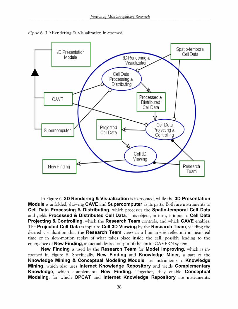
Figure 6. 3D Rendering & Visualization in-zoomed.
In Figure 6, 3D Rendering & Visualization is in-zoomed, while the 3D Presentation
Module is unfolded, showing CAVE and Supercomputer as its parts. Both are instruments to Cell Data Processing & Distributing, which processes the Spatio-temporal Cell Data and yields Processed & Distributed Cell Data. This object, in turn, is input to Cell Data
Projecting & Controlling, which the Research Team controls, and which CAVE enables. The Projected Cell Data is input to Cell 3D Viewing by the Research Team, yielding the desired visualization that the Research Team views as a human-size reflection in near-real time or in slow-motion replay of what takes place inside the cell, possibly leading to the emergence of New Finding, an actual desired output of the entire CAVERN system.
New Finding is used by the Research Team for Model Improving, which is in-zoomed in Figure 8. Specifically, New Finding and Knowledge Miner, a part of the Knowledge Mining & Conceptual Modeling Module, are instruments to Knowledge
Mining, which also uses Internet Knowledge Repository and yields Complementary
Knowledge, which complements New Finding. Together, they enable Conceptual
Modeling, for which OPCAT and Internet Knowledge Repository are instruments.

_____________________________Journal of Multidisciplinary Research____________________________
39
Finally, Conceptual Modeling changes Cell Life Model from partial to improved, in accord to what is expressed at a more abstract level in Figure 4, and even more abstractly, in Figure 3.
Figure 7. Left: a “chip” cellular microarray; right: rod-shaped bacillus.
Using a cellular microarray (Chen et al., 2005), shown in Figure 7, the supercomputer processes the detected cellular activity data it receives from the Scene Inspection & Capture
Module. Cellular microarrays currently allow for multiplexed interrogation of living cells on the surface of a solid support, spotted with varying materials (antibodies, proteins, lipids...), which can interact with the cells, leading to their capture on specific spots. Combinations of different materials can be spotted in a given area, allowing the triggering of a cellular response, change in phenotype, or detection of a specific secreted factor. Statistical algorithms, developed within the Computation & Control Module of CAVERN will be applied to compute spatiotemporal interactions of interest in one representative cell (see Figure 8 left) that will be computed from data obtained from millions of cells, which had been biologically marked as explained by the OPD in Figure 8 (right).
Figure 8. Model Improving (left) and Biological Marking (right) in-zoomed.
The New Finding is input to the Model Improving process, shown in Figure 8. Within
Model Improving, New Finding is instrument to Knowledge Mining, which is aimed at checking whether a similar or related finding exists anywhere in the literature or databases. The potentially found Complementary Knowledge and the New Finding are used to update the Cell Life Model via the Conceptual Modeling process, changing it from partial to improved.
_____________________________Journal of Multidisciplinary Research____________________________
40
Diving yet deeper into the fourth level of detail, Biological Marking from Figure 5 is zoomed into in the OPD in Figure 8, showing its two subprocesses, Cell Coordinate System
Marking and Region-of-Interest Marking. The objective of the first subprocess is to enable the establishment of a 3D Cartesian, cylindrical, or polar coordinate system, depending on the cell shape. For example, since bacillus (see left of Figure 7) is shaped as a rod, it will be marked for a cylindrical coordinate system. Good potential candidates for marking the origin of a coordinate system are specific proteins in the nucleolus, a non-membrane bound structure within the nucleus, where rRNA is transcribed and assembled. A customary way of marking is through fluorescent tagging fluorophores and fluorescent recovery after photobleaching (FRAP). A fluorophore, such as fluorescein isothiocyanate (FITC), coumarin, cyanine (Rietdorf, 2005), and newer, more effective ones (Lakowicz, 2006), is a functional group in a molecule that absorbs energy of a specific wavelength and reemits energy at a specific different wavelength. The database of fluorescent dyes and applications (Database of Fluorescent Dyes, 2010) enables directed search of thousands of fluorophores. The amount and wavelength of emitted energy depend on the fluorophore and its environment. Photobleaching, the photochemical destruction of a fluorophore, can be exploited to study the motion of molecules, their diffusion, or both, for example via FRAP. FRAP is an optical technique capable of examining single cells that has been useful in studies of cell membrane diffusion and protein binding.
Biological Knowledge Mining
The OPM architecture model of CAVERN calls for constructing a conceptual model of the cell which exploits synergy between 3D visualization, using the CAVE, and knowledge creation or discovery through biological text mining. Figure 9 and Figure 10 are two examples of OPCAT-enabled URL hyperlinks documenting the references from the OPM model to the PDF passage or Web page sentence on which any specific modeled fact is based. Based on the work conducted as part of ISO standardization of OPM (ISO, 2012), CAVERN will be able to automatically or semi-automatically construct such links as well as biological OPM model snippets. After being verified by experts, these will be universally accessible for biologists to browse, reuse, and improve. Each model snippet will be paired with semantically organized URL pointers to documents, including scientific papers and multimedia files, such as CAVERN clips, which researchers can replay in their CAVERNs, creating an evolving web of biological knowledge.

_____________________________Journal of Multidisciplinary Research____________________________
41
Figure 9. Example of a URL link from the OPM model to a PDF document on which the model is based.
Challenges and Open Problems
CAVERN is an ambitious complex research project with potentially highly valuable gains in science, health, and education, explained above. Its detailed OPM architecture, of which only part was elaborated upon, suggests that in spite of its highly innovative nature, it definitely is feasible. Like most high-gain disruptive technology developments, there are open questions that require integration, developing new technologies, techniques, interfaces, and algorithms in several cross-disciplinary domains. Below are the main open questions:
How do we best mark cells for establishing their coordinate system? Preliminary
directions for answers are provided in this article. How will the tracing of marked molecules of interest in the microarray and in the
electron microscope be done? Is it possible to put one cell in each well of the microarray by dilution? If not, how do
we track several cells in each well? How to compute the representative cell from cell microarray data? How to combine representative cell data with data from direct observations from the
electron microscope? Should we use Transmission Electron Microscopy (TEM), which provides 2D images,
or Scanning Electron Microscopy (SEM), which provides 3D images? How to mark coordinates on the cell for reference points? What sites and markers
should be used? How to detect that two molecules are combined? How to detect that two combined
molecules separate?

_____________________________Journal of Multidisciplinary Research____________________________
42
Figure 10. Example of a URL link from the OPM model to a website documenting the modeled fact.
Summary
This chapter has laid out the vision, principles, and architecture blueprint of CAVERN –
a Computer-based Augmented Virtual Environment for Realizing Nature. CAVERN is a disruptive intelligent multimedia system-of-systems that opens the door for a completely new type of molecular biology research by translating nano-scale size and time dimensions that characterize molecular level processes in the cell into the human cognitive comfort zone. When operational CAVERN will enable researchers to design, execute, and monitor experiments, they will be able to see the results in near real time in the most direct way conceivable, and update the OPM-based conceptual model with new findings from their own experiments as well as from new literature.
The major technological building blocks of CAVERN are mature, but algorithms, interfaces, and standards need to be developed to make the CAVERN vision a reality for the benefit of humankind. This will require significant intellectual and monetary resources, but the potential gains of an operational CAVERN definitely outweigh the efforts and investments needed to materialize it.
References
Chen, D. S., Soen, Y., Stuge, T. B., Lee, P. P., Weber, J. S., Brown, P. O., & Davis, M. M.
(2005). Marked differences in human melanoma antigen-specific T cell responsiveness after vaccination using a functional microarray. PLoS Med, 2(10), e265. doi:10.1371/journal.pmed.0020265. Available: http://www.plosmedicine.org/article/ info%3Adoi%2F10.1371%2Fjournal.pmed.0020265, accessed December 17, 2012.

_____________________________Journal of Multidisciplinary Research____________________________
43
Cooney, M. (2010). NSF earmarks $30M for game-changing Internet research. Network World, January 28. Retrieved December 17, 2012, from http://www.networkworld.com/ community/node/56553
Database of Fluorescent Dyes, Properties, and Applications. (2010). Retrieved January 10, 2013, from http://www.fluorophores.tugraz.at/
Dori, D. (2002). Object-Process Methodology – A holistic systems paradigm. Berlin, Heidelberg, and New York: Springer Verlag.
Dori, D. (2008). Words from pictures for dual channel processing. Communications of the ACM, 51(5), 47-52.
Dori, D., & Choder, M. (2007). Conceptual modeling in systems biology fosters empirical findings: The mRNA lifecycle. PLoS ONE, 2(9), e872. doi:10.1371/journal.pone.0000872. Retrieved December 17, 2012, from http://www.plosone.org/article/info%3Adoi%2F10.1371%2Fjournal.pone.0000872
Dori, D., Linchevski, C., & Manor, R. (2010). OPCAT – A Software Environment for Object-Process Methodology Based Conceptual Modelling of Complex Systems. In P. Heisig, J. Clarkson, & S. Vajna (Eds.), Proceedings of the 1st International Conference on Modelling and Management of Engineering Processes (pp. 147-151). Cambridge: University of Cambridge.
Estephan, J. (2008). Survey of Model-Based Systems Engineering (MBSE) Methodologies. INCOSE-TD-2007-003-02. Retrieved December 17, 2012, from http://www.incose.org/ProductsPubs/pdf/techdata/MTTC/MBSE_Methodology_Survey_2008-0610_RevB-JAE2.pdf
ISO. (2012). ISO/TC 184/SC 5 Plenary Meeting Resolutions. Retrieved January 10, 2013, from http://esml.iem.technion.ac.il/site/wp-content/uploads/2011/05/ISO-TC184-SC5_N1184_2012_SC5_Plenary_Resolutions_-_Haifa__re.pdf
Kitano, H. (2002). Foundations of systems biology. Cambridge, MA: MIT Press. Lakowicz, J. R. (2006). Principles of fluorescence spectroscopy (3rd ed.). New York: Springer. Mayer, R. E., & Moreno, R. (2003). Nine ways to reduce cognitive load in multimedia learning.
Educational Psychologist, 38(1), 43-52. Rietdorf, J. (2005). Microscopic techniques. Advances in Biochemical Engineering/
Biotechnology, 246-249. Somekh, J., Choder, M., & Dori, D. (2012). Conceptual model-based systems biology: Mapping
knowledge and discovering gaps in the mRNA transcription cycle. PLoS ONE. Retrieved January 10, 2013, from http://www.plosone.org/article/info%3Adoi%2F10.1371%2F journal.pone.0051430

_____________________________Journal of Multidisciplinary Research____________________________
44
About the Author
Dov Dori ([email protected]) is Information and Systems Engineering Professor and Head of the Enterprise System Modeling Laboratory at the Faculty of Industrial Engineering and Management, Technion-Israel Institute of Technology, and Visiting Professor at the Engineering Systems Division, Massachusetts Institute of Technology, where he lectures on a regular basis. His research interests include conceptual modeling of complex systems, systems architecture and design, software and systems engineering, and systems biology. Professor Dori invented and developed Object-Process Methodology (OPM), the emerging 19450 ISO Standard. He has authored or edited five books, and more than 250 journal and conference publications and book chapters. He is Fellow of INCOSE – International Council on Systems Engineering, Fellow of IAPR – International Association for Pattern Recognition, and Senior Member of IEEE and ACM.
Discussion Questions
1. What are the main impediments to full implementation of the CAVERN vision presented in
this chapter? Are they mainly technological, managerial, or related to integration and systems engineering?
2. What potential value to scientists and students lies in the CAVERN system, once
implemented? 3. Assuming the CAVERN vision is implemented, how would you classify and rate this
development in the evolution from the optical microscope to the electron microsphere to further visualization devices?
4. What are the similarities and differences between the former inventions and this one?
To Cite this Article
Dori, D. (2012, Fall). Extending the human spatiotemporal comfort zone with CAVERN – Computer-based Augmented Virtual Environment for Realizing Nature. Journal of Multidisciplinary Research, 4(3), 23-44.