expression of the n-methyl-d-aspartate receptor nr1 splice variants and nr2 subunit subtypes in the...
TRANSCRIPT

Expression of the N-methyl-D-aspartate Receptor NR1 SpliceVariants and NR2 Subunit Subtypes in the Rat Colon
Arseima Y Del Valle-Pinero1,2, Shelby K. Suckow1,2, Qiqi Zhou3, Federico M. Perez2, G.Nicholas Verne3, and Robert M. Caudle.1,2,*
1 Department of Neuroscience, University of Florida College of Medicine, Gainesville, Fl 32610, USA.
2 Department of Oral and Maxillofacial Surgery and Diagnostic Sciences, University of Florida College ofDentistry, Gainesville, Fl 32610, USA.
3 Department of Medicine, Division of Gastroenterology, University of Florida, Gainesville, Fl 32610, USA.
AbstractN-methyl-D-aspartate (NMDA) receptors and the expression of their different splice variants andsubunits were previously characterized in the brain and spinal cord. However, knowledge on theNMDA receptor expression and function in the enteric nervous system is limited. Previous worksuggested that NMDA receptors were involved in a rat model of visceral hypersensitivity. The aimof this study was to characterize the expression of the NMDA receptor NR1 splice variants and theNR2 subunit subtypes in the rat colon. We visualized the expression of NR1 protein in the ratsubmucosal and myenteric plexuses. The NR1 splice variants found in the colon of rats lacked theN1 and C1 cassettes and contained the C2 and C2′cassettes (NR1000 and NR1001). The NR2B andNR2D subunits were also found in the rat colon. Moreover, NMDA receptors in the rat colon wereheteromeric, since NR1 was co-localized with NR2B and NR2D subunits using fluorescentimmunohistochemistry. The identification of the NMDA receptors in the enteric nervous systemcould lead to the development of drugs that selectively modulate bowel function.
Keywordsglutamate receptor; NMDAR1; NMDAR2; myenteric plexus; submucosal plexus;immunohistochemistry
N-Methyl-D-aspartate (NMDA) receptors are the most clearly defined glutamate receptorsubtype. These receptors have two subunit families designated NMDAR1 (NR1) andNMDAR2 (NR2) (Michaelis 1993). In mammals, the functional NMDA receptor is aheteromeric complex containing NR1 and NR2 subunits (McBain and Mayer 1994). However,the exact number of subunits in each heteromeric glutamate receptor channel is still unknown.
The NR1 subunit (Fig. 1) has only one gene which has three regions of alternative splicing,named the N1, C1 and C2 cassettes and results in eight possible splice variants. The N1 (exon5) and C1 (exon 21) cassettes may be either present or absent without affecting the remaining
* Correspondence: Robert M. Caudle, Ph.D., Department of Oral and Maxillofacial Surgery, University of Florida College of Dentistry,PO Box 100416, Gainesville, FL 32610, USA, Email: [email protected], Phone: 352-273-6767, Fax: 352-392-7609Pain Mechanisms Section Editor: Dr. Linda S. Sorkin, Department of Anesthesiology, University of California at San Diego, 9500Gilman Drive, La Jolla, CA 92093, USAPublisher's Disclaimer: This is a PDF file of an unedited manuscript that has been accepted for publication. As a service to our customerswe are providing this early version of the manuscript. The manuscript will undergo copyediting, typesetting, and review of the resultingproof before it is published in its final citable form. Please note that during the production process errors may be discovered which couldaffect the content, and all legal disclaimers that apply to the journal pertain.
NIH Public AccessAuthor ManuscriptNeuroscience. Author manuscript; available in PMC 2008 June 15.
Published in final edited form as:Neuroscience. 2007 June 15; 147(1): 164–173.
NIH
-PA Author Manuscript
NIH
-PA Author Manuscript
NIH
-PA Author Manuscript

NR1 protein. When the C2 cassette (exon 22) is spliced out the first stop codon is lost, resultingin the expression of the C2′ cassette. Therefore, either the C2 or C2′ cassette are present in allNR1 protein, but the two cassettes are not co-expressed within the same NR1 protein (Zukinand Bennett 1995). The N1 cassette is located extracellularly and interacts with variouspharmacological modulators of the channel, including zinc, protons and polyamines(Dingledine et al. 1999). The C-terminal cassettes of the protein control cell-surface expressionof NR1, where proteins with the shortest C-terminal region (lacking the C1 cassette andcontaining C2′; NR1x00) show the highest cell surface expression (Okabe et al. 1999).
The NR2 subunit includes four family members NR2A, 2B, 2C and 2D. Expression of differentNR2 subunits determines the deactivation kinetics, antagonist sensitivity and ligand affinityof recombinant NR1/NR2 receptors (McBain and Mayer 1994,Mori and Mishina 1995).
The presence of L-glutamate receptors, presumably of the NMDA type, was documented inthe myenteric plexus of the guinea pig (Moroni et al. 1986,Luzzi et al. 1988,Shannon andSawyer 1989,Alesiani et al. 1990,Wiley et al. 1991). Furthermore, the expression of the NMDAreceptor NR1 subunit was characterized by in situ hybridization and PCR techniques in boththe rat and guinea-pig enteric plexuses (Burns et al. 1994,Broussard et al. 1994). Alsoglutamatergic receptors, including the NMDA type, were found in the enteric plexuses of thehuman colon (Giaroni et al. 2003). These findings together established the presence of NMDAreceptors in the enteric nervous system. However, the roles of these enteric NMDA receptorsare still not fully understood.
Previous studies showed that the NR1 subunit was co-labeled with the vasoactive intestinalpeptide (VIP) on the inhibitory motor neurons of the ENS using double in situ hybridization(Burns and Stephens 1995). Moreover, glutamate and NMDA enhance acetylcholine andnoradrenaline release from the ENS. Colonic peristaltic reflex was inhibited in vitro whenNMDA was added and these effects were antagonized by 2-Amino-5-phosphonovalerate(APV) (Cosentino et al. 1995,Giaroni et al. 2003). The presence of the NR1 subunit was alsocharacterized in the rat extrinsic primary afferents (EPANs) and the intravenous administrationof memantine inhibited pain responses to noxious mechanical stimuli (McRoberts et al.2001). Intrathecal administration (10 nmol) of the NMDA receptor antagonist; MK-801 weresufficient to significantly attenuate the hyperalgesic visceromotor responses to colonicdistention after zymosan-induced inflammation (Coutinho et al. 2001). In a similarinflammation model, the administration of both intrathecal MK-801 (1.5 nmol) andintraperitoneal MK-801 (0.15 mg/kg) completely abolished the colorectal distension-inducedhypersensitivity of both noxious and innocuous stimuli (Gaudreau and Plourde 2004). Eventhough it is uncertain whether these NMDA receptor antagonists were acting at the level of thecolon, taken together these studies suggested that NMDA receptors were involved in thesemodels of visceral hypersensitivity. Whether visceral hypersensitivity is produced by NMDAreceptors on nociceptive neurons or through the receptor’s actions on visceral motor neuronsis currently not known. However, the identification and characterization of these entericNMDA receptors could provide useful information for the development of drugs thatselectively modulate bowel function. Here we studied the expression of the NMDA receptorNR1 protein splice variants and the NR2 subunit subtypes in the rat colon by using RT-PCR,western blot and immunohistochemistry.
Experimental ProceduresAnimals
Sprague Dawley rats (200–300 g, N=9) were maintained on a 12 hour light/dark cycle and fedstandard rodent chow and water ad libitum. All experiments were approved by the Universityof Florida Institutional Animal Care and Use Committee.
Valle-Pinero et al. Page 2
Neuroscience. Author manuscript; available in PMC 2008 June 15.
NIH
-PA Author Manuscript
NIH
-PA Author Manuscript
NIH
-PA Author Manuscript
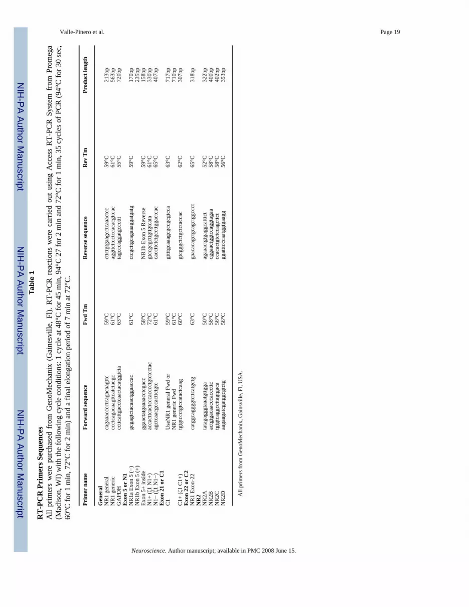
Reverse transcription-PCR (RT-PCR)Animals were euthanized with CO2 and the brain and intestine were quickly removed. TotalRNA was isolated from rat brain and intestine tissues using RNeasy Mini Kit from Qiagen(Valencia, CA.). Target transcripts were amplified with PCR primers from GenoMechanix(Gainesville, Fl) listed on Table 1. RT-PCR reactions were carried out using Access RT-PCRSystem from Promega (Madison, WI) and the following cycle conditions: 1 cycle at 48°C for45 minutes, 94°C for 2 minutes and 72°C for 1 minutes, 35 cycles of PCR (94°C for 30 sec,60°C for 1 minutes, 72°C for 2 minutes) and a final elongation period of 7 minutes at 72°C.PCR products were separated on 1.2% agarose gel with 1X TBE buffer, viewed with ethidiumbromide and analyzed with Bio-Rad Gel Doc EQ Gel Documentation System, Bio-RadLaboratories (Hercules, CA). All the RT-PCR products were sequenced at the GenomeSequencing Services Laboratory (GSSL), part of the Interdisciplinary Center forBiotechnology Research (ICBR) at the University of Florida, Gainesville, FL.
Western blotsSamples of tissue were taken from rat brain and descending colon. Tissue was homogenizedin cold Cell Lysate Buffer [1mM Sodium Ortho-Vanadate, 10 mM Tris and 1% SDS] using aSonics Vibra-Cell Sonicator. Lysates were boiled for 5 minutes and then centrifuged at 16,000g for 5 minutes and the supernatant was collected. Protein concentration was determined bystandard spectrophotometer method. Proteins were separated using 4–20% Tris-Glycine Gelfrom Invitrogen (Carlsbad, CA), each lane was loaded with 15 μg of protein extract. Proteinsin the gel were then transferred to a Millipore (Bedford, MA) Immobilon-P polyvinylidenefluoride (PVDF) membrane using a semi-dry transfer device (Bio-Rad Laboratories, Hercules,CA). The transfer buffer used contains 20% methanol, 48 mM Tris pH 9.2, and 39 mM glycine.The membrane was then placed in TTBS buffer [20 mM Tris pH 7.6, 0.9% NaCl, and 0.05%Tween-20, pH 7.4] containing 5% non-fat dry milk for 1 hour to block non-specific bindingof antibodies. NR1 splice variant specific primary antibodies were provided by Dr. MichaelIadarola from The National Institute of Dental and Craniofacial Research (NIDCR), Bethesda,MD (Antibodies’ selectivity was assessed in Caudle et al. 2005). Antibodies against Actin,NR1, NR2B and NR2D were purchased from different vendors (Table 2). After overnightincubation with primary antibody at 4°C, the membrane was washed three times in TTBS (5minutes each) and then placed in fresh TTBS containing 5% non-fat dry milk and secondaryantibody [dilution 1:4000] for rabbit or mouse IgG coupled to horse radish peroxidase (HRP)for 1 hour. The membrane is then washed three times with TTBS (5 minutes each) and placedin chemiluminescence substrate (LumiGLO®, Cell Signaling Technology, Beverly, MA) andexposed to film at variable times points (15 seconds to 10 minutes) to ensure best resolution.
Immunohistochemistry (IHC)Perfusion fixation—Rats were given a lethal dose of pentobarbital and perfused throughthe heart with cold 0.9% saline followed immediately with cold 4% Paraformaldehyde. Afterfixation the colon of the animal was removed, post-fixed in Paraformaldehyde for 24h at 4°C,and then stored in 30% sucrose at 4°C for at least 24 hours.
Cryostat sections—Tissue was sectioned at 10 μm on a cryostat, serially mounted on aglass slide, and air-dried for 1 hour. All preparations were washed 3 times (10 minutes each)in Phosphate Buffered Saline (PBS) [10 mM sodium phosphate, pH 7.4, 0.9% NaCl] and placedin blocking buffer containing 3% Normal Goat Serum (NGS) with PBS for 1 hour, andincubated in primary antibody in 3% NGS/0.3% tween-20/PBS for 24 hours at 4°C. Most ofprimary antibodies mentioned above in the Western blots section were used. In additionantibodies for neuronal markers; Protein Gene Product 9.5 (PGP 9.5) and Neurofilament (NF)were used (Table 2). The sections were then washed 3 times in PBS (10 minutes each) followed
Valle-Pinero et al. Page 3
Neuroscience. Author manuscript; available in PMC 2008 June 15.
NIH
-PA Author Manuscript
NIH
-PA Author Manuscript
NIH
-PA Author Manuscript
by 1 hour incubation in secondary antibodies, either Alexa Fluor 488 or 495 (1:1000; MolecularProbes, Boston, MA) in 3%NGS/0.3% tween-20/PBS. Negative controls were performed byincubating samples with only secondary antibodies and omitting primary antibodies.
After incubation with secondary antibodies, tissue was washed 3 times (10 minutes each) andcoversliped with ProLong® Antifade Kit mounting media (Molecular Probes, Boston, MA).The sections were visualized with filters for red and green excitation. Images werephotographed on an Olympus BX51 Fluorescence microscope (Olympus, Center Valley, PA).
Whole mount—Segments of tissue from the descending colon (2–3cm) were removed andcut longitudinally. The segments were spread and mounted onto slides containing a siliconebase to enable pinning of the tissue to the slides. To defat the tissue, the segments were washedthrough a series of ethanol dilutions starting with 2 washes in 100% ethanol followed by singlewashes in 95%, 70%, and 50% ethanol for 20 minutes each. Sections were incubated in distilledwater overnight at 4°C. The mucosa, submucosal plexus and circular muscle layers were thenpealed from each tissue section and the remaining myenteric plexus layer placed in 4%paraformaldehyde overnight followed by 3–5 washes (5 minutes each) in PBS. Tissue sectionswere cleared by placing them in KOH (in PBS) and glycerin serial incubations: 3:1 0.5% KOH:glycerin for 1 hour, 1:1 0.5% KOH: glycerin for 1 hour, 1:3 0.5% KOH: glycerin for 1 hour,and 100% glycerin overnight. A few drops of 30% H2O2 were added to both 1:1 and 1:3 KOH:glycerin incubations. After 3–5 washes (5 minutes each) in PBS, the tissue was placed inblocking buffer containing 3% Normal Goat Serum (NGS) with PBS for 30 minutes, thenincubated in the primary antibody [1:500 – 1:100] in 3% NGS/PBS, 1% Triton X-100 overnightat 4°C. After 5 washes (10 minutes each) in PBS, the tissue was incubated for 60 minutes insecondary antibodies, either Alexa Fluor 594 or Alexa Fluor 488 (1:2000, Molecular Probes,Boston, MA) with 1% NGS/PBS, 0.3% Triton X-100. The tissue was then washed 6 times (5minutes each) in PBS and placed in distilled water. Tissues were then mounted into clean slidesand coversliped with ProLong® Antifade Kit mounting media (Molecular Probes, Boston, MA)(Rosa-Molinar et al. 1999). Slides were visualized with filters for red and green excitation.Images were photographed on an Olympus BX51 Fluorescence microscope (Olympus, CenterValley, PA).
ResultsExpression of NR1 protein in the rat colon
The expression of NR1 was examined in the rat colon (Fig. 2). We used two different sets ofprimers to amplify by RT-PCR (Fig. 2A) the RNA extracted from intestine and brain tissues.These primers (arbitrarily named NR1 general and generic) recognized areas in the NR1 outsidethe spliced exons. The expression of NR1 was evident in the colon tissue (gastrointestinal, GI)and the rat brain. Rat brain was used as a positive control. Sample integrity and reactionreliability were validated with the expression of the housekeeping gene, Glyceraldehyde-3-phosphate dehydrogenase (GAPDH) in both the GI and the brain tissues. A negative control(−RT) that lacked reverse transcriptase using GAPDH primers was included to control forgenomic DNA contamination.
Protein expression was confirmed by western blot analyses that showed the NR1 protein in GIand brain tissues using general mouse-NR1 antibody, which recognized a site outside of thealternatively spliced cassettes (Fig. 2B, Top panel). To control for sample loading purposes,an antibody against Actin was used.
Furthermore, we visualized the double label of the NR1 protein with the neuronal markers;Protein Gene Product 9.5 (PGP 9.5) (Fig. 3A) and neurofilament (NF) (Fig. 3B) in the rat
Valle-Pinero et al. Page 4
Neuroscience. Author manuscript; available in PMC 2008 June 15.
NIH
-PA Author Manuscript
NIH
-PA Author Manuscript
NIH
-PA Author Manuscript
myenteric and submucosal plexuses using immunohistochemistry. The co-labeling of NR1 andthe neuronal markers PGP and neurofilament verified that NR1 is localized in enteric neurons.
Expression of splice variants of NR1 proteinThe NR1 subunit gene has a total of 22 exons, three of which (exons 5, 21 and 22) undergoalternative splicing to generate eight NR1 splice variants. The alternatively spliced cassettesof the NR1 protein were named N1, C1 and C2. A detailed scheme of splice variant compositionof NR1 protein is presented in Fig. 1B. The four alternatively spliced cassettes of NR1 proteinare represented as colored boxes. In this model, the N1 cassette (exon 5) is extracellular at theN-terminal, while the three C-terminal cassettes (C1, C2 and C2′) are located intracellularly.The resultant eight splice variants of the NR1 protein in the presence (+) or absence (−) of thethree alternatively spliced exons (N1, C1 and C2) have several nomenclatures (Reviewed byYamakura and Shimoji 1999). The nomenclature used here; NR1xyz, was proposed by Durandand colleagues (Durand et al. 1993), where x, y and z, represent the cassettes N1, C1 and C2,respectively. The values of x, y and z are either 0 or 1 indicating the absence or presence ofthe respective cassette (Fig. 1C).
After verifying the general expression of NR1 protein in the rat intestine, we examined theexpression of the splice variants of NR1 using specific primers and antibodies with RT-PCRand western blots. Rat brain contains all splice variant isoforms (Sugihara et al. 1992), thus,this tissue was used as a control in all our assays.
Expression of the N1 cassette (exon 5) in the rat colonNR1 protein containing and lacking the N1 cassette was known to be present in both rat brain(Sugihara et al. 1992) and spinal cord (Tolle et al. 1995,Prybylowski et al. 2001). To analyzethe expression of the N1 cassette in rat colon we performed RT-PCR (Fig. 4A) using fourdifferent sets of primers (Table 1); “NR1a(b)”, which produced two different sized products,a 274 bp band in the presence of exon 5 or a 211 bp band in the absence of this cassette. Primers“exon 5+” and “N1+”, that were designed inside the N1 cassette and only showed a band ifthis cassette was present. Alternatively “N1−” primers, only showed a band if exon 5 was notpresent. While brain samples expressed both the N1 splice variants, the rat colon only expressedthe variant that lacked N1 (NR10yz). To verify these findings at the protein level, western blotassays were performed. The expected NR1 band around 118 kDa was present in brain but itwas lacking in GI tissues (Fig. 4B).
These results further suggested that only splice variants without the N1 cassette (NR10yz) werepresent in the rat colon.
Expression of C-terminal cassettes in the rat colonThe C-terminal cassettes were found to be critical in determining the cellular localization ofthe NR1 (Okabe et al. 1999). Here we analyzed the expression of the rat colon C1 cassette byRT-PCR, using two sets of primers; “C1” (with NR1 generic, see Table 1) and the “C1+”primers (Liesi et al. 1999). We found with both sets of primers that the expected C1 band waspresent in brain, but was absent from the rat colon (Fig. 4C).
To confirm these findings, we blotted using an antibody targeted towards the C1 cassette. Weobserved the expression of C1 containing NR1 protein in brain but not in GI (Fig. 4D).
Whereas bands corresponding to the C1 cassette in both RT-PCR and western blot analyseswere present as expected in brain, none were visible in GI, suggesting that only the NR1 splicevariants that lacked the C1 cassette (NR1x0z) were present in the rat colon.
Valle-Pinero et al. Page 5
Neuroscience. Author manuscript; available in PMC 2008 June 15.
NIH
-PA Author Manuscript
NIH
-PA Author Manuscript
NIH
-PA Author Manuscript
When the C2 cassette (exon 22) is spliced out the first stop codon is removed, resulting in theexpression of the C2′ cassette. Consequently, either the C2 or C2′ cassette is present in all NR1protein, but the two cassettes are not co-expressed within one NR1 molecule (Zukin andBennett 1995).
RT-PCR and western blot were used to assess the expression of the C2 and C2′cassettes. RT-PCR with primers for exon 22 showed a band for both GI and brain (Fig. 4E). For the westernblot analyses we employed two different antibodies; the C2+ antibody, which targeted the C2cassette and the C2- antibody directed against the C2′ cassette. These antibodies showedexpression of both the C2 and C2′ cassettes in the rat brain and colon (Fig. 4F). These resultssuggested that both NR1 splice variants, the one that has the C2 cassette and the one thatcontains the C2′ cassette (NR1xx0 and NR1xx1) were present in the rat colon. These variantswere visualized in the rat myenteric plexus by immunohistochemistry with Alexa fluor-linkedantibodies (Fig 5).
Expression of NR2 protein subtypesThe second subunit family of the rat NMDA receptor, named NMDAR2 (NR2), has fourmembers the NR2A, NR2B2, NR2C, and NR2D subunits. In mammals functional NMDAreceptor channels are produced when the NR1 subunit is expressed together with one or moreof the four NR2 subunit subtypes (McBain and Mayer 1994). All four NR2 subunit subtypesare expressed in brain therefore we compared the expression of NR2 in the rat colon with thistissue.
We used RT-PCR with four different sets of primers to assess the expression the NR2 subunits(Fig. 6A). Analyses of NR2A and NR2C demonstrated no bands for GI, while both NR2B andNR2D were evident in GI and brain tissues. These results suggested that only NR2B and NR2Dwere present in the rat colon. These subunits were visualized in the rat myenteric plexus byimmunohistochemistry with Alexa fluor-linked antibodies (Fig 6B). Moreover, we were ableto visualize the double label of the NR2B protein with the neuronal marker; Protein GeneProduct 9.5 (PGP 9.5) (Fig. 6C) using immunohistochemistry. The co-labeling of NR2B andthe neuronal marker PGP further verified that NR2B was localized in enteric neurons.
Co-labeling of NR1 with NR2B and NR2D in the rat colonThe functional NMDA receptor is a heteromeric complex containing NR1 and NR2 subunits(McBain and Mayer 1994). In order to verify if NR2B and NR2D were co-localized with NR1in the rat colon we used specific antibodies targeted to NR1, NR2B and NR2D. We visualizedthe double labeling of NR1 with both NR2B and NR2D with immunohistochemistry usingAlexa fluor-linked secondary antibodies (Fig. 7). The co-localization of NR1 with NR2B andNR2D suggested there were heteromeric complexes of these NR2 subtypes with NR1 in therat colon.
DiscussionOur studies confirmed the expression of NR1 protein in the rat myenteric and submucosalplexuses. We found that only the NR1 splice variants that contained the C2 or the C2′ cassette(NR1000 and NR1001) were present in the rat colon. Also, the NR2B and NR2D subunits werethe only NR2 subunit subtypes found in the rat colon. Lastly, the NMDA receptors in the ratcolon were arranged in heteromeric complexes including the NR1 with NR2B and NR2Dsubunits.
The presence of the N1 cassette caused a decrease in the open time of the NMDA receptorchannel (Rumbaugh et al. 2000) and decreased the ability of spermine to potentiate NMDA
Valle-Pinero et al. Page 6
Neuroscience. Author manuscript; available in PMC 2008 June 15.
NIH
-PA Author Manuscript
NIH
-PA Author Manuscript
NIH
-PA Author Manuscript

mediated currents (Mott et al. 1998). Since these enteric receptors lacked the N1 cassette, theywould show an increased pH, Zn2+, and spermine sensitivity similar to receptors with mutationsat the N1 cassette (Traynelis et al. 1998).
NR1 proteins with the shortest C-terminal region (lacking the C1 cassette and containing C2′;NR1x00) showed the highest cell surface expression (Okabe et al. 1999). The lack of the C1cassette also affected the PKC potentiation (Durand et al. 1993,Logan et al. 1999,Zheng et al.1997) and the calmodulin-dependent inhibition (Ehlers et al. 1996) of these receptors’ currents.Receptor clustering may be affected also, since the C1 cassette was shown to have specificinteractions with multiple intracellular proteins, including neurofilament-L (Ehlers et al.1998) and the cytoskeletal protein yotiao (Lin et al. 1998). Therefore, these enteric NMDAreceptors which lacked both the N1 and C1 cassette may have an increased cell-surfaceexpression but poor clustering properties. Moreover, prior studies found that NR1000 receptorswere markedly less active than other splice forms like NR1111 (Durand et al. 1993), indicatingthat the NMDA receptors in the colon may not produce large currents in the neurons whenactivated.
Functional NMDA receptor channels in mammals are considered to be produced only whenthe NR1 subunit is expressed together with one or more of the four NR2 subunits (McBain andMayer 1994). While the NR1 is widely expressed throughout the whole brain, the expressionof the NR2 subunits is highly regulated. The distributions of NR2A and NR2C have temporaland spatial similarities to that of NR1, while the expression of NR2B shows differences in theintensity and distribution (Takai et al. 2003). NR2D expression in the brain is very faintcompared to other NR2s, and mutant mice defective in NR2D expression appear to developnormally (Ikeda et al. 1995). Not only the intensity and distribution of expression of the NR2subunits is unique, but their functional behavior differs. The offset decay time constant of theNRl/NR2B channel is ~400 msec, while the NR1/NR2D channel shows a very long offsetdecay time constant (~5000 msec) (Reviewed by Mori and Mishina 1995).
The NR2B subunit has been the object of great interest as a therapeutic target in a wide rangeof pathologies, including acute and chronic pain. Therefore, a remarkable compilation of drugswere developed which target the NR2B subunit (Chazot 2004).
In conclusion we found that enteric NMDA receptors were heteromeric complexes of eitherNR1000 or NR1001 with the NR2B and NR2D subunits. Moreover, in our studies with a 2,4,6-trinitrobenzene sulfonic acid (TNBS)-induced model of colitis of the rat colon, both theexpression of the N1 and C1 cassettes are enhanced 14 days after inflammation (Zhou et al.2006). Therefore, selective changes in the expression of the NR1 splice variants and possiblythe NR2 subunit subtypes of the NMDA receptors may be an element for the ongoing visceralhypersensitivity in conditions such as irritable bowel syndrome (IBS). It is possible that theenteric NMDA receptors have low activity in the normal healthy intestines and their activityis increased after alternative splicing events that occur during pathophysiological states.Previous studies using NMDA receptor antagonists showed promising inhibitory effects onmodels of visceral hypersensivity (Coutinho et al. 2001,McRoberts et al. 2001,Giaroni et al.2003,Gaudreau and Plourde 2004). The drugs in these studies seemed to be working at thelevels of the spinal cord and primary afferent neurons. Whether these NMDA receptorantagonists could have an effect at the enteric level remains uncertain. To our knowledge thisis the first comprehensive analysis of the expression of the NMDA receptor NR1 protein splicevariants and the NR2 subunit subtypes in the rat colon. Therefore, more studies are needed todetermine the specific properties of these receptors both in normal and pathological conditions.A better understanding of the expression, physiology and pharmacological properties of theNMDA receptors present in the enteric nervous system could lead to the development of drugsthat selectively modulate the bowel function.
Valle-Pinero et al. Page 7
Neuroscience. Author manuscript; available in PMC 2008 June 15.
NIH
-PA Author Manuscript
NIH
-PA Author Manuscript
NIH
-PA Author Manuscript
Acknowledgements
The authors thank Dr. Michael Iadarola, from The National Institute of Dental and Craniofacial Research (NIDCR)in Bethesda, Maryland for supplying the NR1 splice variants specific antibodies. We like to thank Alan Jenkins fromthe Laboratory of Dr. John Neubert at the University of Florida, Department of Orthodontics for providing technicalassistance with the immunohistochemistry protocol. We also like to thank Dr. Eduardo Rosa-Molinar and Jose Serranofrom the Department of Biology at the University of Puerto Rico in San Juan, Puerto Rico for providing us with thewhole mount immunohistochemistry protocol. The work summarized here was supported by The National Institutesof Health NS045614.
ReferencesAbe M, Fukaya M, Yagi T, Mishina M, Watanabe M, Sakimura K. NMDA receptor GluRepsilon/NR2
subunits are essential for postsynaptic localization and protein stability of GluRzeta1/NR1 subunit. JNeurosci 2004;24(2):7292–7304. [PubMed: 15317856]
Alesiani M, Pellegrini Giampietro DE, Cherici G, Galli A, Moroni F. The guinea pig myenteric plexusas a tool to characterize drugs active at the glycine recognition site of the NMDA receptors. PharmacolRes 1990;22(Suppl 1):15–16. [PubMed: 1980945]
Ascher P, Nowak L. Calcium permeability of the channels activated by N,-methyl-D-aspartate (NMDA)in isolated mouse central neurones. J Physiol 1986;377:35P.
Broussard DL, Li X, Pritchett DB, Lynch D, Bannermann PG, Pleasure D. The expression of a NMDAreceptor gene in guinea-pig myenteric plexus. Neuroreport 1994;5(2):973–976. [PubMed: 8061307]
Burns GA, Stephens KE, Benson JA. Expression of mRNA for the N-methyl-D-aspartate (NMDAR1)receptor by the enteric neurons of the rat. Neurosci Lett 1994;170(2):87–90. [PubMed: 8041519]
Burns GA, Stephens KE. Expression of mRNA for the N-methyl-D-aspartate (NMDAR1) receptor andvasoactive intestinal polypeptide (VIP) co-exist in enteric neurons of the rat. J Auton Nerv Syst 1995;55(2):207–210. [PubMed: 8801272]
Caudle RM, Perez FM, Del Valle-Pinero AY, Iadarola MJ. Spinal cord NR1 serine phosphorylation andNR2B subunit suppression following peripheral inflammation. Mol Pain 2005;21:25. [PubMed:16137337]
Chazot PL. The NMDA receptor NR2B subunit: a valid therapeutic target for multiple CNS pathologies.Curr Med Chem 2004;11(2):389–396. [PubMed: 14965239]
Cosentino M, De Ponti F, Marino F, Giaroni C, Leoni O, Lecchini S, Frigo G. N-methyl-D-aspartatereceptors modulate neurotransmitter release and peristalsis in the guinea pig isolated colon. NeurosciLett 1995;183(1–2):139–142. [PubMed: 7746475]
Coutinho SV, Urban MO, Gebhart GF. The role of CNS NMDA receptors and nitric oxide in visceralhyperalgesia. Eur J Pharmacol 2001;429(1–3):319–325. [PubMed: 11698052]
Davies J, Francis AA, Jones AW, Watkins JC. 2-amino-5-phosphonovalerate (2APV), a potent andselective antagonist of amino acid-induced and synaptic excitation. Neurosci Lett 1981;21:77–81.[PubMed: 6111052]
Dingledine R, Borges K, Bowie D, Traynelis SF. The glutamate receptor ion channels. Pharmacol Rev1999;51:7–61. [PubMed: 10049997]
Durand GM, Bennett MV, Zukin RS. Splice variants of the N-methyl-D-aspartate receptor NR1 identifydomains involved in regulation by polyamines and protein kinase C. Proc Natl Acad Sci (USA)1993;90:6731–6735. [PubMed: 8341692]
Ehlers MD, Fung ET, O’Brien RJ, Huganir RL. Splice variant-specific interaction of the NMDA receptorsubunit NR1 with neuronal intermediate filaments. J Neurosci 1998;18:720–730. [PubMed:9425014]
Ehlers MD, Zhang S, Bernhadt JP, Huganir RL. Inactivation of NMDA receptors by direct interactionof calmodulin with the NR1 subunit. Cell 1996;84(2):745–755. [PubMed: 8625412]
Gaudreau GA, Plourde V. Involvement of N-methyl-d-aspartate (NMDA) receptors in a rat model ofvisceral hypersensitivity. Behav Brain Res 2004;150(1–2):185–189. [PubMed: 15033291]
Giaroni C, Zanetti E, Chiaravalli AM, Albarello L, Dominioni L, Capella C, Lecchini S, Frigo G.Evidence for a glutamatergic modulation of the cholinergic function in the human enteric nervoussystem via NMDA receptors. Eur J Pharmacol 2003;476(1–2):63–69. [PubMed: 12969750]
Valle-Pinero et al. Page 8
Neuroscience. Author manuscript; available in PMC 2008 June 15.
NIH
-PA Author Manuscript
NIH
-PA Author Manuscript
NIH
-PA Author Manuscript

Ikeda K, Araki K, Takayama C, Inoue Y, Yagi T, Aizawa S, Mishina M. Reduced spontaneous activityof mice defective in the epsilon 4 subunit of the NMDA receptor channel. Brain Res Mol Brain Res1995;33(2):61–71. [PubMed: 8774946]
Liesi P, Stewart RR, Akinshola BE, Wright JM. Weaver cerebellar granule neurons show alteredexpression of NMDA receptor subunits both in vivo and in vitro. J Neurobiol 1999;38(2):441–454.[PubMed: 10084680]
Liu MT, Rothstein JD, Gershon MD, Kirchgessner AL. Glutamatergic enteric neurons. J Neurosci1997;17(2):4764–4784. [PubMed: 9169536]
Lin JW, Wyszynski M, Madhavan R, Sealock R, Kim JU, Sheng M. Yotiao, a novel protein ofneuromuscular junction and brain that interacts with specific splice variants of NMDA receptorsubunit NR1. J Neurosci 1998;18:2017–2027. [PubMed: 9482789]
Logan SM, Rivera FE, Leonard JP. Protein kinase C modulation of recombinant NMDA receptor currents:roles for the C-terminal C1 exon and calcium ions. J Neurosci 1999;19(2):974–986. [PubMed:9920661]
Luzzi S, Zilletti L, Franchi-Micheli S, Gori AM, Moroni F. Agonists, antagonists and modulators ofexcitatory amino acid receptors in the guinea-pig myenteric plexus. Br J Pharmacol 1988;95(2):1271–1277. [PubMed: 2905914]
MacDermott AB, Mayer ML, Westbrook GL, Smith SJ, Barker JL. NMDA-receptor activation increasescytoplasmic calcium concentration in cultured spinal cord neurones. Nature 1986;321:519–522.[PubMed: 3012362]
Mayer ML, Westbrook GL, Guthrie PB. Voltage-dependent block by Mg2+ of NMDA responses in spinalcord neurons. Nature 1984;309:261–263. [PubMed: 6325946]
McBain CJ, Mayer ML. N-methyl-D-aspartic acid receptor structure and function. Physiol Rev1994;74:723–760. [PubMed: 8036251]
McRoberts JA, Coutinho SV, Marvizon JC, Grady EF, Tognetto M, Sengupta JN, Ennes HS, ChabanVV, Amadesi S, Creminon C, Lanthorn T, Geppetti P, Bunnett NW, Mayer EA. Role of peripheralN-methyl-D-aspartate (NMDA) receptors in visceral nociception in rats. Gastroenterology 2001;120(2):1737–1748. [PubMed: 11375955]
Michaelis EK. Two different families of NMDA receptors in mammalian brain: physiological functionand role in neuronal development and degeneration. Adv Exp Med Biol 1993;341:119–128.[PubMed: 8116482]
Mori H, Mishina M. Structure and function of the NMDA receptor channel. Neuropharmacology 1995;34(2):1219–1237. [PubMed: 8570021]
Moroni F, Luzzi S, Franchi-Micheli S, Zilletti L. The presence of N-methyl-D-aspartate-type receptorsfor glutamic acid in the guinea pig myenteric plexus. Neurosci Lett 1986;68(2):57–62. [PubMed:2873540]
Mott DD, Doherty JJ, Zhang S, Washburn MS, Fendley MJ, Lyuboslavsky P, Traynelis SF, DingledineR. Enhancement of proton inhibition: a novel mechanism of inhibition of NMDA receptors byphenylehanolamines. Nat Neurosci 1998;1:659–667. [PubMed: 10196581]
Mu Y, Otsuka T, Horton AC, Scott DB, Ehlers MD. Activity-dependent mRNA splicing controls ERexport and synaptic delivery of NMDA receptors. Neuron 2003;40(2):581–594. [PubMed:14642281]
Nowak L, Bregestovski P, Ascher P, Herbet A, Prochiantz A. Magnesium gates glutamate-activatedchannels in mouse central neurones. Nature 1984;307:462–465. [PubMed: 6320006]
Okabe S, Miwa A, Okado H. Alternative splicing of the C-terminal domain regulates cell surfaceexpression of the NMDA receptor NR1 subunit. J Neurosci 1999;19:7781–7792. [PubMed:10479681]
Prybylowski KL, Grossman SD, Wrathall JR, Wolfe BB. Expression of splice variants of the NR1 subunitof the N-methyl-D-aspartate receptor in the normal and injured rat spinal cord. J Neurochem 2001;76(2):797–805. [PubMed: 11158251]
Rosa-Molinar E, Proskocil BJ, Ettel M, Fritzsch B. Whole-mount procedures for simultaneousvisualization of nerves, neurons, cartilage and bone. Brain Res Brain Res Protoc 1999;4(2):115–123.[PubMed: 10446405]
Valle-Pinero et al. Page 9
Neuroscience. Author manuscript; available in PMC 2008 June 15.
NIH
-PA Author Manuscript
NIH
-PA Author Manuscript
NIH
-PA Author Manuscript

Rumbaugh G, Prybylowski K, Wang JF, Vicini S. Exon 5 and spermine regulate deactivation of NMDAreceptor subtypes. J Neurophys 2000;83:1300–1307.
Shannon HE, Sawyer BD. Glutamate receptors of the N-methyl-D-aspartate subtype in the myentericplexus of the guinea pig ileum. J Pharmacol Exp Ther 1989;251(2):518–523. [PubMed: 2572691]
Sugihara H, Moriyoshi K, Ishii T, Masu M, Nakanishi S. Structures and properties of seven isoforms ofthe NMDA receptor generated by alternative splicing. Biochem Biophys Res Commun1992;185:826–832. [PubMed: 1352681]
Takai H, Katayama K, Uetsuka K, Nakayama H, Doi K. Distribution of N-methyl-D-aspartate receptors(NMDARs) in the developing rat brain. Exp Mol Pathol 2003;75(2):89–94. [PubMed: 12834630]
Tolle TR, Berthele A, Laurie DJ, Seeburg PH, Zieglgansberger W. Cellular and subcellular distributionof NMDAR1 splice variant mRNA in the rat lumbar spinal cord. Eur J Neurosci 1995;7(2):1235–1244. [PubMed: 7582097]
Traynelis SF, Hartley M, Heinemann SF. Control of proton sensitivity of the NMDA receptor by RNAsplicing and polyamines. Science 1995;268:873–876. [PubMed: 7754371]
Watkins JC. The synthesis of some acidic amino acids possessing nemopharmacological activity. J MedPharm Chem 1962;5:1187–1199. [PubMed: 14056452]
Wiley JW, Lu YX, Owyang C. Evidence for a glutamatergic neural pathway in the myenteric plexus.Am J Physiol 1991;261(4 Pt 1):G693–700. [PubMed: 1681738]
Yamakura T, Shimoji K. Subunit- and site-specific pharmacology of the NMDA receptor channel. ProgNeurobiol 1999;59(2):279–298. [PubMed: 10465381]
Zheng X, Zhang L, Wang AP, Bennett MV, Zukin RS. Ca2+ influx amplifies protein kinase C potentiationof recombinant NMDA receptors. J Neurosci 1997;17(2):8676–8686. [PubMed: 9348336]
Zhou Q, Caudle RM, Price DD, Del Valle-Pinero AY, Verne GN. Selective up-regulation of NMDA-NR1 receptor expression in myenteric plexus after TNBS induced colitis in rats. Mol Pain 2006;2(1):3. [PubMed: 16417630]
Zukin RS, Bennett MV. Alternatively spliced isoforms of the NMDAR1 receptor subunit. TrendsNeurosci 1995;18:306–313. [PubMed: 7571011]
Comprehensive List of AbbreviationsAPV
2-Amino-5-phosphonovalerate
ENS Enteric Nervous System
EPAN Extrinsic primary afferents
GAPDH Glyceraldehyde-3-phosphate dehydrogenase
GI Gastrointestinal
HRP Horse radish peroxidase
IBS Irritable Bowel Syndrome
IHC Immunohistochemistry
NMDA N-methyl-D-aspartate
Valle-Pinero et al. Page 10
Neuroscience. Author manuscript; available in PMC 2008 June 15.
NIH
-PA Author Manuscript
NIH
-PA Author Manuscript
NIH
-PA Author Manuscript

NF Neurofilament
NGS Normal Goat Serum
PBS Phosphate Buffered Saline
PGP 9.5 Protein gene product 9.5
PVDF Polyvinylidene fluoride
RT- PCR Reverse transcriptase polymerase chain reaction
TNBS 2,4,6-trinitrobenzene sulfonic acid
VIP Vasoactive intestinal peptide
Valle-Pinero et al. Page 11
Neuroscience. Author manuscript; available in PMC 2008 June 15.
NIH
-PA Author Manuscript
NIH
-PA Author Manuscript
NIH
-PA Author Manuscript
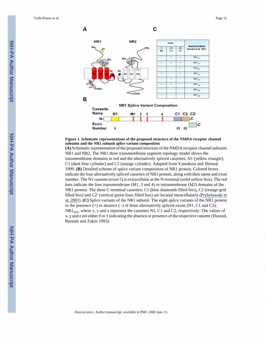
Figure 1. Schematic representations of the proposed structure of the NMDA receptor channelsubunits and the NR1 subunit splice variant composition(A) Schematic representation of the proposed structure of the NMDA receptor channel subunitsNR1 and NR2. The NR1 three transmembrane segment topology model shows thetransmembrane domains in red and the alternatively spliced cassettes; N1 (yellow triangle),C1 (dark blue cylinder) and C2 (orange cylinder). Adapted from Yamakura and Shimoji1999. (B) Detailed scheme of splice variant composition of NR1 protein. Colored boxesindicate the four alternatively spliced cassettes of NR1 protein, along with their name and exonnumber. The N1 cassette (exon 5) is extracellular at the N-terminal (solid yellow box). The redbars indicate the four transmembrane (M1, 3 and 4) or intramembrane (M2) domains of theNR1 protein. The three C-terminal cassettes; C1 (blue diamonds filled box), C2 (orange gridfilled box) and C2′ (vertical green lines filled box) are located intracellularly (Prybylowski etal. 2001). (C) Splice variants of the NR1 subunit. The eight splice variants of the NR1 proteinin the presence (+) or absence (−) of three alternatively spliced exons (N1, C1 and C2);NR1xyz, where x, y and z represent the cassettes N1, C1 and C2, respectively. The values ofx, y and z are either 0 or 1 indicating the absence or presence of the respective cassette (Durand,Bennett and Zukin 1993).
Valle-Pinero et al. Page 12
Neuroscience. Author manuscript; available in PMC 2008 June 15.
NIH
-PA Author Manuscript
NIH
-PA Author Manuscript
NIH
-PA Author Manuscript

Figure 2. Expression of NR1 in rat colon(A) RT-PCR analyses of NR1 in rat colon (named GI, for gastrointestinal) and brain tissuesusing two different set of primers; NR1 general and NR1 generic, that recognize areas in theNR1 outside the spliced exons. Assessment of housekeeping gene, GAPDH(Glyceraldehyde-3-phosphate dehydrogenase) was included as a control for sample integrityand reaction fidelity. A negative control (−RT) was included which lacks Reverse transcriptaseand uses GAPDH primers. (B) Top panel, western blot showing broad expression of NR1protein in GI and brain tissues using general mouse-NR1 antibody that recognizes a site outsideof the alternatively spliced cassettes. Bottom panel, an antibody against actin was use as acontrol for loading purposes.
Valle-Pinero et al. Page 13
Neuroscience. Author manuscript; available in PMC 2008 June 15.
NIH
-PA Author Manuscript
NIH
-PA Author Manuscript
NIH
-PA Author Manuscript
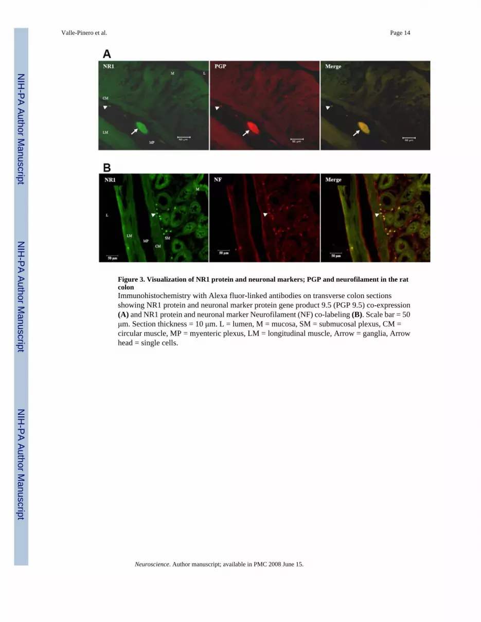
Figure 3. Visualization of NR1 protein and neuronal markers; PGP and neurofilament in the ratcolonImmunohistochemistry with Alexa fluor-linked antibodies on transverse colon sectionsshowing NR1 protein and neuronal marker protein gene product 9.5 (PGP 9.5) co-expression(A) and NR1 protein and neuronal marker Neurofilament (NF) co-labeling (B). Scale bar = 50μm. Section thickness = 10 μm. L = lumen, M = mucosa, SM = submucosal plexus, CM =circular muscle, MP = myenteric plexus, LM = longitudinal muscle, Arrow = ganglia, Arrowhead = single cells.
Valle-Pinero et al. Page 14
Neuroscience. Author manuscript; available in PMC 2008 June 15.
NIH
-PA Author Manuscript
NIH
-PA Author Manuscript
NIH
-PA Author Manuscript

Figure 4. Expression of NR1 splice variants in the rat colon(A) Expression of N1 cassette (exon 5) in the rat colon. RT-PCR analyses of N1 cassette(exon 5) expression in rat colon and brain tissues using four different sets of primers; “NR1a(b)”, “exon 5+”, “N1+” and “N1−”. (B) Western Blot showing expression of N1 cassettecontaining NR1 protein in GI and brain tissues using antibody against N1. (C) Expression ofC1 cassette (exon 21) in the rat colon. RT-PCR results of C1 cassette (exon 21) expressionin rat colon and brain tissues using two different set of primers; C1 and C1+. (D) Western blotshowing expression of C1 cassette containing NR1 protein using antibody targeted towardsthe C-terminal of the C1 cassette (exon 21). (E) Expression of C2 cassette (exon 22) in therat colon. RT-PCR results of C2 cassette (exon 22) expression in rat colon and brain tissuesusing primers targeted to the C2 cassette. (F) Western blot showing probing of C2+ antibodywhich is targeted to the C2 cassette and C2− antibody directed against the C2′ cassette.
Valle-Pinero et al. Page 15
Neuroscience. Author manuscript; available in PMC 2008 June 15.
NIH
-PA Author Manuscript
NIH
-PA Author Manuscript
NIH
-PA Author Manuscript

Figure 5. Visualization of the C2 and C2′cassettes in the rat myenteric plexusWhole mount immunohistochemistry showing the C2 cassette expression and the C2′ cassetteexpression in the rat myenteric plexus using an Alexa fluor antibody. Scale bar = 50 μm.
Valle-Pinero et al. Page 16
Neuroscience. Author manuscript; available in PMC 2008 June 15.
NIH
-PA Author Manuscript
NIH
-PA Author Manuscript
NIH
-PA Author Manuscript

Figure 6. Expression of NR2 subtypes in the rat colon(A) RT-PCR analyses of NR2A, NR2B, NR2C and NR2D expression in the rat colon and braintissues using primers to detect the presence of each different NR2 subtype respectively. (B)Whole mount immunohistochemistry showing the presence of the NR2B and NR2D proteinsin the rat myenteric plexus using Alexa fluor-linked antibodies. (C) Double-labeling of NR2Band neuronal marker PGP 9.5. Scale bar = 50 μm.
Valle-Pinero et al. Page 17
Neuroscience. Author manuscript; available in PMC 2008 June 15.
NIH
-PA Author Manuscript
NIH
-PA Author Manuscript
NIH
-PA Author Manuscript
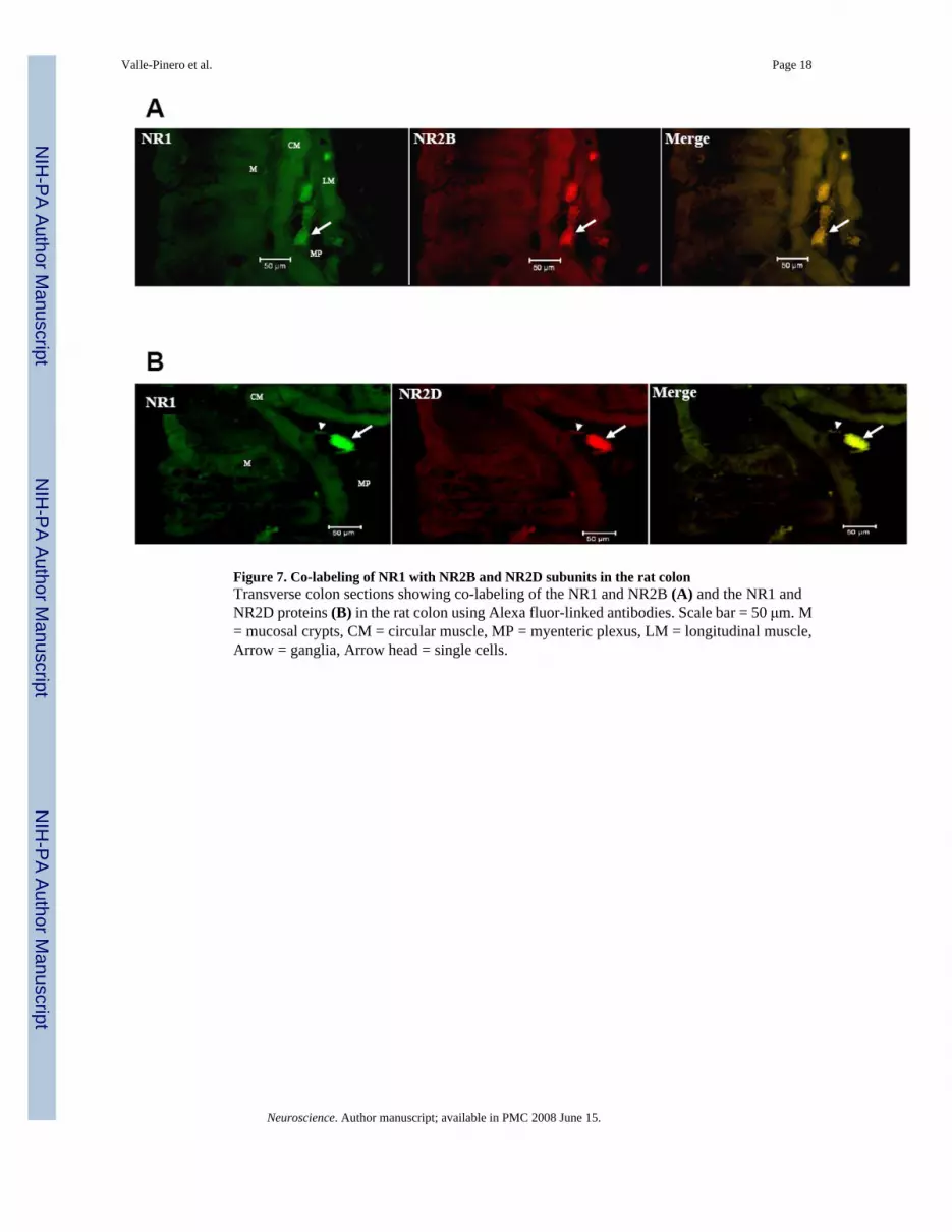
Figure 7. Co-labeling of NR1 with NR2B and NR2D subunits in the rat colonTransverse colon sections showing co-labeling of the NR1 and NR2B (A) and the NR1 andNR2D proteins (B) in the rat colon using Alexa fluor-linked antibodies. Scale bar = 50 μm. M= mucosal crypts, CM = circular muscle, MP = myenteric plexus, LM = longitudinal muscle,Arrow = ganglia, Arrow head = single cells.
Valle-Pinero et al. Page 18
Neuroscience. Author manuscript; available in PMC 2008 June 15.
NIH
-PA Author Manuscript
NIH
-PA Author Manuscript
NIH
-PA Author Manuscript
NIH
-PA Author Manuscript
NIH
-PA Author Manuscript
NIH
-PA Author Manuscript
Valle-Pinero et al. Page 19Ta
ble
1R
T-P
CR
Pri
mer
s Seq
uenc
esA
ll pr
imer
s w
ere
purc
hase
d fr
om G
enoM
echa
nix
(Gai
nesv
ille,
Fl).
RT-
PCR
rea
ctio
ns w
ere
carr
ied
out u
sing
Acc
ess
RT-
PCR
Sys
tem
fro
m P
rom
ega
(Mad
ison
, WI)
with
the
follo
win
g cy
cle
cond
ition
s: 1
cyc
le a
t 48°
C fo
r 45
min
, 94°
C 2
7 fo
r 2 m
in a
nd 7
2°C
for 1
min
, 35
cycl
es o
f PC
R (9
4°C
for 3
0 se
c,60
°C fo
r 1 m
in, 7
2°C
for 2
min
) and
a fi
nal e
long
atio
n pe
riod
of 7
min
at 7
2°C
.
Prim
er n
ame
Forw
ard
sequ
ence
Fwd
Tm
Rev
erse
sequ
ence
Rev
Tm
Prod
uct l
engt
h
Gen
eral
NR
1 ge
nera
lca
gaaa
cccc
tcag
acaa
gttc
59°C
cttc
tgtg
aagc
ctca
aact
cc59
°C21
3bp
NR
1 ge
neric
ccct
caga
caag
ttcat
ctac
gc61
°Cag
gttc
ttcct
ccac
acgt
tcac
61°C
563b
pG
APD
Hcc
ttcat
tgac
ctca
acta
catg
gtct
a63
°Cta
gccc
agga
tgcc
cttt
55°C
720b
pE
xon
5 or
N1
NR
1a E
xon
5 (−
)gc
gagt
ctac
aact
ggaa
ccac
61°C
ctcg
cttg
caga
aagg
atga
tg59
°C17
0bp
NR
1b E
xon
5 (+
)23
5bp
Exon
5+
insi
degg
aact
atga
aaac
ctcg
acc
58°C
NR
1b E
xon
5 R
ever
se59
°C15
8bp
N1+
(ζ1
N1+
)ac
cact
tcac
tccc
accc
ctgt
ctcc
tac
72°C
gtcc
gcgc
ttgttg
tcat
a61
°C33
0bp
N1−
(ζ1
N1−
)ag
ctca
acgc
cact
tctg
tc61
°Cca
ccttc
tctg
ccttg
gact
cac
65°C
407b
pE
xon
21 o
r C
1C
1U
seN
R1
gene
ral F
wd
or59
°Cgt
tttgc
aaag
cgcc
gcgt
cca
63°C
717b
pN
R1
gene
ric F
wd
61°C
710b
pC
1+ (ζ
1 C
1+)
tgtg
tccc
tgtc
cata
ctca
ag60
°Cgt
cggg
ctct
gctc
tacc
ac62
°C30
7bp
Exo
n 22
or
C2
NR
1 Ex
on-2
2ca
tggc
aggg
gtct
tcat
gctg
63°C
gaac
acag
ctgc
agct
ggcc
ct65
°C31
8bp
NR
2N
R2A
tata
gagg
gtaa
atgt
tgga
50°C
agaa
actg
tgag
gcat
ttct
52°C
322b
pN
R2B
actg
tgac
aacc
cacc
cttc
58°C
cgga
actg
gtcc
aggt
agaa
58°C
400b
pN
R2C
tgtg
tcag
gcct
tagt
gaca
56°C
ccac
actg
tctc
cagc
ttct
58°C
402b
pN
R2D
aaga
agat
cgat
ggcg
tctg
56°C
ggat
ttccc
aatg
gtga
agg
56°C
353b
p
All
prim
ers f
rom
Gen
oMec
hani
x, G
ains
ville
, Fl,
USA
.
Neuroscience. Author manuscript; available in PMC 2008 June 15.

NIH
-PA Author Manuscript
NIH
-PA Author Manuscript
NIH
-PA Author Manuscript
Valle-Pinero et al. Page 20
Table 2Antibodies used for Western Blots and Immunohistochemical AssaysAntibodies were purchased from different vendors and dilutions were prepared according to vendors’specifications. Incubation time for primary antibodies was 24 hours and 1 hour for secondary antibodies.
Antibody Host Company Cat. No.
Primary AntibodiesActin Mouse Chemicon, Temecula, CA. MAB1501Neurofilament Mouse Sigma, Saint Louis, Missouri. N-5389NR1 Mouse BD Biosciences Pharmigen, San Diego, CA. 556308NR1 Rabbit Santa Cruz Biotechnology, Santa Cruz, CA. SC-9058NR1 splice variants Rabbit Provided by Dr. Michael Iadarola, The National Institute of
Dental and Craniofacial Research NIDCR, Bethesda, MD.N/A
NR2B Rabbit Santa Cruz Biotechnology, Santa Cniz, CA. SC-9057NR2B Mouse BD Biosciences Pharmigen, San Diego, CA. 610417NR2D Rabbit Santa Cruz Biotechnology, Santa Cruz, CA. SC-10727Protein Gene Product 9.5 Rabbit Chemicon, Temecula, CA. AB1761Fluorescent Secondary AntibodiesAlexa Fluor 488 Mouse Molecular Probes, Eugene, OR. A21202Alexa Fluor 488 Rabbit Molecular Probes, Eugene, OR. A21206Alexa Fluor 594 Mouse Molecular Probes, Eugene, OR. A21201Alexa Fluor 594 Rabbit Molecular Probes, Eugene, OR. A21442
Neuroscience. Author manuscript; available in PMC 2008 June 15.