expression, genetic localization and phylogenic analysis of naplr in piscine streptococcus...
TRANSCRIPT
Journal of Advanced Research (2014) xxx, xxx–xxx
Cairo University
Journal of Advanced Research
ORIGINAL ARTICLE
Expression, genetic localization and phylogenic
analysis of NAPlr in piscine Streptococcusdysgalactiae subspecies dysgalactiae isolates
and their patterns of adherence
* Corresponding authors. Tel.: +20 2 1122671243, +20 2 35720399;
fax: +20 2 35725240, +20 2 35710305.E-mail addresses: [email protected] (M. Abdelsalam),
[email protected] (M. Warda).
Peer review under responsibility of Cairo University.
Production and hosting by Elsevier
2090-1232 ª 2014 Production and hosting by Elsevier B.V. on behalf of Cairo University.
http://dx.doi.org/10.1016/j.jare.2014.05.005
Please cite this article in press as: Abdelsalam M et al., Expression, genetic localization and phylogenic analysis of NAPlr in piscine Streptdysgalactiae subspecies dysgalactiae isolates and their patterns of adherence, J Adv Res (2014), http://dx.doi.org/10.1016/j.jare.2014.05.0
M. Abdelsalam a,*, M. Fujino b, A.E. Eissa a,c, S.C. Chen d,e, M. Warda f,*
a Department of Fish Diseases and Management, Faculty of Veterinary Medicine, Cairo University, 12211 Giza, Egyptb AIDS Research Center, National Institute of Infectious Diseases, Tokyo, Japanc Departments of Poultry and Fish Diseases, Faculty of Veterinary Medicine, Tripoli University, Tripoli, Libyad Graduate Institute of Animal Vaccine Technology, National Pingtung University of Science and Technology, Pingtung, Taiwane Department of Veterinary Medicine, National Pingtung University of Science and Technology, Pingtung, Taiwanf Department of Biochemistry, Biotechnology Center for Services and Researches, Faculty of Veterinary Medicine,Cairo University, 12211 Giza, Egypt
A R T I C L E I N F O
Article history:
Received 27 February 2014
Received in revised form 16 May 2014
Accepted 16 May 2014
Available online xxxx
Keywords:
NAPlr gene
a-enolase gene
Piscine S. dysgalactiae subsp.
dysgalactiae
Virulence traits
Adherence pattern
A B S T R A C T
Streptococcus dysgalactiae, the long recognized mammalian pathogen, has currently received a
major concern regarding fish bacterial infection. Adhesion to host epithelial cells and the pres-
ence of wall-associated plasminogen binding proteins are prerequisites to Streptococcus infec-
tion. This is the first study of the occurrence of nephritis-associated plasminogen-binding
receptor (NAPlr) and a-enolase genes in piscine S. dysgalactiae subspecies dysgalactiae (SDSD)
isolates. Further characterization of surface localized NAPlr of fish SDSD revealed a similar
immune-reactive band of 43 KDa as that from porcine S. dysgalactiae subsp. equisimilis
(SDSE). The phylogenetic analysis revealed that NAPlr of fish SDSD is more associated with
those of mammalian SDSE and Streptococcus pyogenes rather than of other streptococci.
Our findings warrant public attention to the possible implication of these virulence genes in dis-
semination of SDSD to different tissues of infected hosts and to get advantage to new niches.
The SDSD adherence patterns were also studied to better understand their pathogenicity.
ococcus05
2 M. Abdelsalam et al.
Please cite this article in press as: Abdelsalam Mdysgalactiae subspecies dysgalactiae isolates an
The patterns of adherence of SDSD on two different cell lines showed a different pattern of
adherence. Such difference gives an insight about the variance in host susceptibility to infection.
ª 2014 Production and hosting by Elsevier B.V. on behalf of Cairo University.
Introduction
Streptococcus dysgalactiae was discovered by Diernhofer in1932 [1], and officially recognized as a new species in 1983 [2].S. dysgalactiae was subdivided into two genetically similar
subspecies: the animal subspecies dysgalactiae (belongs toLancefield group C (GCS)) and human subspecies equisimilis(belongs toGCS orGGS orGLS) [3]. The a-hemolytic S. dysga-
lactiae subsp. dysgalactiae (SDSD) is a strict animal pathogen ofpyrogenic streptococcus [4]. SDSD is responsible for diverseproblems such as mastitis, toxic shock like syndrome, subcuta-neous cellulitis in cows [5], extensive fibrinous pleurisy in ewes
[6], suppurative polyarthritis in lambs [7], neonatal mortalitiesin dogs [8], severe septicemia in fish [9], and bacteremia andmen-ingitis in immunocompromised individuals [10,11]. SDSD is
potentially considered as an emerging zoonotic agent since itis implicated in cutaneous cellulitis in humans engaged eitherin cleaning fish [12] or handling livestock [13].
SDSD has been associated with high mortalities in Kingfish(Seriola lalandi), amberjack (S. dumerili) and yellowtail(S. quinqueradiata) in Japan [9,14–17], Nile tilapia (Oreochr-omis niloticus) in Brazil [18], Amur sturgeon (Acipenser
schrenckii), the Siberian sturgeon (A. baerii), golden pomfret(Trachinotus ovatus), Soiny mullet (Liza haematocheila) grasscarp (Ctenopharyngodon idella), crucian carp (Carassius caras-
sius) and pompano (Trachinotus blochii) in China [19–22]. Ithas been recovered from cobia (Rachycentron canadum),basket mullet (Liza alata) and grey mullet (Mugil cephalus)
in Taiwan, hybrid red tilapia (Oreochromis sp.) in Indonesia,white spotted snapper (Lutjanus stellatus) and pompano(T. blochii) in Malaysia [9,16,17,23], and rainbow trout
(Oncorhynchus mykiss) in Iran [24]. The infected fish revealedsystemic pyrogranulomatous inflammation with a severenecrotic lesion in their caudal peduncles [25]. Despite its clini-cal significance, the complete sequence revelation and virulence
characterization are generally unknown for SDSD. Fish SDSDwas found to possess some virulence factors such as streptoly-sin S structural gene (sagA), streptococcal pyrogenic exotoxin
G gene (spegg) and serum opacity factor (SOF-FD) [17,26].Fish SDSD strongly adheres to and invades fish epithelial cellline as Epithelial Papiloma of Carp (EPC) in vitro [14]. How-
ever, the adherence patterns and the surface structures impli-cated in adhesion are still uncovered. The M/M-like proteins(emm), surface dehydrogenase (SDH) and a-enolase are the
most important wall-associated plasminogen-binding proteinsof pathogenic streptococci [27]. The ability of pathogenicstreptococci to bind host plasminogen system empowers theirinvasiveness through utilizing the fibrinolytic activity of plas-
min and promoting the adherence of streptococci to host cells[27]. Plasminogen-binding glycoproteins, such as a-enolase andSDH, are generally found in the cytosolic compartment and
are transported to the bacterial cell wall by a yet unknownmechanism that comprised moonlighting functions [28–30].The surface protein SDH displays ADP-ribosylating activities
and glyceraldehyde 3-phosphate dehydrogenase (GAPDH)
et al., Expression, geneticd their patterns of adheren
[31], and has been recognized as a potential nephritogenic pro-
tein under the name nephritis-associated plasminogen-bindingreceptor (NAPlr) [32]. Streptococcal cell wall a-enolase is asso-ciated with streptococcal infection and post-streptococcalautoimmune disease in human [28,30].
Hence, NAPlr and a-enolase genes are important virulencefactors in Streptococcus pyogenes [33,34], S. agalactiae [35], S.iniae [30], and S. pneumoniae [28,29] due to its contribution to
the establishment of infections and colonization by bacterialpathogens [27,36]. This is the first study to investigate theoccurrence of gapdh/naplr/sdh and a-enolase genes in piscine
isolates of SDSD. We also investigated the adherence patternsof selected SDSD strains to EPC and CHSE-214 (Chinook sal-mon embryo) cell lines in vitro.
Material and methods
Bacterial isolates
Twenty-three bacterial isolates were used in this study. Thea-hemolytic SDSD isolates (n = 18) were recovered from mor-
ibund fishes obtained from various fish farms in Japan (n = 9;three from king fish, three from amberjack and three from yel-lowtail), Taiwan (n = 5; three from grey mullet, one from cobia
and one from basket mullet),Malaysia (n=2; one from pompanoand one from snapper), China (n= 1; one from pompano) andIndonesia (n = 1; one from tilapia). For comparative purpose,
b-hemolytic S. dysgalactiae subsp. equisimilis (SDSE) isolates(n = 5) were collected from pigs with endocarditis (Kumamotomeat inspection office in Japan).
DNA extraction
The pure stock isolates were stored in Todd-Hewitt broth(THB; Difco, Sparks, MD, USA) at �80 �C. All isolates were
cultured aerobically on Todd Hewitt agar (THA; Difco,Sparks, MD, USA), and on 5% sheep blood agar (Columbiaagar base; Becton Dickinson, Cockeysville, MD, USA), and
then incubated at 37 �C for 24 h. Genomic DNA was extractedfrom cultivated strains using a DNAzol� reagent (Invitrogen,Carlsbad, USA) [37]. The fish SDSD isolates were discrimi-
nated from pig SDSE isolates by using sodA gene primers spe-cific for fish SDSD detection. PCR was performed as describedpreviously [37].
PCR detection of virulence genes
PCR amplification of emm was performed using specific pri-mer pairs; A: (50-TATTAGCTTAGAAAATTAA-30) and B:
(50-GCAAGTTCTTCAGCTTGTTT-30) as described previ-ously by Zhao et al., [38]. To amplify a 963-bp fragmentof NAPlr; the specific primer pairs of Plr 1:
50-GTTAAAGTTGGTATTAACGGT-30, and Plr 2: 50-TTGA-GCAGTGTAAGACATTTC-30 were designed based on nephritis
localization and phylogenic analysis of NAPlr in piscine Streptococcusce, J Adv Res (2014), http://dx.doi.org/10.1016/j.jare.2014.05.005
Virulence determinants of Streptococcus dysgalactiae 3
associated plasminogen receptor gene of SDSE (GenBankaccession number AB217852). PCR was performed with thefollowing parameters: an initial denaturation cycle at 94 �Cfor 5 min, followed by 35 cycles of denaturation at 94 �C for30 s, primer annealing at 52 �C for 30 s, elongation at 72 �Cfor 50 s, and a final cycle at 72 �C for 10 min. To amplify a
1308-bp fragment of a-enolase; the primer pairs of Eno1:50-ATGTCAATTATTACTGATGT-30, and Eno2: 50-CTATTTTTTTAAGTTATAGA-30 were designed based on a-eno-lase gene of SDSE (AP012976). The thermal scheme of PCRwas performed as described for the NAPlr gene, except thatthe primer annealing was adjusted at 50 �C and the primerextension was set for 1 min.
Cloning and sequencing of NAPlr and a-enolase
The NAPlr and a-enolase genes were sequenced according to
Abdelsalam et al. [17]. The amplified products were cloned intopGEM-T easy vector (Promega, Madison, WI, USA), and therecombinant plasmid was introduced into Escherichia coli
DH5a. The QIAprep Spin Miniprep kit (Qiagen, German-town, MD, USA) was used to purify the plasmid DNA.Sequencing reactions were performed by using the oligonu-
cleotide primers SP6 (5-ATTTAGGTGACACTATAGAA-3)and T7 (5-TAATACGACTCACTATAGGG-3) with theGenomeLab DTCS Quick Start Kit (Beckman Coulter, Fuller-ton, CA, USA). The samples were then loaded into the CEQ
8000 Genetic Analysis System (Beckman Coulter) and thenucleotide sequence was determined. The nucleotide sequenceswere analyzed by using BioEdit version 7.0 [39]. The phyloge-
netic analysis was performed by the neighbor joining methodusing MEGA version 5 [40]. The nucleotide sequences of theNAPlr and a-enolase genes were submitted to the DNA Data
Bank of Japan (DDBJ) nucleotide sequence database.
Surface protein extraction
Bacterial surface proteins were extracted according to the pro-tocol described by Fujino et al. [32] with some modifications.Briefly, bacteria were inoculated onto Todd Hewitt agar andthe culture was incubated for 16 h at 37 �C. Then, bacterialcolonies were harvested from the surface of the grow med-ium/agar plates by loops and were suspended in phosphate-buffered saline (PBS, pH 7.5) in a tube. The bacterial cells were
then centrifuged at 10,000g for 20 min. The bacterial cell pelletwas then resuspended in PBS. Bacterial cell pellets werewashed three times with sterile PBS, and surface proteins were
extracted using sodium dodecyl sulfate (SDS; Bio-Rad, Hercu-les, CA, USA, 30 mg wet weight of bacteria per 100 ll of 0.2%SDS) for 1 h at 4 �C. Extraction mixture was centrifuged and
supernatant protein samples were recovered. The SDS extractof bacterial surface proteins was filtered consecutively through0.45-lm (Millex-HV, Millipore) and 0.22-lm (Millex-GX, Mil-lipore) sterile Millipore filters to remove bacteria. Protein con-
centration was determined using Bradford assay kit (Bio-Rad,Hercules, CA, USA).
Production of anti-NAPlr monoclonal antibody
Anti-NAPlr monoclonal antibody (mAb) was produced aspreviously described [32]. Briefly, the specific pathogen-free
Please cite this article in press as: Abdelsalam M et al., Expression, geneticdysgalactiae subspecies dysgalactiae isolates and their patterns of adheren
BALB/c mice were injected intraperitoneal (IP) with 100 mgrecombinant NAPlr emulsified in Freund’s complete adjuvant.Three weeks later, the mice were given a booster immunization
with 100 mg of recombinant Plr emulsified in Freund’s incom-plete adjuvant. Thirty days later, the mice were injected intra-peritoneally with 100 mg of recombinant PH in PBS. After
3 days, the mice were sacrificed and their spleens removed.The splenocytes were fused with P3U1 myeloma cells. Hybrid-oma cultures that secreted anti-Plr antibody were cloned by
limiting dilution and the resulting monoclonal antibodies(mAbs) were rescreened to determine the specificity and reac-tivity with Plr.). The gained Anti-NAPlr mAb from hybridomacultured cells was evaluated by ELISA using rNAPlr.
Western blots for NAPlr
Protein extract (10 lg protein/lane) of three SDSD isolates
(12–06, KNH07808, T11358) and another (5 lg protein/lane)of three SDSE isolates (PAGU656, PAGU706, PAGU707)were separated by sodium dodecyl sulfate polyacrylamide gel
electrophoresis (SDS–PAGE) on 12.5% polyacrylamide gels(SuperSep 12.5%. Wako Pure Chemical, Osaka, Japan), andthen transferred to PVDF (Millipore, Bedford, MA, USA)
using a semi-dry blotter (ATTO Bioscience, Tokyo, Japan).SDS–PAGE ‘‘wide range’’ (200–6.5 kDa) molecular weightstandard was purchased from Sigma. NAPlr was identifiedby the use of the previously prepared anti-NAPlr mAb com-
bined with peroxidase-labeled anti-mouse IgG (AmericanQualex, San Clemente, CA, USA) and ECL Advance WesternBlotting Detection Kit (GE Healthcare, Buckinghamshire,
UK). Blot of E. coli was included as a negative control. NAPlrexpression in each strain was quantified based on the strengthof the luminescence of the mAb – specific band with the
densitometry system (Atto).
Adherence pattern of SDSD
This assay was performed according to the method describedby Duary et al. [41] with some modifications. Briefly, a sterile12 mm diameter glass cover-slip coated with poly-L-lysine(NeuVitro, El Monte, CA, USA) was placed in each well of
the 24-well tissue culture plate (Costar, Corning, Inc., NY,USA) and the wells were seeded with EPC or CHSE-214 cells.The seeded cells of the EPC or CHSE-214 were grown in
Leibovitz-15 (L-15) medium (Gibco Invitrogen, USA) contain-ing 10% (v/v) fetal bovine serum and penicillin (5 lg/ml;Sigma–Aldrich Inc., USA), and incubated at 25 �C and
18 �C respectively, in 5% CO2, and inspected daily until theyattained semi-Confluency (2 · 105 cells/well). The SDSD iso-lates (12–06, KNH07808, T11358) were incubated in THB
overnight at 37 �C to midlogarithmic phase (108 CFU/ml),and then centrifuged at 2190g for 30 min. Pellets were washedtwice with phosphate-buffered saline (PBS; pH 7.2), and thecell concentration/counts were adjusted to approximately
108 CFU/ml. 100 ll of the bacterial suspension was inoculatedto the wells containing EPC and CHSE-214 cells (final bacte-rial cell concentration in the wells was approximately
107 CFU/ml) and the culture plates were incubated for30 min at 25 �C and 18 �C for EPC and CHSE-214, respec-tively. The monolayers were then carefully washed several
times with L-15 medium to remove non-adherent bacteria by
localization and phylogenic analysis of NAPlr in piscine Streptococcusce, J Adv Res (2014), http://dx.doi.org/10.1016/j.jare.2014.05.005
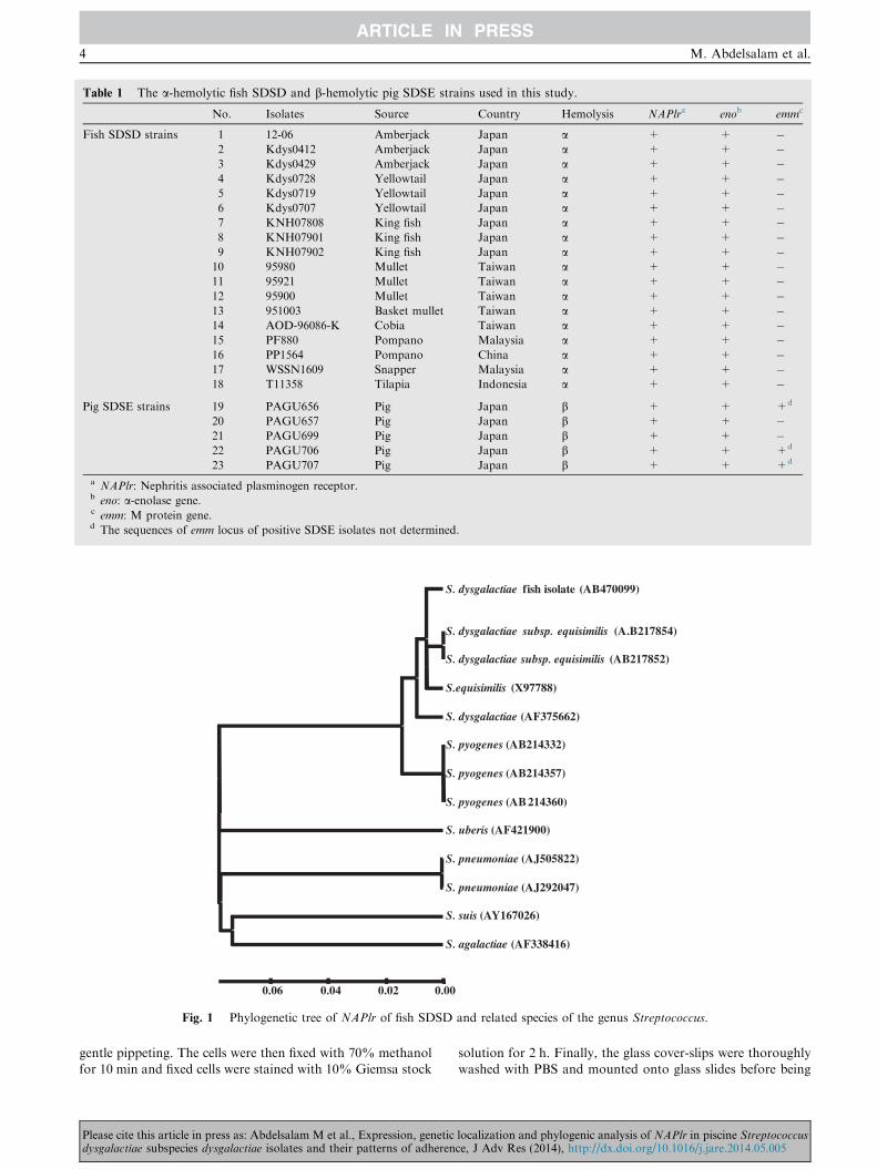
Table 1 The a-hemolytic fish SDSD and b-hemolytic pig SDSE strains used in this study.
No. Isolates Source Country Hemolysis NAPlra enob emmc
Fish SDSD strains 1 12-06 Amberjack Japan a + + �2 Kdys0412 Amberjack Japan a + + �3 Kdys0429 Amberjack Japan a + + �4 Kdys0728 Yellowtail Japan a + + �5 Kdys0719 Yellowtail Japan a + + �6 Kdys0707 Yellowtail Japan a + + �7 KNH07808 King fish Japan a + + �8 KNH07901 King fish Japan a + + �9 KNH07902 King fish Japan a + + �10 95980 Mullet Taiwan a + + �11 95921 Mullet Taiwan a + + �12 95900 Mullet Taiwan a + + �13 951003 Basket mullet Taiwan a + + �14 AOD-96086-K Cobia Taiwan a + + �15 PF880 Pompano Malaysia a + + �16 PP1564 Pompano China a + + �17 WSSN1609 Snapper Malaysia a + + �18 T11358 Tilapia Indonesia a + + �
Pig SDSE strains 19 PAGU656 Pig Japan b + + +d
20 PAGU657 Pig Japan b + + �21 PAGU699 Pig Japan b + + �22 PAGU706 Pig Japan b + + +d
23 PAGU707 Pig Japan b + + +d
a NAPlr: Nephritis associated plasminogen receptor.b eno: a-enolase gene.c emm: M protein gene.d The sequences of emm locus of positive SDSE isolates not determined.
S. dysgalactiae subsp. equisimilis (A.B217854)
S. dysgalactiae fish isolate (AB470099)
S. dysgalactiae subsp. equisimilis (AB217852)
S.equisimilis (X97788)
S. dysgalactiae (AF375662)
S. pyogenes (AB214332)
S. pyogenes (AB214357)
S. pyogenes (AB 214360)
S. uberis (AF421900)
S. pneumoniae (AJ505822)
S. pneumoniae (AJ292047)
S. suis (AY167026)
S. agalactiae (AF338416)
0.000.020.040.06
Fig. 1 Phylogenetic tree of NAPlr of fish SDSD and related species of the genus Streptococcus.
4 M. Abdelsalam et al.
gentle pippeting. The cells were then fixed with 70% methanol
for 10 min and fixed cells were stained with 10% Giemsa stock
Please cite this article in press as: Abdelsalam M et al., Expression, geneticdysgalactiae subspecies dysgalactiae isolates and their patterns of adheren
solution for 2 h. Finally, the glass cover-slips were thoroughly
washed with PBS and mounted onto glass slides before being
localization and phylogenic analysis of NAPlr in piscine Streptococcusce, J Adv Res (2014), http://dx.doi.org/10.1016/j.jare.2014.05.005
Virulence determinants of Streptococcus dysgalactiae 5
examined by light microscope and photographed. The bacte-rial adherence patterns were categorized according to the fol-lowing criteria: localized-like-adherence (LAL), when the
bacteria adhered to the cell surface, forming loose clusters;localized adherence (LA), when the bacteria adhered to the cellsurface as tight clusters; diffuse adherence (DA), when the
bacteria adhered diffuse to the cell surface; and aggregativeadherence (AA), when the bacteria adhered to the cell surfaceand to the cover slip in a stacked-brick pattern [42]. The adher-
ence rate was expressed as the number of adhering bacteria per50 cells of EPC or CHSE-214. The results were expressed as aweak adherence (6100 adherent bacteria), moderate adherence(100–200 adherent bacteria) and strong adherence (P200
adherent bacteria) [43].
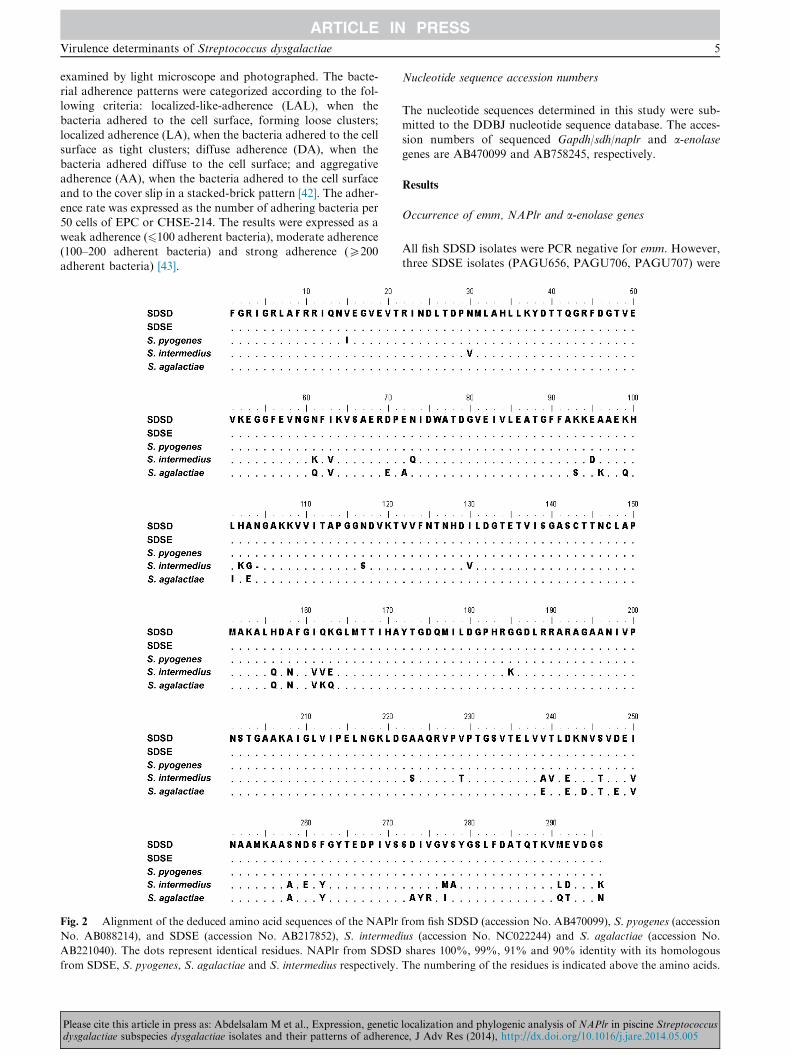
Fig. 2 Alignment of the deduced amino acid sequences of the NAPlr
No. AB088214), and SDSE (accession No. AB217852), S. intermed
AB221040). The dots represent identical residues. NAPlr from SDSD
from SDSE, S. pyogenes, S. agalactiae and S. intermedius respectively.
Please cite this article in press as: Abdelsalam M et al., Expression, geneticdysgalactiae subspecies dysgalactiae isolates and their patterns of adheren
Nucleotide sequence accession numbers
The nucleotide sequences determined in this study were sub-mitted to the DDBJ nucleotide sequence database. The acces-sion numbers of sequenced Gapdh/sdh/naplr and a-enolasegenes are AB470099 and AB758245, respectively.
Results
Occurrence of emm, NAPlr and a-enolase genes
All fish SDSD isolates were PCR negative for emm. However,three SDSE isolates (PAGU656, PAGU706, PAGU707) were
from fish SDSD (accession No. AB470099), S. pyogenes (accession
ius (accession No. NC022244) and S. agalactiae (accession No.
shares 100%, 99%, 91% and 90% identity with its homologous
The numbering of the residues is indicated above the amino acids.
localization and phylogenic analysis of NAPlr in piscine Streptococcusce, J Adv Res (2014), http://dx.doi.org/10.1016/j.jare.2014.05.005
6 M. Abdelsalam et al.
PCR positive for emm. All SDSD and SDSE isolates containedhomologous segments of NAPlr and a-enolase (Table 1). ThePCR products of distinct strains were of the expected size,
963 bp and 1308 bp, respectively.
Nucleotide sequence analyses of NAPlr
The NAPlr gene of SDSD collected from diseased fish wassequenced under the GenBank accession number AB470099.The NAPlr gene obtained from SDSD strain (T11358) was
963 bp long. The NAPlr was found to have 99% similarity toNAPlr (AB217852) of SDSE and 97% similarity to NAPlr(AB214357) of S. pyogenes, and has one ORF encoding 336
aminoacids. Therefore, phylogenetic analysis revealed thatNAP-lr of piscine SDSD isolate was related to that of SDSE and S.pyogenes and separated from other gapdh/sdh/naplr clusters ofother streptococci (Fig. 1). The deduced amino acid sequence of
fish SDSD NAPlr was identical to the previous investigatednephritogenic strains of SDSE and S. pyogenes (Fig. 2).
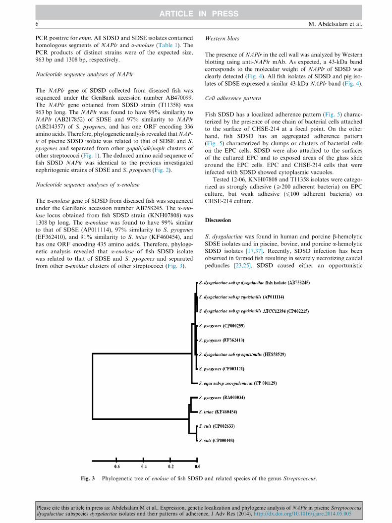
Nucleotide sequence analyses of a-enolase
The a-enolase gene of SDSD from diseased fish was sequencedunder the GenBank accession number AB758245. The a-eno-lase locus obtained from fish SDSD strain (KNH07808) was1308 bp long. The a-enolase was found to have 99% similarto that of SDSE (AP011114), 97% similarity to S. pyogenes(EF362410), and 91% similarity to S. iniae (KF460454), and
has one ORF encoding 435 amino acids. Therefore, phyloge-netic analysis revealed that a-enolase of fish SDSD isolatewas related to that of SDSE and S. pyogenes and separated
from other a-enolase clusters of other streptococci (Fig. 3).
Fig. 3 Phylogenetic tree of enolase of fish SDSD
Please cite this article in press as: Abdelsalam M et al., Expression, geneticdysgalactiae subspecies dysgalactiae isolates and their patterns of adheren
Western blots
The presence of NAPlr in the cell wall was analyzed by Westernblotting using anti-NAPlr mAb. As expected, a 43-kDa bandcorresponds to the molecular weight of NAPlr of SDSD was
clearly detected (Fig. 4). All fish isolates of SDSD and pig iso-lates of SDSE expressed a similar 43-kDa NAPlr band (Fig. 4).
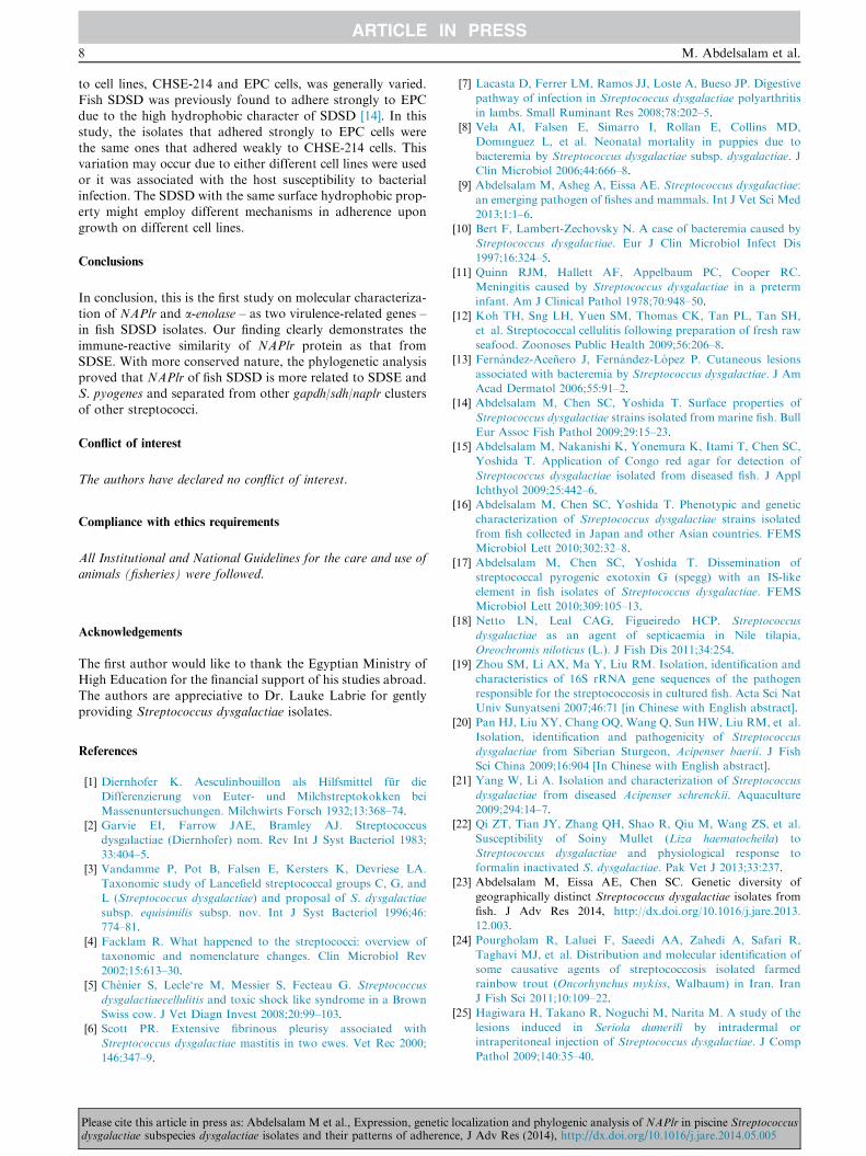
Cell adherence pattern
Fish SDSD has a localized adherence pattern (Fig. 5) charac-terized by the presence of one chain of bacterial cells attachedto the surface of CHSE-214 at a focal point. On the other
hand, fish SDSD has an aggregated adherence pattern(Fig. 5) characterized by clumps or clusters of bacterial cellson the EPC cells. SDSD were also attached to the surfaces
of the cultured EPC and to exposed areas of the glass slidearound the EPC cells. EPC and CHSE-214 cells that wereinfected with SDSD showed cytoplasmic vacuoles.
Tested 12-06, KNH07808 and T11358 isolates were catego-rized as strongly adhesive (P200 adherent bacteria) on EPCculture, but weak adhesive (6100 adherent bacteria) onCHSE-214 culture.
Discussion
S. dysgalactiae was found in human and porcine b-hemolytic
SDSE isolates and in piscine, bovine, and porcine a-hemolyticSDSD isolates [17,37]. Recently, SDSD infection has beenobserved in farmed fish resulting in severely necrotizing caudal
peduncles [23,25]. SDSD caused either an opportunistic
and related species of the genus Streptococcus.
localization and phylogenic analysis of NAPlr in piscine Streptococcusce, J Adv Res (2014), http://dx.doi.org/10.1016/j.jare.2014.05.005
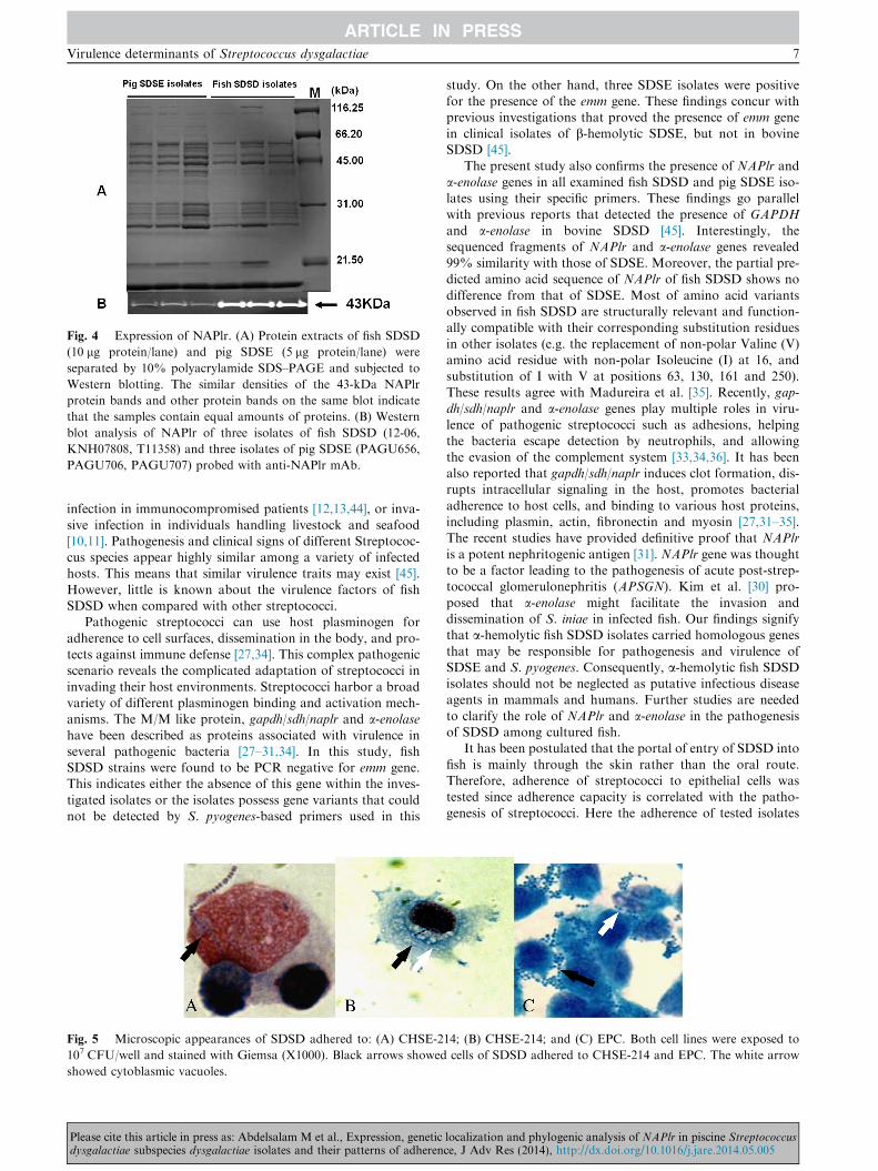
Fig. 4 Expression of NAPlr. (A) Protein extracts of fish SDSD
(10 lg protein/lane) and pig SDSE (5 lg protein/lane) were
separated by 10% polyacrylamide SDS–PAGE and subjected to
Western blotting. The similar densities of the 43-kDa NAPlr
protein bands and other protein bands on the same blot indicate
that the samples contain equal amounts of proteins. (B) Western
blot analysis of NAPlr of three isolates of fish SDSD (12-06,
KNH07808, T11358) and three isolates of pig SDSE (PAGU656,
PAGU706, PAGU707) probed with anti-NAPlr mAb.
Virulence determinants of Streptococcus dysgalactiae 7
infection in immunocompromised patients [12,13,44], or inva-
sive infection in individuals handling livestock and seafood[10,11]. Pathogenesis and clinical signs of different Streptococ-cus species appear highly similar among a variety of infectedhosts. This means that similar virulence traits may exist [45].
However, little is known about the virulence factors of fishSDSD when compared with other streptococci.
Pathogenic streptococci can use host plasminogen for
adherence to cell surfaces, dissemination in the body, and pro-tects against immune defense [27,34]. This complex pathogenicscenario reveals the complicated adaptation of streptococci in
invading their host environments. Streptococci harbor a broadvariety of different plasminogen binding and activation mech-anisms. The M/M like protein, gapdh/sdh/naplr and a-enolasehave been described as proteins associated with virulence inseveral pathogenic bacteria [27–31,34]. In this study, fishSDSD strains were found to be PCR negative for emm gene.This indicates either the absence of this gene within the inves-
tigated isolates or the isolates possess gene variants that couldnot be detected by S. pyogenes-based primers used in this
Fig. 5 Microscopic appearances of SDSD adhered to: (A) CHSE-2
107 CFU/well and stained with Giemsa (X1000). Black arrows showed
showed cytoblasmic vacuoles.
Please cite this article in press as: Abdelsalam M et al., Expression, geneticdysgalactiae subspecies dysgalactiae isolates and their patterns of adheren
study. On the other hand, three SDSE isolates were positivefor the presence of the emm gene. These findings concur withprevious investigations that proved the presence of emm gene
in clinical isolates of b-hemolytic SDSE, but not in bovineSDSD [45].
The present study also confirms the presence of NAPlr and
a-enolase genes in all examined fish SDSD and pig SDSE iso-lates using their specific primers. These findings go parallelwith previous reports that detected the presence of GAPDH
and a-enolase in bovine SDSD [45]. Interestingly, thesequenced fragments of NAPlr and a-enolase genes revealed99% similarity with those of SDSE. Moreover, the partial pre-dicted amino acid sequence of NAPlr of fish SDSD shows no
difference from that of SDSE. Most of amino acid variantsobserved in fish SDSD are structurally relevant and function-ally compatible with their corresponding substitution residues
in other isolates (e.g. the replacement of non-polar Valine (V)amino acid residue with non-polar Isoleucine (I) at 16, andsubstitution of I with V at positions 63, 130, 161 and 250).
These results agree with Madureira et al. [35]. Recently, gap-dh/sdh/naplr and a-enolase genes play multiple roles in viru-lence of pathogenic streptococci such as adhesions, helping
the bacteria escape detection by neutrophils, and allowingthe evasion of the complement system [33,34,36]. It has beenalso reported that gapdh/sdh/naplr induces clot formation, dis-rupts intracellular signaling in the host, promotes bacterial
adherence to host cells, and binding to various host proteins,including plasmin, actin, fibronectin and myosin [27,31–35].The recent studies have provided definitive proof that NAPlr
is a potent nephritogenic antigen [31]. NAPlr gene was thoughtto be a factor leading to the pathogenesis of acute post-strep-tococcal glomerulonephritis (APSGN). Kim et al. [30] pro-
posed that a-enolase might facilitate the invasion anddissemination of S. iniae in infected fish. Our findings signifythat a-hemolytic fish SDSD isolates carried homologous genes
that may be responsible for pathogenesis and virulence ofSDSE and S. pyogenes. Consequently, a-hemolytic fish SDSDisolates should not be neglected as putative infectious diseaseagents in mammals and humans. Further studies are needed
to clarify the role of NAPlr and a-enolase in the pathogenesisof SDSD among cultured fish.
It has been postulated that the portal of entry of SDSD into
fish is mainly through the skin rather than the oral route.Therefore, adherence of streptococci to epithelial cells wastested since adherence capacity is correlated with the patho-
genesis of streptococci. Here the adherence of tested isolates
14; (B) CHSE-214; and (C) EPC. Both cell lines were exposed to
cells of SDSD adhered to CHSE-214 and EPC. The white arrow
localization and phylogenic analysis of NAPlr in piscine Streptococcusce, J Adv Res (2014), http://dx.doi.org/10.1016/j.jare.2014.05.005
8 M. Abdelsalam et al.
to cell lines, CHSE-214 and EPC cells, was generally varied.Fish SDSD was previously found to adhere strongly to EPCdue to the high hydrophobic character of SDSD [14]. In this
study, the isolates that adhered strongly to EPC cells werethe same ones that adhered weakly to CHSE-214 cells. Thisvariation may occur due to either different cell lines were used
or it was associated with the host susceptibility to bacterialinfection. The SDSD with the same surface hydrophobic prop-erty might employ different mechanisms in adherence upon
growth on different cell lines.
Conclusions
In conclusion, this is the first study on molecular characteriza-tion of NAPlr and a-enolase – as two virulence-related genes –in fish SDSD isolates. Our finding clearly demonstrates the
immune-reactive similarity of NAPlr protein as that fromSDSE. With more conserved nature, the phylogenetic analysisproved that NAPlr of fish SDSD is more related to SDSE andS. pyogenes and separated from other gapdh/sdh/naplr clusters
of other streptococci.
Conflict of interest
The authors have declared no conflict of interest.
Compliance with ethics requirements
All Institutional and National Guidelines for the care and use ofanimals (fisheries) were followed.
Acknowledgements
The first author would like to thank the Egyptian Ministry ofHigh Education for the financial support of his studies abroad.
The authors are appreciative to Dr. Lauke Labrie for gentlyproviding Streptococcus dysgalactiae isolates.
References
[1] Diernhofer K. Aesculinbouillon als Hilfsmittel fur die
Differenzierung von Euter- und Milchstreptokokken bei
Massenuntersuchungen. Milchwirts Forsch 1932;13:368–74.
[2] Garvie EI, Farrow JAE, Bramley AJ. Streptococcus
dysgalactiae (Diernhofer) nom. Rev Int J Syst Bacteriol 1983;
33:404–5.
[3] Vandamme P, Pot B, Falsen E, Kersters K, Devriese LA.
Taxonomic study of Lancefield streptococcal groups C, G, and
L (Streptococcus dysgalactiae) and proposal of S. dysgalactiae
subsp. equisimilis subsp. nov. Int J Syst Bacteriol 1996;46:
774–81.
[4] Facklam R. What happened to the streptococci: overview of
taxonomic and nomenclature changes. Clin Microbiol Rev
2002;15:613–30.
[5] Chenier S, Lecle‘re M, Messier S, Fecteau G. Streptococcus
dysgalactiaecellulitis and toxic shock like syndrome in a Brown
Swiss cow. J Vet Diagn Invest 2008;20:99–103.
[6] Scott PR. Extensive fibrinous pleurisy associated with
Streptococcus dysgalactiae mastitis in two ewes. Vet Rec 2000;
146:347–9.
Please cite this article in press as: Abdelsalam M et al., Expression, geneticdysgalactiae subspecies dysgalactiae isolates and their patterns of adheren
[7] Lacasta D, Ferrer LM, Ramos JJ, Loste A, Bueso JP. Digestive
pathway of infection in Streptococcus dysgalactiae polyarthritis
in lambs. Small Ruminant Res 2008;78:202–5.
[8] Vela AI, Falsen E, Simarro I, Rollan E, Collins MD,
Domınguez L, et al. Neonatal mortality in puppies due to
bacteremia by Streptococcus dysgalactiae subsp. dysgalactiae. J
Clin Microbiol 2006;44:666–8.
[9] Abdelsalam M, Asheg A, Eissa AE. Streptococcus dysgalactiae:
an emerging pathogen of fishes and mammals. Int J Vet Sci Med
2013;1:1–6.
[10] Bert F, Lambert-Zechovsky N. A case of bacteremia caused by
Streptococcus dysgalactiae. Eur J Clin Microbiol Infect Dis
1997;16:324–5.
[11] Quinn RJM, Hallett AF, Appelbaum PC, Cooper RC.
Meningitis caused by Streptococcus dysgalactiae in a preterm
infant. Am J Clinical Pathol 1978;70:948–50.
[12] Koh TH, Sng LH, Yuen SM, Thomas CK, Tan PL, Tan SH,
et al. Streptococcal cellulitis following preparation of fresh raw
seafood. Zoonoses Public Health 2009;56:206–8.
[13] Fernandez-Acenero J, Fernandez-Lopez P. Cutaneous lesions
associated with bacteremia by Streptococcus dysgalactiae. J Am
Acad Dermatol 2006;55:91–2.
[14] Abdelsalam M, Chen SC, Yoshida T. Surface properties of
Streptococcus dysgalactiae strains isolated from marine fish. Bull
Eur Assoc Fish Pathol 2009;29:15–23.
[15] Abdelsalam M, Nakanishi K, Yonemura K, Itami T, Chen SC,
Yoshida T. Application of Congo red agar for detection of
Streptococcus dysgalactiae isolated from diseased fish. J Appl
Ichthyol 2009;25:442–6.
[16] Abdelsalam M, Chen SC, Yoshida T. Phenotypic and genetic
characterization of Streptococcus dysgalactiae strains isolated
from fish collected in Japan and other Asian countries. FEMS
Microbiol Lett 2010;302:32–8.
[17] Abdelsalam M, Chen SC, Yoshida T. Dissemination of
streptococcal pyrogenic exotoxin G (spegg) with an IS-like
element in fish isolates of Streptococcus dysgalactiae. FEMS
Microbiol Lett 2010;309:105–13.
[18] Netto LN, Leal CAG, Figueiredo HCP. Streptococcus
dysgalactiae as an agent of septicaemia in Nile tilapia,
Oreochromis niloticus (L.). J Fish Dis 2011;34:254.
[19] Zhou SM, Li AX, Ma Y, Liu RM. Isolation, identification and
characteristics of 16S rRNA gene sequences of the pathogen
responsible for the streptococcosis in cultured fish. Acta Sci Nat
Univ Sunyatseni 2007;46:71 [in Chinese with English abstract].
[20] Pan HJ, Liu XY, Chang OQ, Wang Q, Sun HW, Liu RM, et al.
Isolation, identification and pathogenicity of Streptococcus
dysgalactiae from Siberian Sturgeon, Acipenser baerii. J Fish
Sci China 2009;16:904 [In Chinese with English abstract].
[21] Yang W, Li A. Isolation and characterization of Streptococcus
dysgalactiae from diseased Acipenser schrenckii. Aquaculture
2009;294:14–7.
[22] Qi ZT, Tian JY, Zhang QH, Shao R, Qiu M, Wang ZS, et al.
Susceptibility of Soiny Mullet (Liza haematocheila) to
Streptococcus dysgalactiae and physiological response to
formalin inactivated S. dysgalactiae. Pak Vet J 2013;33:237.
[23] Abdelsalam M, Eissa AE, Chen SC. Genetic diversity of
geographically distinct Streptococcus dysgalactiae isolates from
fish. J Adv Res 2014, http://dx.doi.org/10.1016/j.jare.2013.
12.003.
[24] Pourgholam R, Laluei F, Saeedi AA, Zahedi A, Safari R,
Taghavi MJ, et al. Distribution and molecular identification of
some causative agents of streptococcosis isolated farmed
rainbow trout (Oncorhynchus mykiss, Walbaum) in Iran. Iran
J Fish Sci 2011;10:109–22.
[25] Hagiwara H, Takano R, Noguchi M, Narita M. A study of the
lesions induced in Seriola dumerili by intradermal or
intraperitoneal injection of Streptococcus dysgalactiae. J Comp
Pathol 2009;140:35–40.
localization and phylogenic analysis of NAPlr in piscine Streptococcusce, J Adv Res (2014), http://dx.doi.org/10.1016/j.jare.2014.05.005
Virulence determinants of Streptococcus dysgalactiae 9
[26] Nishiki I, Minami T, Chen SC, Itami T, Yoshida T. Expression
of the serum opacity factor gene and the variation in its
upstream region in Streptococcus dysgalactiae isolates from fish.
J Gen Appl Microbiol 2012;58:457–63.
[27] Fulde M, Steinert M, Bergmann S. Interaction of streptococcal
plasminogen binding proteins with the host fibrinolytic system.
Front Cell Infect Microbiol 2013;3:85.
[28] Bergmann S, Rohde M, Chhatwal GS, Hammerschmidt S.
Alpha-Enolase of Streptococcus pneumoniae is a plasmin(ogen)-
binding protein displayed on the bacterial cell surface. Mol
Microbiol 2001;40:1273–87.
[29] Bergmann S, Rohde M, Hammerschmidt S. Glyceraldehyde-3-
phosphate dehydrogenase of Streptococcus pneumoniae is a
surface-displayed plasminogen-binding protein. Infect Immun
2004;72:2416–9.
[30] Kim MS, Choi SH, Lee EH, Nam YK, Kim SK, Kim KH. a-enolase, a plasmin(ogen) binding protein and cell wall
associating protein from a fish pathogenic Streptococcus iniae
strain. Aquaculture 2007;265:55–60.
[31] Pancholi V, Fischetti VA. Glyceraldehyde-3-phosphate
dehydrogenase on the surface of group A streptococci is also
an ADP-ribosylating enzyme. Proc Natl Acad Sci USA
1993;90:8154–8.
[32] Fujino M, Yamakami K, Oda T, Omasu F, Murai T, Yoshizawa
N. Sequence and expression of NAPlr is conserved among group
A streptococci isolated from patients with acute
poststreptococcal glomerulonephritis (APSGN) and non-
APSGN. J Nephrol 2007;20:364–9.
[33] Terao Y, Yamaguchi M, Hamada S, Kawabata S.
Multifunctional glyceraldehyde-3-phosphate dehydrogenase of
Streptococcus pyogenes is essential for evasion from neutrophils.
J Biol Chem 2006;281:14215–23.
[34] Jin H, Agarwal S, Pancholi V. Surface export of GAPDH/SDH,
a glycolytic enzyme, is essential for Streptococcus pyogenes
virulence. MBio 2011;2:e00011–68.
[35] Madureira P, Baptista M, Vieira M, Magalhaes V, Camelo A,
Oliveira L, et al. Streptococcus agalactiae GAPDH is a
virulence-associated immunomodulatory protein. J Immunol
2007;178:1379–87.
[36] Pancholi V. Multifunctional a-enolase: its role in diseases. Cell
Mol Life Sci 2001;58:902–20.
Please cite this article in press as: Abdelsalam M et al., Expression, geneticdysgalactiae subspecies dysgalactiae isolates and their patterns of adheren
[37] Nomoto R, Kagawa H, Yoshida T. Partial sequencing of sodA
Gene and its application to identification of Streptococcus
dysgalactiae subsp. dysgalactiae isolated from farmed fish. Lett
Appl Microbiol 2008;46:95–100.
[38] Zhao J, Hayashi T, Saarinen S, Papageorgiou AC, Kato H,
Imanishi K, et al. Cloning, expression, and characterization of
the superantigen streptococcal pyrogenic exotoxin G from
Streptococcus dysgalactiae. Infect Immun 2007;75:1721–9.
[39] Hall TA. Bioedit: a user friendly biological sequence alignment
editor and analysis program for Windows 95/98/NT. Nucleic
Acids Symp Ser 1999;41:95–8.
[40] Tamura K, Peterson D, Peterson N, Stecher G, Nei M, Kumar
S. MEGA5: molecular evolutionary genetics analysis using
maximum likelihood, evolutionary distance, and maximum
parsimony methods. Mol Biol Evol 2011;28:2731–9.
[41] Duary RK, Rajput YS, Batish VK, Grover S. Assessing the
adhesion of putative indigenous probiotic lactobacilli to human
colonic epithelial cells. Indian J Med Res 2011;134:664–71.
[42] Mora A, Blanco M, Yamamoto D, Dahbi G, Blanco JE, Lopez
C, et al. HeLa-cell adherence patterns and actin aggregation of
enteropathogenic Escherichia coli (EPEC) and Shiga-toxin-
producing E. coli (STEC) strains carrying different eae and tir
alleles. Int Microbiol 2009;12:243–51.
[43] Allgaier A, Wisselink HJ, Smith HE, Valentin-Weigand P.
Relatedness of Streptococcus suis isolates of various serotypes
and clinical backgrounds as evaluated by macrorestriction
analysis and expression of potential virulence traits. J Clin
Microbiol 2001;39:445–53.
[44] Park M-J, Eun I-S, Jung C-Y, Ko Y-C, Kim Y-J, Kim C-k.
Streptococcus dysgalactiae subspecies dysgalactiae infection after
total knee arthroplasty: a case report. Knee Surg Relat Res
2012;24:120–3.
[45] Rato MG, Nerlich A, Bergmann R, Bexiga R, Nunes SF, Vilela
CL, et al. Virulence gene pool detected in bovine group C
Streptococcus dysgalactiae subsp. dysgalactiae solates by use of a
group A S. pyogenes virulence microarray. J Clin Microbiol
2011;49:2470–9.
localization and phylogenic analysis of NAPlr in piscine Streptococcusce, J Adv Res (2014), http://dx.doi.org/10.1016/j.jare.2014.05.005