evolving genomics of pulmonary fibrosis
TRANSCRIPT

Abstract Genomic-scale transcript profi ling approaches provide an unbiased view of the transcriptome of organs, tissues, and cells. Such technologies have been applied to the study of lungs and cells of patients with fi brotic lung disease and animal models of lung disease with the goal of detecting key molecules that play a signifi cant role in pathogenesis, identifying potential drug targets, and developing biomarkers of disease presence, progression, and outcome. Genomic profi ling stud-ies have also been used to classify and distinguish different interstitial lung diseases such as IPF, nonspecifi c interstitial pneumonia (NSIP), lung fi brosis associated with scleroderma, and hypersensitivity pneumonitis (HP). In this chapter, we describe the progress and insights derived from applying genomic-scale transcript profi ling approaches to fi brotic lung diseases as well as the potential impact of new technolo-gies and NIH-funded projects on the fi eld of genomics.
Keywords Interstitial lung diseases (ILD) • Idiopathic pulmonary fi brosis (IPF) • Genome-scale transcript profi ling
Introduction
The central dogma of gene expression in eukaryote cells assumes that a process is initiated by a signal that triggers the transcription of a DNA sequence into messen-ger RNA (mRNA), which is then translated to create a protein. Recent analyses suggest that this initial dogma may have been oversimplifi ed. Many other factors
Chapter 19 Evolving Genomics of Pulmonary Fibrosis
Jose D. Herazo-Maya and Naftali Kaminski
J. D. Herazo-Maya , M.D. • N. Kaminski , M.D. (*) Pulmonary, Critical Care and Sleep Medicine , Yale School of Medicine , New Haven , CT , USA
379K.C. Meyer and S.D. Nathan (eds.), Idiopathic Pulmonary Fibrosis: A Comprehensive Clinical Guide, Respiratory Medicine 9, DOI 10.1007/978-1-62703-682-5_19, © Springer Science+Business Media New York 2014
380
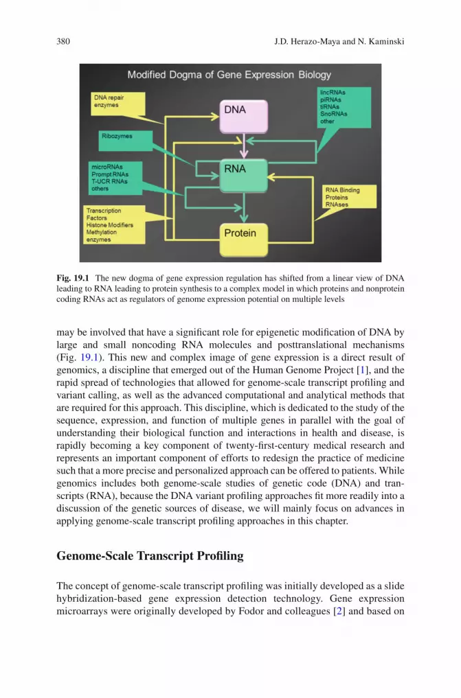
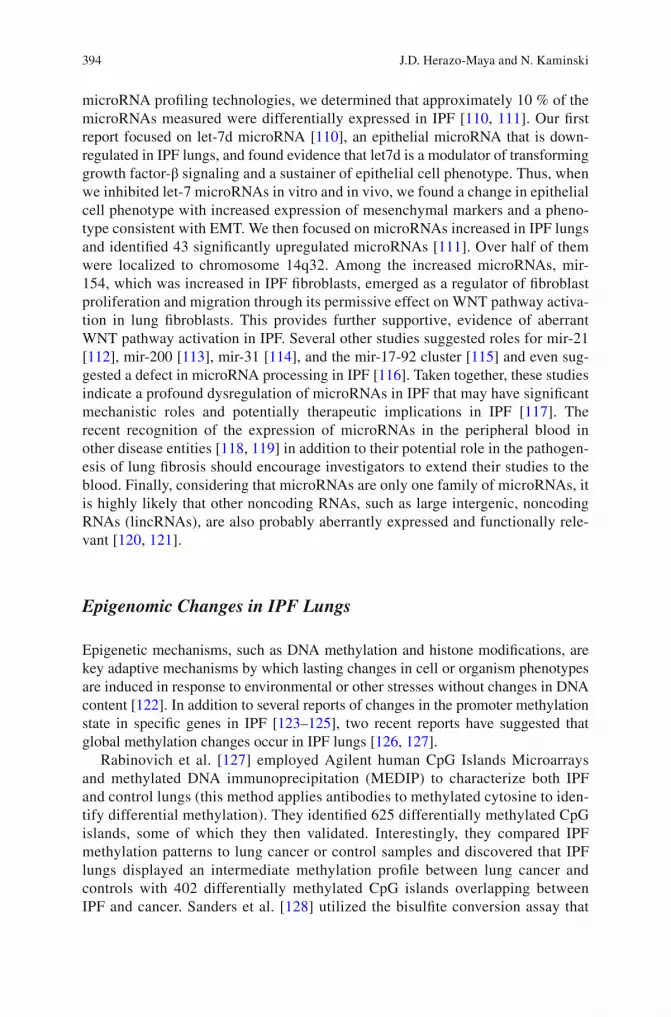
may be involved that have a signifi cant role for epigenetic modifi cation of DNA by large and small noncoding RNA molecules and posttranslational mechanisms (Fig. 19.1 ). This new and complex image of gene expression is a direct result of genomics, a discipline that emerged out of the Human Genome Project [ 1 ], and the rapid spread of technologies that allowed for genome-scale transcript profi ling and variant calling, as well as the advanced computational and analytical methods that are required for this approach. This discipline, which is dedicated to the study of the sequence, expression, and function of multiple genes in parallel with the goal of understanding their biological function and interactions in health and disease, is rapidly becoming a key component of twenty-fi rst-century medical research and represents an important component of efforts to redesign the practice of medicine such that a more precise and personalized approach can be offered to patients. While genomics includes both genome-scale studies of genetic code (DNA) and tran-scripts (RNA), because the DNA variant profi ling approaches fi t more readily into a discussion of the genetic sources of disease, we will mainly focus on advances in applying genome-scale transcript profi ling approaches in this chapter.
Genome-Scale Transcript Profi ling
The concept of genome-scale transcript profi ling was initially developed as a slide hybridization-based gene expression detection technology. Gene expression microarrays were originally developed by Fodor and colleagues [ 2 ] and based on
Fig. 19.1 The new dogma of gene expression regulation has shifted from a linear view of DNA leading to RNA leading to protein synthesis to a complex model in which proteins and nonprotein coding RNAs act as regulators of genome expression potential on multiple levels
J.D. Herazo-Maya and N. Kaminski
381
the principle of light-directed, in situ oligonucleotide synthesis with the later development of cDNA and oligonucleotide arrays [ 3 , 4 ]. More recently, novel methods that utilize high-throughput sequencing technologies have been applied to genome-scale transcript profi ling. Such technologies, which will soon render gene arrays obsolete, provide transcript-level information that can be combined with gene structure information such as alternative splicing, information about noncoding RNAs, and posttranscriptional modifi cations, as well as genomic vari-ants at the base-level resolution [ 5 ].
Regardless of the technology, experiments are performed with RNA extracted from the tissue or cell of interest and depend on the purity and integ-rity of the RNA. Genome-scale transcript profiling experiments measure the expression of a large number of transcripts (typically around 40,000–50,000), which generates a large amount of information that has to be preprocessed, analyzed, and validated before the results can be used. Obtaining the right information out of these large datasets represents the major challenge when analyzing such large genomic datasets. Before describing the most significant results obtained from genomic studies in lung fibrosis, it is critical to under-stand the steps required after the completion of microarray experiments. These steps can be summarized as comprising three broad categories: quality assess-ment, normalization, and statistical analysis. Because quality assessment and normalization approaches vary greatly with technology, they will not be dis-cussed here.
Once the genomic dataset is (1) assessed for quality and normalized and (2) outliers and batch effects (if present) are handled satisfactorily, investigators can then proceed to perform statistical analyses. Different algorithms for statistical analyses can be used for genome-scale transcriptome data, and their use depends on the objectives of the study. Typically, the statistical algorithms used for gene expression profi ling in human disease can be grouped into four major study objec-tives as defi ned by Simon and colleagues [ 6 ]: class comparison, class prediction, class discovery, and pathway analysis. Two additional study objectives may also be considered for inclusion in this group: outcome analysis and meta-analysis. Table 19.1 provides a description of the types of transcriptome study objectives and lists some of the available algorithms that can be applied to each type of objective. Some of these algorithms can be used independently, be part of a com-putational software program (such as GeneSpring GX, Bioconductor [ 43 ], and BRB array tools [ 44 ]), or be used in a statistical environment, the most widely used being the R statistical environment [ 45 ]. However, regardless of the tools, attention to testing multiple hypotheses and using effective visualization are criti-cally important.
After the statistical analysis is completed, the number of differentially expressed transcripts may still be too large to validate and study in depth. Traditionally, two different approaches have been used to deal with this issue. One can use the reductionist or “cherry picking” approach versus the global or
19 Evolving Genomics of Pulmonary Fibrosis

382 Ta
ble
19.1
Su
mm
ary
of th
e ty
pe o
f tr
ansc
ript
pro
fi lin
g ex
peri
men
tal o
bjec
tives
and
rel
evan
t alg
orith
ms
for
stat
istic
al a
naly
sis
Stud
y ob
ject
ives
D
escr
iptio
n St
atis
tical
alg
orith
ms
for
each
cat
egor
y
Cla
ss c
ompa
riso
n C
lass
com
pari
son
anal
yses
foc
us o
n th
e id
entifi
cat
ion
of d
iffe
rent
ially
ex
pres
sed
gene
s am
ong
pred
efi n
ed c
lass
es o
f sa
mpl
es
• t -
test
•
Ana
lysi
s of
var
ianc
e (A
NO
VA
) [ 7
] •
Sign
ifi ca
nce
anal
ysis
of
mic
roar
rays
(SA
M)
[ 8 , 9
] •
Ran
dom
var
ianc
e m
odel
(R
VM
) [ 1
0 ]
• L
asso
ed p
rinc
ipal
com
pone
nt (
LPC
) [ 1
1 ]
Cla
ss p
redi
ctio
n C
lass
pre
dict
ion
stud
ies
are
also
bas
ed o
n pr
edefi
ned
cla
sses
of
sam
ples
, alth
ough
its
goal
is to
dev
elop
a s
tatis
tical
pre
dict
ion
mod
el b
ased
on
the
expr
essi
on o
f a
grou
p of
gen
es to
allo
w th
e pr
edic
tion
of th
e cl
ass
in e
ach
sam
ple
• T
hres
hold
num
ber
of m
iscl
assi
fi cat
ions
(T
NoM
) [ 1
2 , 1
3 ]
• C
ompo
und
cova
riat
e pr
edic
tor
[ 14 ,
15 ]
•
Part
ial l
east
squ
are
[ 16 ]
•
k-N
eare
st n
eigh
bor
(KN
N)
[ 17 ,
18 ]
•
Supp
ort v
ecto
r m
achi
ne (
SVM
) [ 1
9 ]
• N
eare
st s
hrun
ken
cent
roid
(PA
M)
[ 20 ]
•
Top
scor
ing
pair
s [ 2
1 ]
Cla
ss d
isco
very
C
lass
dis
cove
ry e
mph
asiz
es o
n th
e de
tect
ion
of a
n un
iden
tifi e
d cl
ass
base
d on
the
co-e
xpre
ssio
n of
gen
es. T
ypic
ally
thes
e st
udie
s ar
e pe
rfor
med
to c
hara
cter
ize
an u
nkno
wn
clin
ical
dis
ease
sub
phen
o-ty
pe b
ased
on
the
expr
essi
on o
f cl
uste
rs o
f ge
nes
• K
-Mea
ns c
lust
erin
g [ 2
2 ]
• H
iera
rchi
cal c
lust
erin
g [ 2
3 ]
• B
iclu
ster
ing
[ 24 ]
•
Self
-org
aniz
ing
map
s (S
OM
) [ 2
5 ]
• M
odel
-bas
ed c
lust
erin
g [ 2
6 ]
• G
ene
expr
essi
on d
ynam
ic in
spec
tor
(GE
DI)
[ 27
] Pa
thw
ay a
naly
sis
Path
way
ana
lysi
s st
udie
s fo
cuse
s on
the
iden
tifi c
atio
n of
dif
fere
ntia
lly
expr
esse
d ge
nes
that
occ
ur in
the
sam
e m
olec
ular
pat
hway
in
pred
efi n
ed c
lass
es o
f sa
mpl
es
• G
loba
l tes
t for
gro
ups
of g
enes
[ 28
] •
Gen
e se
t enr
ichm
ent a
naly
sis
(GSE
A)
[ 29 ]
•
SAM
-GS
[ 30 ]
•
Gen
e se
t ana
lysi
s (G
SA)
[ 31 ]
•
Inte
grat
ive
mic
roar
ray
anal
ysis
of
path
way
s (I
MA
P) [
31 ]
• G
ene
set e
xpre
ssio
n co
mpa
riso
n [ 3
2 ]
Out
com
e an
alys
is
Out
com
e an
alys
is s
tudi
es e
xplo
re th
e as
soci
atio
n of
gen
e ex
pres
sion
s w
ith a
pre
defi n
ed o
utco
me
(i.e
., su
rviv
al, t
rans
plan
t-fr
ee s
urvi
val,
dise
ase
prog
ress
ion)
• C
ox m
odel
[ 33
] •
Part
ial l
east
squ
ares
pro
port
iona
l haz
ard
regr
essi
on [
34 ]
• M
ultip
le r
ando
m v
alid
atio
n [ 3
5 ]
• Pr
edic
tion
by s
uper
vise
d pr
inci
pal c
ompo
nent
(Su
perP
C)
[ 36 ]
M
eta-
anal
ysis
G
ene
expr
essi
on m
eta-
anal
ysis
stu
dies
com
bine
mul
tiple
and
sim
ilar
gene
exp
ress
ion
data
sets
to in
crea
se th
e st
atis
tical
pow
er a
nd
accu
racy
of
the
resu
lts
• T
runc
ated
pro
duct
met
hod
for
com
bini
ng P
-val
ues
[ 37 ]
•
t -ba
sed
mod
elin
g [ 3
8 ]
• R
ankP
rod
[ 39 ]
•
Met
a-an
alys
is b
ased
on
cont
rol o
f fa
lse
disc
over
y ra
te [
40 ]
• Pr
edic
tor-
base
d ap
proa
ch [
41 ]
• M
etaO
mic
s [ 4
2 ]
J.D. Herazo-Maya and N. Kaminski
383
“systems” approach [ 46 ]. In the “cherry picking” approach, researchers select differentially expressed transcripts for which there is prior biological knowl-edge. Such transcripts are validated at the RNA and protein level if a given transcript is a coding RNA. Following this validation, in-depth in vitro and in vivo studies are required to determine its relevance to the disease. The fi ndings are then translated back to humans to confi rm an association with the disease of interest, and the transcripts’ potential use as a biomarker or as a therapeutic target is assessed. When using the global or “systems” approach, researchers try to study gene expression profi les as a unit by using the concept that differen-tially expressed genes belong to a common pathway that is relevant to disease or that a number of genes can interact with each other depending on their pattern of expression. The global approach has been made possible with the use of gene ontology annotations, previously published knowledge of gene interactions, and pathway analysis with a focus on the identifi cation of differentially expressed genes occurring in the same molecular pathway.
Finally, the selection of relevant genes for validation can be facilitated with the integration of patient clinical information with the analysis of gene expres-sion data, which can facilitate the identifi cation of profi les that characterize a clinical variable of interest. This is typically used to study gene profi les associ-ated with response to drug therapy, disease severity and progression, and subse-quent outcomes.
In summary, the analysis of genome-scale transcript profi ling experiments requires dedicated quality control, data normalization, and statistical analysis that are based on the objectives of the study. The selection of gene(s) for validation and potential translation to patient care can be facilitated using a reductionist approach, a global approach, or both. In addition, depending on the ultimate goal of the study, clinical variables could be introduced to the analysis of gene expression to ensure an easier translation to clinical practice.
The Contribution of Genomics to Our Mechanistic Understanding of Lung Fibrosis
In contrast to hypothesis-driven experimental approaches that are based on what is known, the results of genome-scale transcript profiling experiments often contain results that were unforeseen or even contrary to currently accepted paradigms. When one considers that many breakthroughs in modern medicine were the result of serendipity [ 47 , 48 ], one could consider large-scale genomic profiling experiments as a means that can introduce serendipity into pulmonary research and thereby identify new hypotheses and provide new insights.
19 Evolving Genomics of Pulmonary Fibrosis
384
The Lung Phenotype in IPF Is Not a Result of Passive Accumulation of Extracellular Matrix
A passive accumulation of extracellular matrix was the dominant paradigm that was perceived to explain pulmonary fi brosis in the last decade of the twentieth century, and this explanation assumed that fi brosis and accumulation of extracellular matrix were the result of a protease–antiprotease imbalance. This concept was accompa-nied by an apparent increase in the activity of naturally occurring inhibitors of metalloproteases accompanied by an associated reduction in the activity of matrix metalloproteases that was thought to lead to the accumulation of extracellular matrix [ 49 ]. This paradigm was supported by observations from a limited set of hypothesis based, albeit carefully designed experiments, but it was never tested in a global non- biased analysis of the lung environment in IPF.
When Zuo et al. [ 50 ] analyzed lung tissue of patients with IPF and compared them with healthy controls, they immediately noticed that multiple members of the matrix metalloproteinase (MMP) family (including MMP 1, 7, and 9) were upreg-ulated at the mRNA and protein level in IPF lungs. Among the overexpressed genes in IPF, MMP-7 was the most informative and was localized to the alveolar epithelium, a fi nding that suggested an active role of the alveolar epithelium in the lung remodeling that characterizes IPF. Interestingly, MMP-7 knockout mice were relatively protected from bleomycin-induced fi brosis, suggesting the potential role of this protease as a regulator of fi brosis. Indeed, it is impressive that despite the fact that these original observations were obtained on a very small number of tissues, they have been repeatedly verifi ed [ 51 – 53 ].
The proteolytic effects of MMP-7 can mediate the cleavage of molecules such as collagen type IV, aggrecan, laminin, fi bronectin, gelatin, entactin, decorin, tenascin, vitronectin, osteonectin, elastin, and SPP1 (among others) [ 54 ]. MMP-7 is also an example of a metalloprotease that may have regulatory effects that can be inferred by looking at its bioactive substrates that potentially include fi brosis-relevant proteins such as FAS ligand, β4 integrin, E-cadherin, pro-HB-epidermal growth factor, plas-minogen, pro-TNF-α, pro-α-defensin, endostatin, syndecan, and insulin growth fac-tor-binding protein-3 (IGFBP-3) [ 55 ]. While the local effects of MMP-7 overexpression in the alveolar epithelium in humans are not clear, evidence from mice concerning its regulation of neutrophil egress, regulation of dendritic cells, and activation of defen-sins [ 56 – 58 ] suggests that it may have a signifi cant role in regulating the local infl am-matory milieu. Its effect on SPP1 provides additional support in this regard [ 59 ].
Other MMPs, including MMP 1, 2, 3, 9, 10, and 19 [ 49 , 60 – 62 ], have been consis-tently found to be increased in IPF lungs, and some of these proteins have been shown to be relevant to the pathogenesis of pulmonary fi brosis. As an example, Yamashita et al. demonstrated that rats transfected with an adeno-MMP-3 vector developed tran-sient pulmonary fi brosis, and in vitro treatment of lung epithelial cells with MMP-3 resulted in activation of the β-catenin signaling pathway followed by subsequent induction of epithelial-mesenchymal transition (EMT), which is one of the proposed mechanisms for the development of lung fi brosis [ 61 ]. More recently, after performing
J.D. Herazo-Maya and N. Kaminski
385
microarray expression studies of the lung microenvironment obtained by laser capture microdissected lung tissue from IPF patients, our group identifi ed MMP-19 overex-pression in hyperplastic epithelial cells from patients with IPF when compared with normal appearing epithelial cells. The presence of MMP-19 was confi rmed by immu-nohistochemistry in hyperplastic epithelial cells that were overlying fi brotic areas, but in contrast to what was observed with MMP-7, MMP-19 knockout mice developed worse fi brosis when exposed to bleomycin, suggesting that MMP19 overexpression failed to provide protection. Thus, genome-scale transcript profi ling studies have led to a paradigm shift in the perception of the role of proteases in lung fi brosis, and instead of the simplistic protease–antiprotease imbalance paradigm, we now have a more complex understanding that suggests that proteases have multiple and some-times opposing roles in lung fi brosis. The roles of these MMPs depend on their tem-poral expression, the MMP-producing cell type and spatial distribution, and the availability of substrates [ 63 ].
Genome-scale transcript profi ling studies have not only generated relevant infor-mation regarding the presence and potential role of some of the MMP family mem-bers in IPF but also opened a new biomarker fi eld for their use in IPF diagnosis, disease monitoring, and mortality prediction. Based on our previous fi ndings [ 50 ], our group applied a targeted proteomic approach and identifi ed a protein signature that includes MMP-1, MMP-7, MMP-8, IGFBP-1, and TNFRSA1F [ 53 ], and this signature was able to distinguish IPF from healthy controls with a sensitivity of 98.6 % and specifi city of 98.1 %. Two members of this signature, MMP-1 and MMP-7, differentiated IPF patients from those with subacute/chronic hypersensi-tivity pneumonitis (HP) with a sensitivity of 96.3 % and specifi city of 87.2 %. Increased concentrations of MMPs, including MMP-7, have also been shown in the bronchoalveolar lavage (BAL) of IPF patients [ 53 , 64 ], confi rming that these mol-ecules not only participate in disease pathogenesis but can also be used as makers of disease presence. More recently, we also demonstrated that increased plasma con-centrations of MMP-7 at the initial clinical presentation were predictive of subse-quent increased mortality in IPF [ 65 ], especially when MMP-7 was used along with clinical variables. These fi ndings have potential implications for risk stratifi cation, patient counseling, and prioritization for lung transplantation in the future.
Thus, the emergence of MMPs as mechanistically important in determining the lung phenotype in IPF and other interstitial lung diseases (as well as their role as new peripheral blood biomarker candidates) can be fully attributed to unbiased genome-scale transcript profi ling.
The Wnt Pathway in IPF
As previously discussed, one of the advantages of the “systems” approach over the “cherry picking” approach for genome-scale transcript profi ling is that by group-ing differentially expressed genes in gene sets based on their attributes, research-ers are allowed to identify pathways (and genes within the pathways) that
19 Evolving Genomics of Pulmonary Fibrosis
386
characterize the differences between the analyzed groups. This procedure enables the generation of new hypotheses regarding disease pathogenesis by focusing on pathways that were not considered relevant to the disease. This also helps research-ers to focus on differentially expressed genes within a pathway that might other-wise have been missed.
Following a “systems” approach, we reanalyzed the microarray datasets gener-ated by our group using more powerful pathway analysis tools and, surprisingly, identifi ed a large number of developmental pathway genes [ 66 ]. Some of the development- related genes that were found overexpressed in IPF included members of transcription factor families (such as the Sry-related high-mobility group box and forkhead box) and genes related to the Wnt/β-catenin pathway [ 67 ]. In our analysis, the Wnt pathway was one of the most signifi cantly overexpressed pathways in IPF, and, interestingly, this was not the case in HP.
The Wnt pathway consists of a network of glycoproteins that are involved in embryogenesis and development, and this pathway was especially characterized after the identifi cation that a mutation in one of its genes, “Wingless,” was associ-ated with the development of wingless Drosophila melanogaster fl ies (fruit fl y) [ 68 ]. The key player of the canonical Wnt signaling is β-catenin, which, after it accumulates in the cytoplasm, eventually translocates into the nucleus and interacts with transcription factors of the LEF/TCF family, which affects gene transcription [ 69 ]. Most Wnt proteins bind to the frizzled family of receptors and LRP5/6 co- receptors, and these, in turn, inhibit the phosphorylation and degradation of β-catenin, which allows its translocation into the nucleus [ 70 ]. Without Wnt signal-ing, β-catenin is degraded by its destruction complex.
Experiments in mice have demonstrated that β-catenin is required for the normal differentiation of the bronchiolar and alveolar epithelium [ 71 ]. Wnt7b-defi cient mice exhibit impaired alveolar type I cell differentiation, have hypoplastic lungs, and die at birth of respiratory failure [ 72 ]. Similarly, Wnt5a-defi cient mice exhibit increased proliferation of lung epithelial and mesenchymal compartments and die shortly after birth due to respiratory failure [ 73 , 74 ]. In humans, mutations and genetic variations in genes of the Wnt pathway have been associated with condi-tions such as cancer, neuropsychiatric disorders, cardiac diseases, and bone disor-ders [ 75 ]. Several reports that appeared following publication of our microarray fi ndings validated the increase of functional Wnt in IPF. Chilosi et al. demonstrated β-catenin accumulation in fi broblastic foci of IPF lungs and its expression co- localized with two Wnt downstream target genes (cyclin-D1 and MMP-7) in prolif-erative bronchiolar lesions [ 76 ]. This report was followed by the fi ndings of Königshoff et al. who demonstrated the overexpression of Wnt1, 7b and 10b, Fzd2 and 3, beta-catenin, and Lef1 expression in IPF lungs by qRT-PCR and localized Wnt1, Wnt3a, β-catenin, and Gsk-3β expression to alveolar and bronchial epithe-lium by immunohistochemistry [ 77 ]. Along with the discovery of increased func-tional Wnt in IPF, there is evidence of reversal of pulmonary fi brosis after the inhibition of Wnt/β-catenin [ 78 ]. Interestingly, MMP7, which was recently men-tioned as both a mechanistically relevant molecule and a peripheral blood biomarker, is a repeatedly validated Wnt pathway target gene [ 79 ].
J.D. Herazo-Maya and N. Kaminski
387
In summary, the observation of overexpression of Wnt signaling in IPF suggests an aberrant activation of developmental pathways that are not usually involved in normal lung health. A better understanding of these mechanisms could lead to potentially effective therapeutic strategies for this devastating lung disease.
Apoptosis in Lung Fibrosis from a Genomic Perspective
Two studies using genomics-based approaches (and published only a month apart) confi rmed the role of apoptosis in IPF pathogenesis. Bridges et al. [ 80 ] performed microarray gene expression experiments with cells obtained from normal lung sam-ples and compared them with cells from IPF lungs (including samples obtained from microdissected fi broblastic foci). They used class discovery (unsupervised clustering) and class comparison (t-test) analyses and identifi ed Twist 1 as one of the most consistently up-regulated transcription factors in the IPF lung. In this study, researchers determined that overexpression of Twist1 led to increased viabil-ity of rat lung fi broblasts exposed to pro-apoptotic molecules (lipid 4-HNE and thapsigargin). However, lower concentrations of these proapoptotic stimuli resulted in a reduction of Twist1, which in turn resulted in increased activity of caspase-3, which is a marker of apoptosis.. They also demonstrated that profi brotic growth fac-tors such as platelet derived growth factor (PDGF) induced Twist1 expression in rat lung fi broblasts, which was necessary to protect these cells from apoptosis, particu-larly in the continued presence of these growth factors. In summary, the results demonstrated an anti-apoptotic role of Twist1 by promoting fi broblast viability when these cells where exposed to growth factors.
Our group corroborated the fi ndings confi rming the role of apoptosis in the pathogenesis of acute exacerbations of IPF [ 81 ]. We performed microarray experi-ments and compared lung tissue of IPF patients with acute exacerbation of IPF, lung tissue from IPF subjects with stable disease, and non-IPF lungs with normal histology using a class comparison approach (signifi cance analysis of microarrays). A total of 579 genes were found to be differentially expressed between the lungs of patients with acute exacerbations of IPF versus patients with stable IPF; specifi -cally, cyclin A2 (CCNA2), a cell cycle regulatory gene, was one of the top overex-pressed genes in this signature and was localized to alveolar epithelial cells in subjects with acute exacerbations of IPF. Increased CCNA2 protein expression was localized to proliferating epithelial cells, and this fi nding suggests the presence of accelerated epithelial cell proliferation, which could potentially refl ect a compensa-tory response to injured epithelium. The fi nding that lungs of IPF patients showed widespread apoptosis by in situ TUNEL assay was of even greater interest. Taken together, these observations suggest an aberrant proliferative response of the alveo-lar epithelium in reaction to apoptosis during acute exacerbations of IPF.
19 Evolving Genomics of Pulmonary Fibrosis
388
Global Analysis of IPF Lungs Reveals Dramatic Changes in Epithelial Cell Phenotype
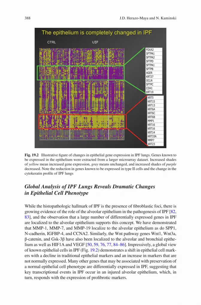
While the histopathologic hallmark of IPF is the presence of fi broblastic foci, there is growing evidence of the role of the alveolar epithelium in the pathogenesis of IPF [ 82 , 83 ], and the observation that a large number of differentially expressed genes in IPF are localized to the alveolar epithelium supports this concept. We have demonstrated that MMP-1, MMP-7, and MMP-19 localize to the alveolar epithelium as do SPP1, N-cadherin, IGFBP-4, and CCNA2. Similarly, the Wnt pathway genes Wnt1, Wnt3a, β-catenin, and Gsk-3β have also been localized to the alveolar and bronchial epithe-lium as well as HIF1A and VEGF [ 50 , 59 , 76 , 77 , 84 – 86 ]. Impressively, a global view of known epithelial cells in IPF (Fig. 19.2 ) demonstrates a shift in epithelial cell mark-ers with a decline in traditional epithelial markers and an increase in markers that are not normally expressed. Many other genes that may be associated with preservation of a normal epithelial cell phenotype are differentially expressed in IPF, suggesting that key transcriptional events in IPF occur in an injured alveolar epithelium, which, in turn, responds with the expression of profi brotic markers.
Fig. 19.2 Illustrative fi gure of changes in epithelial gene expression in IPF lungs. Genes known to be expressed in the epithelium were extracted from a larger microarray dataset. Increased shades of yellow mean increased gene expression, gray means unchanged, and increased shades of purple decreased. Note the reduction in genes known to be expressed in type II cells and the change in the cytokeratin profi le of IPF lungs
J.D. Herazo-Maya and N. Kaminski

389
Gene Expression Profi ling and the Classifi cation of Interstitial Lung Diseases
The diagnosis of interstitial lung diseases in clinical practice can be challenging at times given the fact that some of the patients can present with radiological patterns that are inconclusive [ 87 – 89 ]. Further, in some cases, lung histology may show discordant pat-terns such as a usual interstitial pneumonia (UIP) pattern in one lobe and a nonspecifi c interstitial pneumonia (NSIP) pattern in a different lobe from the same patient [ 90 , 91 ].
The diagnostic dilemma usually is more common when comparing cases of chronic HP, NSIP, and ILD associated with collagen vascular disease from those with IPF. One of the goals of genomic studies in ILD has been to fi nd transcript profi les that could dif-ferentiate these entities in order to develop more accurate diagnostic strategies. We will discuss the gene expression studies addressing these issues in the following section.
Differences in Gene Expression Between IPF and HP
To study gene expression differences in lung tissue from patients with IPF versus patients with HP, our group performed gene expression microarrays and compared transcript levels using a class comparison ( t -test) and class prediction (threshold number of misclassifi cations—TNoM) approach and identifi ed 407 genes that accurately distinguished IPF from HP [ 84 ]. The pathway analysis of this signature confi rmed the prior knowledge regarding the pathogenesis of these two entities. While the HP signature is characterized by enrichment of pathways associated with cytokine and T-cell activation, infl ammation, and humoral immune responses, the IPF signature is characterized by cell adhesion, extracel-lular matrix, and smooth muscle differentiation as well as genes associated with lung development, heparin binding, enzyme inhibitor activity, and insulin growth factor binding [ 46 ]. It is clear after looking at the gene pathway differ-ences between these two conditions that gene expression associated with infl am-mation is more pronounced in HP, while increased expression of genes involved in matrix turnover and developmental pathways is more characteristic of IPF. These fi ndings are consistent with the knowledge that evidence of infl ammation in IPF is not as prominent as initially thought [ 92 ].
IPF and Familial Pulmonary Fibrosis (FPF) Are Unexpectedly Different While IPF and NSIP Are Unexpectedly Similar
Yang et al. [ 93 ] performed gene expression microarrays of lung tissue from patients with sporadic IPF, FPF, NSIP, and normal controls. However, because these investigators were unable to identify statistically signifi cant differences between IPF and NSIP, the results were considered somewhat disappointing but were in agreement with our prior observations [ 84 ]. An interesting fi nding was
19 Evolving Genomics of Pulmonary Fibrosis
390
the identifi cation of differentially expressed genes between sporadic IPF and FPF, diseases that have considerably more similarities than differences. While the genes distinguishing familial cases from the sporadic ones were part of the same functional pathways as genes distinguishing IPF from normal samples, they seemed to exhibit larger changes. One conclusion was that familial pulmo-nary fi brosis may represent a more extreme molecular phenotype of the same disease process as sporadic IPF. However, while this is certainly possible, we suggest that the stage in the natural history of the disease when tissue sampling was performed may have played a role in these differences [ 94 ], because 50 % of the familial samples were obtained from open lung biopsies, whereas 90 % of sporadic cases were collected from explant or autopsy, suggesting that the dif-ferences may be due to differences in disease stage.
Different Forms of UIP Share Very Similar Gene Expression Patterns
The UIP pattern in lung biopsies of patients with systemic sclerosis (SSc) can be indistinguishable from the UIP pattern of IPF patients [ 95 ], a fi nding that contrasts with the major clinical differences between these two entities. In an attempt to better elucidate the molecular mechanisms behind the differences in SSc and IPF, Hsu and colleagues [ 96 ] performed gene expression profi ling in patients with SSc and clas-sifi ed patients as having a predominance of pulmonary fi brosis (with a UIP pattern) or as a pulmonary arterial hypertension (PAH) phenotype and compared them with lung tissue from IPF and idiopathic pulmonary arterial hypertension (IPAH) patients. Using a class comparison approach (effi ciency analysis and signifi cance analysis of microarrays), they identifi ed 242 differentially expressed genes between the studied subclasses of SSc patients. Focusing on the comparison that is relevant to our discussion and similar to what was observed between IPF and NSIP, the gene expression profi le of the UIP lung of IPF patients was very similar to the UIP lung of systemic SSc patients, with only 25 genes being uniquely expressed in IPF lung tissue and 20 genes uniquely expressed in the UIP lung tissue of SSc patients.
The authors of this study acknowledge that one of their limitations was the use of explanted lung tissue of patients undergoing lung transplant, which could repre-sent end-stage disease, suggesting that comparisons in gene expression between SSc and IPF at earlier stages could potentially provide a better molecular character-ization of these two entities.
Identifi cation of Gene Expression Profi les Associated with Disease Severity in the IPF Lung and Peripheral Blood
It has been shown that IPF patients have different patterns of disease progression. Although some patients can be stable for long period of time, others can quickly deteriorate or have an acute exacerbation and die as a consequence of an accelerated
J.D. Herazo-Maya and N. Kaminski
391
disease course [ 97 ]. The recognition of this erratic clinical behavior of some IPF patients prompted Selman et al. [ 85 ] to study gene expression profi les of IPF patients with evidence of rapid progression (defi ned as symptoms starting 6 months prior to initial presentation) and compare them with IPF patients with slow progression (defi ned as symptoms present for more than 24 months) using a class comparison and class prediction approach. The investigators identifi ed a group of 437 differen-tially expressed genes between these two patient groups. When a pathway analysis was performed, patients with evidence of rapid progression had overexpression of genes involved in morphogenesis, cancer, oxidative stress, cell proliferation, apop-tosis, and genes from fi broblast/smooth muscle cells. The discovery of overexpres-sion of genes associated with cell proliferation and apoptosis preceded the fi ndings by Konishi et al. [ 81 ] who demonstrated evidence of overexpression of cyclins (cell cycle regulators) along with overwhelming apoptosis in the lung of patients with an acute exacerbation. This observation again suggested the potential presence of aber-rant proliferative responses in response to apoptosis in patients with rapid progres-sion of their IPF.
Konishi et al. [ 81 ] discovered another interesting fi nding in the lung tissue of IPF patients with acute exacerbations; alpha-defensins, particularly defensin alpha 3 (DEFA3) and 4 (DEFA4), were overexpressed, and these authors also demonstrated increased levels in the serum of these natural antimicrobial peptides, which are a component of innate immunity and participate in host defense [ 98 ]. Interestingly, defensins released in response to microbial invasion can activate an adaptive immune response [ 99 ], a mechanism that has been described in IPF [ 100 ], by attracting antigen-presenting dendritic cells to the site of invasion. Defensins are mostly expressed by neutrophils, epithelial cells, and paneth cells, and, interest-ingly, they are activated via proteolytic cleavage by MMP-7 [ 58 ]. In summary, these fi ndings support the notion that defensins are not only surrogates of disease activity and severity, but they may also be closely associated with IPF pathogenesis.
The overexpression of defensins has been validated in the peripheral blood tran-scriptome of IPF patients with evidence of advanced disease by Yang et al. [ 101 ]. These investigators performed gene expression profi ling of patients with IPF who were stratifi ed according to disease severity. They defi ned severe disease as DLCO ≤35 % or FVC ≤50 % and compared them with IPF patients with mild disease that was defi ned as DLCO ≥65 % or FVC ≥75 %. They also compared these two sub-classes of IPF patients with age and gender-matched healthy controls using a class comparison approach (signifi cance analysis of microarrays). When comparing patients categorized by percent-predicted DLCO ≥65 % with patients with DLCO ≤35 %, the authors identifi ed 13 differentially expressed transcripts including DEFA3 and DEFA4. DEFA3 also differentiated mild and severe IPF cases from healthy controls, confi rming the relevance of defensins in IPF progression.
The functional analysis performed in the study by Yang et al. [ 101 ] using the 13 differentially expressed transcripts differentiating mild and severe cases of IPF revealed a fi nding that is contradictory to our prior observations in lung tissue of IPF individuals. Specifi cally, they reported overexpression of genes associated with infl ammatory responses and immune traffi cking in the severe IPF group. While this
19 Evolving Genomics of Pulmonary Fibrosis
392
could represent evidence that infl ammatory responses are indeed potentially relevant in IPF, it can also indicate that a more infl ammatory phenotype is present in patients with more rapid disease progression.
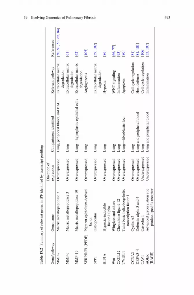
Boon and colleagues [ 102 ] also studied and compared gene expression profi les from lung tissue of IPF patients with evidence of disease progression or relatively stable disease, defi ned respectively as FVC% and DLCO% decline ≥10 % and ≥15 % versus decline of <10 % and <15 % over a 12-month period. For this study, the investigators used serial analysis of gene expression (SAGE), a technique that has the same goal of microarrays with the difference that SAGE sampling is based on sequencing of short tags of mRNA, while microarrays are based on hybridization of mRNAs to probes. Using a class comparison ( t -test) and class discovery (hierar-chical clustering) approach, 134 differentially expressed transcripts distinguished the two cohorts. While this study is also limited by the small number of samples (six in each group), it certainly provided interesting fi ndings, because some of the over-expressed genes in the group of patients with evidence of IPF progression included surfactant protein A1 (SFTPA1), SPP1, and heat shock 70 KDa protein 1A (HSPA1A) among others. These fi ndings correlated with previously noted associa-tions of surfactant protein A levels in serum and autoantibodies against heat shock protein 70 (HSP70) with worse survival in IPF [ 103 , 104 ]. We have previously reported consistent overexpression of SPP1 when analyzing gene expression pro-fi les of IPF lung tissue compared to normal controls [ 59 ] and have also demon-strated increased SPP1 levels in BAL of IPF patients. We also found evidence suggesting that SPP1 activates MMP-7 and co-localizes with this molecule in alveo-lar epithelial cells of IPF patients, resulting in a profi brotic effect on lung fi broblasts and epithelial cells. Others have demonstrated the relative protection to bleomycin- induced fi brosis in SPP1 knockout mice and increased SPP1 levels in serum of patients with interstitial lung disease. In summation, this body of evidence suggests that SPP1 is not only relevant to the pathogenesis of IPF but could also be a poten-tial biomarker for disease progression. Table 19.2 includes a summary of some of the most relevant molecules identifi ed in IPF (based on gene expression studies).
Noncoding RNAs in IPF
One of the direct results of the Human Genome Project and the large next- generation sequencing studies that followed, including ENCODE [ 108 ], was the recognition that noncoding RNAs are critically important in determining cell and organ phenotype through their effects on gene and protein expression (see Fig. 19.1 ). While the data are only now emerging, it is already obvious that at least one family of noncoding RNAs, that of microRNAs, is critically important in IPF. MicroRNAs are small, noncoding RNAs (21–25 nucleotides) that bind via base pairing to the 3′ untranslated region of their target mRNAs. In most cases they repress gene expression by increasing mRNA degradation or by disrupting trans-lation initiation [ 109 ]. In two recent studies that utilized different generations of
J.D. Herazo-Maya and N. Kaminski
393
Tabl
e 19
.2
Sum
mar
y of
rel
evan
t gen
es in
IPF
iden
tifi e
d by
tran
scri
pt p
rofi l
ing
Gen
e/pa
thw
ay
Gen
e na
me
Dir
ectio
n of
ex
pres
sion
C
ompa
rtm
ent i
dent
ifi ed
R
elev
ant p
athw
ay
Ref
eren
ces
MM
P-7
Mat
rix
met
allo
pept
idas
e 7
Ove
rexp
ress
ed
Lun
g, p
erip
hera
l blo
od, a
nd B
AL
E
xtra
cellu
lar
mat
rix
degr
adat
ion
[ 50 ,
51 ,
53 ,
65 ,
84 ]
MM
P-3
Mat
rix
met
allo
pept
idas
e 3
Ove
rexp
ress
ed
Lun
g E
xtra
cellu
lar
mat
rix
degr
adat
ion
[ 61 ]
MM
P-19
M
atri
x m
etal
lope
ptid
ase
19
Ove
rexp
ress
ed
Lun
g—hy
perp
last
ic e
pith
elia
l cel
ls
Ext
race
llula
r m
atri
x de
grad
atio
n [ 6
2 ]
SER
PIN
F1 (
PED
F)
Pigm
ent e
pith
eliu
m- d
eriv
ed
fact
or
Ove
rexp
ress
ed
Lun
g A
ngio
gene
sis
[ 105
]
SPP1
O
steo
pont
in
Ove
rexp
ress
ed
Lun
g E
xtra
cellu
lar
mat
rix
degr
adat
ion
[ 59 ,
102
]
HIF
1A
Hyp
oxia
-ind
ucib
le
fact
or-1
alph
a O
vere
xpre
ssed
L
ung
Hyp
oxia
[ 8
6 ]
Wnt
W
ingl
ess
and
othe
rs
Ove
rexp
ress
ed
Lun
g W
NT
sig
nalin
g [ 6
6 , 7
7 ]
CX
CL
12
Che
mok
ine
ligan
d 12
O
vere
xpre
ssed
L
ung
Infl a
mm
atio
n [ 9
3 ]
TW
IST
1 Tw
ist b
asic
hel
ix-l
oop-
helix
tr
ansc
ript
ion
fact
or 1
O
vere
xpre
ssed
L
ung—
fi bro
blas
tic f
oci
Apo
ptos
is
[ 80 ]
CC
NA
2 C
yclin
A2
Ove
rexp
ress
ed
Lun
g C
ell c
ycle
reg
ulat
ion
[ 81 ]
D
EFA
3–4
Def
ensi
n al
pha
3 an
d 4
Ove
rexp
ress
ed
Lun
g an
d pe
riph
eral
blo
od
Hos
t def
ense
[ 8
1 , 1
01 ]
CA
V1
Cav
eolin
1
Und
erex
pres
sed
Lun
g C
ell c
ycle
reg
ulat
ion
[ 106
] A
GE
R
(RA
GE
) A
dvan
ced
glyc
osyl
atio
n en
d pr
oduc
t- sp
ecifi
c re
cept
or
Und
erex
pres
sed
Lun
g an
d pe
riph
eral
blo
od
Infl a
mm
atio
n [ 5
3 , 1
07 ]
19 Evolving Genomics of Pulmonary Fibrosis
394
microRNA profi ling technologies, we determined that approximately 10 % of the microRNAs measured were differentially expressed in IPF [ 110 , 111 ]. Our fi rst report focused on let-7d microRNA [ 110 ], an epithelial microRNA that is down-regulated in IPF lungs, and found evidence that let7d is a modulator of transforming growth factor-β signaling and a sustainer of epithelial cell phenotype. Thus, when we inhibited let-7 microRNAs in vitro and in vivo, we found a change in epithelial cell phenotype with increased expression of mesenchymal markers and a pheno-type consistent with EMT. We then focused on microRNAs increased in IPF lungs and identifi ed 43 signifi cantly upregulated microRNAs [ 111 ]. Over half of them were localized to chromosome 14q32. Among the increased microRNAs, mir-154, which was increased in IPF fi broblasts, emerged as a regulator of fi broblast proliferation and migration through its permissive effect on WNT pathway activa-tion in lung fi broblasts. This provides further supportive, evidence of aberrant WNT pathway activation in IPF. Several other studies suggested roles for mir-21 [ 112 ], mir-200 [ 113 ], mir-31 [ 114 ], and the mir-17-92 cluster [ 115 ] and even sug-gested a defect in microRNA processing in IPF [ 116 ]. Taken together, these studies indicate a profound dysregulation of microRNAs in IPF that may have signifi cant mechanistic roles and potentially therapeutic implications in IPF [ 117 ]. The recent recognition of the expression of microRNAs in the peripheral blood in other disease entities [ 118 , 119 ] in addition to their potential role in the pathogen-esis of lung fi brosis should encourage investigators to extend their studies to the blood. Finally, considering that microRNAs are only one family of microRNAs, it is highly likely that other noncoding RNAs, such as large intergenic, noncoding RNAs (lincRNAs), are also probably aberrantly expressed and functionally rele-vant [ 120 , 121 ].
Epigenomic Changes in IPF Lungs
Epigenetic mechanisms, such as DNA methylation and histone modifi cations, are key adaptive mechanisms by which lasting changes in cell or organism phenotypes are induced in response to environmental or other stresses without changes in DNA content [ 122 ]. In addition to several reports of changes in the promoter methylation state in specifi c genes in IPF [ 123 – 125 ], two recent reports have suggested that global methylation changes occur in IPF lungs [ 126 , 127 ].
Rabinovich et al. [ 127 ] employed Agilent human CpG Islands Microarrays and methylated DNA immunoprecipitation (MEDIP) to characterize both IPF and control lungs (this method applies antibodies to methylated cytosine to iden-tify differential methylation). They identifi ed 625 differentially methylated CpG islands, some of which they then validated. Interestingly, they compared IPF methylation patterns to lung cancer or control samples and discovered that IPF lungs displayed an intermediate methylation profi le between lung cancer and controls with 402 differentially methylated CpG islands overlapping between IPF and cancer. Sanders et al. [ 128 ] utilized the bisulfi te conversion assay that
J.D. Herazo-Maya and N. Kaminski
395
converts unmethylated cytosine into uracil and determined that 870 genes were differentially methylated. These authors identifi ed 16 genes with inversely related signifi cant changes in gene methylation and expression, 8 of which were previously shown to be associated with fi brosis. While at this stage it is impos-sible to draw any fi nal conclusions from the small number of samples [ 129 ], both studies suggest that the changes in methylation, which represents one aspect of epigenetics, are indeed relatively signifi cant and justify additional forays into genome-scale profi ling of epigenetic changes in IPF.
Summary and Future Direction
In this chapter, we discussed the discipline of genomics and its impact on our understanding of fi brotic lung diseases with a focus on IPF, the most common and lethal idiopathic interstitial lung disease. Of genomic technologies, the technique that has had the greatest impact to date is genome-scale transcript profi ling using microarrays. Indeed, most of the signifi cant fi ndings (including the role of matrix metalloproteases, the role of developmental pathways such as the Wnt pathway and apoptosis, and the role of the alveolar epithelium) that have emerged from microarray experiments have fostered a paradigm shift in our understanding of the pathogenesis of IPF.
The contribution of genomic studies in fi brotic lung disease is not limited to pathogenesis. The transcript profi ling fi ndings also led to the identifi cation of MMP- 7, one of the emerging peripheral blood biomarkers for IPF diagnosis and outcome prediction, as well as many other markers. Similarly, we reviewed the differences and similarities in gene expression profi ling between IPF and other forms of ILD as well as the identifi cation of gene expression profi les associated with disease severity in lung tissue and the peripheral blood of IPF patients. Although these results are less developed, they still highlight the depth of information that is relevant to this disease and can be gleaned from genome-scale transcript profi les, and these exciting fi ndings should serve to encourage larger and more detailed studies.
While the fi eld of genomics is continuously evolving and new discoveries in ILD are constantly appearing, extensively analysis and review of the available data, and at least from a “systems” perspective, has greatly advanced our knowledge of what characterizes abnormalities in the IPF lung. It is clear that new studies are required to provide an in-depth look into other less common forms of ILD and to explore the differences between the two physiologic extremes of pulmonary ailments, namely, the obstructive versus the restrictive lung diseases. Future study designs that may potentially impact the care of patients should include the use of large transcriptomic analyses of peripheral blood on a serial basis, because this could lead to the devel-opment of biomarkers that provide a closer representation of disease activity and progression at the molecular level [ 130 ]. Additionally, it is also important to develop studies that integrate clinical data as well as other regulatory portions of the genome such as microRNAs and long intergenic noncoding RNAs. This integration has
19 Evolving Genomics of Pulmonary Fibrosis
396
already started with the fi nancial support of the National Institute of Health (NIH) through their funding of the Lung Tissue Research Consortium ( http://www.ltrcpublic.com/ ) and the Lung Genomics Research Consortium ( http://www.lung- genomics.org/ ), which have generated genetic and genomic information of more than 700 lung tissues that are available to the public for data analysis.
Finally, it is critical for investigators and clinicians to push the integration of the genetic and genomic information into patient care. In addition to the fi ndings described within this chapter, several recent studies have applied genome-wide association studies to identify novel variants associated with IPF [ 131 , 132 ]. So that the progress achieved in the discipline of genomics can materialize into meaningful change in how we manage ILD patients, it is essential that a concerted effort is undertaken, including “buy-in” and collaboration among clinicians, the scientifi c community, industry, and regulatory agencies. This chapter presents strong evi-dence of the importance of the genomic fi eld to the study of lung fi brosis, its poten-tial for translation into patient care, and the potential for ongoing development and discovery. However, all these efforts will be for naught if they are not focused on a specifi c cause and goal, which remains the optimal evaluation and treatment of patients with fi brotic lung diseases.
References
1. Yang IV, Schwartz DA. The next generation of complex lung genetic studies. Am J Respir Crit Care Med. 2012;186(11):1087–94.
2. Fodor SP, Read JL, Pirrung MC, Stryer L, Lu AT, Solas D. Light-directed, spatially address-able parallel chemical synthesis. Science. 1991;251(4995):767–73.
3. Lipshutz RJ, Morris D, Chee M, Hubbell E, Kozal MJ, Shah N, et al. Using oligonucleotide probe arrays to access genetic diversity. Biotechniques. 1995;19(3):442–7.
4. Schena M, Heller RA, Theriault TP, Konrad K, Lachenmeier E, Davis RW. Microarrays: biotechnology’s discovery platform for functional genomics. Trends Biotechnol. 1998;16(7):301–6.
5. Cloonan N, Grimmond SM. Transcriptome content and dynamics at single-nucleotide resolu-tion. Genome Biol. 2008;9(9):234.
6. Simon R, Korn E, McShane L, Radmacher M, Wright G, Zhao Y. Design and analysis of DNA microarray investigations. New York, NY: Springer; 2003.
7. Churchill GA. Using ANOVA to analyze microarray data. Biotechniques. 2004;37(2):173–5. 177.
8. Consortium M, Shi L, Reid LH, Jones WD, Shippy R, Warrington JA, et al. The MicroArray Quality Control (MAQC) project shows inter- and intraplatform reproducibility of gene expression measurements. Nat Biotechnol. 2006;24(9):1151–61.
9. Tusher VG, Tibshirani R, Chu G. Signifi cance analysis of microarrays applied to the ionizing radiation response. Proc Natl Acad Sci USA. 2001;98(9):5116–21.
10. Wright GW, Simon RM. A random variance model for detection of differential gene expression in small microarray experiments. Bioinformatics. 2003;19(18):2448–55.
11. Witten DM, Tibshirani R. Testing signifi cance of features by lassoed principal components. Ann Appl Stat. 2008;2(3):986–1012.
12. Bittner M, Meltzer P, Chen Y, Jiang Y, Seftor E, Hendrix M, et al. Molecular classifi cation of cutaneous malignant melanoma by gene expression profi ling. Nature. 2000;406(6795):536–40.
J.D. Herazo-Maya and N. Kaminski
397
13. Ben-Dor A, Bruhn L, Friedman N, Nachman I, Schummer M, Yakhini Z. Tissue classifi ca-tion with gene expression profi les. J Comput Biol. 2000;7(3–4):559–83.
14. Tukey JW. Tightening the clinical trial. Control Clin Trials. 1993;14(4):266–85. 15. Hedenfalk I, Duggan D, Chen Y, Radmacher M, Bittner M, Simon R, et al. Gene-expression
profi les in hereditary breast cancer. N Engl J Med. 2001;344(8):539–48. 16. Nguyen DV, Rocke DM. Tumor classifi cation by partial least squares using microarray gene
expression data. Bioinformatics. 2002;18(1):39–50. 17. Cover TM, Hart PE. Nearest neighbor pattern classifi cation. IEEE Trans Inf Theory.
1967;13:21–7. 18. Parry RM, Jones W, Stokes TH, Phan JH, Moffi tt RA, Fang H, et al. k-Nearest neighbor
models for microarray gene expression analysis and clinical outcome prediction. Pharmacogenomics J. 2010;10(4):292–309.
19. Brown MP, Grundy WN, Lin D, Cristianini N, Sugnet CW, Furey TS, et al. Knowledge-based analysis of microarray gene expression data by using support vector machines. Proc Natl Acad Sci USA. 2000;97(1):262–7.
20. Tibshirani R, Hastie T, Narasimhan B, Chu G. Diagnosis of multiple cancer types by shrunken centroids of gene expression. Proc Natl Acad Sci USA. 2002;99(10):6567–72.
21. Geman D, d’Avignon C, Naiman DQ, Winslow RL. Classifying gene expression profi les from pairwise mRNA comparisons. Stat Appl Genet Mol Biol. 2004;3:19.
22. Hartigan JA, Wong MA. Algorithm AS 136: a K-means clustering algorithm. J R Stat Soc Ser C Appl Stat. 1979;28(1):100–8.
23. Eisen MB, Spellman PT, Brown PO, Botstein D. Cluster analysis and display of genome- wide expression patterns. Proc Natl Acad Sci USA. 1998 Dec 8;95(25):14863–8.
24. Cheng Y, Church GM. Biclustering of expression data. Proc Int Conf Intell Syst Mol Biol. 2000;8:93–103.
25. Tamayo P, Slonim D, Mesirov J, Zhu Q, Kitareewan S, Dmitrovsky E, et al. Interpreting pat-terns of gene expression with self-organizing maps: methods and application to hematopoi-etic differentiation. Proc Natl Acad Sci USA. 1999;96(6):2907–12.
26. Yeung KY, Fraley C, Murua A, Raftery AE, Ruzzo WL. Model-based clustering and data transformations for gene expression data. Bioinformatics. 2001;17(10):977–87.
27. Eichler GS, Huang S, Ingber DE. Gene Expression Dynamics Inspector (GEDI): for integra-tive analysis of expression profi les. Bioinformatics. 2003;19(17):2321–2.
28. Goeman JJ, van de Geer SA, de Kort F, van Houwelingen HC. A global test for groups of genes: testing association with a clinical outcome. Bioinformatics. 2004;20(1):93–9.
29. Subramanian A, Tamayo P, Mootha VK, Mukherjee S, Ebert BL, Gillette MA, et al. Gene set enrichment analysis: a knowledge-based approach for interpreting genome-wide expression profi les. Proc Natl Acad Sci USA. 2005;102(43):15545–50.
30. Dinu I, Potter JD, Mueller T, Liu Q, Adewale AJ, Jhangri GS, et al. Improving gene set analy-sis of microarray data by SAM-GS. BMC Bioinformatics. 2007;8:242.
31. Setlur SR, Royce TE, Sboner A, Mosquera JM, Demichelis F, Hofer MD, et al. Integrative microarray analysis of pathways dysregulated in metastatic prostate cancer. Cancer Res. 2007;67(21):10296–303.
32. Xu X, Zhao Y, Simon R. Gene set expression comparison kit for BRB-ArrayTools. Bioinformatics. 2008;24(1):137–9.
33. Cox DR. Regression models and life-tables. J R Stat Soc Series B. 1972;34(2):187–220. 34. Nguyen DV, Rocke DM. Partial least squares proportional hazard regression for application
to DNA microarray survival data. Bioinformatics. 2002;18(12):1625–32. 35. Michiels S, Koscielny S, Hill C. Prediction of cancer outcome with microarrays: a multiple
random validation strategy. Lancet. 2005;365(9458):488–92. 36. Bair E, Hastie T, Paul D, Tibshirani R. Prediction by supervised principal components. J Am
Stat Assoc. 2006;101(473):119–37. 37. Zaykin DV, Zhivotovsky LA, Westfall PH, Weir BS. Truncated product method for combin-
ing P-values. Genet Epidemiol. 2002;22(2):170–85.
19 Evolving Genomics of Pulmonary Fibrosis
398
38. Choi JK, Yu U, Kim S, Yoo OJ. Combining multiple microarray studies and modeling interstudy variation. Bioinformatics. 2003;19 Suppl 1:i84–90.
39. Hong F, Breitling R, McEntee CW, Wittner BS, Nemhauser JL, Chory J. RankProd: a biocon-ductor package for detecting differentially expressed genes in meta-analysis. Bioinformatics. 2006;22(22):2825–7.
40. Pyne S, Futcher B, Skiena S. Meta-analysis based on control of false discovery rate: combin-ing yeast ChIP-chip datasets. Bioinformatics. 2006;22(20):2516–22.
41. Fishel I, Kaufman A, Ruppin E. Meta-analysis of gene expression data: a predictor-based approach. Bioinformatics. 2007;23(13):1599–606.
42. Wang X, Kang DD, Shen K, Song C, Lu S, Chang LC, et al. An R package suite for microar-ray meta-analysis in quality control, differentially expressed gene analysis and pathway enrichment detection. Bioinformatics. 2012;28(19):2534–6.
43. Reimers M, Carey VJ. Bioconductor: an open source framework for bioinformatics and com-putational biology. Methods Enzymol. 2006;411:119–34.
44. Simon R, Lam A, Li MC, Ngan M, Menenzes S, Zhao Y. Analysis of gene expression data using BRB-ArrayTools. Cancer Inform. 2007;3:11–7.
45. Raponi M, Zhang Y, Yu J, Chen G, Lee G, Taylor JM, et al. Gene expression signatures for predicting prognosis of squamous cell and adenocarcinomas of the lung. Cancer Res. 2006;66(15):7466–72.
46. Kaminski N, Rosas IO. Gene expression profi ling as a window into idiopathic pulmonary fi brosis pathogenesis: can we identify the right target genes? Proc Am Thorac Soc. 2006;3(4):339–44.
47. Ban TA. The role of serendipity in drug discovery. Dialogues Clin Neurosci. 2006;8(3):335–44. 48. Steinberg D. Chance and serendipity in science: two examples from my own career. J Biol
Chem. 2011;286(44):37895–904. 49. Selman M, Ruiz V, Cabrera S, Segura L, Ramirez R, Barrios R, et al. TIMP-1, -2, -3, and -4
in idiopathic pulmonary fi brosis. A prevailing nondegradative lung microenvironment? Am J Physiol Lung Cell Mol Physiol. 2000;279(3):L562–74.
50. Zuo F, Kaminski N, Eugui E, Allard J, Yakhini Z, Ben-Dor A, et al. Gene expression analysis reveals matrilysin as a key regulator of pulmonary fi brosis in mice and humans. Proc Natl Acad Sci USA. 2002;99(9):6292–7.
51. Cosgrove GP, Schwarz MI, Geraci MW, Brown KK, Worthen GS. Overexpression of matrix metalloproteinase-7 in pulmonary fi brosis. Chest. 2002;121(3 Suppl):25S–6.
52. Fujishima S, Shiomi T, Yamashita S, Yogo Y, Nakano Y, Inoue T, et al. Production and activa-tion of matrix metalloproteinase 7 (matrilysin 1) in the lungs of patients with idiopathic pul-monary fi brosis. Arch Pathol Lab Med. 2010;134(8):1136–42.
53. Rosas IO, Richards TJ, Konishi K, Zhang Y, Gibson K, Lokshin AE, et al. MMP1 and MMP7 as potential peripheral blood biomarkers in idiopathic pulmonary fi brosis. PLoS Med. 2008;5(4):e93.
54. Pardo A, Selman M. Matrix metalloproteases in aberrant fi brotic tissue remodeling. Proc Am Thorac Soc. 2006;3(4):383–8.
55. Pardo A, Selman M. Role of matrix metaloproteases in idiopathic pulmonary fi brosis. Fibrogenesis Tissue Repair. 2012;5 Suppl 1:S9.
56. Manicone AM, Huizar I, McGuire JK. Matrilysin (Matrix Metalloproteinase-7) regulates anti-infl ammatory and antifi brotic pulmonary dendritic cells that express CD103 (alpha(E)beta(7)-integrin). Am J Pathol. 2009;175(6):2319–31.
57. Swee M, Wilson CL, Wang Y, McGuire JK, Parks WC. Matrix metalloproteinase-7 (matrily-sin) controls neutrophil egress by generating chemokine gradients. J Leukoc Biol. 2008;83(6):1404–12.
58. Wilson CL, Schmidt AP, Pirila E, Valore EV, Ferri N, Sorsa T, et al. Differential processing of {alpha}- and {beta}-defensin precursors by matrix metalloproteinase-7 (MMP-7). J Biol Chem. 2009;284(13):8301–11.
59. Pardo A, Gibson K, Cisneros J, Richards TJ, Yang Y, Becerril C, et al. Up-regulation and profi brotic role of osteopontin in human idiopathic pulmonary fi brosis. PLoS Med. 2005;2(9):e251.
J.D. Herazo-Maya and N. Kaminski
399
60. Checa M, Ruiz V, Montano M, Velazquez-Cruz R, Selman M, Pardo A. MMP-1 polymor-phisms and the risk of idiopathic pulmonary fi brosis. Hum Genet. 2008;124(5):465–72.
61. Yamashita CM, Dolgonos L, Zemans RL, Young SK, Robertson J, Briones N, et al. Matrix metalloproteinase 3 is a mediator of pulmonary fi brosis. Am J Pathol. 2011;179(4):1733–45.
62. Yu G, Kovkarova-Naumovski E, Jara P, Parwani A, Kass D, Ruiz V, et al. Matrix metallopro-teinase- 19 is a key regulator of lung fi brosis in mice and humans. Am J Respir Crit Care Med. 2012;186(8):752–62.
63. Pardo A, Selman M, Kaminski N. Approaching the degradome in idiopathic pulmonary fi bro-sis. Int J Biochem Cell Biol. 2008;40(6–7):1141–55.
64. McKeown S, Richter AG, O’Kane C, McAuley DF, Thickett DR. Matrix metalloproteinase expression and abnormal lung permeability are important determinants of outcome in IPF. Eur Respir J. 2009;33(1):77–84.
65. Richards TJ, Kaminski N, Baribaud F, Flavin S, Brodmerkel C, Horowitz D, et al. Peripheral blood proteins predict mortality in idiopathic pulmonary fi brosis. Am J Respir Crit Care Med. 2012;185(1):67–76.
66. Studer SM, Kaminski N. Towards systems biology of human pulmonary fi brosis. Proc Am Thorac Soc. 2007;4(1):85–91.
67. Selman M, Pardo A, Kaminski N. Idiopathic pulmonary fi brosis: aberrant recapitulation of developmental programs? PLoS Med. 2008;5(3):e62.
68. Sharma RP, Chopra VL. Effect of the Wingless (wg1) mutation on wing and haltere develop-ment in Drosophila melanogaster. Dev Biol. 1976;48(2):461–5.
69. Rao TP, Kuhl M. An updated overview on Wnt signaling pathways: a prelude for more. Circ Res. 2010;106(12):1798–806.
70. Blankesteijn WM, van de Schans VA, ter Horst P, Smits JF. The Wnt/frizzled/GSK-3 beta pathway: a novel therapeutic target for cardiac hypertrophy. Trends Pharmacol Sci. 2008;29(4):175–80.
71. Stripp BR, Reynolds SD. Maintenance and repair of the bronchiolar epithelium. Proc Am Thorac Soc. 2008;5(3):328–33.
72. Rajagopal J, Carroll TJ, Guseh JS, Bores SA, Blank LJ, Anderson WJ, et al. Wnt7b stimu-lates embryonic lung growth by coordinately increasing the replication of epithelium and mesenchyme. Development. 2008;135(9):1625–34.
73. Behrens J. The role of the Wnt signalling pathway in colorectal tumorigenesis. Biochem Soc Trans. 2005;33(Pt 4):672–5.
74. Vuga LJ, Ben-Yehudah A, Kovkarova-Naumovski E, Oriss T, Gibson KF, Feghali-Bostwick C, et al. WNT5A is a regulator of fi broblast proliferation and resistance to apoptosis. Am J Respir Cell Mol Biol. 2009;41(5):583–9.
75. Luo J, Chen J, Deng ZL, Luo X, Song WX, Sharff KA, et al. Wnt signaling and human dis-eases: what are the therapeutic implications? Lab Invest. 2007;87(2):97–103.
76. Chilosi M, Poletti V, Zamo A, Lestani M, Montagna L, Piccoli P, et al. Aberrant Wnt/beta- catenin pathway activation in idiopathic pulmonary fi brosis. Am J Pathol. 2003;162(5):1495–502.
77. Konigshoff M, Balsara N, Pfaff EM, Kramer M, Chrobak I, Seeger W, et al. Functional Wnt signaling is increased in idiopathic pulmonary fi brosis. PLoS One. 2008;3(5):e2142.
78. Henderson Jr WR, Chi EY, Ye X, Nguyen C, Tien YT, Zhou B, et al. Inhibition of Wnt/beta- catenin/CREB binding protein (CBP) signaling reverses pulmonary fi brosis. Proc Natl Acad Sci USA. 2010;107(32):14309–14.
79. Schmalhofer O, Spaderna S, Brabletz T. Native promoter reporters validate transcriptional targets. Methods Mol Biol. 2008;468:111–28.
80. Bridges RS, Kass D, Loh K, Glackin C, Borczuk AC, Greenberg S. Gene expression profi ling of pulmonary fi brosis identifi es Twist1 as an antiapoptotic molecular “rectifi er” of growth factor signaling. Am J Pathol. 2009;175(6):2351–61.
81. Konishi K, Gibson KF, Lindell KO, Richards TJ, Zhang Y, Dhir R, et al. Gene expression profi les of acute exacerbations of idiopathic pulmonary fi brosis. Am J Respir Crit Care Med. 2009;180(2):167–75.
19 Evolving Genomics of Pulmonary Fibrosis
400
82. Selman M, Pardo A. Role of epithelial cells in idiopathic pulmonary fi brosis: from innocent targets to serial killers. Proc Am Thorac Soc. 2006;3(4):364–72.
83. Selman M, Pardo A. Idiopathic pulmonary fi brosis: an epithelial/fi broblastic cross-talk disor-der. Respir Res. 2002;3:3.
84. Selman M, Pardo A, Barrera L, Estrada A, Watson SR, Wilson K, et al. Gene expression profi les distinguish idiopathic pulmonary fi brosis from hypersensitivity pneumonitis. Am J Respir Crit Care Med. 2006;173(2):188–98.
85. Selman M, Carrillo G, Estrada A, Mejia M, Becerril C, Cisneros J, et al. Accelerated variant of idiopathic pulmonary fi brosis: clinical behavior and gene expression pattern. PLoS One. 2007;2(5):e482.
86. Tzouvelekis A, Harokopos V, Paparountas T, Oikonomou N, Chatziioannou A, Vilaras G, et al. Comparative expression profi ling in pulmonary fi brosis suggests a role of hypoxia- inducible factor-1alpha in disease pathogenesis. Am J Respir Crit Care Med. 2007;176(11):1108–19.
87. MacDonald SL, Rubens MB, Hansell DM, Copley SJ, Desai SR, du Bois RM, et al. Nonspecifi c interstitial pneumonia and usual interstitial pneumonia: comparative appear-ances at and diagnostic accuracy of thin-section CT. Radiology. 2001;221(3):600–5.
88. Sverzellati N, Wells AU, Tomassetti S, Desai SR, Copley SJ, Aziz ZA, et al. Biopsy-proved idiopathic pulmonary fi brosis: spectrum of nondiagnostic thin-section CT diagnoses. Radiology. 2010;254(3):957–64.
89. Silva CI, Muller NL, Lynch DA, Curran-Everett D, Brown KK, Lee KS, et al. Chronic hyper-sensitivity pneumonitis: differentiation from idiopathic pulmonary fi brosis and nonspecifi c interstitial pneumonia by using thin-section CT. Radiology. 2008;246(1):288–97.
90. Flaherty KR, Travis WD, Colby TV, Toews GB, Kazerooni EA, Gross BH, et al. Histopathologic variability in usual and nonspecifi c interstitial pneumonias. Am J Respir Crit Care Med. 2001;164(9):1722–7.
91. Monaghan H, Wells AU, Colby TV, du Bois RM, Hansell DM, Nicholson AG. Prognostic implications of histologic patterns in multiple surgical lung biopsies from patients with idio-pathic interstitial pneumonias. Chest. 2004;125(2):522–6.
92. Selman M, King TE, Pardo A, American Thoracic Society, European Respiratory Society, American College of Chest Physicians. Idiopathic pulmonary fi brosis: prevailing and evolv-ing hypotheses about its pathogenesis and implications for therapy. Ann Intern Med. 2001;134(2):136–51.
93. Yang IV, Burch LH, Steele MP, Savov JD, Hollingsworth JW, McElvania-Tekippe E, et al. Gene expression profi ling of familial and sporadic interstitial pneumonia. Am J Respir Crit Care Med. 2007;175(1):45–54.
94. Rosas IO, Kaminski N. When it comes to genes–IPF or NSIP, familial or sporadic–they’re all the same. Am J Respir Crit Care Med. 2007;175(1):5–6.
95. Lamblin C, Bergoin C, Saelens T, Wallaert B. Interstitial lung diseases in collagen vascular diseases. Eur Respir J Suppl. 2001;32:69s–80.
96. Hsu E, Shi H, Jordan RM, Lyons-Weiler J, Pilewski JM, Feghali-Bostwick CA. Lung tissues in patients with systemic sclerosis have gene expression patterns unique to pulmonary fi brosis and pulmonary hypertension. Arthritis Rheum. 2011;63(3):783–94.
97. Martinez FJ, Safrin S, Weycker D, Starko KM, Bradford WZ, King Jr TE, et al. The clinical course of patients with idiopathic pulmonary fi brosis. Ann Intern Med. 2005;142(12 Pt 1):963–7.
98. Oppenheim JJ, Biragyn A, Kwak LW, Yang D. Roles of antimicrobial peptides such as defen-sins in innate and adaptive immunity. Ann Rheum Dis. 2003;62 Suppl 2:ii17–21.
99. Lillard Jr JW, Boyaka PN, Chertov O, Oppenheim JJ, McGhee JR. Mechanisms for induction of acquired host immunity by neutrophil peptide defensins. Proc Natl Acad Sci USA. 1999 Jan 19;96(2):651–6.
J.D. Herazo-Maya and N. Kaminski
401
100. Feghali-Bostwick CA, Tsai CG, Valentine VG, Kantrow S, Stoner MW, Pilewski JM, et al. Cellular and humoral autoreactivity in idiopathic pulmonary fi brosis. J Immunol. 2007;179(4):2592–9.
101. Yang IV, Luna LG, Cotter J, Talbert J, Leach SM, Kidd R, et al. The peripheral blood tran-scriptome identifi es the presence and extent of disease in idiopathic pulmonary fi brosis. PLoS One. 2012;7(6):e37708.
102. Boon K, Bailey NW, Yang J, Steel MP, Groshong S, Kervitsky D, et al. Molecular phenotypes distinguish patients with relatively stable from progressive idiopathic pulmonary fi brosis (IPF). PLoS One. 2009;4(4):e5134.
103. Greene KE, King Jr TE, Kuroki Y, Bucher-Bartelson B, Hunninghake GW, Newman LS, et al. Serum surfactant proteins-A and -D as biomarkers in idiopathic pulmonary fi brosis. Eur Respir J. 2002;19(3):439–46.
104. Kahloon RA, Xue J, Bhargava A, Csizmadia E, Otterbein L, Kass DJ, et al. Idiopathic pulmo-nary fi brosis patients with antibodies to heat shock protein 70 have poor prognoses. Am J Respir Crit Care Med. 2013;187(7):768–75.
105. Cosgrove GP, Brown KK, Schiemann WP, Serls AE, Parr JE, Geraci MW, et al. Pigment epithelium-derived factor in idiopathic pulmonary fi brosis: a role in aberrant angiogenesis. Am J Respir Crit Care Med. 2004;170(3):242–51.
106. Wang XM, Zhang Y, Kim HP, Zhou Z, Feghali-Bostwick CA, Liu F, et al. Caveolin-1: a criti-cal regulator of lung fi brosis in idiopathic pulmonary fi brosis. J Exp Med. 2006;203(13):2895–906.
107. Englert JM, Hanford LE, Kaminski N, Tobolewski JM, Tan RJ, Fattman CL, et al. A role for the receptor for advanced glycation end products in idiopathic pulmonary fi brosis. Am J Pathol. 2008;172(3):583–91.
108. Consortium EP, Dunham I, Kundaje A, Aldred SF, Collins PJ, Davis CA, et al. An integrated encyclopedia of DNA elements in the human genome. Nature. 2012;489(7414):57–74.
109. Bazzini AA, Lee MT, Giraldez AJ. Ribosome profi ling shows that miR-430 reduces transla-tion before causing mRNA decay in zebrafi sh. Science. 2012;336(6078):233–7.
110. Pandit KV, Corcoran D, Yousef H, Yarlagadda M, Tzouvelekis A, Gibson KF, et al. Inhibition and role of let-7d in idiopathic pulmonary fi brosis. Am J Respir Crit Care Med. 2010;182(2):220–9.
111. Milosevic J, Pandit K, Magister M, Rabinovich E, Ellwanger DC, Yu G, et al. Profi brotic role of miR-154 in pulmonary fi brosis. Am J Respir Cell Mol Biol. 2012;47(6):879–87.
112. Liu G, Friggeri A, Yang Y, Milosevic J, Ding Q, Thannickal VJ, et al. miR-21 mediates fi bro-genic activation of pulmonary fi broblasts and lung fi brosis. J Exp Med. 2010;207(8):1589–97.
113. Yang S, Banerjee S, de Freitas A, Sanders YY, Ding Q, Matalon S, et al. Participation of miR- 200 in pulmonary fi brosis. Am J Pathol. 2012;180(2):484–93.
114. Yang S, Xie N, Cui H, Banerjee S, Abraham E, Thannickal VJ, et al. miR-31 is a negative regulator of fi brogenesis and pulmonary fi brosis. FASEB J. 2012;26(9):3790–9.
115. Dakhlallah D, Batte K, Wang Y, Cantemir-Stone CZ, Yan P, Nuovo G, et al. Epigenetic regu-lation of miR-17 92 contributes to the pathogenesis of pulmonary fi brosis. Am J Respir Crit Care Med. 2013;187(4):397–405.
116. Oak SR, Murray L, Herath A, Sleeman M, Anderson I, Joshi AD, et al. A micro RNA processing defect in rapidly progressing idiopathic pulmonary fi brosis. PLoS One. 2011;6(6):e21253.
117. Pandit KV, Milosevic J, Kaminski N. MicroRNAs in idiopathic pulmonary fi brosis. Translational Res. 2011;157(4):191–9.
118. Dai Y, Huang YS, Tang M, Lv TY, Hu CX, Tan YH, et al. Microarray analysis of microRNA expression in peripheral blood cells of systemic lupus erythematosus patients. Lupus. 2007;16(12):939–46.
119. Hausler SF, Keller A, Chandran PA, Ziegler K, Zipp K, Heuer S, et al. Whole blood-derived miRNA profi les as potential new tools for ovarian cancer screening. Br J Cancer. 2010;103(5):693–700.
19 Evolving Genomics of Pulmonary Fibrosis

402
120. Cabili MN, Trapnell C, Goff L, Koziol M, Tazon-Vega B, Regev A, et al. Integrative annota-tion of human large intergenic noncoding RNAs reveals global properties and specifi c sub-classes. Genes Dev. 2011;25(18):1915–27.
121. Rinn JL, Chang HY. Genome regulation by long noncoding RNAs. Annu Rev Biochem. 2012;81:145–66.
122. Yang IV. Epigenomics of idiopathic pulmonary fi brosis. Epigenomics. 2012;4(2):195–203. 123. Sanders YY, Pardo A, Selman M, Nuovo GJ, Tollefsbol TO, Siegal GP, et al. Thy-1 promoter
hypermethylation: a novel epigenetic pathogenic mechanism in pulmonary fi brosis. Am J Respir Cell Mol Biol. 2008;39(5):610–8.
124. Coward WR, Watts K, Feghali-Bostwick CA, Jenkins G, Pang L. Repression of IP-10 by interactions between histone deacetylation and hypermethylation in idiopathic pulmonary fi brosis. Mol Cell Biol. 2010;30(12):2874–86.
125. Cisneros J, Hagood J, Checa M, Ortiz-Quintero B, Negreros M, Herrera I, et al. Hypermethylation-mediated silencing of p14ARF in fi broblasts from idiopathic pulmonary fi brosis. Am J Physiol Lung Cell Mol Physiol. 2012;303(4):L295–303.
126. Sanders YY, Tollefsbol TO, Varisco BM, Hagood JS. Epigenetic regulation of thy-1 by his-tone deacetylase inhibitor in rat lung fi broblasts. Am J Respir Cell Mol Biol. 2011;45(1):16–23.
127. Rabinovich EI, Kapetanaki MG, Steinfeld I, Gibson KF, Pandit KV, Yu G, et al. Global meth-ylation patterns in idiopathic pulmonary fi brosis. PLoS One. 2012;7(4):e33770.
128. Sanders YY, Ambalavanan N, Halloran B, Zhang X, Liu H, Crossman DK, et al. Altered DNA methylation profi le in idiopathic pulmonary fi brosis. Am J Respir Crit Care Med. 2012;186(6):525–35.
129. Rabinovich EI, Selman M, Kaminski N. Epigenomics of idiopathic pulmonary fi brosis: eval-uating the fi rst steps. Am J Respir Crit Care Med. 2012;186(6):473–5.
130. Herazo-Maya JD, Kaminski N. Personalized medicine: applying ‘omics’ to lung fi brosis. Biomark Med. 2012;6(4):529–40.
131. Fingerlin TE, Murphy E, Zhang W, Peljto AL, Brown KK, Steele MP, et al. Genome-wide association study identifi es multiple susceptibility loci for pulmonary fi brosis. Nat Genet. 2013;45(6):613–20.
132. Noth I, Zhang Y, Ma S-F, Flores C, Barber M, Huang Y, et al. Genetic variants associated with idiopathic pulmonary fi brosis susceptibility and mortality: a genome-wide association study. Lancet. 2013;1(4):309–17.
J.D. Herazo-Maya and N. Kaminski