evolutionary analysis points to divergent physiological roles of type 1 fimbriae in salmonella and...
TRANSCRIPT

doi:10.1128/mBio.00625-12. 4(2): .mBio. Escherichia coli
and SalmonellaPhysiological Roles of Type 1 Fimbriae in Evolutionary Analysis Points to Divergent2013.
Tchesnokova, et al.Dagmara I. Kisiela, Sujay Chattopadhyay, Veronika Escherichia coli
and Salmonella1 Fimbriae in Divergent Physiological Roles of Type Evolutionary Analysis Points to
http://mbio.asm.org/content/4/2/e00625-12.full.htmlUpdated information and services can be found at:
MATERIALSUPPLEMENTAL http://mbio.asm.org/content/4/2/e00625-12.full.html#SUPPLEMENTAL
REFERENCES
http://mbio.asm.org/content/4/2/e00625-12.full.html#ref-list-1This article cites 60 articles, 36 of which can be accessed free at:
CONTENT ALERTS
more>>article), Receive: RSS Feeds, eTOCs, free email alerts (when new articles cite this
http://journals.asm.org/subscriptions/To subscribe to another ASM Journal go to:
http://mbio.asm.org/misc/contentdelivery.xhtmlInformation about Print on Demand and other content delivery options:
http://mbio.asm.org/misc/reprints.xhtmlInformation about commercial reprint orders:
m
bio.asm.org
on April 25, 2013 - P
ublished by m
bio.asm.org
Dow
nloaded from
Evolutionary Analysis Points to Divergent Physiological Roles of Type1 Fimbriae in Salmonella and Escherichia coli
Dagmara I. Kisiela,a Sujay Chattopadhyay,a Veronika Tchesnokova,a Sandip Paul,a Scott J. Weissman,b Irena Medenica,a
Steven Clegg,c Evgeni V. Sokurenkoa
Department of Microbiology, University of Washington, Seattle, Washington, USAa; Seattle Children’s Research Institute, Seattle, Washington, USAb; Department ofMicrobiology, University of Iowa, Iowa City, Iowa, USAc
D.I.K. and S.C. contributed equally to this article.
ABSTRACT Salmonella and Escherichia coli mannose-binding type 1 fimbriae exhibit highly similar receptor specificities, mor-phologies, and mechanisms of assembly but are nonorthologous in nature, i.e., not closely related evolutionarily. Their operonsdiffer in chromosomal location, gene arrangement, and regulatory components. In the current study, we performed a compara-tive genetic and structural analysis of the major structural subunit, FimA, from Salmonella and E. coli and found that FimA pi-lins undergo diverse evolutionary adaptation in the different species. Whereas the E. coli fimA locus is characterized by highallelic diversity, frequent intragenic recombination, and horizontal movement, Salmonella fimA shows structural diversity thatis more than 5-fold lower without strong evidence of gene shuffling or homologous recombination. In contrast to SalmonellaFimA, the amino acid substitutions in the E. coli pilin heavily target the protein regions that are predicted to be exposed on theexternal surface of fimbriae. Altogether, our results suggest that E. coli, but not Salmonella, type 1 fimbriae display a high level ofstructural diversity consistent with a strong selection for antigenic variation under immune pressure. Thus, type 1 fimbriae inthese closely related bacterial species appear to function in distinctly different physiological environments.
IMPORTANCE E. coli and Salmonella are enteric bacteria that are closely related from an evolutionary perspective. They are bothnotorious human pathogens, though with somewhat distinct ecologies and virulence mechanisms. Type 1 fimbriae are rod-shaped surface appendages found in most E. coli and Salmonella isolates. In both species, they mediate bacterial adhesion tomannose receptors on host cells and share essentially the same morphology and assembly mechanisms. Here we show that de-spite the strong resemblances in function and structure, they are exposed to very different natural selection environments. Se-quence analysis indicates that E. coli, but not Salmonella, fimbriae are subjected to strong immune pressure, resulting in a highlevel of major fimbrial protein gene shuffling and interbacterial transfer. Thus, evolutionary analysis tools can provide evidenceof divergent physiological roles of functionally similar traits in different bacterial species.
Received 24 December 2012 Accepted 5 February 2013 Published 5 March 2013
Citation Kisiela DI, Chattopadhyay S, Tchesnokova V, Paul S, Weissman SJ, Medenica I, Clegg S, Sokurenko EV. 2013. Evolutionary analysis points to divergent physiological rolesof type 1 fimbriae in Salmonella and Escherichia coli. mBio 4(2):e00625-12. doi:10.1128/mBio.00625-12.
Invited Editor Harry Mobley, University of Michigan Medical School Editor Olaf Schneewind, The University of Chicago
Copyright © 2013 Kisiela et al. This is an open-access article distributed under the terms of the Creative Commons Attribution-Noncommercial-ShareAlike 3.0 Unportedlicense, which permits unrestricted noncommercial use, distribution, and reproduction in any medium, provided the original author and source are credited.
Address correspondence to Evgeni V. Sokurenko, [email protected].
Type 1 fimbriae are fibrillar surface appendages that mediatemannose-sensitive bacterial interactions with host cells. They
are expressed by many members of the Enterobacteriaceae, includ-ing Salmonella enterica and Escherichia coli (1, 2). These adhesivestructures are encoded by fim gene clusters and assembled on thebacterial surface via the chaperone/usher pathway (3–5). The shaftof type 1 fimbriae is formed by helically arranged major structuralprotein FimA subunits (up to 3,000 copies) and distally locatedminor structural subunits, including a single copy of the tip-associated adhesin FimH. The FimH adhesin is responsible forbinding to target receptors, exhibiting specificity for oligosaccha-rides containing terminal mannose residues (6, 7).
Despite similarities in function, morphology, and biogenesis,Salmonella and E. coli type 1 fimbriae have been found to be non-orthologous and independently acquired by these bacteria (8, 9).Their fim operons are distinctly located on chromosomes and dif-
fer in gene organization and composition. The differences applyespecially to loci encoding regulatory proteins that, consequently,determine diverse mechanisms controlling type 1 fimbrial phase-variable expression (10–13). It is still unclear why, in these twoclosely related bacteria, the same adhesive properties are per-formed by independently acquired fimbrial operons. This is espe-cially puzzling because an ortholog of the Salmonella fim genecluster designated sfm (Salmonella-like fimbriae) is present at thecorresponding location in the chromosome of many E. coli strains.However, sfm fimbriae in E. coli were shown to display no affinityfor �-D-mannosides when expressed in a recombinant system(14) and are either nonfunctional or have acquired a differentadhesive specificity.
Type 1 fimbriae of both Salmonella and E. coli were demon-strated to contribute to pathogenesis by mediating adhesion to avariety of host cells, including epithelial, endothelial, and lym-
RESEARCH ARTICLE
March/April 2013 Volume 4 Issue 2 e00625-12 ® mbio.asm.org 1
m
bio.asm.org
on April 25, 2013 - P
ublished by m
bio.asm.org
Dow
nloaded from
phoidal cells, and subsequent internalization in these cells (15–20). Recently, interactions of type 1 fimbriae with specific host cellreceptors were also shown to play a critical role in initiation andmodulation of innate and adaptive immune responses (21–23).Type 1 fimbriae, and particularly FimA, as an abundant surfaceprotein, were shown to be potent targets for host immunity (24–28). Although type 1 fimbriae from both species were shown toelicit a strong immune response, the use of E. coli type 1 fimbriaeas vaccine antigens in most cases failed to confer efficient protec-tion against infections (28–30). This was suggested to be due tohigh antigenic heterogeneity of E. coli FimA. In contrast, consid-erable antigenic conservation was observed for type 1 fimbriae ofmany different Salmonella serovars (2, 31, 32). These observationstogether may indicate that the major structural components ofthese fimbriae evolve under diverse (host) environmental condi-tions in different species. However, little direct evidence exists tosupport this hypothesis.
In the present study, we performed a comparative phylogeneticand structural analysis of the fimA genes from S. enterica and E. coliand investigated possible mechanisms of adaptive evolution inthese two major structural subunits.
RESULTS
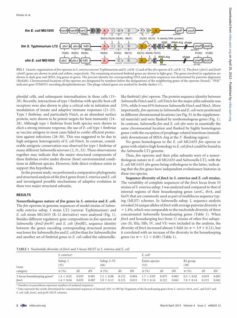
Nonorthologous nature of fim genes in S. enterica and E. coli.The fim operons in genome sequences of model strains of Salmo-nella enterica subsp. I strain LT2 (serovar Typhimurium) andE. coli strain MG1655 (K-12 derivative) were analyzed (Fig. 1).Besides different regulatory gene compositions in fim operons ofSalmonella (fimZ-fimW) and E. coli (fimBE), sequence identitybetween the genes encoding corresponding structural proteinswas lower for Salmonella fim and E. coli fim than for Salmonella fimand another set of fimbrial genes in E. coli called the salmonella-
like fimbrial (sfm) operon. The protein sequence identity betweenSalmonella FimA and E. coli FimA for the major pilin subunits was53%, while it was 65% between Salmonella FimA and SfmA. Moreimportantly, fim operons in Salmonella and E. coli were positionedin different chromosomal locations (see Fig. S1 in the supplemen-tal material) and were flanked by nonhomologous genes (Fig. 1).In contrast, Salmonella fim and E. coli sfm were in essentially thesame chromosomal location and flanked by highly homologousgenes (with the exception of prophage-related insertions immedi-ately downstream of tRNA-Arg at the 3=-flanking region).
No genes homologous to the E. coli MG1655 fim operon orgenes with relative high homology to E. coli fimA could be found inthe Salmonella LT2 genome.
Thus, fim operons and their pilin subunits were of a nonor-thologous nature in E. coli MG1655 and Salmonella LT2, with theE. coli MG1655 sfm genes being orthologous to the latter, indicat-ing that the fim genes have independent evolutionary histories inthese two species.
Sequence diversity of fimA in S. enterica and E. coli strains.The variability of complete sequences of the fimA locus from 55strains of S. enterica subsp. I was analyzed and compared to that ofinternal regions of their housekeeping genes (aroC, thrA, andhisD) that are commonly used as part of multilocus sequence typ-ing (MLST) schemes. In Salmonella subsp. I, sequence analysisrevealed 24 unique alleles of fimA with average pairwise diversity �� 1.4%, which was comparable to the nucleotide diversity of threeconcatenated Salmonella housekeeping genes (Table 1). WhenfimA and housekeeping loci from 11 strains of other five subspe-cies (II, IIIa, IIIb, IV, and VI) were included in the analysis, thediversity of fimA increased almost 3-fold (to � � 3.9 � 0.12), butit correlated with an increase of the diversity in the housekeepinggenes (to � � 3.2 � 0.08) (Table 1).
59(53) 55(34) 59(46) 58(47) 53(30) 42(15) % identity DNA (protein)
fim E. coli MG1655
fim S. Typhimurium LT2
……….. 4536808…………………………………………………………………………………………………………………..…4547976…
………..556101………..……………………………………………………………………….………….…563946…………………………………..…601182….
sfm E. coli MG1655
75(80) 85(93) 85(95) 65(65) 67(62) 69(72) 65(72) 60(58) 70(72) 81(92) 81(88)
ybcI ybcJ folD A I C D H F Z Y W tRNA-Arg phage
pheP ybdG
pheP ybdGybcI ybcJ folD A C D H F Z tRNA-Arg phage
…….602702………………………………………………………………………………………………………………..613558……….......….624199…
nanS nanM nanC B E A I C D F G H gntP uxuA
PDE
FIG 1 Genetic organization of fim operons in S. enterica serovar Typhimurium and E. coli K-12 and of the sfm operon of E. coli K-12. The fimA (sfmA) and fimH(sfmH) genes are shown in pink and yellow, respectively. The remaining structural fimbrial genes are shown in light gray. The genes involved in regulation areshown in dark gray and tRNA-Arg genes in green. The percent identity for corresponding DNA and protein sequences was determined by pairwise alignment(BioEdit). Chromosomal locations of the operons are designated by numbers below the designations of the neighboring genes of the operons (boxed). “PDE”indicates gene STM0551 encoding phosphodiesterase. The phage-related genes are marked by double slashes (//).
TABLE 1 Nucleotide diversity of fimA and 3-locus MLST in S. enterica and E. coli
Genecategory
S. entericaa E. colia
Subsp. I(55)
Subsp. I–VI(66)
Entire species(53)
B2 group(38)
� (%) dS dN � (%) dS dN � (%) dS dN � (%) dS dN
3-locus housekeeping genesb 1.4 � 0.02 0.055 0.001 3.2 � 0.08 0.132 0.004 1.7 � 0.05 0.075 0.001 0.5 � 0.02 0.019 0.001fimA 1.4 � 0.04 0.035 0.007 3.9 � 0.12 0.125 0.015 7.9 � 0.14 0.222 0.041 7.8 � 0.14 0.215 0.041a Numbers in parentheses represent numbers of analyzed sequences.b Data represent the results determined for concatenated sequences of internal (450- to 500-bp) fragments of the housekeeping genes from S. enterica (thrA, aroC, and hisD) andE. coli (adk, fumC, and gyrB) MLST schemes.
Kisiela et al.
2 ® mbio.asm.org March/April 2013 Volume 4 Issue 2 e00625-12
m
bio.asm.org
on April 25, 2013 - P
ublished by m
bio.asm.org
Dow
nloaded from
The nucleotide diversity of fimA in 53 strains of E. coli (com-prising representative strains of the entire species) was on averagefour times higher (� � 7.9%) than the diversity of housekeepinggenes from the E. coli MLST scheme (adk, gyrB, and fumC). Whilethe housekeeping gene diversity of E. coli (1.7%) was only slightlyhigher than that of S. enterica subsp. I (1.4%), the E. coli fimAdiversity was as much as 5-fold higher (Table 1). Importantly,while the housekeeping gene diversity of all S. enterica subspecieswas twice as high as in E. coli, the fimA diversity of the former washalf that of the latter. In addition, E. coli fimA was characterized bythe highest rates of synonymous (dS) and nonsynonymous (dN)values compared to E. coli and Salmonella genes (Table 1). Inter-estingly, when a subgroup of E. coli strains belonging to phyloge-netic B2 group was analyzed, the average nucleotide diversity oftheir housekeeping genes was much lower than that seen even inS. enterica subsp. I strains (� � 0.5%) but fimA diversity remainedas high as in the entire E. coli species (7.8%).
Thus, while fimA diversity in Salmonella is on par with thediversity of housekeeping genes, E. coli fimA is significantly morediverse and its diversity appears to be independent of the level ofhousekeeping gene diversity within individual phylogeneticgroups of E. coli.
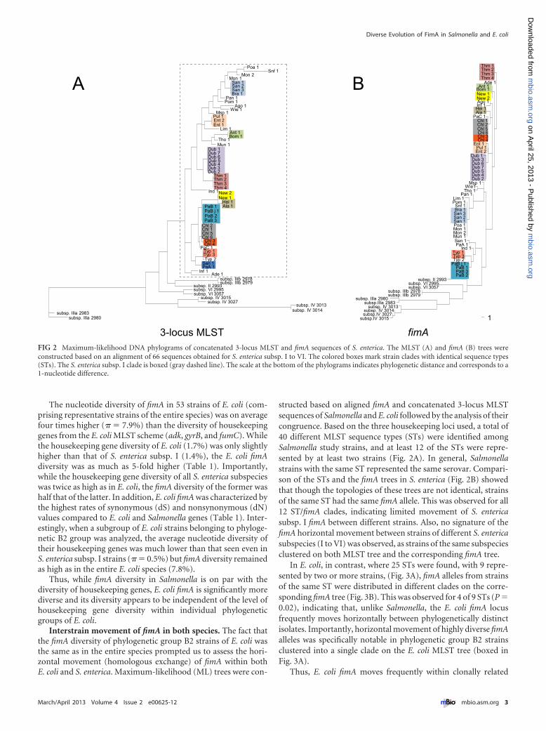
Interstrain movement of fimA in both species. The fact thatthe fimA diversity of phylogenetic group B2 strains of E. coli wasthe same as in the entire species prompted us to assess the hori-zontal movement (homologous exchange) of fimA within bothE. coli and S. enterica. Maximum-likelihood (ML) trees were con-
structed based on aligned fimA and concatenated 3-locus MLSTsequences of Salmonella and E. coli followed by the analysis of theircongruence. Based on the three housekeeping loci used, a total of40 different MLST sequence types (STs) were identified amongSalmonella study strains, and at least 12 of the STs were repre-sented by at least two strains (Fig. 2A). In general, Salmonellastrains with the same ST represented the same serovar. Compari-son of the STs and the fimA trees in S. enterica (Fig. 2B) showedthat though the topologies of these trees are not identical, strainsof the same ST had the same fimA allele. This was observed for all12 ST/fimA clades, indicating limited movement of S. entericasubsp. I fimA between different strains. Also, no signature of thefimA horizontal movement between strains of different S. entericasubspecies (I to VI) was observed, as strains of the same subspeciesclustered on both MLST tree and the corresponding fimA tree.
In E. coli, in contrast, where 25 STs were found, with 9 repre-sented by two or more strains, (Fig. 3A), fimA alleles from strainsof the same ST were distributed in different clades on the corre-sponding fimA tree (Fig. 3B). This was observed for 4 of 9 STs (P �0.02), indicating that, unlike Salmonella, the E. coli fimA locusfrequently moves horizontally between phylogenetically distinctisolates. Importantly, horizontal movement of highly diverse fimAalleles was specifically notable in phylogenetic group B2 strainsclustered into a single clade on the E. coli MLST tree (boxed inFig. 3A).
Thus, E. coli fimA moves frequently within clonally related
3-locus MLST fimA1
Thm 1Thm 2Thm 3Thm 4
Ade 1Ant 1
Bom 1New 1New 2Ago 1Inf 1Hei 1 Ala 1
PaC 1Chl 1
Chl 6Chl 5Chl 2
Chl 3Chl 4
Ent 1Pul 1Ent 2
Dub 1Dub 3Dub 6Dub 7Dub 5Dub 4Dub 2
Msp 1Wie1Tho 1
Pan 1Lim 1Pom 1Snf 1Bra 1San 3San 2San 1Poa 1Mon 1Mon 2Mun 1Sen 1PaA 1
Ind 1Typ 1Typ 3Typ 2
PaB j 1PaB 1PaB 3PaB 2
subsp. II 2993subsp. VI 2995subsp. VI 3057
subsp. IIIb 2978subsp. IIIb 2979
subsp. IIIa 2980subsp.IIIa 2983
subsp. IV 3013subsp. IV 3014subsp.IV 3027
subsp.IV 3015
Poa 1Snf 1
Mon 2Mon 1
San 1
Bra 1San 3San 2
Pan 1Pom 1
Ago 1Wie 1
Msp 1Pul 1Ent 2Ent 1
Lim 1Ant 1Bom 1
Tho 1Mun 1
Dub 1Dub 7Dub 6Dub 5Dub 4Dub 3Dub 2
Thm 1Thm 2Thm 3Thm 4
Ind 1New 2New 1
Hei 1Ala 1PaB 1
PaB j 1
PaB 3PaB 2
Chl 2
Chl 5Chl 6
Chl 1
Chl 3Chl 4
PaC 1Typ 1Typ 3Typ 2
Sen 1PaA 1
Inf 1Ade 1
subsp. IIIb 2978subsp. IIIb 2979
subsp. II 2993subsp. VI 2995subsp. VI 3057
subsp. IV 3015subsp. IV 3027
subsp. IV 3013subsp. IV 3014subsp. IIIa 2983
subsp. IIIa 2980
A B
FIG 2 Maximum-likelihood DNA phylograms of concatenated 3-locus MLST and fimA sequences of S. enterica. The MLST (A) and fimA (B) trees wereconstructed based on an alignment of 66 sequences obtained for S. enterica subsp. I to VI. The colored boxes mark strain clades with identical sequence types(STs). The S. enterica subsp. I clade is boxed (gray dashed line). The scale at the bottom of the phylograms indicates phylogenetic distance and corresponds to a1-nucleotide difference.
Diverse Evolution of FimA in Salmonella and E. coli
March/April 2013 Volume 4 Issue 2 e00625-12 ® mbio.asm.org 3
m
bio.asm.org
on April 25, 2013 - P
ublished by m
bio.asm.org
Dow
nloaded from
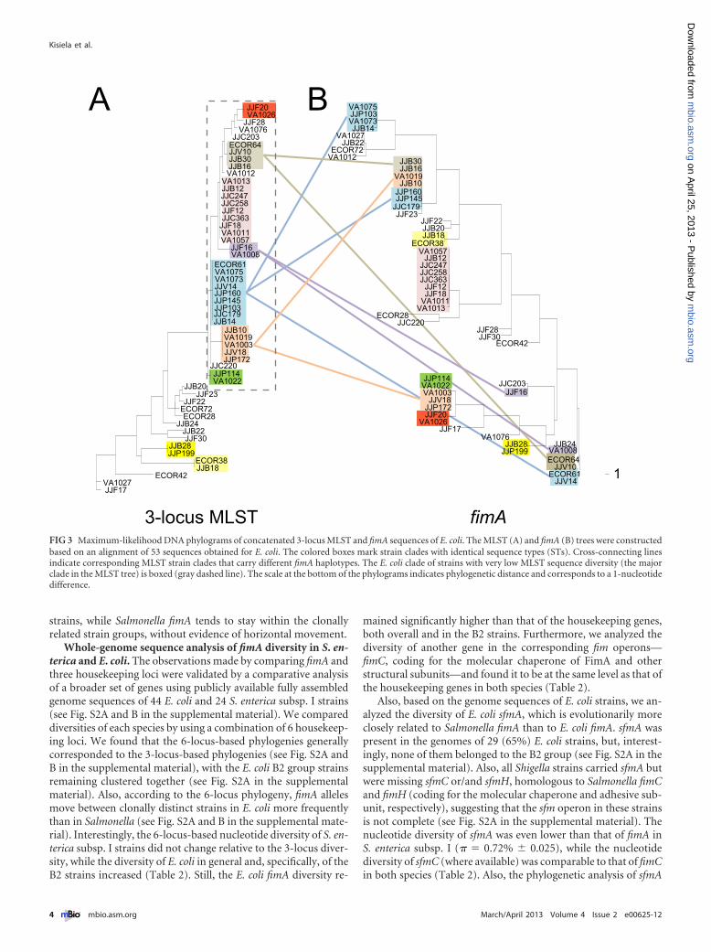
strains, while Salmonella fimA tends to stay within the clonallyrelated strain groups, without evidence of horizontal movement.
Whole-genome sequence analysis of fimA diversity in S. en-terica and E. coli. The observations made by comparing fimA andthree housekeeping loci were validated by a comparative analysisof a broader set of genes using publicly available fully assembledgenome sequences of 44 E. coli and 24 S. enterica subsp. I strains(see Fig. S2A and B in the supplemental material). We compareddiversities of each species by using a combination of 6 housekeep-ing loci. We found that the 6-locus-based phylogenies generallycorresponded to the 3-locus-based phylogenies (see Fig. S2A andB in the supplemental material), with the E. coli B2 group strainsremaining clustered together (see Fig. S2A in the supplementalmaterial). Also, according to the 6-locus phylogeny, fimA allelesmove between clonally distinct strains in E. coli more frequentlythan in Salmonella (see Fig. S2A and B in the supplemental mate-rial). Interestingly, the 6-locus-based nucleotide diversity of S. en-terica subsp. I strains did not change relative to the 3-locus diver-sity, while the diversity of E. coli in general and, specifically, of theB2 strains increased (Table 2). Still, the E. coli fimA diversity re-
mained significantly higher than that of the housekeeping genes,both overall and in the B2 strains. Furthermore, we analyzed thediversity of another gene in the corresponding fim operons—fimC, coding for the molecular chaperone of FimA and otherstructural subunits—and found it to be at the same level as that ofthe housekeeping genes in both species (Table 2).
Also, based on the genome sequences of E. coli strains, we an-alyzed the diversity of E. coli sfmA, which is evolutionarily moreclosely related to Salmonella fimA than to E. coli fimA. sfmA waspresent in the genomes of 29 (65%) E. coli strains, but, interest-ingly, none of them belonged to the B2 group (see Fig. S2A in thesupplemental material). Also, all Shigella strains carried sfmA butwere missing sfmC or/and sfmH, homologous to Salmonella fimCand fimH (coding for the molecular chaperone and adhesive sub-unit, respectively), suggesting that the sfm operon in these strainsis not complete (see Fig. S2A in the supplemental material). Thenucleotide diversity of sfmA was even lower than that of fimA inS. enterica subsp. I (� � 0.72% � 0.025), while the nucleotidediversity of sfmC (where available) was comparable to that of fimCin both species (Table 2). Also, the phylogenetic analysis of sfmA
JJF20VA1026
JJF28VA1076
JJC203ECOR64JJV10JJB30JJB16VA1012
VA1013JJB12JJC247JJC258JJF12JJC363
JJF18VA1011VA1057
JJF16VA1008
ECOR61VA1075VA1073JJV14JJP160JJP145JJP103JJC179JJB14
JJB10VA1019VA1003JJV18JJP172
JJC220JJP114VA1022JJB20
JJF23JJF22
ECOR72ECOR28
JJB24JJB22JJF30
JJB28JJP199
ECOR38JJB18
ECOR42VA1027JJF17
VA1075JJP103VA1073JJB14
VA1027JJB22
ECOR72VA1012
JJB10JJP160
VA1019
JJB30
JJP145
JJB16
JJC179JJF23
JJF22JJB20JJB18
ECOR38VA1057
JJB12JJC247JJC258JJC363
JJF12JJF18
VA1011VA1013
ECOR28JJC220
JJF28JJF30
ECOR42
JJP114VA1022VA1003
JJV18JJP172JJF20
VA1026JJF17
VA1076JJB28
JJP199
JJC203JJF16
JJB24VA1008ECOR64
JJV10ECOR61
JJV141
3-locus MLST fimA
A B
FIG 3 Maximum-likelihood DNA phylograms of concatenated 3-locus MLST and fimA sequences of E. coli. The MLST (A) and fimA (B) trees were constructedbased on an alignment of 53 sequences obtained for E. coli. The colored boxes mark strain clades with identical sequence types (STs). Cross-connecting linesindicate corresponding MLST strain clades that carry different fimA haplotypes. The E. coli clade of strains with very low MLST sequence diversity (the majorclade in the MLST tree) is boxed (gray dashed line). The scale at the bottom of the phylograms indicates phylogenetic distance and corresponds to a 1-nucleotidedifference.
Kisiela et al.
4 ® mbio.asm.org March/April 2013 Volume 4 Issue 2 e00625-12
m
bio.asm.org
on April 25, 2013 - P
ublished by m
bio.asm.org
Dow
nloaded from
showed limited horizontal movement of sfmA between differentE. coli strains (data not shown).
Considering the patchy distribution of sfm operon in E. coli, wealso looked throughout the publicly available S. enterica genomesfor possible homologues of the E. coli-like fim operon. Based onsequence identity, operon structure, and chromosomal position,no complete or partial presence of an E. coli-like fim operon wasdetected in any of the 24 S. enterica subsp. I genomes or in the onlyavailable genome for other subspecies—that of S. enterica subsp.IIIa strain RKS 2980 (not shown).
Thus, based on the expanded set of genes from strains withwhole-genome sequence available, we confirmed the much higher
diversity and more frequent horizontal movement of E. coli fimArelative to Salmonella fimA and E. coli sfmA.
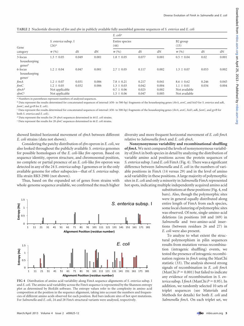
Nonsynonymous variability and recombinational shufflingof fimA. We next compared the levels of nonsynonymous variabil-ity of fimA in both species in detail by analyzing the distribution ofvariable amino acid positions across the protein sequences ofS. enterica subsp. I and E. coli FimA (Fig. 4). There was a significantdifference between Salmonella and E. coli in the numbers of vari-able positions in FimA (14 versus 29) and in the level of aminoacid variability in these positions. A large majority of polymorphicsites in E. coli and only a minority in Salmonella FimA representedhot spots, indicating multiple independently acquired amino acid
substitutions at these positions (Fig. 4, redbars). Also, though the polymorphic siteswere in general equally distributed alongentire length of FimA from each species,some local clustering of polymorphic siteswas observed. Of note, single-amino-aciddeletions (in positions 168 and 169) inSalmonella and two-amino-acid inser-tions (between residues 26 and 27) inE. coli were also present.
To analyze to what extent the struc-tural polymorphism in pilin sequencesresults from mutation versus recombina-tion (intragenic shuffling) events, wetested the presence of intragenic recombi-nation regions in fimA using the MaxChistatistic (33). The analysis showed strongsignals of recombination in E. coli fimA(MaxChi P � 0.001) but failed to indicateany evidence of recombination in S. en-terica subsp. I fimA (MaxChi P � 0.95). Inaddition, we randomly selected 10 sets oftriplet sequences (see Materials andMethods for details) for both E. coli andSalmonella fimA. On each triplet set, we
TABLE 2 Nucleotide diversity of fim and sfm in publicly available fully assembled genome sequences of S. enterica and E. coli
Genecategory
S. enterica subsp. I(24)a
E. colia
Entire species(44)
B2 group(15)
� (%) dS dN � (%) dS dN � (%) dS dN
3-locushousekeepinggenesb
1.3 � 0.05 0.049 0.001 1.8 � 0.05 0.077 0.001 0.5 � 0.04 0.02 0.001
6-locushousekeepinggenesc
1.2 � 0.04 0.047 0.001 2.7 � 0.05 0.117 0.002 1.3 � 0.07 0.053 0.002
fimA 1.2 � 0.07 0.031 0.006 7.8 � 0.21 0.217 0.041 8.6 � 0.62 0.246 0.045fimC 1.2 � 0.05 0.032 0.006 1.3 � 0.03 0.042 0.004 1.1 � 0.01 0.034 0.004sfmAd Not applicable 0.7 � 0.06 0.025 0.002 Not availablesfmCe Not applicable 1.5 � 0.06 0.047 0.005 Not availablea Numbers in parentheses represent numbers of analyzed sequences.b Data represent the results determined for concatenated sequences of internal (450- to 500-bp) fragments of the housekeeping genes (thrA, aroC, and hisD for S. enterica and adk,fumC, and gyrB for E. coli).c Data represent the results determined for concatenated sequences of internal (450- to 500-bp) fragments of the housekeeping genes (thrA, aroC, hisD, adk, fumC, and gyrB forboth S. enterica and E. coli).d Data represent the results for 29 sfmA sequences determined in 44 E. coli strains.e Data represent the results for 28 sfmC sequences determined in 44 E. coli strains.
0
0.5
1
1.5
1 11 21 31 41 51 61 71 81 91 101 111 121 131 141 151 161 171 181
Entr
opy
(Hx)
Alignment Posi�on (residue number)
0
0.5
1
1.5
1 11 21 31 41 51 61 71 81 91 101 111 121 131 141 151 161 171 181
Entr
opy
(Hx)
Alignment Posi�on (residue number)
E. coli
S. enterica subsp. I
FIG 4 Distribution of amino acid variability along FimA sequence alignments of S. enterica subsp. Iand E. coli. The amino acid variability across the FimA sequence is represented by the Shannon entropyplot as determined by BioEdit software. The entropy values refer to the complexity in amino acidcomposition at the position in the sequence alignment, taking into account the numbers and frequen-cies of different amino acids observed for each position. Red bars indicate sites of hot spot mutations.For Salmonella and E. coli, 24 and 28 FimA structural variants were analyzed, respectively.
Diverse Evolution of FimA in Salmonella and E. coli
March/April 2013 Volume 4 Issue 2 e00625-12 ® mbio.asm.org 5
m
bio.asm.org
on April 25, 2013 - P
ublished by m
bio.asm.org
Dow
nloaded from
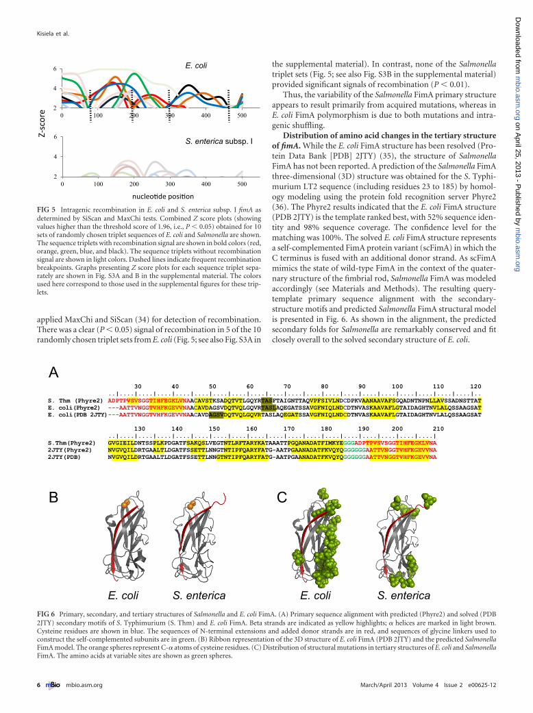
applied MaxChi and SiScan (34) for detection of recombination.There was a clear (P � 0.05) signal of recombination in 5 of the 10randomly chosen triplet sets from E. coli (Fig. 5; see also Fig. S3A in
the supplemental material). In contrast, none of the Salmonellatriplet sets (Fig. 5; see also Fig. S3B in the supplemental material)provided significant signals of recombination (P � 0.01).
Thus, the variability of the Salmonella FimA primary structureappears to result primarily from acquired mutations, whereas inE. coli FimA polymorphism is due to both mutations and intra-genic shuffling.
Distribution of amino acid changes in the tertiary structureof fimA. While the E. coli FimA structure has been resolved (Pro-tein Data Bank [PDB] 2JTY) (35), the structure of SalmonellaFimA has not been reported. A prediction of the Salmonella FimAthree-dimensional (3D) structure was obtained for the S. Typhi-murium LT2 sequence (including residues 23 to 185) by homol-ogy modeling using the protein fold recognition server Phyre2(36). The Phyre2 results indicated that the E. coli FimA structure(PDB 2JTY) is the template ranked best, with 52% sequence iden-tity and 98% sequence coverage. The confidence level for thematching was 100%. The solved E. coli FimA structure representsa self-complemented FimA protein variant (scFimA) in which theC terminus is fused with an additional donor strand. As scFimAmimics the state of wild-type FimA in the context of the quater-nary structure of the fimbrial rod, Salmonella FimA was modeledaccordingly (see Materials and Methods). The resulting query-template primary sequence alignment with the secondary-structure motifs and predicted Salmonella FimA structural modelis presented in Fig. 6. As shown in the alignment, the predictedsecondary folds for Salmonella are remarkably conserved and fitclosely overall to the solved secondary structure of E. coli.
2
4
6
0 100 200 300 400 500
2
4
6
0 100 200 300 400 500
nucleo�de posi�on
Z-sc
ore
E. coli
S. enterica subsp. I
FIG 5 Intragenic recombination in E. coli and S. enterica subsp. I fimA asdetermined by SiScan and MaxChi tests. Combined Z score plots (showingvalues higher than the threshold score of 1.96, i.e., P � 0.05) obtained for 10sets of randomly chosen triplet sequences of E. coli and Salmonella are shown.The sequence triplets with recombination signal are shown in bold colors (red,orange, green, blue, and black). The sequence triplets without recombinationsignal are shown in light colors. Dashed lines indicate frequent recombinationbreakpoints. Graphs presenting Z score plots for each sequence triplet sepa-rately are shown in Fig. S3A and B in the supplemental material. The colorsused here correspond to those used in the supplemental figures for these trip-lets.
E. coli S. enterica E. coli S. enterica
A
B C
30 40 50 60 70 80 90 100 110 120 ..|....|....|....|....|....|....|....|....|....|....|....|....|....|....|....|....|....|....|....|..
S. Thm (Phyre2) ADPTPVSVSGGTIHFEGKLVNAACAVSTKSADQTVTLGQYRTASFTAIGNTTAQVPFSIVLNDCDPKVAANAAVAFSGQADNTNPNLLAVSSADNSTTAT E. coli(Phyre2) ---AATTVNGGTVHFKGEVVNAACAVDAGSVDQTVQLGQVRTASLAQEGATSSAVGFNIQLNDCDTNVASKAAVAFLGTAIDAGHTNVLALQSSAAGSAT E. coli(PDB 2JTY) ---AATTVNGGTVHFKGEVVNAACAVDAGSVDQTVQLGQVRTASLAQEGATSSAVGFNIQLNDCDTNVASKAAVAFLGTAIDAGHTNVLALQSSAAGSAT
130 140 150 160 170 180 190 200 210 ..|....|....|....|....|....|....|....|....|....|....|....|....|....|....|....|....|....|
S.Thm(Phyre2) GVGIEILDNTSSPLKPDGATFSAKQSLVEGTNTLRFTARYKATAAATTPGQANADATFIMKYEGGGADPTPVSVSGGTIHFEGKLVNA 2JTY(Phyre2) NVGVQILDRTGAALTLDGATFSSETTLNNGTNTIPFQARYFATG-AATPGAANADATFKVQYQGGGGGGAATTVNGGTVHFKGEVVNA 2JTY(PDB) NVGVQILDRTGAALTLDGATFSSETTLNNGTNTIPFQARYFATG-AATPGAANADATFKVQYQGGGGGGAATTVNGGTVHFKGEVVNA
FIG 6 Primary, secondary, and tertiary structures of Salmonella and E. coli FimA. (A) Primary sequence alignment with predicted (Phyre2) and solved (PDB2JTY) secondary motifs of S. Typhimurium (S. Thm) and E. coli FimA. Beta strands are indicated as yellow highlights; � helices are marked in light brown.Cysteine residues are shown in blue. The sequences of N-terminal extensions and added donor strands are in red, and sequences of glycine linkers used toconstruct the self-complemented subunits are in green. (B) Ribbon representation of the 3D structure of E. coli FimA (PDB 2JTY) and the predicted SalmonellaFimA model. The orange spheres represent C-� atoms of cysteine residues. (C) Distribution of structural mutations in tertiary structures of E. coli and SalmonellaFimA. The amino acids at variable sites are shown as green spheres.
Kisiela et al.
6 ® mbio.asm.org March/April 2013 Volume 4 Issue 2 e00625-12
m
bio.asm.org
on April 25, 2013 - P
ublished by m
bio.asm.org
Dow
nloaded from

As viewed by PyMol (Fig. 6B), the predicted Salmonella FimAmodel retains a complete Ig fold, where the added donor strand(red) occupies the hydrophobic groove as observed in E. coli FimA2JTY. Moreover, similar to those in E. coli, cysteine residues in theSalmonella model are juxtaposed in a position suitable for theformation of a stabilizing disulfide bond (Fig. 6B, orange spheres).
Using the E. coli FimA structure and Salmonella FimA model,we next compared spatial distributions of variable positions in themajor subunits from these two species (Fig. 6C). The analysis re-vealed that, in E. coli, the amino acid substitutions predominantlytarget one side of FimA molecule opposite the position of thecomplementing donor strand (red). According to the previouslyreported fimbrial rod model, the donor strand faces the insidecore of the fimbrial rod (37). Thus, the highly polymorphic loopsare predicted to be exposed on the outer surface of the fimbrial rod(Fig. 6C). In contrast, in Salmonella, the variable positions tend tocluster on the top (residues 23, 24, 43, 93, 148, 149, and 150) andthe bottom (residues 167, 168, 169, and 170) of theimmunoglobulin-like fold, with only sporadic occurrence (resi-dues 71, 72, and 107) in external loops.
The helical structure of the type 1 fimbriae with its extensivesubunit-subunit interactions raises the possibility that mutationsin FimA residues might occur in pairs and affect residues thatinteract with one another in the intersubunit interface, represent-ing compensatory intragenic suppression. We determined thenumber of events where mutations were acquired in pairs byE. coli and Salmonella fimA per total number of mutation eventsusing Zonal phylogeny (ZP) software (38) that provides branch-specific information on nonsynonymous substitutions in the phy-logenetic tree. For Salmonella fimA we detected that 2 of 34 mu-tations and, similarly, for E. coli we detected that 2 of 44 mutationsrepresented events where mutations were likely acquired in pairs,indicating that intergenic suppression does not play a significantrole in the structural diversification of FimA in either species.
Thus, there is a distinct structural pattern of the amino acidchanges in the E. coli FimA and the Salmonella FimA, with thevariable residues in E. coli FimA concentrated in the outer surface-exposed epitopes of the subunit, i.e., putative antigenic epitopes.
DISCUSSION
FimA, as the major structural component of type 1 fimbriae, isabundantly expressed on the bacterial surface. This protein is thusexpected to be a major antigen and evolve under strong selectivepressure from the host immune system. In E. coli FimA, the foot-print of such selection can be seen in the great allelic variation ofthe fimA locus evolving under strong diversification selection (8,39). In this report, however, we show that fimA from a closelyrelated pathogen, S. enterica, evolves under a different adaptiveselection pressure with much lower diversity, indicating that fim-briated Salmonella are not subjected to strong immune pressure tovary the fimbrial structure.
In both species, type 1 fimbriae were shown to bind to manno-sylated receptors on target cells, facilitating bacterial adhesion andinvasion, and triggering and modulating the host immune re-sponse (22–24, 40). The mannose-specific interactions involvevery similar molecular mechanisms. Despite very low (15%) se-quence identity, the fimbrial tip adhesive protein of E. coli andSalmonella, FimH, has highly homologous, two-domain tertiarystructures and mediates shear-dependent binding to mannose viaan allosteric catch-bond mechanism (41, 42). Both adhesins
evolve under positive selection for accumulation of structural mu-tations that greatly affect their adhesive properties, with markedlysimilar distribution patterns of functional mutations in the corre-sponding tertiary structures (43–46). In particular, mutations thatenhance mannose binding under static conditions (and are com-mon among uropathogenic E. coli and systemically invasive Sal-monella serovars) are primarily localized to the interdomain in-terface of FimH, exerting an allosteric effect on binding pocketaffinity (46–49).
In contrast to the high structural and functional similarities ofthe adhesive subunits, we found that FimA, the major structuralsubunit from Salmonella and E. coli, displays distinct adaptivepatterns. The E. coli fimA locus is characterized by a high level ofallelic variation, with strong signals for intragenic recombinationand frequent horizontal movement. In contrast, the SalmonellafimA locus exhibits relatively low diversity (on par with that ofneutrally evolving housekeeping loci) without any strong evi-dence of intragenic shuffling or interstrain movement. The differ-ence is especially obvious in comparisons of the S. enterica subsp.I and E. coli group B2 strains that were overrepresented in ouranalysis because of their medical significance. Both represent phy-logenetic clades within the corresponding species, combining rel-atively closely related strains with distinct virulence characteris-tics. S. enterica subsp. I is comprised of serovars that causegastroenteritis and/or systemic infections in humans, while E. coligroup B2 strains are notorious for their ability to cause urinarytract infections, sepsis, meningitis, and other extraintestinal infec-tions. From the perspective of general genomic characteristics,there is a similarity between these subspecies groups, as shown inour previous genome-wide comparative study of Salmonella andE. coli (38). It was shown that S. enterica subsp. I and the E. coli B2group strains are very similar in regard to the level of nucleotidediversity of core genes (1% in both species) and the prevalence ofmosaic versus core genes (33% versus 63% in S. enterica subsp. Iand 25% versus 64% of the total genes in E. coli group B2 strains).
In the data set analyzed here, based on MLST housekeepinggene analyses, B2 E. coli strains had levels of genetic diversity eitherlower than or similar to those of S. enterica subsp. I. However, theaverage diversity of fimA alleles in E. coli was 5- to 6-fold higherthan that of fimA in the Salmonella isolates, with a distinctivelyhigher rate of horizontal allelic exchange in the former. This indi-cates that the lower diversity of S. enterica subsp. I fimA does notsimply result from the overall lower genetic diversity of S. entericasubsp. I strains.
In E. coli, the functional relevance of the diversifying selectionin fimA locus was verified by structural studies. Using a predictedmodel of type 1 fimbriae, it has been demonstrated that the vari-able amino acid residues of FimA are predominantly located onthe external surface of the fimbrial rod (37). In our study, to com-pare the spatial distributions of polymorphic sites of SalmonellaFimA and E. coli FimA, we used 3D structures of the pilin mono-mers that included the recently solved structure of E. coli FimA(PDB 2JTY) (35) and we obtained here by homology modeling aputative structure of Salmonella FimA. This analysis revealed thatthe majority of the amino acid substitutions in E. coli targetedloops on one side of FimA molecule which, according to the pre-viously reported model (37), are exposed on the external surfaceof the fimbrial rod, thereby strongly supporting the view of anti-genic variation in E. coli FimA. In Salmonella, in contrast, thevariable positions were detected on the opposite poles of the
Diverse Evolution of FimA in Salmonella and E. coli
March/April 2013 Volume 4 Issue 2 e00625-12 ® mbio.asm.org 7
m
bio.asm.org
on April 25, 2013 - P
ublished by m
bio.asm.org
Dow
nloaded from
immunoglobulin-like fold, i.e., on the top and the bottom ofFimA, which are more likely to be positioned in the intersubunitinterface than surface exposed. In two proposed models of thetype 1 fimbrial structure (37, 50), the fimbrial shaft is formed by ahelically coiled string of FimA subunits that are connected to eachother “head to tail.” Although at this point we still cannot excludethe possibility of involvement of these mutations in epitope diver-sification, it is clear that there is a significant difference in thestructural variabilities of corresponding FimA segments, suggest-ing that Salmonella FimA evolves under weaker selective pressurefor antigenic diversification than E. coli FimA.
Salmonella and E. coli are known to inhabit many diverseniches (hosts, tissues, cellular environments). It is thus possiblethat immune pressure could act differently on type 1 fimbriae indifferent niches. However, it is equally likely that the differences inthe antigenic diversifications of these two pilins may result fromdistinct mechanisms of regulation of type 1 fimbrial expression inthese bacteria. Both Salmonella and E. coli have been shown toswitch between sparsely and highly fimbriated states in responseto various environmental signals, in part to escape host immunity.In E. coli, on/off switching of fimbrial expression is determined bythe orientation of the promoter-containing DNA region (fimS)(10, 12), where the inversion of the fimS DNA segment is catalyzedby two site-specific recombinases (encoded by fimB and fimE)(51). In Salmonella, in contrast, the promoter is not invertible, butits transcriptional activity is controlled by regulatory proteins en-coded by fimZ, fimY, and fimW located downstream of the operon(13, 52–54). It could be speculated that the differences in the reg-ulatory mechanisms of type 1 fimbrial expression in these twobacteria may determine the different efficiencies of phase switch-ing and thus provide different levels of protection against hostimmune defenses. In this context, the distribution pattern ofstructural mutations observed for Salmonella FimA may suggestthat mutations found at the top and the bottom of the pilin sub-unit (and thus in the subunit contact interface) could affect FimA-FimA interactions during fimbrial polymer formation and conse-quently be functionally adaptive for efficient “phase” switching.
On the other hand, it has been demonstrated that in E. coli, theFimA polymer has the ability to coil and uncoil under the influ-ence of mechanical forces (55). Since bacterial adhesion usuallyoccurs in the presence of flowing bodily fluids that create dragforces on bacteria and their adhesins, the mechanical properties offimbrial shaft exert a significant effect on bacterial adhesion. Inthis aspect, the mutations located in the intersubunit interface ofSalmonella FimA (as well as in that of E. coli FimA) could modu-late mechanical properties of the fimbrial shaft under the influ-ence of shear forces and consequently affect mannose-specific ad-hesion of the bacteria under flow conditions. Although thishypothesis requires experimental verification, a similar observa-tion for enterotoxigenic E. coli (ETEC) class 5 fimbria-mediatedadhesion was previously reported. It was shown that point muta-tions that are localized to the intersubunit interface of the major(nonadhesive) subunit of those fimbriae significantly reduced ad-hesion under flow conditions (56).
Distinct adaptive patterns of fimA in Salmonella and E. coliraise questions about evolutionary trajectories of these distincttypes of type 1 fimbriae. The lack of E. coli-like fim genes in S. en-terica indicates that this operon was either lost from or not ac-quired by the latter. At this point, based on the patchy distributionof either Salmonella-like or E. coli-like fim genes in other entero-
bacterial species (see Table S1 in the supplemental material), it isdifficult to be definitive about that and further analysis is requiredto address the interesting issue about the evolutionary interplay ofthe different types of mannose-specific fimbriae in members of thefamily Enterobacteriaceae. Though E. coli has both fimbrial types,it appears that the Salmonella-like fimbrial operon (sfm) in E. colieither is nonfunctional or has acquired functions distinct fromthose of E. coli and Salmonella fim operons. Our analysis of sfmgenes indicates that they are present in 65% of the E. coli genomes,with none found in strains representing the B2 group and partialinactivation in some of the other E. coli groups. Even when most ofthe operon is present (as in E. coli strain MG1655), the sfm genesappear to diverge more significantly from the corresponding Sal-monella fim genes than the orthologous genes on the flanks. Also,we found that all sfmA genes in E. coli carry a 5= deletion relative tothe Salmonella fimA corresponding to a deletion of 5 amino acidresidues at position 2 to position 6 of the amino acid sequence thatis part of the donor strand crucial for FimA-FimA polymerizationby beta-strand complementation. Such a structural defect couldsignificantly affect fimbrial assembly or even abrogate it. No datawith regard to natural expression of the Sfm fimbriae have beenreported to date. Though it was possible to express the sfm fim-briae of E. coli K-12 from an artificial promoter, the only smallafimbrial structures that were observed were structures whosefunctionality was not established (14). Thus, taken together, thesedata indicate that fim-encoded type 1 fimbriae are the onlymannose-specific organelles in the E. coli species. It remains to bedetermined, however, whether the fim operon was acquired laterthan sfm and functionally replaced Salmonella-like fimbrial genesor, alternatively, whether the two traits shared a long evolutionaryhistory in E. coli and sfm genes functionally diverged over time.
In summary, by using microevolutionary analytical tools, wedemonstrate here that surface organelles that presumably performessentially identical adhesive functions in closely related bacterialpathogens can be under highly dissimilar types of selective pres-sure. While the exact basis of this difference remains to be eluci-dated, this indicates possibly distinct ecological and/or pathogenicenvironments in which these organelles are functioning. Despitethe genes appearing to be under relatively weak immune pressureto diversify, our results also suggest the possibility that type 1fimbria-based vaccines may be more successful for treatment ofinfections by Salmonella, considering the structurally conservednature of the major subunit. Finally and foremost, this reportshows the potential power of comparative population geneticanalysis in determining adaptive and, thus, functional peculiari-ties of specific bacterial traits in different species. This should as-sist us in unraveling the physiological significance and possiblypathogenic roles of the traits, especially in the age of continuousaccumulation of genomic data of a large number of individualstrains from the same species.
MATERIALS AND METHODSBacterial strains. S. enterica and E. coli strains used in this study are listedin Tables S2 and S3 in the supplemental material, respectively. The S. en-terica collection included 53 isolates of subspecies I representing 30 dif-ferent serovars, including 23 strains of systemic and 30 strains of differentintestinal serovars, and 11 strains of other subspecies (subspecies II to VI).The E. coli collection consisted of 53 isolates representing convenient sam-ples of diverse pathotypes and nonpathogenic isolates. Bacteria were rou-tinely grown overnight in LB medium at 37°C without shaking.
Kisiela et al.
8 ® mbio.asm.org March/April 2013 Volume 4 Issue 2 e00625-12
m
bio.asm.org
on April 25, 2013 - P
ublished by m
bio.asm.org
Dow
nloaded from

Gene amplification and sequencing. Genomic DNA was isolatedfrom S. enterica and E. coli strains using a DNeasy Blood & Tissue kit(Qiagen). The fimA was PCR amplified from the genomic DNA using thefollowing pairs of primers: primer pair fimASe F (5=GGATGCCGAAAC-CGGGTG3=) and fimASe R (5=CTGTGGCGACAGCGCAGCC3=) (for S.enterica fimA) and primer pair fimAEc F (5=ACGTTTCTGTGGCTC-GACGCATCT3=) and fimAEc R (5=ACGTCCCTGAACCTGGGTAG-GTTA3=) (for E. coli fimA). S. enterica and E. coli housekeeping locusfragments (from aroC, hisD, and thrA and from fumC, adk, and gyrB,respectively) were amplified in accordance with the protocols available atthe MLST database (http://mlst.ucc.ie/mlst/dbs/Senterica/documents/primersEnterica_html and http://mlst.ucc.ie/mlst/dbs/Ecoli/documents/primersColi_html). The PCR products were purified using ExoSAP-ITreagent (Affymetrix) in accordance with the manufacturer’s instructionsand subjected to sequencing by the GENEWIZ sequencing service(Genewiz Seattle, Seattle, WA).
Phylogenetic analysis. The nucleotide sequences were aligned usingClustalW with default settings (57). Zonal phylogeny (ZP) analysis andassociated statistics were performed using Zonal Phylogeny Software(ZPS) (38). The maximum-likelihood (ML) phylograms as implementedin ZPS were generated by PAUP* 4.0 b using the general time-reversible(GTP) substitution model with codon-position-specific estimated basefrequencies (58). Sequence diversity was measured as the average pairwisediversity index (�) and the rates of nonsynonymous (dN) and synony-mous (dS) mutations (59) using MEGA version 4 (60). Analysis of statis-tical significance was performed using the z test for � and dN/dS values(61). The presence of structural hot spot mutations was determined usingZPS.
Intragenic recombination detection. MaxChi (33) was used to pro-vide a summary statistic for the detection of recombination, where a genedata set showing MaxChi statistic P � 0.05 was considered to have recom-binant sequence(s). For three-sequence-based (triplet) analysis of recom-bination, we applied MaxChi along with SiScan (34), which depicts prob-able recombinant and parent sequences. In SiScan, Z scores were plottedbased on identities of all three sequence pairs of a triplet set across the genelength using a sliding window of 100 nucleotides, a step size of 50 nucle-otides, and 100 randomizations for Monte Carlo sampling. The data setthat showed identity of two of the three sequence pairs with Z scores �1.96 (i.e., significance at P � 0.05) at considerably nonoverlappingstretches across the gene length indicated putative intragenic recombina-tion with the presence of both parents in the data set.
An in-house perl script was used to develop a random sequence num-ber generator. In the aligned fimA allelic variant sets of E. coli and Salmo-nella, we assigned numbers to each gene sequence. Since Salmonella hadlower number of unique sequences (i.e., 24 alleles), the program gener-ated sets of 3 numbers (i.e., triplets) ranging from 1 to 24 sets. We incor-porated the constraint that the numbers within any triplet set would differfrom one another by a value of at least 5 in order to avoid consideringsequences in a triplet that were phylogenetically too close. We generated10 random triplet sets of numbers and used identical sets for the twospecies to choose strain sequences corresponding to each number in thealigned datasets.
FimA structural analysis and modeling. The distribution of aminoacid variability in the sequence alignment was computed by the use ofBioEdit software (http://www.mbio.ncsu.edu/bioedit/bioedit.html) andis represented by Shannon entropy plots (62), where the entropy datarefer to the complexity in the amino acid composition at each position(taking into account the numbers and frequencies of different aminoacids at the position) in the sequence alignment.
Modeling of the S. enterica FimA 3D structure was performed usingPhyre2 fold recognition and template modeling software (36). Briefly, theamino acid sequence of S. Typhimurium SL1344 FimA (including resi-dues 23 to 185) was submitted to the Phyre2 server (http://www.sbg.bio.ic.ac.uk/phyre2/html/page.cgi?id�index). The structure of E. coli FimA(PDB code 2JTY) (35) was selected as the best-ranked template for mod-
eling of S. Typhimurium FimA, with 52% sequence identity, 100% con-fidence, and 98% sequence coverage. As E. coli FimA (template) repre-sents a self-complemented variant of the protein, the C terminus of the S.Typhimurium SL1344 FimA sequence was completed with the corre-sponding self-donor strand sequence (ADPTPVSVSGGTIHFEGKLVNA)via the use of the (Gly)3 linker and resubmitted to the Phyre2 server. Theresulting structures of the S. enterica FimA model and E. coli FimA (PDBcode 2JTY) were viewed and analyzed using the molecular visualizationsystem PyMOL.
Nucleotide sequence accession numbers. The DNA sequences of thenew S. enterica and E. coli fimA alleles have been deposited in GenBankunder accession numbers KC405503 through KC405538. Accession num-bers of all S. enterica and E. coli fimA alleles of the study are presented inTables S2 and S3 in the supplemental material.
SUPPLEMENTAL MATERIALSupplemental material for this article may be found at http://mbio.asm.org/lookup/suppl/doi:10.1128/mBio.00625-12/-/DCSupplemental.
Figure S1, PDF file, 0.1 MB.Figure S2, PDF file, 0.1 MB.Figure S3, PDF file, 0.1 MB.Table S1, DOCX file, 0.1 MB.Table S2, DOCX file, 0.1 MB.Table S3, DOCX file, 0.1 MB.
ACKNOWLEDGMENTS
This work was supported by National Institutes of Health grant RO1GM084318.
We gratefully acknowledge Steve Moseley (University of Washington)for critical reading of the manuscript and helpful advice. We also thank tofollowing individuals for providing Salmonella and E. coli strains: FerricFang, Stephen Libby, Mansour Samadpour, Maciej Ugorski, Roderick I.Mackie, Howard Ochman, and James R. Johnson.
REFERENCES1. Duguid JP, Anderson ES, Campbell I. 1966. Fimbriae and adhesive
properties in salmonellae. J. Pathol. Bacteriol. 92:107–138.2. Duguid JP, Campbell I. 1969. Antigens of the type-1 fimbriae of salmo-
nellae and other enterobacteria. J. Med. Microbiol. 2:535–553.3. Klemm P, Jørgensen BJ, van Die I, de Ree H, Bergmans H. 1985. The
fim genes responsible for synthesis of type 1 fimbriae in Escherichia coli,cloning and genetic organization. Mol. Gen. Genet. 199:410 – 414.
4. Gerlach GF, Clegg S, Ness NJ, Swenson DL, Allen BL, Nichols WA.1989. Expression of type 1 fimbriae and mannose-sensitive hemagglutininby recombinant plasmids. Infect. Immun. 57:764 –770.
5. Waksman G, Hultgren SJ. 2009. Structural biology of the chaperone-usher pathway of pilus biogenesis. Nat. Rev. Microbiol. 7:765–774.
6. Thankavel K, Shah AH, Cohen MS, Ikeda T, Lorenz RG, Curtiss R, III,Abraham SN. 1999. Molecular basis for the enterocyte tropism exhibitedby Salmonella typhimurium type 1 fimbriae. J. Biol. Chem. 274:5797–5809.
7. Jones CH, Pinkner JS, Roth R, Heuser J, Nicholes AV, Abraham SN,Hultgren SJ. 1995. FimH adhesin of type 1 pili is assembled into a fibrillartip structure in the Enterobacteriaceae. Proc. Natl. Acad. Sci. U. S. A.92:2081–2085.
8. Boyd EF, Hartl DL. 1999. Analysis of the type 1 pilin gene cluster fim insalmonella: its distinct evolutionary histories in the 5= and 3= regions. J.Bacteriol. 181:1301–1308.
9. Nuccio SP, Bäumler AJ. 2007. Evolution of the chaperone/usher assem-bly pathway: fimbrial classification goes Greek. Microbiol. Mol. Biol. Rev.71:551–575.
10. Abraham JM, Freitag CS, Clements JR, Eisenstein BI. 1985. An invert-ible element of DNA controls phase variation of type 1 fimbriae of Esch-erichia coli. Proc. Natl. Acad. Sci. U. S. A. 82:5724 –5727.
11. Freitag CS, Abraham JM, Clements JR, Eisenstein BI. 1985. Geneticanalysis of the phase variation control of expression of type 1 fimbriae inEscherichia coli. J. Bacteriol. 162:668 – 675.
Diverse Evolution of FimA in Salmonella and E. coli
March/April 2013 Volume 4 Issue 2 e00625-12 ® mbio.asm.org 9
m
bio.asm.org
on April 25, 2013 - P
ublished by m
bio.asm.org
Dow
nloaded from
12. Eisenstein BI. 1981. Phase variation of type 1 fimbriae in Escherichia coliis under transcriptional control. Science 214:337–339.
13. Clegg S, Hancox LS, Yeh KS. 1996. Salmonella typhimurium fimbrialphase variation and FimA expression. J. Bacteriol. 178:542–545.
14. Korea CG, Badouraly R, Prevost MC, Ghigo JM, Beloin C. 2010.Escherichia coli K-12 possesses multiple cryptic but functional chaperone-usher fimbriae with distinct surface specificities. Environ. Microbiol. 12:1957–1977.
15. Guo A, Lasaro MA, Sirard JC, Kraehenbühl JP, Schifferli DM. 2007.Adhesin-dependent binding and uptake of Salmonella enterica serovartyphimurium by dendritic cells. Microbiology 153:1059 –1069.
16. Abraham S, Shin J, Malaviya R. 2001. Type 1 fimbriated Escherichiacoli-mast cell interactions in cystitis. J. Infect. Dis. 183(Suppl 1):S51–S55.
17. Ponniah S, Abraham SN, Dockter ME, Wall CD, Endres RO. 1989.Mitogenic stimulation of human B lymphocytes by the mannose-specificadhesin on Escherichia coli type 1 fimbriae. J. Immunol. 142:992–998.
18. Althouse C, Patterson S, Fedorka-Cray P, Isaacson RE. 2003. Type 1fimbriae of Salmonella enterica serovar typhimurium bind to enterocytesand contribute to colonization of swine in vivo. Infect. Immun. 71:6446 – 6452.
19. Baorto DM, Gao Z, Malaviya R, Dustin ML, van der Merwe A, LublinDM, Abraham SN. 1997. Survival of FimH-expressing enterobacteria inmacrophages relies on glycolipid traffic. Nature 389:636 – 639.
20. Khan NA, Kim Y, Shin S, Kim KS. 2007. FimH-mediated Escherichiacoli K1 invasion of human brain microvascular endothelial cells. Cell.Microbiol. 9:169 –178.
21. Hase K, Kawano K, Nochi T, Pontes GS, Fukuda S, Ebisawa M,Kadokura K, Tobe T, Fujimura Y, Kawano S, Yabashi A, Waguri S,Nakato G, Kimura S, Murakami T, Iimura M, Hamura K, Fukuoka S,Lowe AW, Itoh K, Kiyono H, Ohno H. 2009. Uptake through glycopro-tein 2 of FimH(�) bacteria by M cells initiates mucosal immune response.Nature 462:226 –230.
22. Ashkar AA, Mossman KL, Coombes BK, Gyles CL, Mackenzie R. 2008.FimH adhesin of type 1 fimbriae is a potent inducer of innate antimicro-bial responses which requires TLR4 and type 1 interferon signalling. PLoSPathog. 4:e1000233.
23. Mossman KL, Mian MF, Lauzon NM, Gyles CL, Lichty B, Mackenzie R,Gill N, Ashkar AA. 2008. Cutting edge: FimH adhesin of type 1 fimbriaeis a novel TLR4 ligand. J. Immunol. 181:6702– 6706.
24. Ochoa-Repáraz J, Sesma B, Alvarez M, Jesús Renedo M, Irache JM,Gamazo C. 2004. Humoral immune response in hens naturally infectedwith salmonella enteritidis against outer membrane proteins and othersurface structural antigens. Vet. Res. 35:291–298.
25. Kassaify ZG, Banat G, Baydoun E, Barbour EK. 2008. Quantitativeassessment of fimbriae-specific serum and egg yolk antibodies induced inchicken layers by a newly developed live salmonella enteritidis vaccine andrelationship to infection. Int. J. Appl. Res. Vet. Med. 6:111–120.
26. Abraham SN, Beachey EH. 1987. Assembly of a chemically synthesizedpeptide of Escherichia coli type 1 fimbriae into fimbria-like antigenicstructures. J. Bacteriol. 169:2460 –2465.
27. Eisenstein BI, Clements JR, Dodd DC. 1983. Isolation and characteriza-tion of a monoclonal antibody directed against type 1 fimbriae organellesfrom Escherichia coli. Infect. Immun. 42:333–340.
28. Levine MM, Black RE, Brinton CC, Jr, Clements ML, Fusco P, HughesTP, O’Donnell S, Robins-Browne R, Wood S, Young CR. 1982. Reac-togenicity, immunogenicity and efficacy studies of Escherichia coli type 1somatic pili parenteral vaccine in man. Scand. J. Infect. Dis. Suppl. 33:83–95.
29. To SC, Moon HW, Runnels PL. 1984. Type 1 pili (F1) of porcine entero-toxigenic Escherichia coli: vaccine trial and tests for production in thesmall intestine during disease. Infect. Immun. 43:1–5.
30. Guerina NG, Woodson K, Hirshfeld D, Goldmann DA. 1989. Heterol-ogous protection against invasive Escherichia coli K1 disease in newbornrats by maternal immunization with purified mannose-sensitive pili. In-fect. Immun. 57:1568 –1572.
31. Crichton PB, Yakubu DE, Old DC, Clegg S. 1989. Immunological andgenetical relatedness of type-1 and type-2 fimbriae in salmonellas of sero-types gallinarum, pullorum and typhimurium. J. Appl. Bacteriol. 67:283–291.
32. Thorns CJ. 1995. Salmonella fimbriae: novel antigens in the detection andcontrol of salmonella infections. Br. Vet. J. 151:643– 658.
33. Smith JM. 1992. Analyzing the mosaic structure of genes. J. Mol. Evol.34:126 –129.
34. Gibbs MJ, Armstrong JS, Gibbs AJ. 2000. Sister-scanning: a Monte Carloprocedure for assessing signals in recombinant sequences. Bioinformatics16:573–582.
35. Puorger C, Vetsch M, Wider G, Glockshuber R. 2011. Structure, foldingand stability of FimA, the main structural subunit of type 1 pili fromuropathogenic Escherichia coli strains. J. Mol. Biol. 412:520 –535.
36. Kelley LA, Sternberg MJ. 2009. Protein structure prediction on the Web:a case study using the Phyre server. Nat. Protoc. 4:363–371.
37. Choudhury D, Thompson A, Stojanoff V, Langermann S, Pinkner J,Hultgren SJ, Knight SD. 1999. X-ray structure of the FimC-FimHchaperone-adhesin complex from uropathogenic Escherichia coli. Science285:1061–1066.
38. Chattopadhyay S, Dykhuizen DE, Sokurenko EV. 2007. ZPS: visualiza-tion of recent adaptive evolution of proteins. BMC Bioinformatics 8:.187.
39. Peek AS, Souza V, Eguiarte LE, Gaut BS. 2001. The interaction of proteinstructure, selection, and recombination on the evolution of the type-1fimbrial major subunit (fimA) from Escherichia coli. J. Mol. Evol. 52:193–204.
40. Ohno H, Hase K. 2010. Glycoprotein 2 (GP2): grabbing the FimH bac-teria into M cells for mucosal immunity. Gut Microbes 1:407– 410.
41. Yakovenko O, Sharma S, Forero M, Tchesnokova V, Aprikian P, KiddB, Mach A, Vogel V, Sokurenko E, Thomas WE. 2008. FimH forms catchbonds that are enhanced by mechanical force due to allosteric regulation.J. Biol. Chem. 283:11596 –11605.
42. Kisiela DI, Kramer JJ, Tchesnokova V, Aprikian P, Yarov-Yarovoy V,Clegg S, Sokurenko EV. 2011. Allosteric catch bond properties of theFimH adhesin from Salmonella enterica serovar typhimurium. J. Biol.Chem. 286:38136 –38147.
43. Sokurenko EV, Chesnokova V, Doyle RJ, Hasty DL. 1997. Diversity ofthe Escherichia coli type 1 fimbrial lectin. Differential binding to manno-sides and uroepithelial cells. J. Biol. Chem. 272:17880 –17886.
44. Sokurenko EV, Chesnokova V, Dykhuizen DE, Ofek I, Wu XR, KrogfeltKA, Struve C, Schembri MA, Hasty DL. 1998. Pathogenic adaptation ofEscherichia coli by natural variation of the FimH adhesin. Proc. Natl.Acad. Sci. U. S. A. 95:8922– 8926.
45. Sokurenko EV, Feldgarden M, Trintchina E, Weissman SJ, Avagyan S,Chattopadhyay S, Johnson JR, Dykhuizen DE. 2004. Selection footprintin the FimH adhesin shows pathoadaptive niche differentiation in Esche-richia coli. Mol. Biol. Evol. 21:1373–1383.
46. Kisiela DI, Chattopadhyay S, Libby SJ, Karlinsey JE, Fang FC, Tches-nokova V, Kramer JJ, Beskhlebnaya V, Samadpour M, Grzymajlo K,Ugorski M, Lankau EW, Mackie RI, Clegg S, Sokurenko EV. 2012.Evolution of Salmonella enterica virulence via point mutations in thefimbrial adhesin. PLoS Pathog. 8:e1002733.
47. Sokurenko EV, Courtney HS, Maslow J, Siitonen A, Hasty DL. 1995.Quantitative differences in adhesiveness of type 1 fimbriated Escherichiacoli due to structural differences in fimH genes. J. Bacteriol. 177:3680 –3686.
48. Thomas WE, Trintchina E, Forero M, Vogel V, Sokurenko EV. 2002.Bacterial adhesion to target cells enhanced by shear force. Cell 109:913–923.
49. Aprikian P, Tchesnokova V, Kidd B, Yakovenko O, Yarov-Yarovoy V,Trinchina E, Vogel V, Thomas W, Sokurenko E. 2007. Interdomaininteraction in the FimH adhesin of Escherichia coli regulates the affinity tomannose. J. Biol. Chem. 282:23437–23446.
50. Hahn E, Wild P, Hermanns U, Sebbel P, Glockshuber R, Häner M,Taschner N, Burkhard P, Aebi U, Müller SA. 2002. Exploring the 3Dmolecular architecture of Escherichia coli type 1 pili. J. Mol. Biol. 323:845– 857.
51. Klemm P. 1986. Two regulatory fim genes, fimB and fimE, control thephase variation of type 1 fimbriae in Escherichia coli. EMBO J.5:1389 –1393.
52. Yeh KS, Hancox LS, Clegg S. 1995. Construction and characterization ofa fimZ mutant of Salmonella typhimurium. J. Bacteriol. 177:6861– 6865.
53. Tinker JK, Hancox LS, Clegg S. 2001. FimW is a negative regulatoraffecting type 1 fimbrial expression in Salmonella enterica serovar typhi-murium. J. Bacteriol. 183:435– 442.
54. Saini S, Pearl JA, Rao CV. 2009. Role of FimW, FimY, and FimZ inregulating the expression of type I fimbriae in Salmonella enterica serovartyphimurium. J. Bacteriol. 191:3003–3010.
Kisiela et al.
10 ® mbio.asm.org March/April 2013 Volume 4 Issue 2 e00625-12
m
bio.asm.org
on April 25, 2013 - P
ublished by m
bio.asm.org
Dow
nloaded from
55. Forero M, Yakovenko O, Sokurenko EV, Thomas WE, Vogel V. 2006.Uncoiling mechanics of Escherichia coli type I fimbriae are optimized forcatch bonds. PLoS Biol. 4:e298.
56. Chattopadhyay S, Tchesnokova V, McVeigh A, Kisiela DI, Dori K,Navarro A, Sokurenko EV, Savarino SJ. 2012. Adaptive evolution of class5 fimbrial genes in enterotoxigenic Escherichia coli and its functional con-sequences. J. Biol. Chem. 287:6150 – 6158.
57. Thompson JD, Higgins DG, Gibson TJ. 1994. Clustal W: improving thesensitivity of progressive multiple sequence alignment through sequenceweighting, position-specific gap penalties and weight matrix choice. Nu-cleic Acids Res. 22:4673– 4680.
58. Swofford D. 2000. PAUP*: phylogenetic analysis using parsimony andother methods. Sinauer Associates, Sunderland, MA.
59. Nei M, Gojobori T. 1986. Simple methods for estimating the numbers ofsynonymous and nonsynonymous nucleotide substitutions. Mol. Biol.Evol. 3:418 – 426.
60. Tamura K, Dudley J, Nei M, Kumar S. 2007. MEGA4: molecular evo-lutionary genetics analysis (MEGA) software version 4.0. Mol. Biol. Evol.24:1596 –1599.
61. Suzuki Y, Gojobori T. 2003. Analysis of coding sequences, p 283–311. InSalemi M, Vandamme A-M (eds.), The phylogenetic handbook—a prac-tical approach to DNA and protein phylogeny, 1st ed. Cambridge Univer-sity Press, Cambridge, MA.
62. Guharoy M, Chakrabarti P. 2005. Conservation and relative importanceof residues across protein-protein interfaces. Proc. Natl. Acad. Sci. U. S. A.102:15447–15452.
Diverse Evolution of FimA in Salmonella and E. coli
March/April 2013 Volume 4 Issue 2 e00625-12 ® mbio.asm.org 11
m
bio.asm.org
on April 25, 2013 - P
ublished by m
bio.asm.org
Dow
nloaded from