evidence that trpc1 contributes to calcium-induced differentiation of human keratinocytes
TRANSCRIPT

Pflugers Arch - Eur J Physiol (2006) 452: 43–52DOI 10.1007/s00424-005-0001-1
EPITHELIAL TRANSPORT
Shiwei Cai . Sahba Fatherazi . Richard B. Presland .Carol M. Belton . Frank A. Roberts .Paul C. Goodwin . Mark M. Schubert .Kenneth T. Izutsu
Evidence that TRPC1 contributes to calcium-induceddifferentiation of human keratinocytes
Received: 3 February 2005 / Revised: 31 March 2005 / Accepted: 10 June 2005 / Published online: 8 November 2005# Springer-Verlag 2005
Abstract External calcium ion concentration is a majorregulator of epidermal keratinocyte differentiation in vitroand probably also in vivo. Regulation of calcium-induceddifferentiation changes is proposed to occur via an externalcalcium-sensing, signaling pathway that utilizes increasesin intracellular calcium ion concentration to activate dif-ferentiation-related gene expression. Calcium ion releasefrom intracellular stores and calcium ion influx via store-operated calcium-permeable channels are key elements inthis proposed signaling pathway; however, the channelsinvolved have not yet been identified. The present reportshows that human gingival keratinocytes (HGKs) also un-dergo calcium-induced differentiation in vitro as indicatedby involucrin expression and morphological changes. More-over, TRPC1, which functions as a store-operated calciumchannel in a number of cell types, including epidermalkeratinocytes, is expressed in both proliferating and differ-entiating HGKs. Transfection of HGKs with TRPC1 siRNAdisrupted expression of TRPC1 mRNA and protein com-
pared with transfection with scrambled TRPC1 siRNA.Cells with disrupted TRPC1 expression showed decreasedcalcium-induced differentiation as measured by involucrinexpression or morphological changes, as well as decreasedthapsigargin-induced calcium ion influx, and a decreased rateof store calcium release. These results indicate that TRPC1 isinvolved in calcium-induced differentiation of HGKs likelyby supporting a store-operated calcium ion influx.
Keywords Differentiation . Involucrin . Gingiva . TRPC .Store-operated calcium influx
Introduction
Epithelial tissues, such as the epidermis and oral mucosa,undergo constant renewal to produce the protective epi-thelial barrier. Basal keratinocytes divide and undergomany phenotypic and biochemical changes as they migratefrom the epithelial basal layer to the tissue surface [24].Basal layer keratinocytes are undifferentiated but highlyproliferative, while keratinocytes in higher layers differen-tiate but generally do not proliferate. The mechanisms thatgovern the orderly expression of differentiation markersthat provide this epithelial barrier are poorly understood.
External [Ca2+] is an important regulator of keratinocyteproliferation and differentiation in vitro [28, 40], but themolecules involved are not known. Keratinocytes culturedin low [Ca2+] (e.g., 0.03 mM) show the proliferation phe-notype but switch to a nonproliferative differentiationphenotype with expression of differentiation-related pro-teins including involucrin [4, 12, 24, 36] when cultured inhigh [Ca2+] (e.g., 1.2 mM). Transepithelial calcium gradi-ents that are consistent with extracellular Ca2+ induction ofkeratinocyte differentiation in vivo have been reported insitu [11, 20]. The increase in external [Ca2+] induces anincrease in intracellular [Ca2+] that is required for inductionof keratinocyte differentiation [18]. The signaling pathwayunderlying the external calcium effect has been shown torequire a calcium receptor to sense the increase in external[Ca2+] [15]. Stimulation of the calcium receptor activates a
S. Cai . S. Fatherazi . R. B. Presland .C. M. Belton . K. T. Izutsu (*)Department of Oral Biology,University of Washington,Box 357132, Seattle, WA 98195, USAe-mail: [email protected]: +01-206-6853162
R. B. PreslandDivision of Dermatology, Department of Medicine,University of Washington,Box 356524, Seattle, WA 98195, USA
F. A. RobertsDepartment of Periodontics, University of Washington,Box 357444, Seattle, WA 98195, USA
P. C. GoodwinApplied Precision,LLC, 1040 12th Ave. NW,Issaquah, WA 98027, USA
M. M. SchubertDepartment of Oral Medicine, University of Washington,Box 356370, Seattle, WA 98195, USA
G-protein-coupled phospholipase C, which generates ino-sitol 1,4,5-trisphosphate (IP3) that interacts with IP3 recep-tors on the calcium store to release Ca2+ from the store.This in turn activates store-operated Ca2+ channels thatsupport a Ca2+ influx that further increases intracellular[Ca2+] [5, 10, 15]. As release of Ca2+ from intracellularstores alone is insufficient to induce differentiation [14],store-operated Ca2+ channels must play a fundamental rolein initiating Ca2+-induced differentiation of keratinocytes.However, the identity of the participating channels is stillnot known.
TRPC1 has been reported to support store-operated Ca2+
currents in salivary gland, endothelial, smooth muscle, DT40, and platelet cells [2, 7, 19] as well as in epidermal kera-tinocytes [33]. Hence, it could function during Ca2+-induceddifferentiation of keratinocytes. Here, we tested whethersiRNA inhibition of TRPC1 expression causes disruption ofCa2+-induced differentiation and/or disruption of thapsigar-gin-induced, store-operated Ca2+ influx in human gingivalkeratinocytes (HGKs).
Materials and methods
Design and synthesis of siRNAThe TRPC1 and scrambledTRPC1 negative control siRNAs were designed with thesiRNA Template Design Tool (Ambion Inc., Austin, TX).Prospective target sequences were checked against the Gen-bank database (NCBI, Washington, DC) to assess homologyto known human genes other than TRPC1. TRPC1 siRNAand scrambled negative control siRNA templates were syn-thesized commercially (Invitrogen, Carlsbad, CA). The se-quences of TPRC1 siRNA were as follows: sense strand,UGACUGUUUUGACAGUAGGUU; antisense strand, CCUACUGUCAAAACAGUCAUU. Sense siRNA oligonu-cleotide template, 5′-AACCTACTGTCAAAACAGTCACCTGTCTC-3′; antisense siRNA oligonucleotide template,5′-AATGACTGTTTTGACAGTAGGCCTGTCTC-3′. TheTRPC1 scrambled negative control sense siRNA templatesequence was 5′-AAATCCTCACAATGACGAACTCCTGTCTC-3′, and the negative control antisense template se-quence was 5′-AAAGTTCGTCATTGTGAGGATCCTGTCTC-3′. TRPC1 siRNA and TRPC1 negative control siRNAwere synthesized using a Silencer siRNA Construction Kit(Ambion Inc.) following the manufacturer’s instructions.
Transfection of HGKs Third-passage HGKs (10,000 cells/well) were plated in a 12-well cell culture plate (CorningInc., Corning, NY) with antibiotic- and serum-free, low-calcium Keratinocyte Basal Medium (KBM, serum free;Cambrex Corp., East Rutherford, NJ) 1 day before trans-fection with siRNA. On the day of transfection, cells wererinsed with fresh antibiotic- and serum-free, low-calciumKBM and transfected with 2 μl/well siPORT Lipid trans-fection reagent (Ambion Inc.). HGKs transfected with 24,36, 48, and 72 nM TRPC1 siRNAwere assessed for TRPC1mRNA by RT-PCR. Nonsense scrambled TRPC1 siRNAwas used as negative control, and a validated GAPDHsiRNA (Ambion Inc.) was used as a positive control for
transfection. After transfection, cells were cultured andharvested at 24, 48, or 72 h. Total RNAwas isolated with aCells-to-cDNA II Kit (Ambion Inc.) for analysis of geneexpression.
RT-PCRTRPC1 mRNA knockdown was analyzed by semi-quantitative RT-PCR. RNA was treated with RNAase-freeDNAase I (Qiagen). The quality of total RNAwas checkedusing an Agilent 2100 Bioanalyzer (Agilent TechnologiesInc., Palo Alto, CA). The TRPC1 primer pair was that ofMcKay et al. [21], and the involucrin mRNA primer pairused was that of LaCelle et al. [17]. The β-actin mRNAprimer pair was from Invitrogen. RT-PCR was performedusing 0.3 μg of RNA as previously described [9], exceptthat PCR was performed by heating at 94°C for 2 min,followed by 25 cycles of 30 s at 94°C, 45 s at 57°C, and45 s at 68°C, adding 5 s/cycle to the elongation step in thelast 25 cycles. PCR reactions were initially done for 20,25, 30, and 35 cycles to determine the range of linearity.PCR product identities were confirmed by sequencing.
Total cell protein Total proteins were extracted from cul-tured HGKs as previously described [25].
Membrane protein Total membrane proteins were preparedas previously described [22].
Western blots For immunoblotting, proteins (50 μg totalprotein, 0.25 μg membrane) were separated on 10% SDS/polyacrylamide gels, transferred to a nitrocellulose mem-brane (Schleicher & Schuell BioScience, Keene, NH), andprobed with either a human TRPC1 antibody (final concen-tration 1 μg/ml; Alomone Labs, Jerusalem, Israel) or invo-lucrin antibody (final concentration 1 μg/ml; LabVision,Fremont, CA). The specificity of the TRPC1 antibody hasbeen demonstrated by Western blots using epitope-tagged protein [1] and an antisense approach [34, 37]and inhibiting Ca2+-activated cell functions using theantibody to decrease TRPC1 activity [16, 26]. Proteinexpression was visualized with HRP-conjugated second-ary antibody (Amersham Biosciences, Piscataway, NJ)and ECL Plus (Amersham).
Fluorescence image analysis HGKs grown on glass cov-erslips were fixed with 4% paraformaldehyde and perme-abilized with 1% Triton X-100 for 5 min at 0°C. Cells wereincubated with anti-TRPC1 antibody (2 μg/ml) followed bygoat anti-rabbit IgG secondary antibody conjugated withfluorescein isothiocyanate (FITC; 1:200 dilution; JacksonImmunoResearch Laboratories, Inc., West Grove, PA) andcounterstained with DAPI (2 μg/ml, Molecular Probes,Eugene, OR). TRPC1 protein and Cy3-labeled siRNA fluo-rescence were imaged with a wide-field deconvolution mi-croscope [3] and analyzed using MetaImaging software(Universal Imaging, West Chester, PA). Four fields per ex-perimental condition, each with a z stack of five sections,were collected, deconvolved, and background-corrected,and the middle section was analyzed for Cy3-siRNA andTRPC1 protein (FITC) intensities. The images were quan-
44
titated using pixel-intensity volumes computed as the sum ofpixel areas containing the markers multiplied by the signalintensities in those pixels.
Calcium imaging Cells transfected as described above werecultured in 0.03mM [Ca2+] KBMmedium for 3 days beforethe Ca2+ imaging studies. Ca2+ imaging experiments wereperformed as previously described [9, 10]. A TRPC1 con-tribution to thapsigargin-induced Ca2+ influx was estimatedby comparing the first derivative of [Ca2+] with respect totime during the initial rise of intracellular [Ca2+] followingelevation of external [Ca2+] to 10 mM in cells transfectedwith TRPC1 siRNA or scrambled (nonsense) TRPC1siRNA (transfected controls). First derivatives were obtainedby copying appropriate records from Excel (Microsoft,Redmond, WA) files generated by the Universal Imagingsoftware package into SigmaPlot (Systat Software, Inc.,Richmond, CA) and generating graphs of individual re-sponses, from which intervals with the greatest slopes wereentered into SigmaStat (Systat Software, Inc.) and analyzedby linear regression analysis to obtain the slopes of the lines.The fitted lines yielded mean R2 values of 0.96. Areas underthe intracellular [Ca2+] vs time curves were obtained usingthe Area Below Curves macro in SigmaPlot.
Statistical analyses Parametric or nonparametric (as ap-propriate) tests were performed utilizing the Sigma Statsoftware.
All procedures in this study were approved by theHuman Subjects Review Committee of the University ofWashington.
Results
[Ca2+]-induced HGK differentiation HGKs incubated in0.03 mM [Ca2+] did not undergo differentiation-relatedchanges and continued to proliferate, provided they didnot become confluent (Fig. 1a,d; n=2). However, HGKsinitially incubated in 0.03 mM [Ca2+] underwent differ-entiation when incubated in 1.2 mM [Ca2+]. These cellsexpressed the differentiation marker involucrin at detect-able levels within 8 h to 2 days of incubation in 1.2 mM[Ca2+] (Fig. 1a,d), at higher levels at 4 days, and at highestlevels at 6–8 days. The cells also showed differentiation-related morphological changes and markedly decreasedproliferation rates within 2 days (e.g., Fig. 3d,b). Hence,HGKs ceased proliferating and underwent differentiationwhen incubated in 1.2 mM [Ca2+].
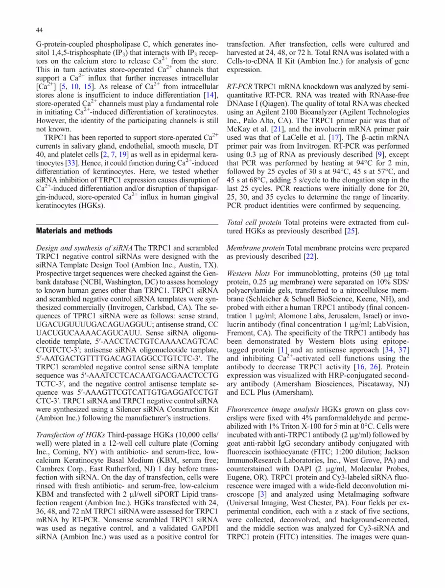
TRPC1 expression in proliferating and differentiatingHGKs Both TRPC1 mRNA and protein were expressed inHGKs incubated in low (0.03 mM) and high (1.2 mM)extracellular [Ca2+] for various times. TRPC1 mRNA waspresent in preconfluent and confluent cells incubated in0.03 mM [Ca2+], but levels were lower in confluent cells(Fig. 1a,a; n=3). TRPC1 mRNA levels remained elevatedbetween 8 h and 2 after increasing external [Ca2+] to1.2 mM, then decreased after 4 to 8 days (Fig. 1a,a).
TRPC1 protein expression was detected with the Alomoneantibody, which showed a single band of about 92 kDa(the expected size of human TRPC1) [35], by Western blotof the membrane fractions prepared from cells incubatedin 0.03 mM [Ca2+] or from fresh tissue (Fig. 1b,a; n=2).Preincubation with the peptide immunogen eliminated theband (Fig. 1b,b), demonstrating the specificity of theantibody. TRPC1 protein expression was relatively con-stant between 8 h and 8 days after incubation in 1.2 mM[Ca2+], although levels decreased slightly (by 9–15%)after 6 or 8 days (Fig. 1a,c). Hence, TRPC1 protein wasexpressed in HGKs incubated in both low and high extra-cellular [Ca2+].
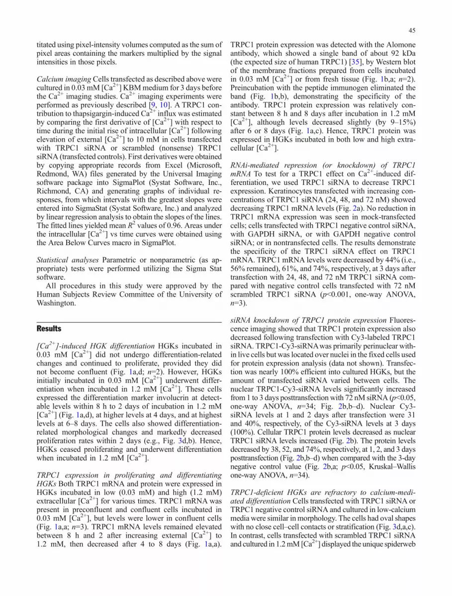
RNAi-mediated repression (or knockdown) of TRPC1mRNA To test for a TRPC1 effect on Ca2+-induced dif-ferentiation, we used TRPC1 siRNA to decrease TRPC1expression. Keratinocytes transfected with increasing con-centrations of TRPC1 siRNA (24, 48, and 72 nM) showeddecreasing TRPC1 mRNA levels (Fig. 2a). No reduction inTRPC1 mRNA expression was seen in mock-transfectedcells; cells transfected with TRPC1 negative control siRNA,with GAPDH siRNA, or with GAPDH negative controlsiRNA; or in nontransfected cells. The results demonstratethe specificity of the TRPC1 siRNA effect on TRPC1mRNA. TRPC1 mRNA levels were decreased by 44% (i.e.,56% remained), 61%, and 74%, respectively, at 3 days aftertransfection with 24, 48, and 72 nM TRPC1 siRNA com-pared with negative control cells transfected with 72 nMscrambled TRPC1 siRNA (p<0.001, one-way ANOVA,n=3).
siRNA knockdown of TRPC1 protein expression Fluores-cence imaging showed that TRPC1 protein expression alsodecreased following transfection with Cy3-labeled TRPC1siRNA. TRPC1-Cy3-siRNAwas primarily perinuclear with-in live cells but was located over nuclei in the fixed cells usedfor protein expression analysis (data not shown). Transfec-tion was nearly 100% efficient into cultured HGKs, but theamount of transfected siRNA varied between cells. Thenuclear TRPC1-Cy3-siRNA levels significantly increasedfrom 1 to 3 days posttransfectionwith 72 nM siRNA (p<0.05,one-way ANOVA, n=34; Fig. 2b,b–d). Nuclear Cy3-siRNA levels at 1 and 2 days after transfection were 31and 40%, respectively, of the Cy3-siRNA levels at 3 days(100%). Cellular TRPC1 protein levels decreased as nuclearTRPC1 siRNA levels increased (Fig. 2b). The protein levelsdecreased by 38, 52, and 74%, respectively, at 1, 2, and 3 daysposttransfection (Fig. 2b,b–d) when compared with the 3-daynegative control value (Fig. 2b,a; p<0.05, Kruskal–Wallisone-way ANOVA, n=34).
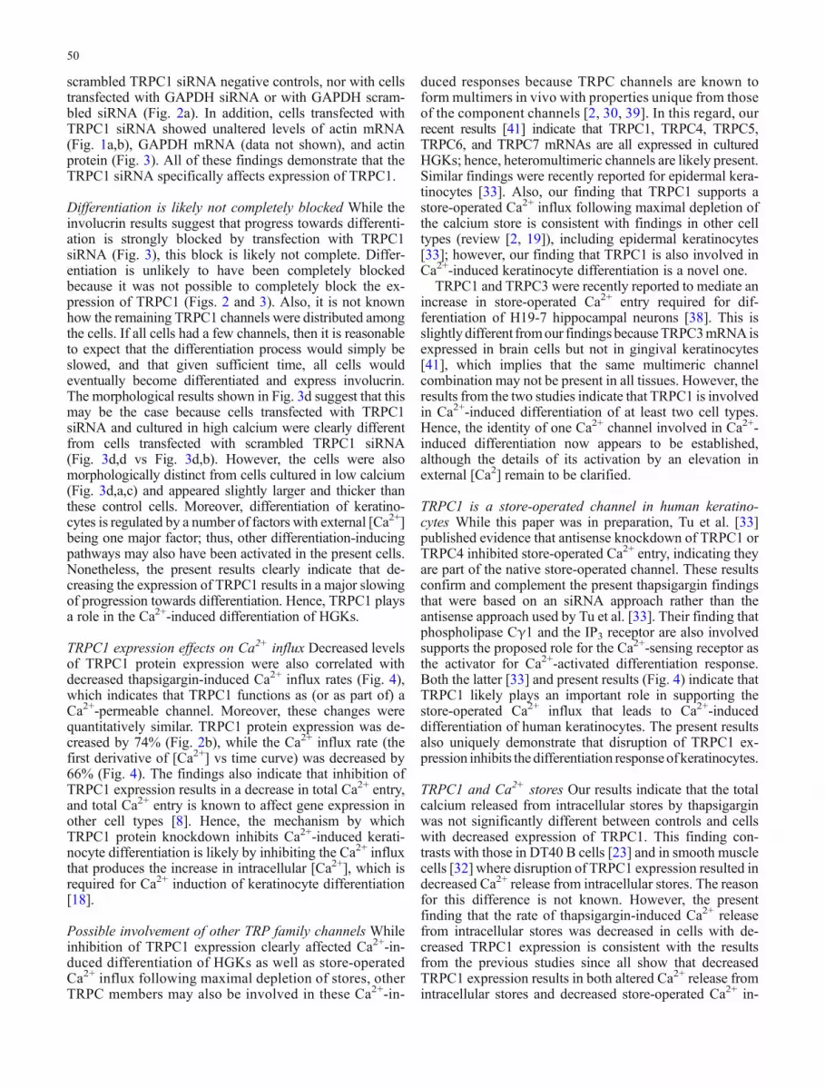
TRPC1-deficient HGKs are refractory to calcium-medi-ated differentiation Cells transfected with TRPC1 siRNA orTRPC1 negative control siRNA and cultured in low-calciummedia were similar inmorphology. The cells had oval shapeswith no close cell–cell contacts or stratification (Fig. 3d,a,c).In contrast, cells transfected with scrambled TRPC1 siRNAand cultured in 1.2mM[Ca2+] displayed the unique spiderweb
45

pattern associated with differentiatedHGKs (Fig. 3d,b). Thesecells were much larger and thicker than cells grown in lowcalcium, and many cells were closely connected to neighbor-ing cells. Cells transfectedwith TRPC1 siRNAand cultured inhigh calcium were intermediate in morphology. Some cellswere small and similar in appearance to cells grown in lowcalcium,while otherswere somewhat larger (Fig. 3d,d) but stillmuch smaller than cells transfected with scrambled TRPC1siRNA and cultured in high calcium (Fig. 3d,b). These mor-phological results qualitatively indicated that HGKstransfected with TRPC1 siRNA did not undergo normalCa2+-induced differentiation.
TRPC1 siRNA inhibits involucrin expression To comple-ment the morphological findings, we tested whether inhi-bition of TRPC1 protein expression affected expression ofthe differentiation marker, involucrin [24, 31]. Hence, cellstransfected with three concentrations of TRPC1 siRNA andincubated for 3 days in low-calcium medium were switchedto high-calcium medium, and involucrin expression wasmeasured after 2 days. Involucrin mRNA expression wasdecreased by 25% (i.e., 75% expression remained), 50%,and 65% (p<0.05, one-way ANOVA, n=3) in HGKs trans-fected with 24, 48, and 72 nM siRNA, respectively, com-pared with cells transfected with control siRNAs (Fig. 3a).
Fig. 1 TRPC1 was expressed inHGKs incubated in low andhigh [Ca2+] in culture and ingingival tissue. a TRPC1mRNA was expressed in cellsincubated in low (0.03 mM) andin high (1.2 mM) [Ca2+] (a).TRPC1 mRNA levels tended todecrease after 4 days in 1.2 mM[Ca2+]. Actin mRNA levels (b)and TRPC1 protein levels (c)were relatively constant inHGKs incubated in low andhigh [Ca2+]. Involucrin proteinexpression, a marker for HGKdifferentiation, was low inHGKs incubated in low [Ca2+]but increased in confluent cellsand in HGKs incubated in high[Ca2+] for more than 2 days (d).b A single band of about 92kDa, the expected size ofTRPC1, was observed in West-ern blots of membrane fractionsprepared from incubated kera-tinocytes and fresh gingival tis-sue (a). Preabsorption ofTRPC1 antibody with peptideimmunogen (at 1 μg peptide/μgantibody) abolished Westernimmunolabeling, demonstratingthe specificity of the antibody(b)
46

Similarly, involucrin protein expression was decreased by62, 67, and 80% (means, n=2), respectively, in HGKs trans-fected with 24, 48, and 72 nM TRPC1 siRNA (Fig. 3b).These results indicated that cells transfected with TRPC1
siRNA for 3 days had an abrogated Ca2+-induced differen-tiation response as indicated by decreased involucrin ex-pression. The specificity of this effect was indicated by theconstancy of β-actin expression.
To test whether the involucrin protein reduction effectwas siRNA transfection time dependent, we transfectedHGKs with a single dose (72 nM) of TRPC1 siRNA andthen incubated them for different times before testing forCa2+-inducible differentiation. Involucrin protein expres-sion was decreased by 48, 69, 74, and 84% (means, n=2)at 1, 2, 3, and 4 days, respectively, after transfection com-pared with negative control transfected cells (Fig. 3c).Hence, inhibiting TRPC1 channel expression greatly de-creased the normal Ca2+-mediated, differentiation-relatedmorphological and biochemical changes.
Effect of TRPC1 siRNA on store-operated Ca2+ influx Theabove results indicated that disruption of TRPC1 expres-sion disrupted Ca2+-induced differentiation of HGKs. Sinceit was previously reported that Ca2+ induction of keratino-cyte differentiation requires a Ca2+ influx [14] and since theexternal Ca2+-sensing receptor signals through the intra-cellular Ca2+ store [5, 6], we tested whether cells withdecreased TRPC1 protein expression also exhibited adecreased thapsigargin-induced Ca2+ influx (Fig. 4). Toseparate cytosolic [Ca2+] changes arising from Ca2+ releasefrom stores and those fromCa2+ influx, we initially exposedcells to thapsigargin (Fig. 4a, TG arrow) in the absence ofextracellular Ca2+ to deplete the intracellular Ca2+ store inHGKs transfected with TRPC1 siRNA or with TRPC1negative control siRNA and to maximally activate and holdthe store-operated Ca2+ channels in an open state whilestore-released Ca2+ ions were cleared from the cytoplasmby plasma membrane Ca2+ pumps [9, 10]. When cytosolic[Ca2+] had returned to near ambient levels, extracellular[Ca2+] was increased to 10 mM (Fig. 4a, Ca arrow), whichallowed Ca2+ ions to rapidly enter cells through the openchannels. The initial rate of change of intracellular [Ca2+]with time after this increase was an estimate of the relativenumber of open store-operated channels. As expected, theintracellular [Ca2+] began to change immediately andsynchronously in all cells when thapsigargin-pretreatedcells were exposed to 10 mM external [Ca2+] (Fig. 4a,b),indicating that the Ca2+ influx channels were open followingthapsigargin pretreatment. The rate of change of intracellular[Ca2+] as estimated using the first derivative of [Ca2+]change with respect to time in TRPC1 scrambled siRNAtransfected control cells (Fig. 4a) was greater than that inTRPC1 siRNA transfected cells (Fig. 4b) by threefold[Fig. 4c: 1.28±0.10 vs 0.43±0.05 (relative fura ratio/minunits) means±SEMs; p<0.001, t test], indicating a greaterCa2+ influx in control cells vs cells with decreased TRPC1expression following thapsigargin depletion of intracellularcalcium stores. The initial rate of change of intracellular[Ca2+] with time after Ca2+ addition in thapsigargin-pre-treated cells is shown in Fig. 4d for HGKs transfected withscrambled TRPC1 siRNA and in Fig. 4e for HGKs trans-fected with TRPC1 siRNA.
Fig. 2 siRNA dose-dependent reduction of TRPC1 expression inHGKs. a TRPC1 mRNA levels decreased 3 days after transfectionwith 24, 48, and 72 nM TRPC1 siRNA compared with scrambled(NC) TRPC1 siRNA, GAPDH, and NC GAPDH siRNA, and mockand no-transfection controls. No RT and No RNA were controlswithout reverse transcriptase or RNA, respectively. mRNA levels aspercent of the scrambledNCTRPC1 transfected value are given beloweach band. bTRPC1 protein expression (quantitated with fluorescein-labeled secondary antibody, right column) was high in HGKstransfected with scrambled TRPC1 Cy3-siRNA (left column; a) andincrementally decreased in HGKs transfected with 72 nM TRPC1Cy3-siRNA for 1 day (b), 2 days (c), and 3 days (d)
47

Effect on total Ca2+ entry The above results indicate thatless total Ca2+ may enter cells with disrupted TRPC1 ex-pression since the Ca2 influx rate is decreased. Other dataalso support this finding. Specifically, the areas under thecurves of intracellular [Ca2+] with time following exposureto 10 mM external [Ca2+] yield the relative total amountsof calcium ions entering the cells for the 9.8-min durationof the experiment (Fig. 4). This amount was 2.43-foldgreater in control cells than in TRPC1-disrupted cells [5.84±0.33 vs 2.40±0.19 (fura ratio units/time), means±SEMs;
p<0.001, t test]. Also, visual inspection of the intracellular[Ca2+] changes following exposure to 10 mM external[Ca2+] indicates that intracellular [Ca2+] decreases towardsbaseline more rapidly in TRPC1 siRNA-transfected cells(Fig. 4b) than in negative control-transfected cells (Fig. 4a).Thus, the total Ca2+ entry into the two cell types was greaterin control cells vs TRPC1-disrupted cells because it wasgreater for the 9.8-min duration of the experiment andbecause [Ca2+] likely subsequently decreased faster tobaseline in TRPC1-disrupted cells than in the control cells.
Fig. 3 Transfection withTRPC1 siRNA inhibited Ca2+-induced differentiation ofHGKs. a HGKs transfected with24, 48, or 72 nM TRPC1 siRNAfor 3 days and then exposed tohigh [Ca2+] for 2 days showedan incremental decrease in in-volucrin mRNA expression. Nodecreases were observed withscrambled TRPC1 siRNA (NC)or other controls. Band intensi-ties expressed as percent ofscrambled TRPC1 siRNA (NC)value are given below the bands.b Involucrin protein levelsshowed a similar TRPC1siRNA-dependent reduction,while actin expression did not.c HGKs transfected with 72 nMTRPC1 siRNA for 1, 2, 3, and 4days and then exposed to high[Ca2+] for 2 days showed anincremental decrease in involu-crin protein expression whencompared with HGKs transfec-ted with scrambled (NC)TRPC1 siRNA for 3 days. Nochanges were observed in actinexpression. d Transfection withTRPC1 siRNA inhibited Ca2+-induced changes in HGK mor-phology. HGKs cultured for 2days in low [Ca2+] followingtransfection with scrambledTRPC1 siRNA were isolated,oval-shaped cells (a). HGKscultured in high [Ca2+] follow-ing transfection with scrambledTRPC1 siRNA showed typicaldifferentiation-related changeswith “spiderweb” morphologyconsisting of closely associated,elongated cells (b). HGKs cul-tured in low [Ca2+] followingtransfection with TRPC1 siRNAshowed no differentiation-re-lated changes (c). HGKs cul-tured in high [Ca2+] followingtransfection with TRPC1 siRNAalso showed no differentiation-related changes (d). These find-ings are representative of threeindependent experiments
48

Hence, cells with decreased TRPC1 expression had adecreased Ca2+ influx at early times after onset of store-operated Ca2+ influx when compared with transfectedcontrols, and these cells also had a decreased total Ca2+
entry when compared with controls.
Effect of TRPC1 knockdown on Ca2+ release from storesKnockdown of TRPC1 did not significantly affect the totalCa2+ released from stores by thapsigargin (Fig. 4a, firstpeak, vs Fig. 4b, first peak; 1.30±0.13 vs 1.30±0.10; mean±SEM; units: fura ratio/min). However, TRPC1 knockdowndecreased the maximum rate of Ca2+ release from stores by40% (0.65±0.04 vs 0.38±0.03; units: fura ratio/min), andthis decrease was statistically significant (p<0.001 by t test).
Discussion
TRPC1 is involved in Ca2+-induced differentiation Themost important finding from the present experiments is theevidence that TRPC1 is involved in Ca2+-induced differen-tiation of HGKs. First, increasing concentrations of TRPC1siRNA resulted in dose-dependent decreases in TRPC1mRNA and protein expression and decreased involucrinmRNAand protein expression.Moreover, the TRPC1 siRNA-induced changes were quantitatively consistent. Thus, trans-fection with 72 nM TRPC1 siRNA for 3 days decreasedTRPC1 mRNA levels by 74% (Fig. 2a), TRPC1 proteinlevels by 74% (Fig. 2b), and involucrin expression levels by80% (Fig. 3b). Such decreases were not obtained with
Fig. 4 HGKs transfected with72 nM TRPC1 siRNA for 3days displayed decreased store-operated Ca2+ influx. a Pre-treatment of HGKs with 1 μMthapsigargin (at TG arrow) re-leased Ca2+ from intracellularstores and transiently increasedcytosolic [Ca2+]. Followingclearance of store-associated[Ca2+] increase, exposure to10 mM external [Ca2+] (at Caarrow) in the continued pres-ence of thapsigargin (from TGarrow) resulted in an immediateand synchronous increase inintracellular [Ca2+] in controlHGKs transfected withscrambled TRPC1 siRNA.b HGKs transfected withTRPC1 siRNA displayed smallerand more transitory intracellular[Ca2+] changes upon externalCa2+ addition. c The initial rateof change of intracellular [Ca2+]with time (i.e., the first timederivative) following addition to10 mM external [Ca2+] wassignificantly greater in controlcells (a) transfected withscrambled TRPC1 siRNA(TRPC1 NC siRNA) than inTRPC1 siRNA transfected cells(b; p<0.001). d A magnifiedview of the intracellular [Ca2+]change following Ca2+ additionin a. Results from 7.9 to 8.5 minare shown. Values from 8.0 to8.5 min were used to estimatethe slope. e A magnified view ofthe intracellular [Ca2+] changefollowing Ca2+ addition in b.Values from 7.9 to 8.4 min wereused to estimate the slope
49
scrambled TRPC1 siRNA negative controls, nor with cellstransfected with GAPDH siRNA or with GAPDH scram-bled siRNA (Fig. 2a). In addition, cells transfected withTRPC1 siRNA showed unaltered levels of actin mRNA(Fig. 1a,b), GAPDH mRNA (data not shown), and actinprotein (Fig. 3). All of these findings demonstrate that theTRPC1 siRNA specifically affects expression of TRPC1.
Differentiation is likely not completely blocked While theinvolucrin results suggest that progress towards differenti-ation is strongly blocked by transfection with TRPC1siRNA (Fig. 3), this block is likely not complete. Differ-entiation is unlikely to have been completely blockedbecause it was not possible to completely block the ex-pression of TRPC1 (Figs. 2 and 3). Also, it is not knownhow the remaining TRPC1 channels were distributed amongthe cells. If all cells had a few channels, then it is reasonableto expect that the differentiation process would simply beslowed, and that given sufficient time, all cells wouldeventually become differentiated and express involucrin.The morphological results shown in Fig. 3d suggest that thismay be the case because cells transfected with TRPC1siRNA and cultured in high calcium were clearly differentfrom cells transfected with scrambled TRPC1 siRNA(Fig. 3d,d vs Fig. 3d,b). However, the cells were alsomorphologically distinct from cells cultured in low calcium(Fig. 3d,a,c) and appeared slightly larger and thicker thanthese control cells. Moreover, differentiation of keratino-cytes is regulated by a number of factors with external [Ca2+]being one major factor; thus, other differentiation-inducingpathways may also have been activated in the present cells.Nonetheless, the present results clearly indicate that de-creasing the expression of TRPC1 results in a major slowingof progression towards differentiation. Hence, TRPC1 playsa role in the Ca2+-induced differentiation of HGKs.
TRPC1 expression effects on Ca2+ influx Decreased levelsof TRPC1 protein expression were also correlated withdecreased thapsigargin-induced Ca2+ influx rates (Fig. 4),which indicates that TRPC1 functions as (or as part of) aCa2+-permeable channel. Moreover, these changes werequantitatively similar. TRPC1 protein expression was de-creased by 74% (Fig. 2b), while the Ca2+ influx rate (thefirst derivative of [Ca2+] vs time curve) was decreased by66% (Fig. 4). The findings also indicate that inhibition ofTRPC1 expression results in a decrease in total Ca2+ entry,and total Ca2+ entry is known to affect gene expression inother cell types [8]. Hence, the mechanism by whichTRPC1 protein knockdown inhibits Ca2+-induced kerati-nocyte differentiation is likely by inhibiting the Ca2+ influxthat produces the increase in intracellular [Ca2+], which isrequired for Ca2+ induction of keratinocyte differentiation[18].
Possible involvement of other TRP family channels Whileinhibition of TRPC1 expression clearly affected Ca2+-in-duced differentiation of HGKs as well as store-operatedCa2+ influx following maximal depletion of stores, otherTRPC members may also be involved in these Ca2+-in-
duced responses because TRPC channels are known toform multimers in vivo with properties unique from thoseof the component channels [2, 30, 39]. In this regard, ourrecent results [41] indicate that TRPC1, TRPC4, TRPC5,TRPC6, and TRPC7 mRNAs are all expressed in culturedHGKs; hence, heteromultimeric channels are likely present.Similar findings were recently reported for epidermal kera-tinocytes [33]. Also, our finding that TRPC1 supports astore-operated Ca2+ influx following maximal depletion ofthe calcium store is consistent with findings in other celltypes (review [2, 19]), including epidermal keratinocytes[33]; however, our finding that TRPC1 is also involved inCa2+-induced keratinocyte differentiation is a novel one.
TRPC1 and TRPC3 were recently reported to mediate anincrease in store-operated Ca2+ entry required for dif-ferentiation of H19-7 hippocampal neurons [38]. This isslightlydifferent fromour findings becauseTRPC3mRNAisexpressed in brain cells but not in gingival keratinocytes[41], which implies that the same multimeric channelcombination may not be present in all tissues. However, theresults from the two studies indicate that TRPC1 is involvedin Ca2+-induced differentiation of at least two cell types.Hence, the identity of one Ca2+ channel involved in Ca2+-induced differentiation now appears to be established,although the details of its activation by an elevation inexternal [Ca2] remain to be clarified.
TRPC1 is a store-operated channel in human keratino-cytes While this paper was in preparation, Tu et al. [33]published evidence that antisense knockdown of TRPC1 orTRPC4 inhibited store-operated Ca2+ entry, indicating theyare part of the native store-operated channel. These resultsconfirm and complement the present thapsigargin findingsthat were based on an siRNA approach rather than theantisense approach used by Tu et al. [33]. Their finding thatphospholipase Cγ1 and the IP3 receptor are also involvedsupports the proposed role for the Ca2+-sensing receptor asthe activator for Ca2+-activated differentiation response.Both the latter [33] and present results (Fig. 4) indicate thatTRPC1 likely plays an important role in supporting thestore-operated Ca2+ influx that leads to Ca2+-induceddifferentiation of human keratinocytes. The present resultsalso uniquely demonstrate that disruption of TRPC1 ex-pression inhibits the differentiation responseof keratinocytes.
TRPC1 and Ca2+ stores Our results indicate that the totalcalcium released from intracellular stores by thapsigarginwas not significantly different between controls and cellswith decreased expression of TRPC1. This finding con-trasts with those in DT40 B cells [23] and in smooth musclecells [32] where disruption of TRPC1 expression resulted indecreased Ca2+ release from intracellular stores. The reasonfor this difference is not known. However, the presentfinding that the rate of thapsigargin-induced Ca2+ releasefrom intracellular stores was decreased in cells with de-creased TRPC1 expression is consistent with the resultsfrom the previous studies since all show that decreasedTRPC1 expression results in both altered Ca2+ release fromintracellular stores and decreased store-operated Ca2+ in-
50

flux. Thus, the present results support the notion that thereis a coupling between the proposed Ca2+ entry channel inthe plasma membrane (TRPC1) and Ca2+ release fromintracellular stores. In this regard, TRPC1 is reported tocouple with the type II IP3 receptor during release of Ca2+
from intracellular stores and the subsequent activation ofstore-operated channels [27]. Moreover, Golovina [13]recently showed that Ca2+ release from intracellular storesby cyclopiazonic acid, an endoplasmic reticulum (ER)Ca2+-ATPase inhibitor, occurred in the same restrictedspace as that affected by store-operated Ca2+ influx. Thisfinding is consistent with our finding that release rate fromthe store is decreased with decreased TRPC1 expressionbecause it indicates that cyclopiazonic acid-induced and,presumably, thapsigargin-induced store Ca2+ release occurat the same plasma membrane/ER interface as that for store-operated Ca2+ entry, i.e., at the IP3R–TRPC1 interface. Ifthis were the case, then it would not be surprising thatdecreased TRPC1 levels would affect Ca2+ release ratesfrom the store. While the details of the interactions betweenplasma membrane and ER components remain to be elu-cidated [2, 29], it is likely that interactions between TRPC1channels on the plasma membrane and IP3 receptors on theER are important in Ca2+ signaling.
Acknowledgements The authors thank Dr. Bertil Hille forinsightful discussions into the semiquantitative analysis of the furaresults. This work is supported by National Institutes of Health GrantAR046254 and a grant from the University of Washington School ofDentistry.
References
1. Ahmmed GU, Mehta D, Vogel S, Holinstat M, Paria BC,Tiruppathi C, Malik AB (2004) Protein kinase Calpha phos-phorylates the TRPC1 channel and regulates store-operated Ca2+
entry in endothelial cells. J Biol Chem 279:20941–209492. Beech DJ, Xu SZ, McHugh D, Flemming R (2003) TRPC1
store-operated cationic channel subunit. Cell Calcium 33:433–440
3. Belton CM, Izutsu KT, Goodwin PC, Park Y, Lamont RJ(1999) Fluorescence image analysis of the association betweenPorphyromonas gingivalis and gingival epithelial cells. CellMicrobiol 1:215–223
4. Bikle DD, Ng D, Tu CL, Oda Y, Xie Z (2001) Calcium- andvitamin D-regulated keratinocyte differentiation. Mol CellEndocrinol 177:161–171
5. Brown EM, MacLeod RJ (2001) Extracellular calcium sensingand extracellular calcium signaling. Physiol Rev 81:239–297
6. Brown EM, Pollak M, Hebert SC (1995) Molecular mecha-nisms underlying the sensing of extracellular Ca2 by parathy-roid and kidney cells. Eur J Endocrinol 132:523–531
7. Brownlow SL, Harper AG, Harper MT, Sage SO (2004) A rolefor hTRPC1 and lipid raft domains in store-mediated calciumentry in human platelets. Cell Calcium 35:107–113
8. Dolmetsch RE, Lewis RS, Goodnow CC, Healy JI (1997)Differential activation of transcription factors induced by Ca2
response amplitude and duration. Nature 386:855–8589. Fatherazi S, Belton CM, Cai S, Zarif S, Goodwin PC, Lamont
RJ, Izutsu KT (2004) Calcium receptor message, expressionand function decrease in differentiating keratinocytes. PflugersArch 448:93–104
10. Fatherazi S, Belton CM, Izutsu KT (2003) Sequential activa-tion of store-operated currents in human gingival keratino-cytes. J Invest Dermatol 121:120–131
11. Forslind B, Lindberg M, Roomans GM, Pallon J, Werner-LindeY (1997) Aspects on the physiology of human skin: studiesusing particle probe analysis. Microsc Res Tech 38:373–386
12. Gibson DF, Ratnam AV, Bikle DD (1996) Evidence for separatecontrol mechanisms at the message, protein, and enzyme acti-vation levels for transglutaminase during calcium-induced dif-ferentiation of normal and transformed human keratinocytes.J Invest Dermatol 106:154–161
13. Golovina VA (2005) Visualization of localized store-operatedcalcium entry in mouse astrocytes. Close proximity to theendoplasmic reticulum. J Physiol 564(3):737–749
14. Jones KT, Sharpe GR (1994) Thapsigargin raises intracellularfree calcium levels in human keratinocytes and inhibits thecoordinated expression of differentiation markers. Exp Cell Res210:71–76
15. Komuves L, Oda Y, Tu CL, Chang WH, Ho-Pao CL, Mauro T,Bikle DD (2002) Epidermal expression of the full-length extra-cellular calcium-sensing receptor is required for normal kera-tinocyte differentiation. J Cell Physiol 192:45–54
16. Kunzelmann-Marche C, Freyssinet JM, Martinez MC (2002)Loss of plasma membrane phospholipid asymmetry requiresraft integrity. Role of transient receptor potential channels andERK pathway. J Biol Chem 277:19876–19881
17. La Celle PT, Polakowska RR (2001) Human homeobox HOXA7regulates keratinocyte transglutaminase type 1 and inhibits dif-ferentiation. J Biol Chem 276:32844–32853
18. Li L, Tucker RW, Hennings H, Yuspa SH (1995) Chelation ofintracellular Ca2+ inhibits murine keratinocyte differentiation invitro. J Cell Physiol 163:105–114
19. Liu X, Singh BB, Ambudkar IS (2003) TRPC1 is required forfunctional store-operated Ca2+ channels. Role of acidic aminoacid residues in the S5–S6 region. J Biol Chem 278:11337–11343
20. Mauro T, Bench G, Sidderas-Haddad E, Feingold K, Elias P,Cullander C (1998) Acute barrier perturbation abolishes the Ca2+
and K+ gradients in murine epidermis: quantitative measurementusing PIXE. J Invest Dermatol 111:1198–1201
21. McKay RR, Szymeczek-Seay CL, Lievremont JP, Bird GS, ZittC, Jungling E, Luckhoff A, Putney JW Jr (2000) Cloning andexpression of the human transient receptor potential 4 (TRP4)gene: localization and functional expression of human TRP4and TRP3. Biochem J 351(Pt 3):735–746
22. Milewski MI, Mickle JE, Forrest JK, Stanton BA, Cutting GR(2002) Aggregation of misfolded proteins can be a selectiveprocess dependent upon peptide composition. J Biol Chem277:34462–34470
23. Mori Y, Wakamori M, Miyakawa T, Hermosura M, Hara Y,NishidaM, Hirose K, Mizushima A, Kurosaki M, Mori E, GotohK, Okada T, Fleig A, Penner R, Iino M, Kurosaki T (2002)Transient receptor potential 1 regulates capacitative Ca(2+) entryand Ca(2+) release from endoplasmic reticulum in B lympho-cytes. J Exp Med 195:673–681
24. Presland RB, Dale BA (2000) Epithelial structural proteins ofthe skin and oral cavity: function in health and disease. Crit RevOral Biol Med 11:383–408
25. Presland RB, Kimball JR, Kautsky MB, Lewis SP, Lo CY, DaleBA (1997) Evidence for specific proteolytic cleavage of the N-terminal domain of human profilaggrin during epidermal dif-ferentiation. J Invest Dermatol 108:170–178
26. Rosado JA, Brownlow SL, Sage SO (2002) Endogenouslyexpressed Trp1 is involved in store-mediated Ca2+ entry byconformational coupling in human platelets. J Biol Chem 277:42157–42163
27. Rosado JA, Sage SO (2001) Activation of store-mediatedcalcium entry by secretion-like coupling between the inositol1,4,5-trisphosphate receptor type II and human transient receptorpotential (hTrp1) channels in human platelets. Biochem J 356:191–198
51

28. Sharpe GR, Gillespie JI, Greenwell JR (1989) An increase inintracellular free calcium is an early event during differentiationof cultured human keratinocytes. FEBS Lett 254:25–28
29. Spassova MA, Soboloff J, He LP, Hewavitharana T, Xu W,Venkatachalam K, van Rossum DB, Patterson RL, Gill DL(2004) Calcium entry mediated by SOCs and TRP channels:variations and enigma. Biochim Biophys Acta 1742:9–20
30. Strubing C, Krapivinsky G, Krapivinsky L, Clapham DE(2001) TRPC1 and TRPC5 form a novel cation channel inmammalian brain. Neuron 29:645–655
31. Sumitomo S, Kumasa S, Iwai Y, Mori M (1986) Involucrinexpression in epithelial tumors of oral and pharyngeal mucosaand skin. Oral Surg Oral Med Oral Pathol 62:155–163
32. Sweeney M, Yu Y, Platoshyn O, Zhang S, McDaniel SS, YuanJX (2002) Inhibition of endogenous TRP1 decreases capacita-tive Ca2+ entry and attenuates pulmonary artery smooth musclecell proliferation. Am J Physiol Lung Cell Mol Physiol 283:L144–155
33. Tu CL, Chang W, Bikle DD (2005) Phospholipase cgamma1 isrequired for activation of store-operated channels in humankeratinocytes. J Invest Dermatol 124:187–197
34. Vanden Abeele F, Lemonnier L, Thebault S, Lepage G, ParysJB, Shuba Y, Skryma R, Prevarskaya N (2004) Two types ofstore-operated Ca2+ channels with different activation modesand molecular origin in LNCaP human prostate cancer epi-thelial cells. J Biol Chem 279:30326–30337
35. WangW, O’Connell B, Dykeman R, Sakai T, Delporte C, SwaimW, Zhu X, Birnbaumer L, Ambudkar IS (1999) Cloning ofTrp1beta isoform from rat brain: immunodetection and local-ization of the endogenous Trp1 protein. Am J Physiol 276:C969–979
36. Watt FM, Mattey DL, Garrod DR (1984) Calcium-induced re-organization of desmosomal components in cultured humankeratinocytes. J Cell Biol 99:2211–2215
37. Wu X, Babnigg G, Zagranichnaya T, Villereal ML (2002) Therole of endogenous human Trp4 in regulating carbachol-in-duced calcium oscillations in HEK-293 cells. J Biol Chem277:13597–13608
38. Wu X, Zagranichnaya TK, Gurda GT, Eves EM, Villereal ML(2004) A TRPC1/TRPC3-mediated increase in store-operatedcalcium entry is required for differentiation of H19-7 hippo-campal neuronal cells. J Biol Chem 279:43392–43402
39. Xu XZ, Li HS, Guggino WB, Montell C (1997) Coassembly ofTRP and TRPL produces a distinct store-operated conductance.Cell 89:1155–1164
40. Yuspa SH, Hennings H, Tucker RW, Jaken S, Kilkenny AE,Roop DR (1988) Signal transduction for proliferation anddifferentiation in keratinocytes. Ann N YAcad Sci 548:191–196
41. Cai S, Fatherazi S, Presland RB, Belton CM, Iźutsu KT (2005)TRPC channel expression during calcium-induced differentia-tion of human gingival keratinocytes. J Dermatol Sci 40:21–28
52