evidence that transgenes encoding components of the wnt signaling pathway preferentially induce...
TRANSCRIPT
Evidence that transgenes encoding components ofthe Wnt signaling pathway preferentially inducemammary cancers from progenitor cellsYi Li*†‡§, Bryan Welm¶, Katrina Podsypanina*, Shixia Huang*, Mario Chamorro*�, Xiaomei Zhang†, Tracey Rowlands**,Mikala Egeblad¶, Pam Cowin**, Zena Werb¶, Lee K. Tan††, Jeffrey M. Rosen‡, and Harold E. Varmus*
*Program in Cell Biology and ††Department of Pathology, Memorial Sloan–Kettering Cancer Center, 1275 York Avenue, New York, NY 10021; †Breast Centerand ‡Department of Molecular and Cellular Biology, Baylor College of Medicine, Houston, TX 77030; ¶Department of Anatomy, University of California,San Francisco, CA 94143; �Weill Medical College, Cornell University, New York, NY 10021; and **Departments of Cell Biology and Dermatology,New York University Medical School, New York, NY 10032
Contributed by Harold E. Varmus, October 21, 2003
Breast cancer is a genetically and clinically heterogeneous disease,and the contributions of different target cells and different onco-genic mutations to this heterogeneity are not well understood.Here we report that mammary tumors induced by components ofthe Wnt signaling pathway contain heterogeneous cell types andexpress early developmental markers, in contrast to tumors in-duced by other signaling elements. Expression of the Wnt-1 pro-tooncogene in mammary glands of transgenic mice expands apopulation of epithelial cells expressing progenitor cell markers,keratin 6 and Sca-1; subsequent tumors express these markers andcontain luminal epithelial and myoepithelial tumor cells that sharea secondary mutation, loss of Pten, implying that they arose froma common progenitor. Mammary tumors arising in transgenic miceexpressing �-catenin and c-Myc, downstream components of thecanonical Wnt signaling pathway, also contain a significant pro-portion of myoepithelial cells and cells expressing keratin 6. Pro-genitor cell markers and myoepithelial cells, however, are lackingin mammary tumors from transgenic mice expressing Neu, H-Ras,or polyoma middle T antigen. These results suggest that mammarystem cells and�or progenitors to mammary luminal epithelial andmyoepithelial cells may be the targets for oncogenesis by Wnt-1signaling elements. Thus, the developmental heterogeneity ofdifferent breast cancers is in part a consequence of differentialeffects of oncogenes on distinct cell types in the breast.
Transgenic (TG) activation of different oncogenic pathways inmouse mammary glands induces tumors with different gene
expression profiles and histopathological features (1–8). Themouse mammary tumor virus (MMTV) promoter usually usedto regulate these transgenes is expressed in a diverse populationof mammary cells (9, 10). Therefore, cells at different develop-mental stages may undergo tumorigenesis as a result of theactivation of different oncogenic pathways.
It has been difficult to define the precise lineage of target cellsof breast cancer because of the lack of animal models expressingoncogenes in specific mammary progenitors. However, studies inthe epidermis and the hematopoietic system have demonstratedthat cancers that arise from stem or progenitor cells usuallyexpress markers of the originating cells (11, 12). Thus, thepresence of stem or progenitor cell markers in mammary tumorsmay suggest that tumors arose from immature cells. Two genes,keratin 6 and stem cell antigen 1 (Sca-1), appear to be prefer-entially expressed in mammary stem and�or progenitor cells.Keratin 6 is expressed in mammary gland anlage at embryonicday 16.5 (ref. 13 and J.M.R., unpublished observations) and insome of the body cells in the terminal end buds (TEBs); butkeratin 6 is not found in the highly proliferative cap cells (14, 15)and rarely in cells in the mature ducts and differentiated alveoli(15), consistent with the distribution of progenitor cells. Inaddition, keratin 6 is associated with the arrested state ofdifferentiation observed in the mammary ducts from C�EBP�-
null mice (13). Sca-1, encoded by Ly-6A�E, is a GPI-linkedprotein also found in hematopoietic stem cells (16); it is ex-pressed on the surface of a population of mammary cellsenriched for stem cells and present in TEBs (17). Depletion ofSca-1-positive cells results in a loss of functional stem cells inmammary gland reconstitution experiments (17). Although re-tained in a population of more mature ductal cells, Sca-1 is notobserved after cells have further differentiated to express theprogesterone receptor (17).
Here we report that keratin 6 and Sca-1 cells are observed inmammary tumors induced by the Wnt-1 signaling pathway,which has been implicated in proliferation and maintenance ofundifferentiated cells in several tissue types, including the mam-mary gland (18–28), but not in tumors induced by oncogenesaffecting other pathways. In addition, we provide other evidenceto suggest that ectopic activation of the Wnt pathway may targetthese undifferentiated mammary progenitors for tumorigenesis.
Materials and MethodsMice. TG mice expressing MMTV-Wnt-1 (29), MMTV-�-catenin(30), MMTV-c-Myc (31), MMTV-Neu (32), MMTV-H-Ras (33),or MMTV-PyMT (34) have been described, as have mice har-boring targeted inactivating mutations of Pten (35). MMTV-Wnt-1 TG mice were a mixture of FVB, SJL, and C57BL�6strains; Pten mutant mice were a mixture of FVB�N, 129, andC57BL�6 strains; all others were in a pure FVB�N background.
Tissue Processing and Immunocytochemistry. Tissues were fixed andprocessed as described (36). Immunohistochemistry was per-formed by using Vector ABC and MOM kits (Vector Labora-tories) according to the manufacturer’s recommendations andas described (35). The following antibodies were used: purifiedrabbit antibodies against mouse keratin 6 (Covance, Princeton)and Pten (NeoMarkers, Fremont, CA); purified mouse mono-clonal antibodies against �-smooth muscle actin (SMA, Dako)and against BrdUrd (BD Biosciences Immunocytometry Sys-tems, product no. 347580); and partially purified rat antibodiesagainst keratin 8 (37, 38), purchased from the DevelopmentalStudies Hybridoma Bank, Iowa City, organized under the aus-pices of the National Institute of Child Health and HumanDevelopment and maintained by the University of Iowa. To labelcells in S-phase of cell cycle, BrdUrd (Sigma, product no.B-5002) at 100 �g per gram of body weight in saline was injectedi.p. 1 h before mice were killed.
Abbreviations: FACS, fluorescence-activated cell sorting; LOH, loss of heterozygosity;MMTV, mouse mammary tumor virus; SMA, smooth muscle actin; TEB, terminal end bud;TG, transgenic.
§To whom correspondence should be addressed at: Breast Center, Baylor College ofMedicine, One Baylor Plaza, Houston, TX 77030. E-mail: [email protected].
© 2003 by The National Academy of Sciences of the USA
www.pnas.org�cgi�doi�10.1073�pnas.2136825100 PNAS � December 23, 2003 � vol. 100 � no. 26 � 15853–15858
MED
ICA
LSC
IEN
CES
Southern Hybridization and Western Analysis. Tumor DNAs weredigested with PstI and processed for Southern blotting by usinga [32P]dCTP-labeled probe made from a 0.9-kb template locatedin intron 5 of Pten as described (36). For Western blotting,tumors were ground to powder in liquid nitrogen and lysed byboiling in the sample-loading buffer for acrylamide gel electro-phoresis. Protein lysates were resolved in 10% polyacrylamidegels containing SDS and transferred to nitrocellulose mem-branes. After incubating with rabbit antibodies against Pten(NeoMarkers, 1:1,000) or mouse monoclonal antibodies against�-tubulin (Sigma, 1:5,000), the specific reaction was visualized byperoxidase-conjugated secondary antibodies (Roche) and achemiluminescent substrate (SuperSignal, Pierce).
Flow Cytometry. Mammary cells and tumors were isolated byusing collagenase as described (17). After incubating with FITC-conjugated rat IgG against Sca-1 (BD Pharmingen, product no.553336, 106 cells per �g), f luorescence-activated cell sorting(FACS) was performed as described (17).
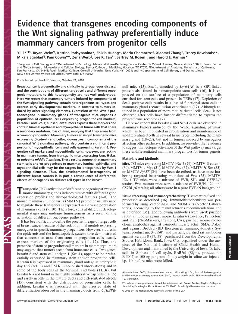
ResultsKeratin 6 and Sca-1 Are Expressed in Preneoplastic and NeoplasticMammary Lesions Induced by Components of the Wnt SignalingPathway but Not by Neu, Ras, and PyMT. While carrying outexperiments designed to identify genes differentially regulated inthe evolution of mammary tumors in MMTV-Wnt-1 TG mice(S.H., unpublished work), we discovered that both keratin 6 andSca-1 are more highly expressed in hyperplastic mammary glandsand tumors from MMTV-Wnt-1 TG mice than in non-TG virginmammary glands (Fig. 6, which is published as supportinginformation on the PNAS web site), suggesting that Wnt-1-induced mammary tumors may originate from progenitor cells.To confirm this observation, we used immunohistochemicalstaining to identify keratin 6-expressing cells. In non-TG mam-mary glands, keratin 6 was detected in some body cells within theterminal end buds (TEBs) and occasionally in mature luminalepithelial cells (Fig. 1 B and D), consistent with previous reports
(14, 15). However, in MMTV-Wnt-1 TG mice, we found keratin6 in a greater number of ductal cells, usually in enlarged ducts,in a heterogeneous pattern (Fig. 1 A and C). Many of the stainedcells are in the luminal layer, but some of them have invaded intothe lumen (Fig. 1 A). Ducts that were not stained were usuallysmaller in diameter and did not appear to be hyperplastic (Fig.1C). Because transgenes regulated by MMTV are known to beexpressed in heterogeneous patterns in the mammary gland (9,39–42), the keratin 6-negative ducts may not express theMMTV-Wnt-1 transgene.
The mammary cells in adult MMTV-Wnt-1 TG mice have agreater rate of proliferation than those in non-TG virgin mice,as measured by immunohistochemical staining for cells labeledwith BrdUrd and for cells expressing Ki-67, another proliferationmarker (data not shown). It is therefore possible that theexpansion of keratin 6-positive cells in the mammary glands ofMMTV-Wnt-1 TG mice may result from an increased proportionof proliferating cells. Indeed, keratin 6 has been associated withhyperproliferation in the suprabasal layer in the skin (43). Todetermine whether the increased expression of keratin 6 is anoncogene- or proliferation-induced effect in the mammarygland, we compared mammary glands from adult MMTV-Wnt-1TG virgins with proliferating mammary glands from mid-pregnancy non-TG mice. Similar numbers of BrdUrd-stainedcells (Fig. 1 E and F) and Ki-67-positive cells (data not shown)were detected in virgin TG and pregnant non-TG mammaryglands. However, only a few cells per field were stained withanti-keratin 6 antibodies in the pregnant non-TG mice (data notshown). Taken together, these results suggest that aberrantWnt-1 expression arrests differentiation of mammary cells at anearly phase and stimulate their proliferation and that prolifer-ation per se is not associated with expression of keratin 6.
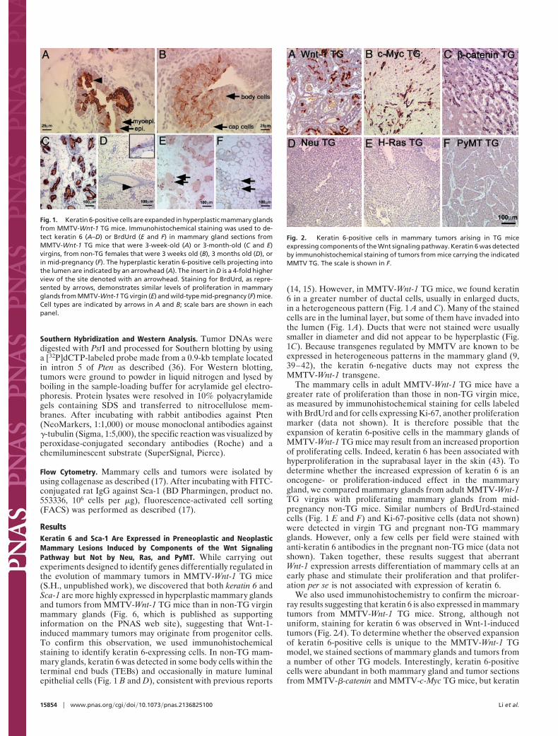
We also used immunohistochemistry to confirm the microar-ray results suggesting that keratin 6 is also expressed in mammarytumors from MMTV-Wnt-1 TG mice. Strong, although notuniform, staining for keratin 6 was observed in Wnt-1-inducedtumors (Fig. 2A). To determine whether the observed expansionof keratin 6-positive cells is unique to the MMTV-Wnt-1 TGmodel, we stained sections of mammary glands and tumors froma number of other TG models. Interestingly, keratin 6-positivecells were abundant in both mammary gland and tumor sectionsfrom MMTV-�-catenin and MMTV-c-Myc TG mice, but keratin
Fig. 1. Keratin 6-positive cells are expanded in hyperplastic mammary glandsfrom MMTV-Wnt-1 TG mice. Immunohistochemical staining was used to de-tect keratin 6 (A–D) or BrdUrd (E and F) in mammary gland sections fromMMTV-Wnt-1 TG mice that were 3-week-old (A) or 3-month-old (C and E)virgins, from non-TG females that were 3 weeks old (B), 3 months old (D), orin mid-pregnancy (F). The hyperplastic keratin 6-positive cells projecting intothe lumen are indicated by an arrowhead (A). The insert in D is a 4-fold higherview of the site denoted with an arrowhead. Staining for BrdUrd, as repre-sented by arrows, demonstrates similar levels of proliferation in mammaryglands from MMTV-Wnt-1 TG virgin (E) and wild-type mid-pregnancy (F) mice.Cell types are indicated by arrows in A and B; scale bars are shown in eachpanel.
Fig. 2. Keratin 6-positive cells in mammary tumors arising in TG miceexpressing components of the Wnt signaling pathway. Keratin 6 was detectedby immunohistochemical staining of tumors from mice carrying the indicatedMMTV TG. The scale is shown in F.
15854 � www.pnas.org�cgi�doi�10.1073�pnas.2136825100 Li et al.
6-positive cells were not detected in premalignant glands ortumors from MMTV-Neu, MMTV-H-Ras, or MMTV-PyMT TGmice (Fig. 2 and Fig. 7, which is published as supportinginformation on the PNAS web site), implying a transgene-specific effect on the expansion of keratin 6-positive cells withkeratin 6 expressed in tumors induced by components of the Wntsignaling pathway. These results confirm that keratin 6 is not aby-product of proliferation, because the frequency of replicatingcells is high in mammary tumors from all these TG mice, asevidenced by staining for Ki67 (data not shown).
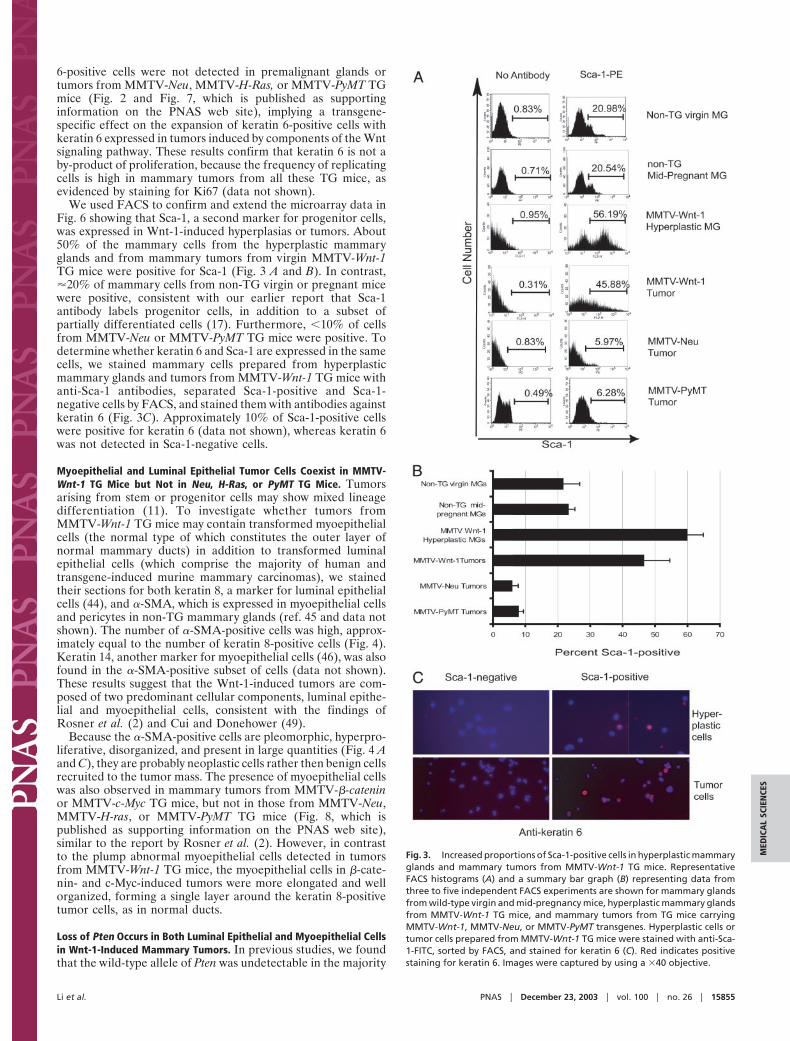
We used FACS to confirm and extend the microarray data inFig. 6 showing that Sca-1, a second marker for progenitor cells,was expressed in Wnt-1-induced hyperplasias or tumors. About50% of the mammary cells from the hyperplastic mammaryglands and from mammary tumors from virgin MMTV-Wnt-1TG mice were positive for Sca-1 (Fig. 3 A and B). In contrast,�20% of mammary cells from non-TG virgin or pregnant micewere positive, consistent with our earlier report that Sca-1antibody labels progenitor cells, in addition to a subset ofpartially differentiated cells (17). Furthermore, �10% of cellsfrom MMTV-Neu or MMTV-PyMT TG mice were positive. Todetermine whether keratin 6 and Sca-1 are expressed in the samecells, we stained mammary cells prepared from hyperplasticmammary glands and tumors from MMTV-Wnt-1 TG mice withanti-Sca-1 antibodies, separated Sca-1-positive and Sca-1-negative cells by FACS, and stained them with antibodies againstkeratin 6 (Fig. 3C). Approximately 10% of Sca-1-positive cellswere positive for keratin 6 (data not shown), whereas keratin 6was not detected in Sca-1-negative cells.
Myoepithelial and Luminal Epithelial Tumor Cells Coexist in MMTV-Wnt-1 TG Mice but Not in Neu, H-Ras, or PyMT TG Mice. Tumorsarising from stem or progenitor cells may show mixed lineagedifferentiation (11). To investigate whether tumors fromMMTV-Wnt-1 TG mice may contain transformed myoepithelialcells (the normal type of which constitutes the outer layer ofnormal mammary ducts) in addition to transformed luminalepithelial cells (which comprise the majority of human andtransgene-induced murine mammary carcinomas), we stainedtheir sections for both keratin 8, a marker for luminal epithelialcells (44), and �-SMA, which is expressed in myoepithelial cellsand pericytes in non-TG mammary glands (ref. 45 and data notshown). The number of �-SMA-positive cells was high, approx-imately equal to the number of keratin 8-positive cells (Fig. 4).Keratin 14, another marker for myoepithelial cells (46), was alsofound in the �-SMA-positive subset of cells (data not shown).These results suggest that the Wnt-1-induced tumors are com-posed of two predominant cellular components, luminal epithe-lial and myoepithelial cells, consistent with the findings ofRosner et al. (2) and Cui and Donehower (49).
Because the �-SMA-positive cells are pleomorphic, hyperpro-liferative, disorganized, and present in large quantities (Fig. 4 Aand C), they are probably neoplastic cells rather then benign cellsrecruited to the tumor mass. The presence of myoepithelial cellswas also observed in mammary tumors from MMTV-�-cateninor MMTV-c-Myc TG mice, but not in those from MMTV-Neu,MMTV-H-ras, or MMTV-PyMT TG mice (Fig. 8, which ispublished as supporting information on the PNAS web site),similar to the report by Rosner et al. (2). However, in contrastto the plump abnormal myoepithelial cells detected in tumorsfrom MMTV-Wnt-1 TG mice, the myoepithelial cells in �-cate-nin- and c-Myc-induced tumors were more elongated and wellorganized, forming a single layer around the keratin 8-positivetumor cells, as in normal ducts.
Loss of Pten Occurs in Both Luminal Epithelial and Myoepithelial Cellsin Wnt-1-Induced Mammary Tumors. In previous studies, we foundthat the wild-type allele of Pten was undetectable in the majority
Fig. 3. Increased proportions of Sca-1-positive cells in hyperplastic mammaryglands and mammary tumors from MMTV-Wnt-1 TG mice. RepresentativeFACS histograms (A) and a summary bar graph (B) representing data fromthree to five independent FACS experiments are shown for mammary glandsfrom wild-type virgin and mid-pregnancy mice, hyperplastic mammary glandsfrom MMTV-Wnt-1 TG mice, and mammary tumors from TG mice carryingMMTV-Wnt-1, MMTV-Neu, or MMTV-PyMT transgenes. Hyperplastic cells ortumor cells prepared from MMTV-Wnt-1 TG mice were stained with anti-Sca-1-FITC, sorted by FACS, and stained for keratin 6 (C). Red indicates positivestaining for keratin 6. Images were captured by using a �40 objective.
Li et al. PNAS � December 23, 2003 � vol. 100 � no. 26 � 15855
MED
ICA
LSC
IEN
CES
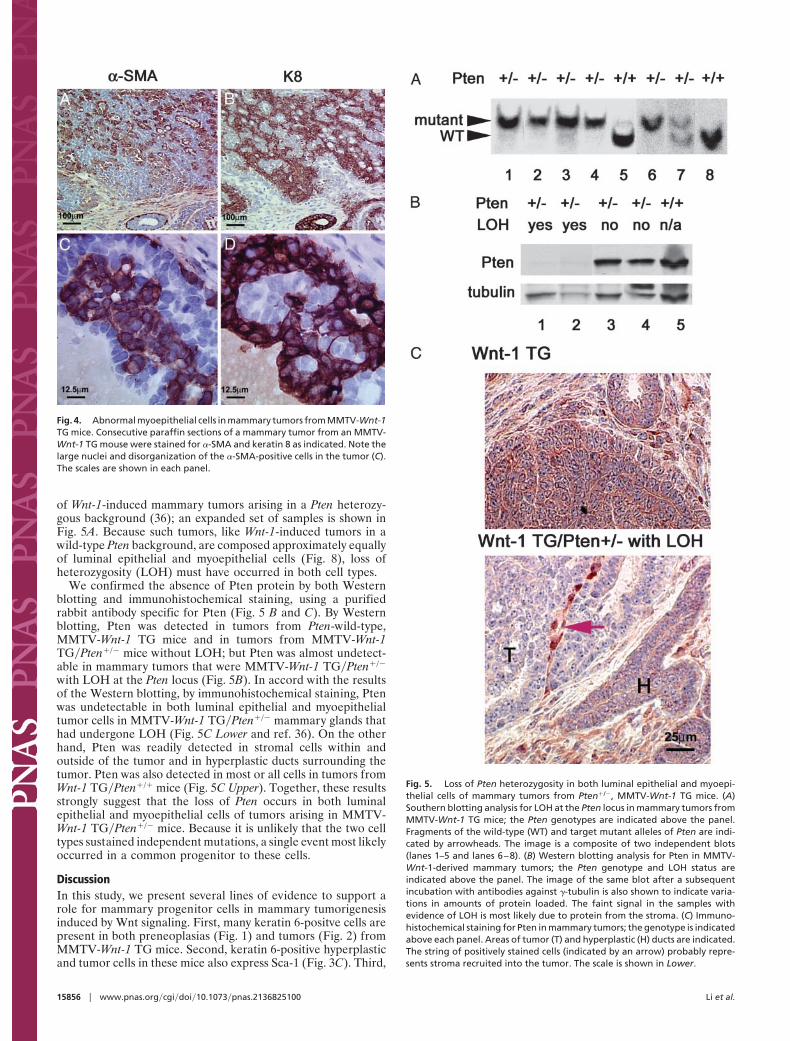
of Wnt-1-induced mammary tumors arising in a Pten heterozy-gous background (36); an expanded set of samples is shown inFig. 5A. Because such tumors, like Wnt-1-induced tumors in awild-type Pten background, are composed approximately equallyof luminal epithelial and myoepithelial cells (Fig. 8), loss ofheterozygosity (LOH) must have occurred in both cell types.
We confirmed the absence of Pten protein by both Westernblotting and immunohistochemical staining, using a purifiedrabbit antibody specific for Pten (Fig. 5 B and C). By Westernblotting, Pten was detected in tumors from Pten-wild-type,MMTV-Wnt-1 TG mice and in tumors from MMTV-Wnt-1TG�Pten�/� mice without LOH; but Pten was almost undetect-able in mammary tumors that were MMTV-Wnt-1 TG�Pten�/�
with LOH at the Pten locus (Fig. 5B). In accord with the resultsof the Western blotting, by immunohistochemical staining, Ptenwas undetectable in both luminal epithelial and myoepithelialtumor cells in MMTV-Wnt-1 TG�Pten�/� mammary glands thathad undergone LOH (Fig. 5C Lower and ref. 36). On the otherhand, Pten was readily detected in stromal cells within andoutside of the tumor and in hyperplastic ducts surrounding thetumor. Pten was also detected in most or all cells in tumors fromWnt-1 TG�Pten�/� mice (Fig. 5C Upper). Together, these resultsstrongly suggest that the loss of Pten occurs in both luminalepithelial and myoepithelial cells of tumors arising in MMTV-Wnt-1 TG�Pten�/� mice. Because it is unlikely that the two celltypes sustained independent mutations, a single event most likelyoccurred in a common progenitor to these cells.
DiscussionIn this study, we present several lines of evidence to support arole for mammary progenitor cells in mammary tumorigenesisinduced by Wnt signaling. First, many keratin 6-positve cells arepresent in both preneoplasias (Fig. 1) and tumors (Fig. 2) fromMMTV-Wnt-1 TG mice. Second, keratin 6-positive hyperplasticand tumor cells in these mice also express Sca-1 (Fig. 3C). Third,
Fig. 5. Loss of Pten heterozygosity in both luminal epithelial and myoepi-thelial cells of mammary tumors from Pten�/�, MMTV-Wnt-1 TG mice. (A)Southern blotting analysis for LOH at the Pten locus in mammary tumors fromMMTV-Wnt-1 TG mice; the Pten genotypes are indicated above the panel.Fragments of the wild-type (WT) and target mutant alleles of Pten are indi-cated by arrowheads. The image is a composite of two independent blots(lanes 1–5 and lanes 6–8). (B) Western blotting analysis for Pten in MMTV-Wnt-1-derived mammary tumors; the Pten genotype and LOH status areindicated above the panel. The image of the same blot after a subsequentincubation with antibodies against �-tubulin is also shown to indicate varia-tions in amounts of protein loaded. The faint signal in the samples withevidence of LOH is most likely due to protein from the stroma. (C) Immuno-histochemical staining for Pten in mammary tumors; the genotype is indicatedabove each panel. Areas of tumor (T) and hyperplastic (H) ducts are indicated.The string of positively stained cells (indicated by an arrow) probably repre-sents stroma recruited into the tumor. The scale is shown in Lower.
Fig. 4. Abnormal myoepithelial cells in mammary tumors from MMTV-Wnt-1TG mice. Consecutive paraffin sections of a mammary tumor from an MMTV-Wnt-1 TG mouse were stained for �-SMA and keratin 8 as indicated. Note thelarge nuclei and disorganization of the �-SMA-positive cells in the tumor (C).The scales are shown in each panel.
15856 � www.pnas.org�cgi�doi�10.1073�pnas.2136825100 Li et al.
luminal epithelial and myoepithelial cells coexist in these tumors,suggesting transformation of a common precursor. Finally, insome mammary tumors arising in Pten heterozygous, MMTV-Wnt-1 TG mice, the wild-type Pten locus is missing in bothluminal epithelial and myoepithelial cells (Fig. 5), suggesting thatloss of Pten occurred in a common precursor to these twopopulations of tumor cells. Efforts are underway to attempt todefine a subset of tumor cells from MMTV-Wnt-1 TG micecapable of cancer regeneration, in light of a recent report (47)that only a small number of human breast cancer cells have thecapacity to regenerate tumors in immunodeficient mice.
Mammary tumors from MMTV-�-catenin and MMTV-c-Mycmice, which express components of the Wnt signaling pathway,are similar to tumors from MMTV-Wnt-1 TG mice. Collectively,our data suggest that deregulated Wnt signaling causes excessproliferation of mammary progenitor cells and predisposes themto cancer. This interpretation is consistent with reports that lossof Wnt signaling blocks early mammary development (25, 26),and it is also consistent with the role of Wnt signaling in othertissues. Wnt signaling is now known to play a critical role in theproliferation of hematopoietic stem cells and those in the skin,colon, and other organs (18–28), and deregulated activation ofthis pathway is a cancer-predisposing factor in several tissuesincluding the colon, skin, and liver (19, 48).
Although progenitor cells are the likely precursors to cancerin mammary glands that have activated the Wnt signalingpathway, keratin 6 and�or Sca-1 are not present in mammaryhyperplasias and tumors from several other TG lines, includingMMTV-Neu, MMTV-H-Ras, and MMTV-PyMT TG mice (Figs.2 and 7). In addition, a detectable second tumor cell type, suchas myoepithelial tumor cells, is absent in these tumors (Fig. 8).These results are consistent with previous reports that initiatinggenetic lesions exert a significant influence on both gene ex-pression patterns and histological features of mammary tumorsfrom both humans and TG animals (1–8). However, our findingsalso suggest that the differentiation status of putative target cellsmay contribute to the heterogeneity of breast cancer. There areseveral possible mechanisms by which target cells may influencethe histopathology and molecular features of tumors resultingfrom different oncogenes. Transgenes encoding Neu, H-Ras,and PyMT may transform progenitor cells, but either fail toarrest them at the progenitor stage or actively induce differen-tiation of the transformed cells. Alternatively, Neu, H-Ras, andPyMT may be able to transform only more differentiated cellsthat no longer express keratin 6 or Sca-1. In addition, there is aremote possibility that different oncogene RNAs expressed from
MMTV are differentially translated in progenitor and differen-tiated cells. Finally, we cannot rule out the possibility that all ofthese oncogenes transform differentiated cells, but Wnt-1 sig-naling leads to dedifferentiation; however, the loss of Pten inboth myoepithelial and luminal epithelial cells argues stronglyagainst this possibility.
Although it remains to be determined whether the Wnt-1signaling pathway and the oncogenic pathways mediated by Neu,H-Ras, and PyMT induce tumors in mammary cells at preciselythe same stage of differentiation, these transgenes are presum-ably expressed in similar populations of mammary cells, becausethey are all under the control of the MMTV LTR. In accord withthis assumption, interbreeding of MMTV-c-Myc and MMTV-H-Ras led to more rapid formation of mammary tumors (33); butdifferentiation markers have not been studied in these tumors toask whether the H-Ras- or c-Myc-induced phenotype predomi-nates. To ask this question in a different context, we haverecently bred MMTV-Wnt-1 with MMTV-Neu TG mice. Theresulting bi-TG females develop mammary tumors as early as 12weeks of age, sooner than in mice carrying either transgenealone, implying that both transgenes were expressed in thesetumors (K.P. and Y.L., unpublished work). Interestingly, neo-plastic cells in these tumors are positive for keratin 6, Sca-1, andmyoepithelial markers, and they are histopathologically moresimilar to Wnt-1-induced tumors than to Neu-induced tumors(K.P. and Y.L., unpublished work). Therefore, it appears thatNeu does not relieve the arrest of differentiation that may beimposed by Wnt-1 signaling in mammary progenitor cells.
In conclusion, we provide several lines of evidence to suggestthat components of the Wnt signaling pathway transform mam-mary progenitors, and that these cells develop into heteroge-neous tumors containing different histological cell types express-ing markers of both mature and immature epithelial cells. Thus,breast cancer heterogeneity may result from transformation ofdistinct cell types by different oncogenes.
We thank Jennifer Doherty and Mary Barrett for excellent technicalassistance, Jose Vargas for animal care, Eric Holland and Gilbert Smithfor stimulating discussions, and William Pao, Brian Lewis, and GaryChamness for critical reading of the manuscript. Y.L. and S.H. weresupported by Department of Defense Breast Cancer Research Programawards, K.P. was supported by a Cancer Research Institute award, B.W.was supported by funds from the National Institutes of Health (GrantsCA057621 and ES07106) to Z.W., and M.E. was supported by the DanishCancer Society. This work was supported in part by National Institutesof Health Grants U01-CA842243 (to J.M.R.) and P01 CA94060-02 (toH.E.V.) and funds from the Martell Foundation (to H.E.V.).
1. Morrison, B. W. & Leder, P. (1994) Oncogene 9, 3417–3426.2. Rosner, A., Miyoshi, K., Landesman-Bollag, E., Xu, X., Seldin, D. C., Moser,
A. R., MacLeod, C. L., Shyamala, G., Gillgrass, A. E. & Cardiff, R. D. (2002)Am. J. Pathol. 161, 1087–1097.
3. Cardiff, R. D., Anver, M. R., Gusterson, B. A., Hennighausen, L., Jensen,R. A., Merino, M. J., Rehm, S., Russo, J., Tavassoli, F. A., Wakefield, L. M.,et al. (2000) Oncogene 19, 968–988.
4. Cardiff, R. D., Sinn, E., Muller, W. & Leder, P. (1991) Am. J. Pathol. 139,495–501.
5. Hedenfalk, I., Duggan, D., Chen, Y., Radmacher, M., Bittner, M., Simon, R.,Meltzer, P., Gusterson, B., Esteller, M., Kallioniemi, O. P., et al. (2001) N. Engl.J. Med. 344, 539–548.
6. Desai, K. V., Xiao, N., Wang, W., Gangi, L., Greene, J., Powell, J. I., Dickson,R., Furth, P., Hunter, K., Kucherlapati, R., et al. (2002) Proc. Natl. Acad. Sci.USA 99, 6967–6972.
7. Perou, C. M., Sorlie, T., Eisen, M. B., van de Rijn, M., Jeffrey, S. S., Rees, C. A.,Pollack, J. R., Ross, D. T., Johnsen, H., Akslen, L. A., et al. (2000) Nature 406,747–752.
8. Sorlie, T., Perou, C. M., Tibshirani, R., Aas, T., Geisler, S., Johnsen, H., Hastie,T., Eisen, M. B., van de Rijn, M., Jeffrey, S. S., et al. (2001) Proc. Natl. Acad.Sci. USA 98, 10869–10874.
9. Wagner, K. U., McAllister, K., Ward, T., Davis, B., Wiseman, R. & Hen-nighausen, L. (2001) Transgenic Res. 10, 545–553.
10. Gunther, E. J., Belka, G. K., Wertheim, G. B., Wang, J., Hartman, J. L., Boxer,R. B. & Chodosh, L. A. (2002) FASEB J. 16, 283–292.
11. Owens, D. M. & Watt, F. M. (2003) Nat. Rev. Cancer 3, 444–451.12. Perez-Losada, J. & Balmain, A. (2003) Nat. Rev. Cancer 3, 434–443.13. Grimm, S. L., Seagroves, T. N., Kabotyanski, E. B., Hovey, R. C., Vonderhaar,
B. K., Lydon, P., Miyoshi, K., Hennighausen, L., Ormandy, C. J., Lee, A. V.,et al. (2002) Mol. Endocrinol. 16, 2675–2691.
14. Sapino, A., Macri, L., Gugliotta, P., Pacchioni, D., Liu, Y. J., Medina, D. &Bussolati, G. (1993) Differentiation 55, 13–18.
15. Smith, G. H., Mehrel, T. & Roop, D. R. (1990) Cell Growth Differ. 1, 161–170.16. Spangrude, G. J., Heimfeld, S. & Weissman, I. L. (1988) Science 241, 58–62.17. Welm, B. E., Tepera, S. B., Venezia, T., Graubert, T. A., Rosen, J. M. &
Goodell, M. A. (2002) Dev. Biol. 245, 42–56.18. Dontu, G., Abdallah, W. M., Foley, J. M., Jackson, K. W., Clarke, M. F.,
Kawamura, M. J. & Wicha, M. S. (2003) Genes Dev. 17, 1253–1270.19. Alonso, L. & Fuchs, E. (2003) Genes Dev. 17, 1189–1200.20. Reya, T., Duncan, A. W., Ailles, L., Domen, J., Scherer, D. C., Willert, K.,
Hintz, L., Nusse, R. & Weissman, I. L. (2003) Nature 423, 409–414.21. van de Wetering, M., Sancho, E., Verweij, C., de Lau, W., Oving, I., Hurlstone,
A., van der Horn, K., Batlle, E., Coudreuse, D., Haramis, A. P., et al. (2002)Cell 111, 241–250.
22. Kielman, M. F., Rindapaa, M., Gaspar, C., van Poppel, N., Breukel, C., vanLeeuwen, S., Taketo, M. M., Roberts, S., Smits, R. & Fodde, R. (2002) Nat.Genet. 32, 594–605.
Li et al. PNAS � December 23, 2003 � vol. 100 � no. 26 � 15857
MED
ICA
LSC
IEN
CES

23. Polesskaya, A., Seale, P. & Rudnicki, M. A. (2003) Cell 113, 841–852.24. Yamashita, Y. M., Jones, D. L. & Fuller, M. T. (2003) Science 301, 1547–1550.25. Andl, T., Reddy, S. T., Gaddapara, T. & Millar, S. E. (2002) Dev. Cell 2, 643–653.26. van Genderen, C., Okamura, R. M., Farinas, I., Quo, R. G., Parslow, T. G.,
Bruhn, L. & Grosschedl, R. (1994) Genes Dev. 8, 2691–2703.27. Willert, K., Brown, J. D., Danenberg, E., Duncan, A. W., Weissman, I. L., Reya,
T., Yates, J. R., III, & Nusse, R. (2003) Nature 423, 448–452.28. Murdoch, B., Chadwick, K., Martin, M., Shojaei, F., Shah, K. V., Gallacher, L.,
Moon, R. T. & Bhatia, M. (2003) Proc. Natl. Acad. Sci. USA 100, 3422–3427.29. Tsukamoto, A. S., Grosschedl, R., Guzman, R. C., Parslow, T. & Varmus, H. E.
(1988) Cell 55, 619–625.30. Imbert, A., Eelkema, R., Jordan, S., Feiner, H. & Cowin, P. (2001) J. Cell Biol.
153, 555–568.31. Stewart, T. A., Pattengale, P. K. & Leder, P. (1984) Cell 38, 627–637.32. Guy, C. T., Webster, M. A., Schaller, M., Parsons, T. J., Cardiff, R. D. & Muller,
W. J. (1992) Proc. Natl. Acad. Sci. USA 89, 10578–10582.33. Sinn, E., Muller, W., Pattengale, P., Tepler, I., Wallace, R. & Leder, P. (1987)
Cell 49, 465–475.34. Guy, C. T., Cardiff, R. D. & Muller, W. J. (1992) Mol. Cell. Biol. 12, 954–961.35. Podsypanina, K., Ellenson, L. H., Nemes, A., Gu, J., Tamura, M., Yamada,
K. M., Cordon-Cardo, C., Catoretti, G., Fisher, P. E. & Parsons, R. (1999) Proc.Natl. Acad. Sci. USA 96, 1563–1568.
36. Li, Y., Podsypanina, K., Liu, X., Crane, A., Tan, L. K., Parsons, R. & Varmus,H. E. (2001) BMC Mol. Biol. 2, 2.
37. Brulet, P., Babinet, C., Kemler, R. & Jacob, F. (1980) Proc. Natl. Acad. Sci. USA77, 4113–4117.
38. Kemler, R., Brulet, P., Schnebelen, M. T., Gaillard, J. & Jacob, F. (1981) J.Embryol. Exp. Morphol. 64, 45–60.
39. Hennighausen, L., Wall, R. J., Tillmann, U., Li, M. & Furth, P. A. (1995) J. Cell.Biochem. 59, 463–472.
40. Hennighausen, L., McKnight, R., Burdon, T., Baik, M., Wall, R. J. & Smith,G. H. (1994) Cell Growth Differ. 5, 607–613.
41. Stocklin, E., Botteri, F. & Groner, B. (1993) J. Cell Biol. 122, 199–208.42. Muller, W. J., Sinn, E., Pattengale, P. K., Wallace, R. & Leder, P. (1988) Cell
54, 105–115.43. Moll, R., Franke, W. W., Schiller, D. L., Geiger, B. & Krepler, R. (1982) Cell
31, 11–24.44. Guelstein, V. I., Tchypysheva, T. A., Ermilova, V. D., Litvinova, L. V.,
Troyanovsky, S. M. & Bannikov, G. A. (1988) Int. J. Cancer 42, 147–153.45. Lazard, D., Sastre, X., Frid, M. G., Glukhova, M. A., Thiery, J. P. &
Koteliansky, V. E. (1993) Proc. Natl. Acad. Sci. USA 90, 999–1003.46. Wetzels, R. H., Holland, R., van Haelst, U. J., Lane, E. B., Leigh, I. M. &
Ramaekers, F. C. (1989) Am. J. Pathol. 134, 571–579.47. Al-Hajj, M., Wicha, M. S., Benito-Hernandez, A., Morrison, S. J. & Clarke,
M. F. (2003) Proc. Natl. Acad. Sci. USA 100, 3983–3988.48. Bienz, M. & Clevers, H. (2000) Cell 103, 311–320.49. Cui, X. S. & Donehower, L. A. (2000) Oncogene 19, 5988–5996.
15858 � www.pnas.org�cgi�doi�10.1073�pnas.2136825100 Li et al.