evidence for induction of dna double strand breaks in the bystander response to targeted soft x-rays...
TRANSCRIPT

Mutation Research 556 (2004) 209–215
Evidence for induction of DNA double strand breaks in thebystander response to targeted soft X-rays in CHO cells
Genro Kashinoa,b,1, Kevin M. Prisea,∗, Giuseppe Schettinoa, Melvyn Folkarda, BorivojVojnovica, Barry D. Michaela, Keiji Suzukib,1, Seiji Kodamab,1, Masami Watanabeb,1
a Gray Cancer Institute, P.O. Box 100, Mount Vernon Hospital, Northwood, Middlesex HA6 2JR, UKb Division of Radiation Biology, Department of Radiology and Radiation Biology, Course of Life Sciences and Radiation Research,
Graduate School of Biomedical Sciences, Nagasaki University, 1-14 Bunkyo-machi, Nagasaki 852-8521, Japan
Received 15 June 2004; received in revised form 17 August 2004; accepted 20 August 2004
Abstract
This study investigated the role of DNA double strand breaks and DNA base damage in radiation-induced bystander responsesin Chinese hamster ovary (CHO) cell lines. Two CHO repair-deficient clones, xrs5 (DNA double strand break repair-deficient)and EM9 (DNA base excision repair-deficient) were used in addition to the wild type (CHO). The Gray Cancer Institute ultrasoftX-ray microprobe is a powerful tool for investigating the bystander response, because it permits the irradiation of only a singlenucleus of a cell, as reported previously. In order to investigate the bystander effect in each repair-deficient cell line, we irradiateda targetedw ell lines,w in EM9.U ell lines,s e effects,w ted cellsw t that DNAd©
K
f mit-ion,der
0
single cell within a population and scored the formation of micronuclei. When a single nucleus in the population wasith 1 Gy, elevated numbers of micronuclei were induced in the neighbouring unirradiated cells in the EM9 and xrs5 chereas induction was not observed in CHO. The induction of micronuclei in xrs5 was significantly higher than thatnder these conditions, the surviving fraction in the neighbouring cells was significantly lower in xrs5 than in the other chowing a higher cell killing effect in xrs5. To confirm that bystander factors secreted from irradiated cells caused these carried out medium transfer experiments using conventional X-irradiation. Medium conditioned for 24 h with irradiaas transferred to unirradiated cells and elevated induction of micronuclei was observed in xrs5. These results suggesouble strand breaks rather than base damage are caused by factors secreted in the medium from irradiated cells.2004 Elsevier B.V. All rights reserved.
eywords:Bystander effect; DNA repair; DNA double strand break; Base damage; Soft X-ray; Microbeam
∗ Corresponding author. Tel.: +44 192 382 8611;ax: +44 192 383 5210.
E-mail address:[email protected] (K.M. Prise).1 Tel.: +81 95 819 2459; fax: +81 95 819 2459.
1. Introduction
It is thought that damage signals may be transted from irradiated to unirradiated cells in a populatleading to a variety of genetic effects via a bystan
027-5107/$ – see front matter © 2004 Elsevier B.V. All rights reserved.doi:10.1016/j.mrfmmm.2004.08.009
210 G. Kashino et al. / Mutation Research 556 (2004) 209–215
effect. It has been reported that the bystander effectcan be mediated via gap junction intercellular commu-nication[1–3] and also factors secreted from irradiatedcells via the culture medium in vitro[3–6]. As it hasbeen reported that bystander cells show a variety ofcellular effects which result in cell death and chromo-some aberrations, DNA damage should be observedin a radiation-induced bystander response[1–6]. How-ever, it is still not clear which type of DNA damage isresponsible for bystander effects.
It is suggested that the bystander effect induced byionizing radiation might lead to carcinogenesis. In vitromutation assays have been useful for the estimation ofthe risk of carcinogenesis. Little et al. have reportedthat the mutation frequency at hprt locus is increasedin bystander Chinese hamster ovary (CHO) cells whichare exposed to very low fluences ofα particles[7].Zhou et al. showed a higher frequency of gene muta-tions in bystander cells at the CD59 locus in a hybridhamster cell line by using alpha particle microbeamirradiation[2]. However, PCR analysis by Zhou et al.showed that a higher yields of deletion mutations wereinduced by microbeam irradiation in bystander cells,whereas most mutations analyzed by Little et al. weresuggested to be mainly point mutations in bystandercells[2,7]. Thus, these two reports are in disagreementregarding the type of mutations produced in bystandercells. Investigations are necessary to resolve the typeof DNA damage resulting from the radiation-inducedbystander effect.
ro-c bles twomh airpr izest NA-P tel ithK re-j isw essl e-c aks[ alsoi iatedw ual
tracks crossing the DNA[16]. Base damage is repairedby base excision repair processes, where XRCC1 isimportant for the activation of ligase III which linksthe digested strands in this repair process[17,18]. Adefect in XRCC1 leads to hypersensitivity to alkyat-ing agents[19]. These repair mechanisms recognize aspecific damage immediately after irradiation and re-move it. Therefore, it is difficult to detect the exact levelof each specific type of DNA damage induced by ra-diation, especially at low doses. DNA repair-deficientChinese hamster ovary (CHO) cell lines, which are de-ficient in Ku80 and XRCC1, have been used to detectDNA damage efficiently, and greatly facilitates the de-tection of small numbers of DNA damages as describedin a previous report[14].
Microbeams are useful for investigation of by-stander responses. The ultrasoft X-ray microprobe atGray Cancer Institute is the first microbeam facilityto use X-rays for radiobiological purposes, and is ableto irradiate a single cell with beams focused down tomicrometer accuracy[20–24]. We reported previouslythat irradiating a single V79 cell with X-rays leads tobystander cell killing in about 10% of the cells in thepopulation[24]. The study of the bystander effect usingthis soft X-ray microbeam is expected to be applicableto not only to survival assays but also to other biologicalendpoints used for detection of chromosome damage,such as the in vitro micronucleus assay, as this assay hasalready been used in bystander studies utilising chargedparticle microbeams[6].
ffecti ellsi air-d tingt ormo ls.
2
2
ellsw iaU edf ,U In-v 0%
DNA damage is repaired by several efficient pesses within cells. For the repair of DNA doutrand breaks, molecular studies have elucidatedain pathways after direct irradiation of cells[8]. Non-omologous end joining (NHEJ) is the main repathways for DNA double strand breaks[9]. In thisepair process the Ku70/80 protein complex stabilhe ends of the fragmented DNA strands and the DK catalytic subunit (DNA-PKcs), which may activa
igase IV with XRCC4, is activated by association wu complex. Finally, activated ligase IV leads to
oining reactions in the two ends of the DNA. Itell known that a defect of any protein in this proc
eads to higher cell killing effect after irradiation bause of less repair ability of DNA double strand bre10–15]. On the other hand, DNA base damage isnduced by irradiation and many of these are associth clustered damage formed at the sites of individ
The present results indicate that the bystander en DNA double strand break repair-deficient xrs5 cs significantly higher than that in base excision repeficient EM9 cells and control CHO cells, sugges
hat DNA double strand breaks are the principal ff damage observed in unirradiated bystander cel
. Materials and methods
.1. Cell culture
Chinese hamster ovary (CHO) cells and xrs5 cere kindly supplied by Dr. Tom K. Hei, Columbniversity, New York, and EM9 cells were purchas
rom ATCC (American Type Culture Collection, VASA). Cells were cultured in MEM alpha medium (itrogen Ltd., Paisley, UK) supplemented with 1

G. Kashino et al. / Mutation Research 556 (2004) 209–215 211
FBS (Helena Biosciences Europe), 100 units/ml peni-cillin and 100�g/ml streptomycin (Invitrogen Ltd.,Paisley, UK). Cells were maintained at 37◦C in a hu-midified atmosphere with 5% CO2.
2.2. Micronucleus assay
To investigate the induction of micronuclei by di-rect X-irradiation, the cells were irradiated with 0.2,0.5, and 1 Gy of conventional X-rays. Exponentiallygrowing cells in T25 flasks were irradiated with X-rays using an X-ray generator (Pantak IV) operating at240 kVp and 13 mA with a filter system composed of0.25 mm Cu plus 1 mm Al filter and 4.3 mm Al flatten-ing filter, at a dose rate of 0.5 Gy/min. Either imme-diately after irradiation or following 24 h incubation,cells were treated with 2�g/ml cytochalasin B for 24 hin a T25 flask. They were then harvested and treatedwith 3 ml of hypotonic (0.1 M) KCl for 20 min, andfixed with 3 ml of methanol–acetic acid (5:1). The cellsuspensions were centrifuged at 1200 rpm for 5 min,the supernatant removed and cells resuspended in 4 mlmethanol–acetic acid solution and incubated on ice for5 min. After further centrifugation, the supernatant wasremoved and 0.5–1 ml methanol–acetic acid solutionwas added. Cells were resuspended and a sample wasdropped onto slides and stained with 7.5% Giemsa for40 min. Micronuclei per 2000 binucleated cells werecounted.
rra-d sti-t urehts ,U 42f ana in or-d cellw ellcp .
oser cellsw im-m erep
2.3. Survival assay
The surviving fraction was determined by a clono-genic survival assay. Individual cells, stained with100 nM Hoechst 33342, were scanned using the GrayCancer Institute X-ray microprobe system, as describedpreviously[20,24]. After 100–200 cells were scanned,a single cell was irradiated with 1 Gy of Alk or Ckproduced by a focused ultrasoft X-ray microprobe.Cells were incubated for 4 days, stained with 100 nMHoechst 33342, and the dishes scanned to revisit theoriginal locations and test for the presence of colonies.Control cells were scanned, without irradiation underthe same conditions, and surviving fractions were cal-culated.
2.4. Medium transfer experiment
Cells (5× 10⁴) were seeded onto six-well platesone day prior to irradiation. Immediately before irradi-ation, medium was changed and cells were irradiatedwith 1 Gy of conventional X-rays. Cells were incubatedfor 24 h following irradiation. The culture medium wasfiltered through a 0.22�m filter and transferred to unir-radiated cultured cells on six-well plates. CytochalasinB was added at the same time as the medium trans-fer, and cells were incubated for 24 h. Micronucleussamples were prepared as described above.
2.5. Statistical analysis
per-f
3
3
X-r neduc easx .F er2 ere2m this
To investigate the bystander effect, localised iiation was carried out using the Gray Cancer In
ute focused ultrasoft X-ray microprobe. The procedas been described in detail elsewhere[20,24]. Briefly,
he day before, the experiment cells (5× 10⁴) wereeeded on 0.9�m-thick Mylar film (Goodfellow Ltd.K). Cells were stained with 100 nM Hoechst 333
or 1 h prior to irradiation. After removal of stain,rea around the centre of the dish was scanneder to identify a precise single nucleus. A singleas irradiated with 1 Gy of aluminum or carbon K-shharacteristic X-rays (Alk = 1.49 keV or Ck = 0.28 keV)roduced by a focused ultrasoft X-ray microprobe
The X-ray microbeam targeted a single cell at a date of 0.1 Gy/s. The medium was changed andere incubated with cytochalasin B for 24 h eitherediately after irradiation or 24 h later. Slides wrepared as described above.
The statistical analysis in the present study wasormed using Student’st-test.
. Results
.1. Direct effect of X-rays on repair-deficient cells
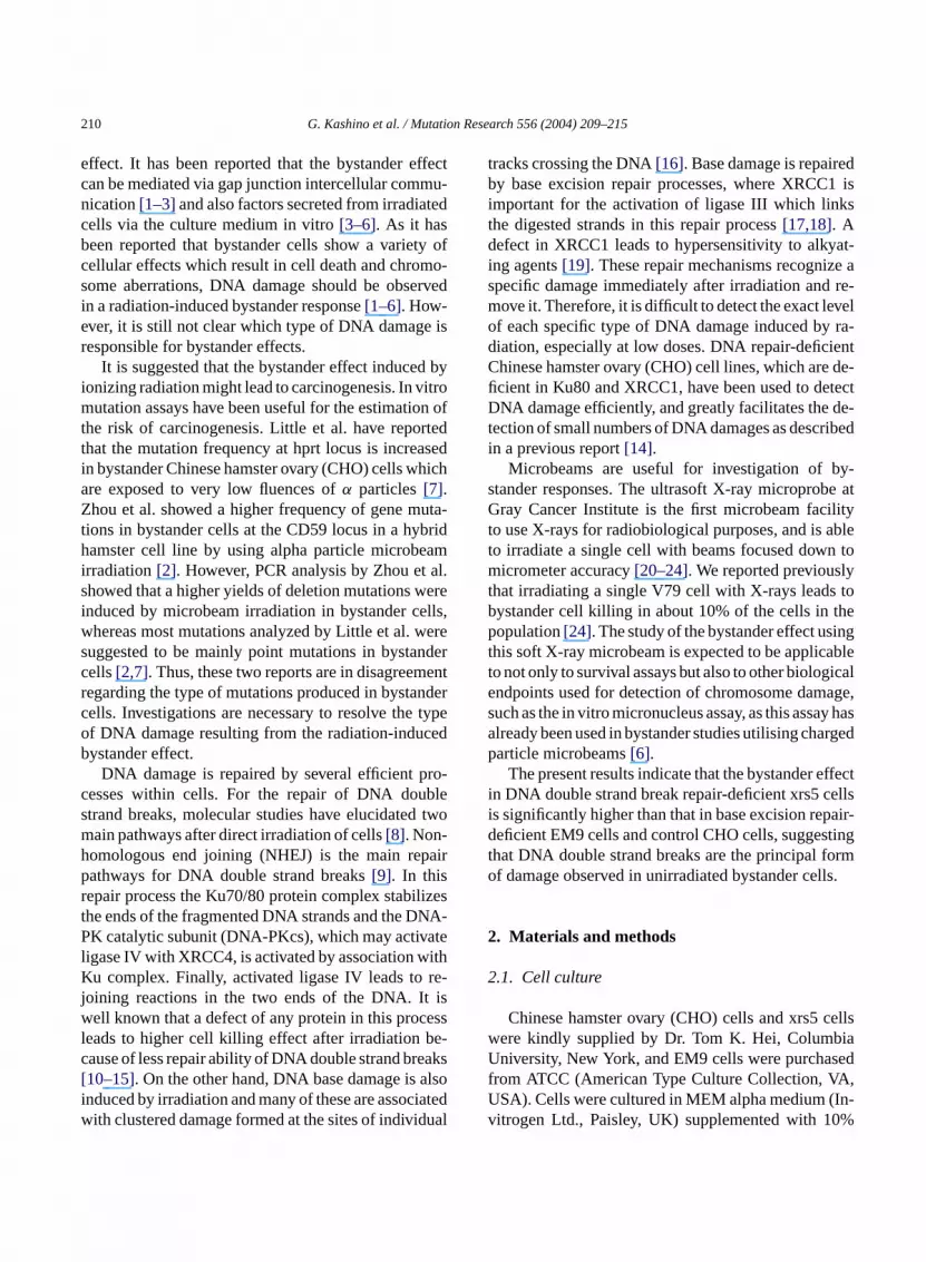
The sensitivity to direct irradiation by low-doseays in repair-deficient CHO cell lines were examising the micronucleus assay. As shown inFig. 1, EM9ells were slightly more sensitive than CHO, wherrs5 cells were significantly more sensitive (p< 0.001)ollowing 1 Gy-irradiation the yield of micronuclei p000 binucleated cells in CHO, EM9 and xrs5 w24, 465 and 1287, respectively (Fig. 1). Induction oficronuclei in xrs5 cells was also detectable in
212 G. Kashino et al. / Mutation Research 556 (2004) 209–215
Fig. 1. Micronuclei induced by conventional X-irradiation in all cellsof CHO (square), EM9 (circle) and xrs5 (triangle). Cells were in-cubated with cytochalasin B for 24 h immediately after irradiation.Results are means± S.D. from three separate experiments.
assay when cells were irradiated at 0.05 Gy of X-ray(data not shown). However, inductions of micronucleiin EM9 cells were not detectable at that dose. There-fore, direct irradiation with low-dose X-rays may causemicronuclei formation through unrepaired or misre-paired DNA double strand breaks in xrs5.
3.2. Bystander effects using X-ray microbeams inCHO repair-deficient cells
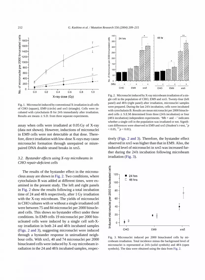
The results of the bystander effect in the micronu-cleus assay are shown inFig. 2. Two conditions, wherecytochalasin B was added at different times, were ex-amined in the present study. The left and right panelsin Fig. 2 show the results following a total incubationtime of 24 and 48 h respectively, after 1 Gy irradiationwith the X-ray microbeam. The yields of micronucleiin CHO cultures with or without a single irradiated cellwere between 75 and 84 micronuclei per 2000 binucle-ated cells. This shows no bystander effect under theseconditions. In EM9 cells 19 micronuclei per 2000 bin-ucleated cells were induced by a single cell soft-X-ray irradiation in both 24 and 48 h incubated samples(Figs. 2 and 3), suggesting micronuclei were inducedthrough a bystander response in unirradiated neigh-bour cells. With xrs5, 40 and 74 micronuclei per 2000binucleated cells were induced by X-ray microbeam ir-radiation in the 24 and 48 h incubated samples, respec-
Fig. 2. Micronuclei induced by X-ray microbeam irradiation of a sin-gle cell in the population of CHO, EM9 and xrs5. Twenty-four (leftpanel) and 48 h (right panel) after irradiation, micronuclei sampleswere prepared. During the last 24 h incubation, cells were incubatedwith cytochalasin B. Results are mean micronuclei per 2000 binucle-ated cells± S.E.M determined from three (24 h incubation) or four(48 h incubation) independent experiments. ‘Mb + and−’ indicateswhether a single cell in the population was irradiated or not. Signifi-cant differences were observed in EM9 and xrs5 (Student’st-test,*p< 0.05,** p < 0.01).
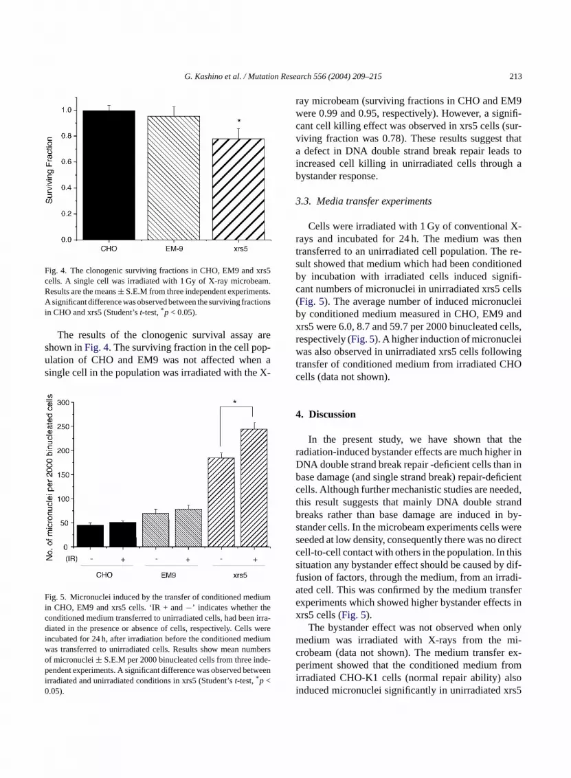
tively (Figs. 2 and 3). Therefore, the bystander effectobserved in xrs5 was higher than that in EM9. Also, theinduced level of micronuclei in xrs5 was increased fur-ther during the 24 h incubation following microbeamirradiation (Fig. 3).
Fig. 3. Micronuclei induced per 2000 binucleated cells by mi-crobeam irradiation. Total incidence minus the background level ofmicronuclei is represented at 24 h (solid symbols) and 48 h (opensymbols). The data were obtained using the data fromFig. 2.
G. Kashino et al. / Mutation Research 556 (2004) 209–215 213
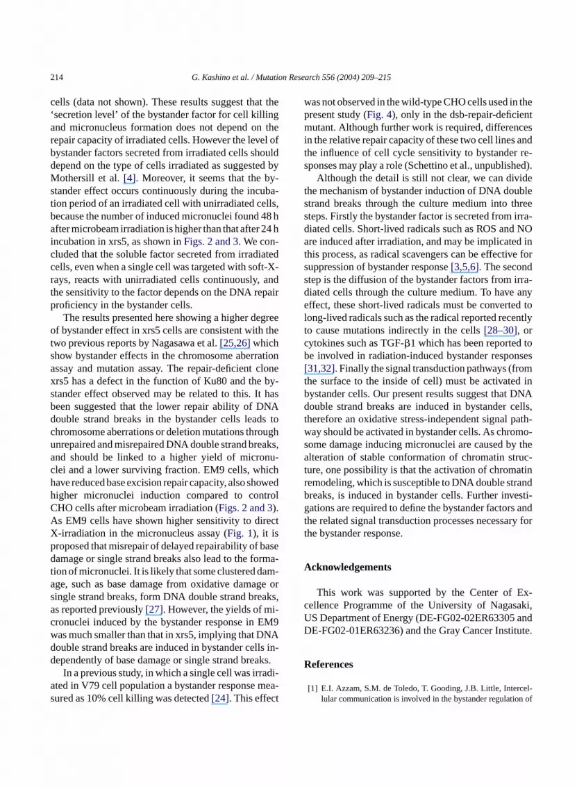
Fig. 4. The clonogenic surviving fractions in CHO, EM9 and xrs5cells. A single cell was irradiated with 1 Gy of X-ray microbeam.Results are the means± S.E.M from three independent experiments.A significant difference was observed between the surviving fractionsin CHO and xrs5 (Student’st-test,*p < 0.05).
The results of the clonogenic survival assay areshown inFig. 4. The surviving fraction in the cell pop-ulation of CHO and EM9 was not affected when asingle cell in the population was irradiated with the X-
Fig. 5. Micronuclei induced by the transfer of conditioned mediumin CHO, EM9 and xrs5 cells. ‘IR + and−’ indicates whether theconditioned medium transferred to unirradiated cells, had been irra-diated in the presence or absence of cells, respectively. Cells wereincubated for 24 h, after irradiation before the conditioned mediumwas transferred to unirradiated cells. Results show mean numbersof micronuclei± S.E.M per 2000 binucleated cells from three inde-pendent experiments. A significant difference was observed betweenirradiated and unirradiated conditions in xrs5 (Student’st-test,*p <0.05).
ray microbeam (surviving fractions in CHO and EM9were 0.99 and 0.95, respectively). However, a signifi-cant cell killing effect was observed in xrs5 cells (sur-viving fraction was 0.78). These results suggest thata defect in DNA double strand break repair leads toincreased cell killing in unirradiated cells through abystander response.
3.3. Media transfer experiments
Cells were irradiated with 1 Gy of conventional X-rays and incubated for 24 h. The medium was thentransferred to an unirradiated cell population. The re-sult showed that medium which had been conditionedby incubation with irradiated cells induced signifi-cant numbers of micronuclei in unirradiated xrs5 cells(Fig. 5). The average number of induced micronucleiby conditioned medium measured in CHO, EM9 andxrs5 were 6.0, 8.7 and 59.7 per 2000 binucleated cells,respectively (Fig. 5). A higher induction of micronucleiwas also observed in unirradiated xrs5 cells followingtransfer of conditioned medium from irradiated CHOcells (data not shown).
4. Discussion
In the present study, we have shown that theradiation-induced bystander effects are much higher inDNA double strand break repair -deficient cells than inb cientc ded,t andb n by-s weres irectc hiss dif-f di-a sfere ts inx
onlym i-c ex-p romi lsoi rs5
ase damage (and single strand break) repair-defiells. Although further mechanistic studies are neehis result suggests that mainly DNA double strreaks rather than base damage are induced itander cells. In the microbeam experiments cellseeded at low density, consequently there was no dell-to-cell contact with others in the population. In tituation any bystander effect should be caused byusion of factors, through the medium, from an irrated cell. This was confirmed by the medium tranxperiments which showed higher bystander effecrs5 cells (Fig. 5).
The bystander effect was not observed whenedium was irradiated with X-rays from the m
robeam (data not shown). The medium transfereriment showed that the conditioned medium f
rradiated CHO-K1 cells (normal repair ability) anduced micronuclei significantly in unirradiated x
214 G. Kashino et al. / Mutation Research 556 (2004) 209–215
cells (data not shown). These results suggest that the‘secretion level’ of the bystander factor for cell killingand micronucleus formation does not depend on therepair capacity of irradiated cells. However the level ofbystander factors secreted from irradiated cells shoulddepend on the type of cells irradiated as suggested byMothersill et al.[4]. Moreover, it seems that the by-stander effect occurs continuously during the incuba-tion period of an irradiated cell with unirradiated cells,because the number of induced micronuclei found 48 hafter microbeam irradiation is higher than that after 24 hincubation in xrs5, as shown inFigs. 2 and 3. We con-cluded that the soluble factor secreted from irradiatedcells, even when a single cell was targeted with soft-X-rays, reacts with unirradiated cells continuously, andthe sensitivity to the factor depends on the DNA repairproficiency in the bystander cells.
The results presented here showing a higher degreeof bystander effect in xrs5 cells are consistent with thetwo previous reports by Nagasawa et al.[25,26]whichshow bystander effects in the chromosome aberrationassay and mutation assay. The repair-deficient clonexrs5 has a defect in the function of Ku80 and the by-stander effect observed may be related to this. It hasbeen suggested that the lower repair ability of DNAdouble strand breaks in the bystander cells leads tochromosome aberrations or deletion mutations throughunrepaired and misrepaired DNA double strand breaks,and should be linked to a higher yield of micronu-clei and a lower surviving fraction. EM9 cells, whichh owedh trolCA ectXp ased rma-t m-a ge ors aks,a i-c M9w NAd ls in-d ks.
di-a ea-s t
was not observed in the wild-type CHO cells used in thepresent study (Fig. 4), only in the dsb-repair-deficientmutant. Although further work is required, differencesin the relative repair capacity of these two cell lines andthe influence of cell cycle sensitivity to bystander re-sponses may play a role (Schettino et al., unpublished).
Although the detail is still not clear, we can dividethe mechanism of bystander induction of DNA doublestrand breaks through the culture medium into threesteps. Firstly the bystander factor is secreted from irra-diated cells. Short-lived radicals such as ROS and NOare induced after irradiation, and may be implicated inthis process, as radical scavengers can be effective forsuppression of bystander response[3,5,6]. The secondstep is the diffusion of the bystander factors from irra-diated cells through the culture medium. To have anyeffect, these short-lived radicals must be converted tolong-lived radicals such as the radical reported recentlyto cause mutations indirectly in the cells[28–30], orcytokines such as TGF-�1 which has been reported tobe involved in radiation-induced bystander responses[31,32]. Finally the signal transduction pathways (fromthe surface to the inside of cell) must be activated inbystander cells. Our present results suggest that DNAdouble strand breaks are induced in bystander cells,therefore an oxidative stress-independent signal path-way should be activated in bystander cells. As chromo-some damage inducing micronuclei are caused by thealteration of stable conformation of chromatin struc-ture, one possibility is that the activation of chromatinr andb esti-g s andt ry fort
A
x-c aki,U andD tute.
R
el-n of
ave reduced base excision repair capacity, also shigher micronuclei induction compared to conHO cells after microbeam irradiation (Figs. 2 and 3).s EM9 cells have shown higher sensitivity to dir-irradiation in the micronucleus assay (Fig. 1), it isroposed that misrepair of delayed repairability of bamage or single strand breaks also lead to the fo
ion of micronuclei. It is likely that some clustered dage, such as base damage from oxidative damaingle strand breaks, form DNA double strand bres reported previously[27]. However, the yields of mronuclei induced by the bystander response in Eas much smaller than that in xrs5, implying that Double strand breaks are induced in bystander celependently of base damage or single strand brea
In a previous study, in which a single cell was irrated in V79 cell population a bystander response mured as 10% cell killing was detected[24]. This effec
emodeling, which is susceptible to DNA double strreaks, is induced in bystander cells. Further invations are required to define the bystander factor
he related signal transduction processes necessahe bystander response.
cknowledgements
This work was supported by the Center of Eellence Programme of the University of NagasS Department of Energy (DE-FG02-02ER63305E-FG02-01ER63236) and the Gray Cancer Insti
eferences
[1] E.I. Azzam, S.M. de Toledo, T. Gooding, J.B. Little, Interclular communication is involved in the bystander regulatio

G. Kashino et al. / Mutation Research 556 (2004) 209–215 215
gene expression in human cells exposed to very low fluences ofalpha particles, Radiat. Res. 150 (1998) 497–504.
[2] H. Zhou, G. Randers-Pehrson, C.A. Waldren, D. Vannais, E.J.Hall, T.K. Hei, Induction of a bystander mutagenic effect ofalpha particles in mammalian cells, Proc. Natl. Acad. Sci. USA97 (2000) 2099–2104.
[3] C. Shao, Y. Furusawa, M. Aoki, K. Ando, Role of gap junctionalintercellular communication in radiation-induced bystander ef-fects in human fibroblasts, Radiat. Res. 160 (2003) 318–323.
[4] C. Mothersill, C. Seymour, Medium from irradiated human ep-ithelial cells but not human fibroblasts reduces the clonogenicsurvival of unirradiated cells, Int. J. Radiat. Biol. 71 (1997)421–427.
[5] C. Shao, Y. Furusawa, M. Aoki, H. Matsumoto, K. Ando, Nitricoxide-mediated bystander effect induced by heavy-ions in hu-man salivary gland tumour cells, Int. J. Radiat. Biol. 78 (2002)837–844.
[6] C. Shao, V. Stewart, M. Folkard, B.D. Michael, K.M. Prise,Nitric oxide-mediated signaling in the bystander response ofindividually targeted glioma cells, Cancer Res. 63 (2003)8437–8442.
[7] L. Huo, H. Nagasawa, J.B. Little, HPRT mutants induced inbystander cells by very low fluences of alpha particles resultprimarily from point mutants, Radiat. Res. 156 (2001) 521–525.
[8] J.S. Bedford, W.C. Dewey, Historical and current highlightsin radiation biology: has anything important been learned byirradiating cells? Radiat. Res. 158 (2002) 251–291.
[9] K. Valerie, L.F. Povirk, Regulation and mechanisms ofmammalian double-strand break repair, Oncogene 22 (2003)5792–5812.
[10] Y. Gu, K.J. Seidl, G.A. Rathbun, C. Zhu, J.P. Manis, N. van derStoep, L. Davidson, H.L. Cheng, J.M. Sekiguchi, K. Frank, P.Stanhope-Baker, M.S. Schlissel, D.B. Roth, F.W. Alt, Growthretardation and leaky SCID phenotype of Ku70-deficient mice,
[ er-m-de-
593.[ es a
.[ , An
ina-alian
[ nes:ir 2
[ .R.ionlym-
[ atin
[ ist,NA
repair protein XRCC1 and DNA ligase III, Mol. Cell Biol. 14(1994) 68–76.
[18] E. Cappelli, R. Taylor, M. Cevasco, A. Abbondandolo, K.Caldecott, G. Frosina, Involvement of XRCC1 and DNA ligaseIII gene products in DNA base excision repair, J. Biol. Chem.272 (1997) 23970–23975.
[19] M.R. Shen, M.Z. Zdzienicka, H. Mohrenweiser, L.H. Thomp-son, M.P. Thelen, Mutations in hamster single-strand break re-pair gene XRCC1 causing defective DNA repair, Nucleic AcidsRes. (1998) 26.
[20] M. Folkard, G. Schettino, B. Vojnovic, S. Gilchrist, A.G.Michette, S.J. Pfauntsch, K.M. Prise, B.D. Michael, A focusedultrasoft X-ray microbeam for targeting cells individually withsubmicrometer accuracy, Radiat. Res. 156 (2001) 796–804.
[21] K.M. Prise, M. Folkard, B.D. Michael, Bystander responsesinduced by low LET radiation, Oncogene 22 (2003) 7043–7049.
[22] K.M. Prise, M. Folkard, B.D. Michael, A review of the by-stander effect and its implications for low-dose exposure, Ra-diat. Prot. Dosimetry 104 (2003) 347–355.
[23] G. Schettino, M. Folkard, K.M. Prise, B. Vojnovic, B.D.Michael, Upgrading of the Gray Laboratory soft X-ray micro-probe and V79 survival measurements following irradiation ofone or all cells with a CK X-ray beam of different size, Radiat.Prot. Dosimetry 99 (2002) 287–288.
[24] G. Schettino, M. Folkard, K.M. Prise, B. Vojnovic, K.D. Held,B.D. Michael, Low-dose studies of bystander cell killing withtargeted soft X-rays, Radiat. Res. 160 (2003) 505–511.
[25] H. Nagasawa, J.B. Little, Bystander effect for chromosomalaberrations induced in wild-type and repair-deficient CHO cellsby low fluences of alpha particles, Mutat. Res. 508 (2002)129–129.
[26] H. Nagasawa, L. Huo, J.B. Little, Increased bystander muta-genic effect in DNA double strand break repair-deficient mam-malian cells, Int. J. Radiat. Biol. 79 (2003) 35–41.
[27] B.M. Sutherland, P.V. Bennett, O. Sidorkina, J. Laval, Clustereds by97
[ n-mma-at 775 K,
[ aki,hich998)
[ , M.ced
zed
[ er-ted
9.[ cell
ancer
Immunity 7 (1997) 653–665.11] A. Nussenzweig, K. Sokol, P. Burgman, L. Li, G.C. Li, Hyp
sensitivity of Ku80-deficient cell lines and mice to DNA daage: the effects of ionizing radiation on growth, survival, andvelopment, Proc. Natl. Sci. Acad. USA 94 (1997) 13588–13
12] G.M. Fulop, R.A. Phillips, The scid mutation in mice causgeneral defect in DNA repair, Nature 343 (1990) 479–482
13] F. Delacote, M. Han, T.D. Stamato, M. Jasin, B.S. Lopezxrcc4 defect or wortmannin stimulates homologous recombtion specifically induced by double-strand breaks in mammcells, Nucleic Acids Res. 30 (2002) 3454–3463.
14] J. Thacker, M.Z. Zdzienicka, The mammalian XRCC getheir roles in DNA repair and genetic stability, DNA Repa(2003) 655–672.
15] U. Grawunder, D. Zimmer, S. Fugmann, K. Schwarz, MLieber, DNA ligase IV is essential for V(D)J recombinatand DNA double-strand break repair in human precursorphocytes, Mol. Cell. 2 (1998) 477–484.
16] B. Rydberg, Radiation-induced DNA damage and chromstructure, Acta Oncol. 40 (2001) 682–685.
17] K.W. Caldecott, C.K. McKeown, J.D. Tucker, S. LjungquL.H. Thompson, An interaction between the mammalian D
DNA damages induced in isolated DNA and in human celllow doses of ionizing radiation, Proc. Natl. Acad. Sci. USA(2000) 103–108.
28] T. Yoshimura, K. Matsuo, T. Miyazaki, K. Suzuki, M. Wataabe, Electron spin resonance studies of free radicals in gairradiated golden hamster embryo cells: radical formationand 295 K, and radioprotective effects of vitamin C at 29Radiat. Res. 136 (1993) 361–365.
29] S. Koyama, S. Kodama, K. Suzuki, T. Matsumoto, T. MiyazM. Watanabe, Radiation-induced long-lived radicals wcause mutation and transformation, Mutat. Res. 421 (145–54.
30] J. Kuamagai, K. Matsui, Y. Itagaki, M. Shiotani, S. KodamaWatanabe, T. Miyazaki, Long-lived mutagenic radicals induin mammalian cells by ionizing radiation are mainly localito proteins, Radiat. Res. 160 (2003) 95–102.
31] M.H. Barcellos-Hoff, R. Denynck, M.L. Tsang, J.A. Weathbee, Transforming growth factor-beta activation in irradiamurine mammary gland, J. Clin. Invest. 93 (1994) 892–89
32] R. Iyer, B.E. Lehnert, R. Svensson, Factors underlying thegrowth-related bystander responses to alpha particles, CRes. 60 (2000) 1290–1298.