evaluation of image reconstruction methods for 123i-mibg-spect: a rank-order study
TRANSCRIPT

Original article
Evaluation of image reconstruction methods for123I-MIBG-SPECT: a rank-order study
Marcus Soderberg1, Soren Mattsson1, Jenny Oddstig1, Helena Uusijarvi-Lizana1, Sven Valind2,
Ola Thorsson2, Sabine Garpered2, Tilmann Prautzsch3, Oleg Tischenko4
and Sigrid Leide-Svegborn1
1Medical Radiation Physics, Department of Clinical Sciences Malmo, Lund University, Skane University Hospital, SE-205 02 Malmo,
Sweden; 2Department of Clinical Physiology, Skane University Hospital, SE-205 02 Malmo, Sweden; 3Scivis wissenschaftlice
Bildverarbeitung GmbH, GE-370 85 Gottingen, Germany; 4Research Unit Medical Radiation Physics and Diagnostics (AMSD),
Helmholtz ZentrumMuenchen – German Research Center for Environmental Health, Neuherberg, Germany
Correspondence to: Marcus Soderberg. Email: [email protected]
AbstractBackground: There is an opportunity to improve the image quality and lesion detectability in single photon
emission computed tomography (SPECT) by choosing an appropriate reconstruction method and optimal
parameters for the reconstruction.
Purpose: To optimize the use of the Flash 3D reconstruction algorithm in terms of equivalent iteration (EI)
number (number of subsets times the number of iterations) and to compare with two recently developed
reconstruction algorithms ReSPECT and orthogonal polynomial expansion on disc (OPED) for application on123I-metaiodobenzylguanidine (MIBG)-SPECT.
Material and Methods: Eleven adult patients underwent SPECT 4 h and 14 patients 24 h after injection of
approximately 200 MBq 123I-MIBG using a Siemens Symbia T6 SPECT/CT. Images were reconstructed from
raw data using the Flash 3D algorithm at eight different EI numbers. The images were ranked by three
experienced nuclear medicine physicians according to their overall impression of the image quality. The
obtained optimal images were then compared in one further visual comparison with images reconstructed
using the ReSPECT and OPED algorithms.
Results: The optimal EI number for Flash 3D was determined to be 32 for acquisition 4 h and 24 h after
injection. The average rank order (best first) for the different reconstructions for acquisition after 4 h was:
Flash 3D32 . ReSPECT . Flash 3D64 . OPED, and after 24 h: Flash 3D16 . ReSPECT . Flash 3D32 . OPED.
A fair level of inter-observer agreement concerning optimal EI number and reconstruction algorithm was
obtained, which may be explained by the different individual preferences of what is appropriate image
quality.
Conclusion: Using Siemens Symbia T6 SPECT/CT and specified acquisition parameters, Flash 3D32 (4 h)
and Flash 3D16 (24 h), followed by ReSPECT, were assessed to be the preferable reconstruction algorithms
in visual assessment of 123I-MIBG images.
Keywords: SPECT, image reconstruction, iterative method, analytic method, human observer
Submitted October 22, 2011; accepted for publication June 12, 2012
Single photon emission computed tomography (SPECT)images are reconstructed from projection data acquiredusing rotating gamma cameras. The technique is based oneither analytical or iterative methods (1). Historically, fil-tered back projection (FBP) has been the main method ofreconstruction of SPECT images, but in recent years FBP
has more and more been replaced by iterative algorithms(2). The reason for this is the computational power availabletoday and the possibility to model the emission and detec-tion process better. In addition, improved compensationfor image-degrading effects such as attenuation, scatter,and variation of spatial resolution with distance between
Acta Radiologica 2012; 53: 778–784. DOI: 10.1258/ar.2012.120078

the collimator and the patient, can be included in the recon-struction process (3, 4).
Nowadays, several different iterative reconstruction tech-niques are available, but the most well-known is themaximum likelihood expectation maximization (5, 6) andits accelerated version: ordered subsets expectation maxi-mization (OSEM) (7). For the OSEM category of iterativereconstruction algorithms, the projection data are groupedinto subsets. One iteration is completed when all of thesubsets have been processed. The number of subsets deter-mines the reconstruction speed. The higher value the fasterthe reconstruction will be performed. A trade-off existsbetween contrast/resolution and noise as the number ofsubsets times the number of iterations (equivalent iterations;EI) increases. Noise increases as the number of subsets anditerations increases and the effect is additive; thus, it is poss-ible to define the EI (8). The convergence speed is dependenton the size of the objects and the total number of counts(4, 9). Noise amplification can be avoided by early termin-ation of the algorithm (using fewer EI) or by smoothingthe images with a reconstruction filter (10). The drawbackis the loss of spatial resolution.
Various studies have shown improvements in imagequality and lesion detectability by optimizing the numberof EI and choosing an appropriate reconstruction methodby doing phantom measurements, as well as human obser-ver studies (11–14). However, the optimal number of EI andreconstruction method is dependent on the specific situationand needs to be optimized for the specific clinical task.Receiver operating characteristic (ROC) studies or deriva-tives thereof are widely used in medical X-ray imaging todefine differences in imaging procedures, but ROC studiesare time-consuming, require a large patient cohort withnormal and pathological subjects, and the truth needs tobe known for each case. No current image quality criteriaare established for nuclear medicine examinations asthere are for X-ray images. However, if the diagnosticperformances are comparable or the differences are small,a side-by-side review (rank-order study) may be useful(15, 16).
The purpose of this study was to optimize the use of theFlash 3D (Siemens Medical Solutions, Forchheim, Germany)reconstruction algorithm in terms of the number of EI and tocompare with two recently developed reconstruction algor-ithms ReSPECT (Scivis GmbH, Gottingen, Germany) andorthogonal polynomial expansion on disc (OPED) for appli-cation on 123I-metaiodobenzylguanidine (MIBG)-SPECT. Asecondary purpose was to evaluate the potential for recentlydeveloped viewer software to be used for visual assessmentstudies in nuclear medicine.
Material and Methods
Acquisition process
Eleven patients (7 men and 4 women, mean age 57 years,age range 23–84 years, 3 pathologically positive) underwentSPECT 4 h after intravenous injection of approximately200 MBq 123I-MIBG and 14 patients (8 men and 6 women;mean age 62 years, age range 37–84 years, 4 pathologically
positive) 24 h post-injection (p.i.). Ten patients were exam-ined both after 4 h and 24 h. The patients included in thestudy were referred for the detection of the neuroendocrinetumors, such as pheochromocytomas or neuroblastomas,and their metastases. These tumors commonly occur inthe abdomen or the adrenal glands. MIBG is a pharma-ceutical with high uptake both in normal sympatheticallyinnervated tissues (e.g. heart and salivary glands) andabnormal tissues (tumors of neuroendocrine origin associ-ated with expression of neurohormone transporters) (17).123I-MIBG SPECT has good sensitivity and high specificity,both greater than 90% for the diagnosis of neuroblastomaand of pheochromocytoma (18).
Data were acquired by use of a Siemens Symbia T6SPECT/CT system (Siemens Medical Solutions, Forchheim,Germany). The system is a combination of a dual-detectorgamma camera and a 6-slice CT scanner. For all acquisi-tions the same protocol was used: 128 � 128 matrix, stepand shoot, non-circular orbit (auto contour), 3608 SPECTwith 64 projections/detector and 30 s/frame. A low energyhigh resolution collimator was used and a symmetrical15% wide energy window was centered at 159 keV.Additionally, one lower and one upper 15% wide energywindow were used in order to perform the scattercorrection.
Image reconstruction process
The reconstruction algorithms considered in this study werethe iterative algorithms Flash 3D (19), ReSPECT (20), andthe analytical algorithm OPED (21, 22).
The Flash 3D algorithm is based on the maximum likeli-hood reconstruction using ordered subsets. The algorithmincludes the variation of resolution with source-detector dis-tance in both the axial direction and the transverse plane(19). The raw data (projection images) were used to recon-struct images of eight different EI numbers: 8, 16, 32, 64,80 (the department’s default setting), 96, 128, and 256(Table 1). The post-filtering was a three-dimensionalGaussian filter with a full-width-at-half-maximum(FWHM) selected to be fixed at 8.4 mm. The higher valueof FWHM, the smoother the image will be. With a toolarge value, the resolution and contrast are diminished.Attenuation correction (attenuation map from the CT scan)and scatter correction (triple-energy window scatter correc-tion) were applied. The reconstructions were performed onthe image processing workstation (Siemens Syngo) that isused routinely for SPECT reconstructions.
Table 1 The number of iterations and subsets that each equivalentiteration contains
EI Iterations Subsets
8 2 4
16 4 4
32 4 8
64 8 8
80 10 8
96 12 8
128 8 16
256 8 32
Evaluation of image reconstruction methods for 123I-MIBG-SPECT 779. . . . . . . . . . . . . . . . . . . . . . . . . . . . . . . . . . . . . . . . . . . . . . . . . . . . . . . . . . . . . . . . . . . . . . . . . . . . . . . . . . . . . . . . . . . . . . . . . . . . . . . . . . . . . . . . . . . . . . . . . . . . . . . . . . . . . . . . . . . . . . . . .
All images were also reconstructed by the iterative algor-ithm ReSPECT version 3 (20). ReSPECT is in principal basedon maximum likelihood reconstruction using orderedsubsets. The difference from OSEM is the way in whichthe correction factors are calculated. The correction factorsare formed as a geometric mean value of each projectionin one subset. Triple energy window scatter correction andattenuation correction by a homogeneous attenuation mapwere applied (m/r ¼ 0.14 cm2/g). A body contour mapwas determined automatically by scatter data. The algor-ithm includes depth-dependent resolution recovery inboth the axial direction and the transverse plane. Noisereduction is done by transient algorithms specialized forregularization of Poisson noise. The reconstruction par-ameters (8 iterations and 32 subsets) for ReSPECT weretaken from default values, which were optimized by experi-ence from Scivis.
The reconstruction was also performed using OPED,which is based on the orthogonal polynomial expansionon the disc. By this method, the Radon data are decom-posed in a special polynomial basis. The reconstruction isrepresented by a sum of components of this decompositionmultiplied by the factors, which play the role of amplifica-tion (hard kernel) or suppression (soft kernel) of thehigher order components (22). The OPED algorithm didnot take into account attenuation and scatter corrections.Instead of applying the soft reconstruction kernel, the orig-inal hard reconstruction kernel was applied. However,
before reconstruction with OPED, the raw data were de-noised with a method described in (23).
Visual assessment
The SPECT images were interpreted by three experiencedobservers, all nuclear medicine physicians, by showing theimage sets in the software package Scientific Visualizer(Scivis GmbH, Gottingen, Germany). The ScientificVisualizer has been developed within the EuropeanCommission research project MADEIRA (MinimizingActivities and Doses by Enhancing Image quality inRadiopharmaceutical Administration) (24). The softwarewas adapted for observer studies with the possibility ofshowing up to eight unlabeled image sets side-by-side.The SPECT data for each patient were presented side byside in sagittal, coronal, and transversal views and as amaximum intensity projection, in random order andunlabeled. The Scientific Visualizer ran on one of thedepartment’s regular workstations in a black and whitescale and resolution set to 3280�2048. The observers werefree to change the window level settings. No time limitwas imposed on the observers’ evaluation.
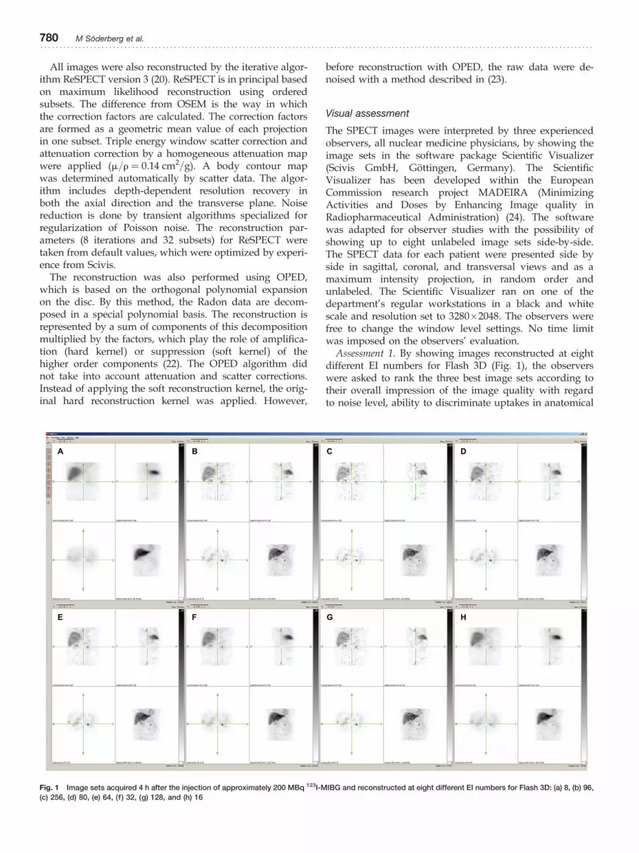
Assessment 1. By showing images reconstructed at eightdifferent EI numbers for Flash 3D (Fig. 1), the observerswere asked to rank the three best image sets according totheir overall impression of the image quality with regardto noise level, ability to discriminate uptakes in anatomical
Fig. 1 Image sets acquired 4 h after the injection of approximately 200 MBq 123I-MIBG and reconstructed at eight different EI numbers for Flash 3D: (a) 8, (b) 96,
(c) 256, (d) 80, (e) 64, (f ) 32, (g) 128, and (h) 16
780 M Soderberg et al.. . . . . . . . . . . . . . . . . . . . . . . . . . . . . . . . . . . . . . . . . . . . . . . . . . . . . . . . . . . . . . . . . . . . . . . . . . . . . . . . . . . . . . . . . . . . . . . . . . . . . . . . . . . . . . . . . . . . . . . . . . . . . . . . . . . . . . . . . . . . . . . . .

structures (e.g. liver, adrenal glands, kidneys, and spleen),introduction of artifacts, and if possible delineation of sus-pected pathology. The rank order was 1 (best) to 3 and allthe remaining image sets obtained a rank order of 4. Thisprocedure was performed for images acquired 4 h as wellas 24 h after injection. The average distribution of imagequality ranking for all observers was calculated for thedifferent EI numbers. In addition, the rank order wasobtained based on the calculated average of scores a givenimage set received, i.e. the lower the value the better theimage quality.
Assessment 2. The two images considered to be the bestin the Flash 3D series (as identified by their EI numbers)were compared with images reconstructed using theReSPECT and OPED algorithms. The four image sets(Fig. 2) were displayed in the Scientific Visualizer andthe observers were in the same manner as in the firstvisual assessment asked to rank the image sets accordingto their overall impression of the image quality. A rankorder of 1 was again assigned to the image set judged tohave the best overall image quality and a rank of 4 wasassigned to the worst. The same procedure was performedfor images acquired 4 h and 24 h after injection. Theaverage distribution of image quality ranking for all obser-vers was calculated for the different reconstructionmethods. In addition, the rank order was obtained basedon the calculated average of scores that a given image setreceived.
Statistical analysis
The inter-observer agreement for multiple observers wasdetermined using generalized kappa (k) statistics (25). Thek can vary between 21.0 and 1.0. A k of 1.0 indicatestotal agreement, whereas a k of 0 means agreement bychance. The non-parametric Friedman test (26) wasapplied on the ranking data to evaluate if a statistically sig-nificant difference between the different number of EI andreconstruction algorithms could be established (a ¼ 0.05).
Results
The software Scientific Visualizer was found to be a userfriendly system for quick opening and closing views,which makes it easy to compare different image sets. Theprocessing time for the three reconstruction algorithms iscomparable and is with today’s available computationalpower not a limitation.
Assessment 1. Fig. 1 shows image sets acquired 4 h p.i.reconstructed at eight different EI numbers using Flash3D. As the number of EI increases, the contrast/resolutionincreases but simultaneously the image noise increases.The average distributions of image quality ranking for allobservers to the different EI numbers are shown inTable 2. Table 3 contains the single observer ranking ofoverall image quality using different numbers of EI. Theobservers concluded that the optimal EI number was 32
Fig. 2 Image sets acquired 4 h after the injection of approximately 200 MBq 123I-MIBG and reconstructed using different reconstruction methods: (a) ReSPECT,
(b) Flash 3D64, (c) OPED, and (d) Flash 3D32
Evaluation of image reconstruction methods for 123I-MIBG-SPECT 781. . . . . . . . . . . . . . . . . . . . . . . . . . . . . . . . . . . . . . . . . . . . . . . . . . . . . . . . . . . . . . . . . . . . . . . . . . . . . . . . . . . . . . . . . . . . . . . . . . . . . . . . . . . . . . . . . . . . . . . . . . . . . . . . . . . . . . . . . . . . . . . . .

for both 4 h and 24 h p.i. measurements, followed by 64 EIand 16 EI, respectively. The strength of the inter-observeragreement was considered as fair (k4h ¼ 0.30, k24h ¼ 0.25).The Friedman test could not establish statistically significantdifference between 32, 64, and 80 EI (4 h p.i.) and between16 and 32 EI (24 h p.i.). However, there was a significantdifference between 80 (the department’s default setting)and 16 (24 h p.i.), and between 80 and 32 EI (24 h p.i.).
Assessment 2. Fig. 2 shows examples of the evaluatedreconstruction methods. The average distributions ofimage quality ranking for all observers to the differentreconstruction methods are shown in Table 4. The threeobservers average rank order of overall image qualityusing four different reconstruction methods for acquisitions4 h p.i. were (best first): Flash 3D32 . ReSPECT . Flash3D64 . OPED, and for acquisitions 24 h p.i.: Flash 3D16 .
ReSPECT . Flash 3D32 . OPED (Table 5). Images recon-structed using the OPED algorithm were consequently byall observers selected as having worst image quality.Fig. 2C shows that the OPED reconstructed images werenoisy and had streak-like artifacts. The strength of the inter-observer agreement was considered as fair (k4h ¼ 0.27,k24h ¼ 0.25). The results from the Friedman tests areshown in Table 6. When considering the rating data for allobservers, a significant difference between OPED and theremaining reconstruction algorithms was found for acqui-sitions after 4 h and 24 h. No statistically significant differ-ence was found between Flash 3D32, Flash 3D64, andReSPECT for acquisitions 4 h after injection. As well, no stat-istically significant difference was found between Flash 3D16
and ReSPECT for acquisitions 24 h after injection.
Discussion
There is an opportunity to improve the image qualityand lesion detectability by choosing an appropriate
reconstruction method and optimal reconstruction par-ameters (11–13). The optimal settings depend on, e.g. theclinical task, the target organs, the patient, and the prefer-ences of the observer. Optimal reconstruction conditionsand selection of algorithm need to be evaluated in eachdepartment for each system and clinical task since thetype of detector, crystal size, correction methods, etc.differ among the various systems. Fewer optimizationtrials in the form of observer performance studies havebeen performed for nuclear medicine imaging than forX-ray imaging. This study shows an example of how anoptimization of this kind can be performed.
The most important parameters in iterative algorithms,e.g. Flash 3D are the number of iterations, subsets, and thechoice of reconstruction filter. In this study the EI numberin the Flash 3D algorithm was varied from 8 to 256, result-ing in eight kinds of reconstructed images. It was not poss-ible to change the type of reconstruction filter. The filterFWHM was selected to be fixed at 8.4, aimed to minimizethe number of variable parameters. This value is the depart-ment’s default value. The ideal would be to perform anoptimization study that incorporates all acquisition andreconstruction parameters, but that is very time consumingand require participation from physicians, technologists,and medical physicists.
The preferred EI number for Flash 3D in the first visualassessment was 32 for acquisitions 4 h and 24 h after injec-tion. This is lower than the department’s default setting of80. Consequently the physicians preferred a more smoothedappearance. With higher EI number the contrast isimproved but simultaneous the noise increases andreduces the achievable image quality. Due to the inter-observer variability and the absence of significant differencebetween the two images considered being the best in theFlash 3D series (as identified by their EI numbers), bothimages were included for the second visual assessment. Inthe second assessment the preferred EI number for acqui-sitions 24 h p.i. were 16 instead of 32. When consideringthe ranking data from all observers, no statistically sig-nificant difference was found between 16 and 32 EI.However, the results indicate that we should lower thenumber of EI, especially for acquisitions 24 h after injection.Acquisitions 24 h p.i. compared to acquisitions 4 h afterinjection have a lower number of counts, resulting in morenoise in the reconstructed images for higher EI. However,using too few EI is undesirable as the algorithm may not
Table 2 Distribution of image quality ranking as a function of rank and the number of equivalent iterations in the Flash 3D reconstruction. Thetable summarizes results for all observers. Each entry is the fraction of observers that assigned the actual rank to the specific equivalent iteration.The best three images were ranked 1–3 and all the remaining image sets obtained a rank order of 4
Acquisition Rank order 8 16 32 64 80 96 128 256
4 h 1 0.00 0.09 0.45 0.18 0.18 0.06 0.03 0.00
2 0.00 0.03 0.21 0.48 0.18 0.06 0.03 0.00
3 0.00 0.06 0.15 0.21 0.33 0.24 0.00 0.00
4 1.00 0.82 0.18 0.12 0.30 0.64 0.94 1.00
24 h 1 0.19 0.26 0.50 0.02 0.02 0.00 0.00 0.00
2 0.07 0.40 0.24 0.19 0.07 0.10 0.00 0.00
3 0.05 0.07 0.24 0.43 0.10 0.12 0.00 0.00
4 0.69 0.26 0.02 0.36 0.81 0.86 1.00 1.00
Table 3 Single observer ranking of overall image quality using theFlash 3D algorithm. Each entry in the table contains the equivalentiteration number that the observer assigned to a specific rank
4 h 24 h
Observer Rank 1 Rank 2 Rank 3 Rank 1 Rank 2 Rank 3
A 32/64 80 32 16 64
B 32 64 80 16 8 32
C 80 64 96 32 64 16
Average 32 64 80 32 16 64
782 M Soderberg et al.. . . . . . . . . . . . . . . . . . . . . . . . . . . . . . . . . . . . . . . . . . . . . . . . . . . . . . . . . . . . . . . . . . . . . . . . . . . . . . . . . . . . . . . . . . . . . . . . . . . . . . . . . . . . . . . . . . . . . . . . . . . . . . . . . . . . . . . . . . . . . . . . .

reach convergence everywhere in the reconstructed volumeand small lesions will not be visible.
When considering data from all observers, Flash 3D wasthe preferred algorithm for acquisitions 4 h and 24 h afterinjection. However, analyzed individually one of the obser-vers preferred ReSPECT for acquisitions 4 h after injection.The observer variability may be explained by differencesin individual preferences of what is appropriate imagequality. The fair inter-observer reliability and the limitedpatient cohort explain the absence of statistically significantdifference between Flash 3D and ReSPECT, when consider-ing ranking data from all observers.
There is an opportunity to further improve the recon-structed images using ReSPECT by performing an optimiz-ation study of the reconstruction parameters, as performedfor Flash 3D. It may also be possible to improve the imagequality if CT based attenuation correction are implementedin ReSPECT. ReSPECT was evaluated in a previous study ofparathyroid scanning with 99mTc-MIBI (27). The studydemonstrated better image quality using ReSPECT com-pared to the algorithm HOSEM (Hermes MedicalSolutions, Stockholm, Sweden).
OPED is a fast exact mathematical reconstruction methodbut its hard kernel version is not capable of dealing withhigh levels of noise as is found in SPECT images. This orig-inal version of OPED applied to the denoised raw data wasranked as worst image quality. The soft kernel is planned tobe tested in the future. The reconstructed images were noisy
and had streak-like artifacts due to the geometry of theSPECT data, and no attenuation and scatter correction areimplemented yet. Due to the collimation in SPECT, themeasured data are parallel in the classical sense, i.e. uni-formly distributed projections with equi-spaced lateralsampling. OPED requires sinusoidal lateral sampling andconsequently the SPECT data needs to be re-sampled. Ingeneral, OPED is more suitable for use in PET and CT (28).
Based on experience from the visual assessment, theScientific Visualizer software will be further improved andimplemented with settings such as linking the windowlevel and slice orientation between the different imagesets. The viewer has potential as a very useful tool in theframework of optimizing nuclear medicine imaging.
The results obtained in studies like this are specific forthe system and parameter settings. The outcome mightvary by the amount of radionuclide activity used, acqui-sition parameters, acquisition time, and examined bodyarea. Another possible limitation is that the observersmight have recognized and were familiar with the Flash3D reconstructions. The primary selection of preferred EInumber for Flash 3D in the first visual assessment couldalso have formed a bias in its favor. To reduce this sourceof bias, the second assessment was carried out a couple ofweeks after the first assessment.
The result of this rank-order study gives an indication ofpreferred reconstruction parameters and algorithms, whichneed to be further investigated with a larger patient
Table 4 Distribution of image quality ranking as a function of the rank and the SPECT reconstruction method. Each entry is the fraction ofobservers that assigned the actual rank to the specific reconstruction method. The rank order was 1 (best) to 4
Acquisition Rank order Flash 3D16 Flash 3D32 Flash 3D64 ReSPECT OPED
4 h 1 – 0.39 0.21 0.39 0.00
2 – 0.52 0.27 0.21 0.00
3 – 0.09 0.52 0.39 0.00
4 – 0.00 0.00 0.00 1.00
24 h 1 0.55 0.17 – 0.29 0.00
2 0.36 0.33 – 0.31 0.00
3 0.10 0.50 – 0.40 0.00
4 0.00 0.00 – 0.00 1.00
Table 5 Single observer ranking of overall image quality using Flash 3D, ReSPECT and OPED. Each entry in the table contains the reconstructionmethod that the observer assigned to a specific rank
4 h 24 h
Observer Rank 1 Rank 2 Rank 3 Rank 4 Rank 1 Rank 2 Rank 3 Rank 4
A ReSPECT Flash 3D32 Flash 3D64 OPED Flash 3D16 ReSPECT Flash 3D32 OPED
B Flash 3D32 Flash 3D64/ReSPECT OPED Flash 3D16 Flash 3D32 ReSPECT OPED
C Flash 3D64 Flash 3D32 ReSPECT OPED Flash 3D32 Flash 3D16 ReSPECT OPED
Average Flash 3D32 ReSPECT Flash 3D64 OPED Flash 3D16 ReSPECT Flash 3D32 OPED
Table 6 Results of the Friedman tests (27). If a statistically significant difference was established between two reconstruction algorithms, it isindicated for each observer A, B, and C, respectively. The shaded areas indicate that a statistically significant difference was established based onthe ranking data for all observers
4 h 24 h
Flash 3D32 Flash 3D64 ReSPECT Flash 3D16 Flash 3D32 ReSPECT
OPED A, B, C B, C A, B, C OPED A, B, C B, C A, B, C
ReSPECT A ReSPECT B A
Flash 3D64 Flash 3D32 A
Evaluation of image reconstruction methods for 123I-MIBG-SPECT 783. . . . . . . . . . . . . . . . . . . . . . . . . . . . . . . . . . . . . . . . . . . . . . . . . . . . . . . . . . . . . . . . . . . . . . . . . . . . . . . . . . . . . . . . . . . . . . . . . . . . . . . . . . . . . . . . . . . . . . . . . . . . . . . . . . . . . . . . . . . . . . . . .

cohort. The impact on diagnostic performance was notinvestigated, and there are variations in patient size,shape, and uptake affinity for the radiopharmaceuticalthat may influence the result. However, our results areuseful for future optimization of reconstruction methodsand parameter settings.
In conclusion, using Siemens Symbia T6 SPECT/CT andspecified acquisition parameters, Flash 3D32 (4 h p.i.) andFlash 3D16 (24 h p.i.), followed by ReSPECT were assessedto be the preferable reconstruction algorithms in visualassessment of 123I-MIBG images. In our department thenumber of EI has been reduced from 80 to 32.
ACKNOWLEDGEMENTS
The work was carried out within the Collaborative Project“MADEIRA” (www.madeira-project.eu), cofounded bythe European Commission through EURATOM SeventhFramework Programme (Grant Agreement FP7-212100).The authors would like to thank Dr Gernot Ebel, Dr TimoAspelmeier, and Dr Bjorn Thiel for support and adaptationwith ReSPECT and the Scientific Visualizer.
Conflict of interest: None.
REFERENCES
1 Bruyant PP. Analytic and iterative reconstruction algorithms in SPECT.J Nucl Med 2002;43:1343–58
2 Vandenberghe S, D’Asseler Y, Van de Walle R, et al. Iterativereconstruction algorithms in nuclear medicine. Comput Med ImagingGraph 2001;25:105–11
3 Groch MW, Erwin WD. SPECT in the year 2000: basic principles. J NuclMed Technol 2000;28:233–44
4 Hutton BF, Hudson HM, Beekman FJ. A clinical perspective ofaccelerated statistical reconstruction. Eur J Nucl Med 1997;24:797–808
5 Lange K, Carson R. EM reconstruction algorithms for emission andtransmission tomography. J Comput Assist Tomogr 1984;8:306–16
6 Shepp LA, Vardi Y. Maximum likelihood reconstruction for emissiontomography. IEEE Trans Med Imaging 1982;1:113–22
7 Hudson HM, Larkin RS. Accelerated image reconstruction using orderedsubsets of projection data. IEEE Trans Med Imaging 1994;13:601–9
8 Brambilla M, Cannillo B, Dominietto M, et al. Characterization ofordered-subsets expectation maximization with 3D post-reconstructionGauss filtering and comparison with filtered backprojection in 99mTcSPECT. Ann Nucl Med 2005;19:75–82
9 Liow JS, Strother SC. The convergence of object dependent resolution inmaximum likelihood based tomographic image reconstruction. Phys MedBiol 1993;38:55–70
10 Beekman FJ, Slijpen ET, Niessen WJ. Selection of task-dependentdiffusion filters for the post-processing of SPECT images. Phys MedBiol 1998;43:1713–30
11 Gutman F, Gardin I, Delahaye N, et al. Optimisation of the OS-EMalgorithm and comparison with FBP for image reconstruction on adual-head camera: a phantom and a clinical 18F-FDG study. Eur J NuclMed Mol Imaging 2003;30:1510–9
12 Inoue K, Sato T, Kitamura H, et al. Improvement of the diagnosticaccuracy of lymph node metastases of colorectal cancer in 18F-FDG-PET/
CT by optimizing the iteration number for the image reconstruction. AnnNucl Med 2008;22:465–73
13 Koch W, Hamann C, Welsch J, et al. Is iterative reconstruction analternative to filtered backprojection in routine processing of dopaminetransporter SPECT studies? J Nucl Med 2005;46:1804–11
14 Zeintl J, Vija AH, Chapman JT, et al. Quantifying the Effects ofAcquisition Parameters in Cardiac SPECT Imaging and Comparisonwith Visual Observers. 2006 IEEE Nuclear Science Symposium ConferenceRecord 2006;6:3251–57
15 Good WF, Sumkin JH, Dash N, et al. Observer sensitivity to smalldifferences: a multipoint rank-order experiment. Am J Roentgenol1999;173:275–78
16 Towers JD, Holbert JM, Britton CA, et al. Multipoint rank-order studymethodology: observer issues. Invest Radiol 2000;35:125–30
17 Vallabhajosula S, Nikolopoulou A. Radioiodinatedmetaiodobenzylguanidine (MIBG): radiochemistry, biology, andpharmacology. Semin Nucl Med 2011;41:324–33
18 Jacobson AF, Deng H, Lombard J, et al. 123I-meta-iodobenzylguanidinescintigraphy for the detection of neuroblastoma andpheochromocytoma: results of a meta-analysis. J Clin Endocrinol Metab2010;95:2596–606
19 Hawman E, Vija H, Daffach R, et al. Flash 3D technology OptimizingSPECT quality and accuracy. Whitepaper Flash 3D. Siemens MedicalSolutions, 2003:1–6
20 Scivis GmbH. ReSPECT – Technical description. Scivis, 2006:1–2721 Tischenko O, Xu Y, Hoeschen C. Main features of the
tomographic reconstruction algorithm OPED. Radiat Prot Dosimetry2010;139:204–7
22 Xu Y, Tischenko O, Hoeschen C. Image reconstruction by OPEDalgorithm with averaging. Numer Algor 2007;45:179–93
23 Tischenko O, Hoeschen C, Buhr E. Reduction of anatomicalnoise in medical X-ray images. Radiat Prot Dosimetry 2005;114:69–74
24 Hoeschen C, Mattsson S, Cantone MC, et al. Minimisingactivity and dose with enhanced image quality byradiopharmaceutical administrations. Radiat Prot Dosimetry2010;139:250–3
25 Fleiss JL. Measuring nominal scale agreement among many raters.Psychological Bulletin 1971;76:378–82
26 Friedman M. The use of ranks to avoid the assumption of normalityimplicit in the analysis of variance. Journal of the American StatisticalAssociation 1937;32:675–701
27 Van Hoorn R, Vriens D, Postema J, et al. Evaluation of the advancedReSPECT image reconstruction software in a phantom model and inparathyroid scanning. J Nucl Med 2010;51 (Suppl. 2): 1352
28 Xu Y, Tischenko O, Hoeschen C. A new reconstruction algorithm forradon data. Proc SPIE 2006;6142:791–8
784 M Soderberg et al.. . . . . . . . . . . . . . . . . . . . . . . . . . . . . . . . . . . . . . . . . . . . . . . . . . . . . . . . . . . . . . . . . . . . . . . . . . . . . . . . . . . . . . . . . . . . . . . . . . . . . . . . . . . . . . . . . . . . . . . . . . . . . . . . . . . . . . . . . . . . . . . . .