eumeces schneideri - université de mons
TRANSCRIPT
Contents lists available at ScienceDirect
Vision Research
journal homepage: www.elsevier.com/locate/visres
Comparative study of the visual system of two psammophilic lizards (Scincusscincus & Eumeces schneideri)
Jérôme Caneia, Carmen Burteab, Denis Nonclercqa,⁎
a Laboratory of Histology, Biosciences Institute, Faculty of Medicine and Pharmacy, University of Mons, 23, Place du Parc, B-7000 Mons, BelgiumbDepartment of General, Organic and Biomedical Chemistry, NMR and Molecular Imaging Laboratory, University of Mons, B-7000 Mons, Belgium
A R T I C L E I N F O
Keywords:SkinksEyeRetinaImmunohistochemistryMRIScincidae
A B S T R A C T
Sand deserts are common biotopes on the earth’s surface. Some specialized vertebrate species have colonizedthese ecological habitats by living buried in the sand. Among these so called psammophilic species are theScincidae sand dune living species Scincus scincus and Eumeces schneideri. These two skinks share a relativelysimilar behavioral ecology by living buried in sand, almost all the time for S. scincus and at least for some part ofthe day for E. schneideri. The visual system of these two lizards was investigated by histological, im-munohistochemical, Magnetic Resonance Imaging (MRI) and morphometric techniques. Both skink species ex-hibit a retina lacking fovea, composed predominantly of cones presenting two types of oil droplets (pale blue-green and colorless). Both species possess a subset of rod like-photoreceptors (about 1 rod for 30 cones) evi-denced by anti-rhodopsin immunoreactivity. A ratio 1:1–1:2 between ganglion cells and photoreceptors points toa linear connection (photoreceptors/bipolar neurons/ganglion cells) in the retina and indicates that both skinksmore likely possess good visual acuity, even in the peripheral retina. The MRI analysis revealed differencesbetween the species concerning the eye structures, with a more spherical eye shape for S. scincus, as well as amore flattened lens. The relative lens diameter of both species seems to correspond to a rather photopic pattern.Beside the fact that S. scincus and E. schneideri have different lifestyles, their visual capacities seem similar, and,generally speaking, these two psammophilic species theoretically exhibit visual capacities not far away fromnon-fossorial species.
1. Introduction
Light is a major environmental factor which has exerted a powerfulselective pressure on living creatures. This factor has led to variousevolutionary innovations, including the appearance of highly complexand specialized organs: the eyes. Vertebrates, including lizard speciesdescribed in this article, possess very efficient paired chambered eyeswith two lenses (Fernald, 2004).
Vertebrates have two types of photoreceptors at the retina level:cones and rods. The first are related to visual acuity and color vision,the second to visual sensitivity (Gerhing, 2014). These two types ofphotoreceptors operate in different light intensity ranges. The conesbeing active at high intensity while the rods are specialized in per-ception under low light intensities. However, there is a range of inter-mediate intensities where these two types of photoreceptor are active.Photopic is the light intensity range in which vision is purely cone-mediated corresponding generally to daylight conditions, scotopic isthe range of low light intensities encountered generally in nocturnal
conditions in which it is rod-mediated, and mesopic vision is the in-between in which both photoreceptor types are functional. Naturally,this ranges vary between species (Kelber & Lind, 2010; Kelber,Yovanovich, & Olsson, 2017). Concerning the color perception, it re-quires the presence of at least two different spectral photoreceptortypes (Kelber, Vorobyev, & Osorio, 2003; Yovanovich et al., 2017).Depending on the species, vertebrates have monochromatic vision(making them color-blind – like for most aquatic mammals) (Peichl,Behrmann, & Kroger, 2001), dichromatic vision (observed in a largenumber of terrestrial mammals) (Kelber et al., 2003; Peichl et al.,2001), and trichromatic vision (like for catarrhine primates, geckos andcrocodiles) (Kelber et al., 2003). Moreover, many teleost fishes(Neumeyer, 1992), reptiles (Ammermüller, Muller, & Kolb, 1995) andbirds (Chen & Goldsmith, 1986; Kelber, 2019) have tetrachromatic vi-sion. Finally, regarding the proportion and the nature of Squamataphotoreceptors, the different lizard families of Iguanidae, Chameleo-nidae, Agamidae, Scincidae, Lacertidae, Anguidae, Pygopodidae andVaranidae commonly possess a pure cone retina, as referred to in the
https://doi.org/10.1016/j.visres.2020.04.004Received 21 November 2019; Received in revised form 17 April 2020; Accepted 17 April 2020
⁎ Corresponding author at: Laboratory of Histology, University of Mons 6, Avenue du Champ de Mars, Mons, 7000, Belgium.E-mail address: [email protected] (D. Nonclercq).
Vision Research 171 (2020) 17–30
0042-6989/ © 2020 The Author(s). Published by Elsevier Ltd. This is an open access article under the CC BY-NC-ND license (http://creativecommons.org/licenses/BY-NC-ND/4.0/).
T
study of Röll (2001a).In bright light conditions, numerous vertebrate species have de-
veloped high visual acuity involving retinal specific structures as thefovea, a retinal pit located in the optical axis with high cone density.Foveae are present in a great number of vertebrates, including mam-mals (Rodieck, 1998), birds, reptiles, (Moore et al., 2017) and evenTeleost fishes (Collin & Collin, 1999; Easter, 1992). Some reptile spe-cies, like the well-studied group of lizards of the genus Anolis, possess acomplex visual system including two morphologically different andspatially separated foveae (Fite & Lister, 1981). Another visual struc-ture is the oil droplets present in cones. These are widespread amongdiurnal birds and reptiles (Vorobyev, 2003) and act like individualfilters of the photoreceptors allowing more acute vision (Stavenga &Wilts, 2014) and erase the flickering effect on the surface of the water(Muntz, 1972; Zeigler & Bischof, 1993). The pigmentation of thesestructures also seems important, because when pigmented they reducethe light spectrum range reaching the outer segments of the cones andpromote the excitation of the specific opsin of the cone. Some conespossess only transparent oil-droplets that improve how light is caughtand increase visual sensitivity (Wilby & Roberts, 2017).
Reptiles, and more precisely Squamata, are generally diurnal species(Hall, 2008). The biggest group of nocturnal lizard species are found inthe Gekkonidae, but it is noteworthy that their ancestors are believed tohave been diurnal (Röll, 2001a). Beside these general adaptations tolight conditions, numerous adaptations have appeared among verte-brates to cope with peculiar lifestyles, like for fossorial species. Amongmammals, some fossorial species, like the rodent Spalax ehrenbergi, haveatrophied eyes and have lost some types of photoreceptors (Hunt &Collin, 2014). These features are also encountered in Squamata, like infossorial Amphisbaenia, which have some common characteristicslinked to this specific lifestyle, such as a reduced eyeball size and a lenswith amorphous nucleate cells (Foureaux et al., 2010). It is noteworthythat atrophied eyes are also found in burrowing caecilian amphibians(Mohun et al., 2010).
Our study focuses on two species of Scincidae family: Scincus scincus(Linnaeus, 1758) and Eumeces schneideri (Daudin, 1802). These twoskinks are phylogenetically closely related (Pyron, Burbrink, & Wiens,2013). Moreover, they share a common geographical distribution, fromthe Mediterranean coast to Saheli band through all Sahara desert andalso in the Arabian peninsula into Sinai, and desertic zones of Syria,Iraq, Jordan and west part of Iran (Arnold & Leviton, 1977; Ayaz, Çiçek,Tok, & Dinçaslan, 2011). The omnivorous sandfish S. scincus hunts in-sects, by the detection of vibratory cues (Hetherington, 1989) in hotdeserts composed of few dry shrubs (Attum, Covell, & Eason, 2004).The omnivorous Berber skink (E. schneideri) is also found in such bio-topes, but with relatively dense vegetation (Allam, Abo-Eleneen, &Othman, 2017). These two diurnal skinks are psammophilic and thuslive buried in the sand, almost all the time for S. scincus (Stadler et al.,2016) while E. schneideri spends more time laps at the surface of thesand during the diurnal period for hunting or mating (Saleh, 1997). Thesandfish is famous for its morphological adaptations as a shovel-shapedsnout, a subquadrangular cross section of the body, a reduced earopenings and fringed toes and digits allowing to move easily under thesand (sand swimming) (Baumgartner et al., 2008) and for the remark-able properties of its skin which resist the intense abrasion of sandparticles (Vihar, Hanisch, & Baumgartner, 2016). To our knowledge, noarticles found in the literature have mentioned the visual system ofthese two burrowing skink species (with the noticeable exception ofKhattab, Khattab, Fares, & Zaki, 2004). These authors attest by ultra-structural approach that the retina of the Eumeces schneideri is largelydominated by cones with present oil droplet. These cones are onlysingle cone, indeed double cones are not present in this species. On theother hand, Khattab et al. (2004) have observed some scattered rods inthe retina of E. schneideri. The Scincidae family (Lepidosauria) com-prises a vast majority of diurnal species, with a few exceptions. Insidethis family, the visual system has been very poorly investigated. Apart
from the study of Khattab et al. (2004) cited just above, the few pub-lished data on Scincidae’s ocular adaptations concern the followingspecies: Tiliqua rugosa (Braekevelt, 1989; New, Hemmi, Kerr, & Bull,2012), Cryptoblepharus boutonii (Röll, 2001b), Acontias orientalis,Acontias rieppeli, Typhlosaurus vermis and Trachylepis punctatissima(Zhao, Goedhals, Verdú-Ricoy, Jordaan, & Heideman, 2019). Recently,the eye morphology of one burrowing species (Calyptommatus nicterus,Gymnophthalmidae) and one surface-dwelling lizard (Ameivula ocelli-fera, Teiidae) has been studied (Yovanovich, Pierotti, Rodrigues, &Grant, 2019). These two species are part of the Infraorder Scinco-morpha and belong to two families close to the Scincidae, from aphylogenic point of view (Pyron et al., 2013).
The aim of this study is to compare the eye morphology and retinalstructure of burrowing Scincidae to other diurnal members of this fa-mily, and to pinpoint the evolutionary adaptations of the eye structureof fossorial psammophilic Scincidae in relation to environmental lightconditions. Considering the lifestyle of both studied species, which arerestricted to very dim light conditions when they are buried, we couldexpect that their visual system is more sensitive and scotopic thanphotopic and acute. However, the Sahara Desert is one of the most il-luminated areas on Earth, with a theoretical daylight of approximately100,000 Lux of illuminance (without clouds) as highlighted by typicalcalculated values (Kandilli & Ulgen, 2008). We expect that S. scincusmay have less acute vision compared to the semi-fossorial Berber Skink,which could possess an intermediate visual system adapted to both preydetection (in bright light conditions) and sand diving (with almost nolight).
2. Material and methods
2.1. Animals
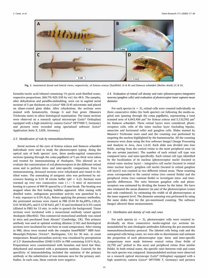
Three adult individuals (two males and one female) (mean snout–-tail length, STL: 16.25 cm ± 2 mm) of the species Scincus scincus(Fig. 1a, b) were used in the experiments. These specimens were ob-tained from the Animal House “Animalerie 2000” (Mouscron, Belgium).Three other adult individuals (three males) (mean snout-tail, STL:32.66 cm ± 2 mm) of the species Eumeces schneideri (Fig. 1c, d) werealso used. These specimens were obtained from the Animal House“Reptiles Univers” (Dour, Belgium). The individuals of these two spe-cies were maintained in the animal housing facility of the University ofMons (UMONS, Belgium). The animals were treated according to theguidelines specified by the Belgian Ministry of Trade and Agricultureand under the control of the UMONS ethical commission (agreementLA1500021). The lizards were placed individually in terraria(120 × 60 × 60 cm) whose bottoms were covered with a 10–12 cmthick layer of fine sand (with a particle diameter comprised between0.75 and 1.3 mm). Each terrarium was equipped with an UV rich lamp(5.0 UVB, 26 W; ZooMed®) and an infrared bulb for heating (Ceramicinfrared, 60 W; ZooMed®). Both light bulbs were placed at a height of35 cm. The animals were maintained in a controlled environment with12 h of daylight and a temperature of 35 °C during the day, and 22 °C atnight. The animals were fed with live adult house crickets (Acheta do-mesticus), and vegetables twice a week and received water ad libitum.The animals were euthanized by a lethal intramuscular injection ofketamine hydrochloride (200 mg/kg body mass, Ketalar®). Each subjectwas considered euthanized when it lost the ability to respond to feetand tail pinching. This was done under the control of the UMONSCommittee for Survey of Experimental Studies and Animal Welfare.
2.2. Histology
After euthanasia, all individuals were transversally cut at the levelof the head-trunk junction. Once the head was isolated, a carefulchirurgical removal of the ocular globes was performed. These werethen fixed by immersion in Duboscq–Brazil fluid (composition:
J. Canei, et al. Vision Research 171 (2020) 17–30
18
formalin/acetic acid/ethanol containing 1% picric acid/distilled water,respective proportions: 260/70/425/245 by vol.) for 48 h. The samples,after dehydration and paraffin-embedding, were cut in sagittal serialsections of 5 µm thickness on a Leica® RM 2145 microtome and placedon silane-coated glass slides. After rehydration, the sections werestained with hematoxylin, Orange G and Fast green (Masson'sTrichrome stain) to allow histological examination. The tissue sectionswere observed on a research optical microscope (Leitz® Orthoplan)equipped with a high sensitivity camera (Leica® DFT7000 T, Germany)and pictures were recorded using specialized software (Leica®Application Suite X, LASX, Germany).
2.3. Identification of rods by immunohistochemistry
Serial sections of the eyes of Scincus scincus and Eumeces schneideriindividuals were used to study the photoreceptor typing. Along theoptical axis of both species’ eyes, three medio-sagittal consecutivesections (passing through the conus papillaris) of 5 µm thick were takenand treated for immunostaining of rhodopsin. This allowed us toevaluate the concentration of rod photoreceptors in the different retinalareas and to perform intra- and inter-specific comparisons. Prior toimmunostaining, dewaxed sections were rehydrated and rinsed in dis-tilled water. The unmasking of antigenic sites was performed by mi-crowave heating in 0.01 M citrate buffer (pH = 6.2). Sections werewarmed up over two consecutive runs (+/−5 min) of microwaveheating at a power of 900 W spaced by a 15 min break. The heating wasstopped when the first boiling bubbles appeared. After rinsing withdistilled water, endogenous peroxidase activity was quenched by a5 min. exposure to 0.5% H2O2. Before applying the primary antibodies,the pretreated sections were rinsed in PBS (0.04 M Na2HPO4.12H₂O,0.01 M KH2PO4 and 0.12 M NaCl, pH 7.4) and incubated in 0.5% casein(diluted in PBS) for 15 min. in order to quench the background. Tissuesections were incubated with a monoclonal antibody raised againstrhodopsin (Rho4D2). This commercial monoclonal antibody was raisedin mice and purchased from Abcam® (Cambridge, UK). This primaryantibody was used at optimal working dilution (1:50) and histologicalsections were incubated for one hour at room temperature. After rinsingin PBS, slices were treated with the complex ImmPRESS™ HRP/Anti-MouseIgG/Polymer (Vector®, Burlingame, CA) for 30 min. at roomtemperature. Bound peroxidase activity was visualized by precipitationof 3.3′ diaminobenzidine (DAB) 0.02% in PBS containing 0.01% H2O2.Preparations were counterstained with hemalun and luxol fast blue,dehydrated and mounted with a permanent medium. Controls for thespecificity of immunolabeling included the omission of the primaryantibody or the substitution of non-immune sera for the primary anti-bodies. In each case, these controls were negative.
2.4. Evaluation of retinal cell density and ratio (photoreceptors/integrativeneurons/ganglion cells) and evaluation of photoreceptor inner segment meandiameter.
For each species (n = 3), retinal cells were counted individually onthree consecutive slides (for both species) cut following the medio-sa-gittal axis (passing through the conus papillaris), representing a totalscanned area of 6,843,456 µm2 for Scincus scincus and 5,132,592 µm2
for Eumeces schneideri. Three retinal layers were considered: photo-receptors cells, cells of the inner nuclear layer (including bipolar,amacrine and horizontal cells) and ganglion cells. Slides stained byMasson’s Trichrome were used and the counting was performed bytargeting the nucleus highlighted by the haematoxylin. All the countingmeasures were done using the free software ImageJ (Image Processingand Analysis in Java, Java 1.6.0). Each slide was divided into fourfields, starting from the central retina to the most peripheral zone (atthe ora serrata junction). The number of each retinal cell type wascompared intra- and inter-specifically. Each retinal cell type identifiedby the localization of its nucleus (photoreceptor nuclei (located inretinal outer nuclear layer) – integrative cell nuclei (located in retinalinner nuclear layer)– ganglion cell nuclei (located in retinal ganglioncell layer)) was counted in two different retinal areas. These countingareas corresponded to the central retina (two central fields) and theperipheral retina (two outmost fields) to investigate intra- and inter-specific differences. The ratio between ganglion cells and photo-receptors was estimated by dividing the former by the latter. We havealso estimated the mean diameter (in µm) of the photoreceptors (conesand rods combined), by estimating the width of all photoreceptors atthe inner segment level. The diameter assessing was performed by usingthe same slides that for the pre-mentioned counting. The softwareImageJ allowed these measurements.
2.5. Distribution and density of rods and cones
For each species (n = 3), photoreceptor cells were counted in-dividually on three consecutive medio-sagittal eye sections im-munolabeled by anti-rhodopsin antibodies following the pre-mentionedimmunohistochemistry protocol. The labeled cells being rods and theuntargeted cells being cones, we were able to discriminate and estimatethe proportion of each photoreceptor type. Intra- and inter-specificcomparisons were made between central retina (four fields of22,750 µm2 picked in this area) and peripheral retina (four similarfields). In both retinal zones, the specific ratio between rods and conesfor each species was compared. Immunolabeled sections were observedon a research optical microscope (Leitz® Orthoplan) equipped with ahigh sensitivity camera (Leica® DFT7000 T, Germany) and pictures
Fig. 1. Anatomical dorsal and lateral views, respectively, of Scincus scincus [Sandfish] (A & B) and Eumeces schneideri [Berber skink] (C & D).
J. Canei, et al. Vision Research 171 (2020) 17–30
19
were recorded using specialized software (Leica® Application Suite X,LAS X, Germany).
2.6. Cornea morphometric analyses
For each species (n = 3), the thickness of the cornea was quantifiedby morphometric analysis using the pre-mentioned software, ImageJ.Three consecutive slides stained by Masson’s Trichrome were used, andthe absolute thickness of the cornea was calculated by choosing themidpoint (located just in the optical axis). The structures taken intoaccount were the corneal epithelium, Bowman's layer, the cornealstroma, Descemet’s membrane and the corneal endothelium. Inter-specific comparisons were made for the relative thickness of this fixedlens. Finally, the focus distance between the cornea and central retina,as well as the curving ray of the cornea, were estimated using aMagnetic Resonance Imaging (MRI) of the medio-frontal plane.
2.7. Magnetic Resonance Imaging
For the Magnetic Resonance Imaging (MRI), each lizard was an-aesthetized using an intramuscular injection of Ketamine (20 mg.kg−1
body mass, Ketalar® 50 mg/ml). The subject was considered anaes-thetized when it lost the ability to push itself back on its feet after being
turned on its back and responded weakly to tail pinching. MRI ex-periments were performed on a 300 MHz (7 T) Bruker Biospec imagingsystem (Bruker, Ettlingen, Germany) equipped with a Pharmascanhorizontal magnet. Ocular globe images for S. scincus were acquiredwith a rapid-acquisition relaxation-enhanced (RARE) imaging protocolusing the following parameters: TR/TE = 3000/59.4 ms, RAREfactor = 4, NEX = 8, matrix = 256 × 256, FOV = 2 cm, slicethickness = 1 mm, 20 coronal slices, spatial resolution = 79 µm,TA = 25 min 36 s. The parameters for E. schneideri were TR/TE = 3000/60 ms, RARE factor = 4, NEX = 4, matrix = 512 × 512,FOV = 2.5 cm, slice thickness = 1 mm, 20 coronal slices, spatial re-solution = 49 µm, TA = 25 min 36 s.
The pictures were used to evaluate the relative proportion of thelens volume when compared to the vitreous body volume. The imagesobtained were also used to assess the diameter (perpendicular to opticalaxis) and the thickness (parallel to optical axis) of the lens and theocular globe. This non-invasive method allowed us to evaluate the insitu anatomy and morphology of eyes in both species, following variousplanes without any potential artefacts provoked by eye fixation.
2.8. Oil droplet characterization
After euthanasia, one supplementary individual by species were
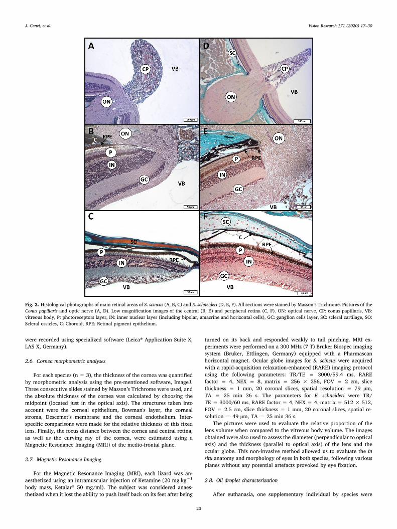
Fig. 2. Histological photographs of main retinal areas of S. scincus (A, B, C) and E. schneideri (D, E, F). All sections were stained by Masson’s Trichrome. Pictures of theConus papillaris and optic nerve (A, D). Low magnification images of the central (B, E) and peripheral retina (C, F). ON: optical nerve, CP: conus papillaris, VB:vitreous body, P: photoreceptors layer, IN: inner nuclear layer (including bipolar, amacrine and horizontal cells), GC: ganglion cells layer, SC: scleral cartilage, SO:Scleral ossicles, C: Choroid, RPE: Retinal pigment epithelium.
J. Canei, et al. Vision Research 171 (2020) 17–30
20
used for oil droplet characterization. The eyes were enucleated and cutto flatten the retina. The retina was surgically cut into a PBS filled cupand was then placed on a glass slide. A drop of PBS was put into theretina which was then coverslipped. Oil droplets in the fresh cut tissueswere identified on the basis of their localization inside the photo-receptors at high magnification (Leitz® Orthoplan microscope equippedwith Leica® DFT7000 camera). The regions located between the centraland the most peripheral retina were chosen to allow examination of oildroplets.
2.9. Statistical analysis
All the statistical analyses were carried out using of the software forstatistical computing R (version 3.2.1). In function of their distribution,the quantitative data obtained in this study were submitted to para-metric (ANOVA one-way or Student's t-test) or non-parametric(Kruskal-Wallis or Wilcoxon) tests with a limit of significance set atp < 0.05.
3. Results
3.1. Histology
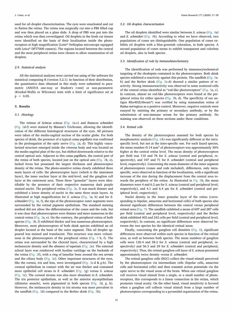
The retinas of Scincus scincus (Fig. 2a-c) and Eumeces schneideri(Fig. 2d-f) were stained by Masson’s Trichrome, allowing the identifi-cation of the different histological structures of the eyes. All pictureswere taken of the medio-sagittal section of the ocular globe. For bothspecies of skink, the presence of a typical conus papillaris was confirmedin the prolongation of the optic nerve (Fig. 2a, d). This highly vascu-larized structure emerged inside the vitreous body and was located onthe medio-sagittal plan of the eye, but shifted downwards relative to thecentral optical axis. Adjacent to this conus papillaris, the central part ofthe retina of both species, located just on the optical axis (Fig. 2b, e),lacked fovea but possessed the largest thickness and photoreceptordensity of the retina. The photo-sensitive retina clearly exhibited threemain layers of cells: the photoreceptor layer (which is the innermostlayer), the inner nuclear layer at the mid-level, and the ganglion celllayer at the outermost area. These three “granular” layers were iden-tifiable by the presence of their respective numerous dark purplestained nuclei. The peripheral retina (Fig. 2c, f) was much thinner andexhibited a lower density of nuclei in the same three main layers. Asillustrated at high magnification for S. scincus (Fig. 3a, b) and for E.schneideri (Fig. 3e, f), the tips of the photoreceptor outer segments weresurrounded by the retinal pigment epithelium. The standard stainingmethod did not allow the differentiation of the cones and the rods, butit was clear that photoreceptors were thinner and more numerous in thecentral retina (Fig. 3a, e). On the contrary, the peripheral retina of bothspecies (Fig. 3b, f) exhibited thicker and less numerous photoreceptors.Moreover, most photoreceptors of both skink species exhibited an oildroplet located at the basis of the outer segment. This oil droplet ap-peared less stained and translucent. This structure was more volumi-nous in the photoreceptors of the peripheral retina (Fig. 3 b, f). Theretina was surrounded by the choroid layer, characterized by a highmelanocyte density and the absence of tapetum (Fig. 2e). The externalscleral layer was reinforced with hyaline cartilage on the backside ofthe retina (Fig. 2f), with a ring of lamellar bone around the ora serrataand the ciliary body (Fig. 2c). Other important structures of the eyes,like the cornea, iris and lens, were investigated (Fig. 3c-d & g-h). Thecorneal epithelium in contact with the air was thicker and containedmore epithelial cell strata in E. schneideri (Fig. 3g) versus S. scincus(Fig. 3c). The corneal stroma was also more abundant in E. schneideri.The iris posterior epithelium, as well as the anterior myoepithelium(dilatator muscle), were pigmented in both species (Fig. 3d, g, h).However, the melanocyte density in iris stroma was more prevalent inthe sandfish (Fig. 3d) compared to the Berber skink (Fig. 3h).
3.2. Oil droplets characterization
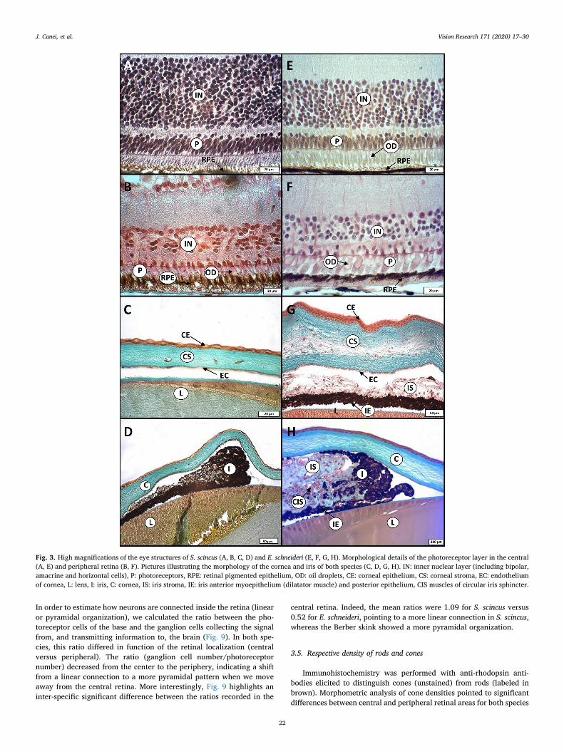
The oil droplets identified were similar between S. scincus (Fig. 4a)and E. schneideri (Fig. 4b). According to what we have observed, twopopulations of cones are distinguishable. One population of cones ex-hibits oil droplets with a blue-greenish coloration, in both species. Asecond population of cones seems to exhibit transparent and colorlessoil droplets, also in both species.
3.3. Identification of rods by immunohistochemistry
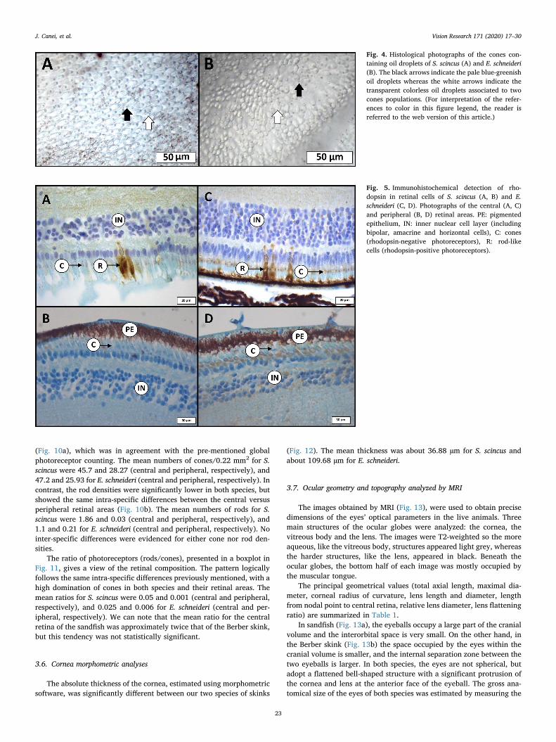
The identification of rods was performed by immunocytochemicaltargeting of the rhodopsin contained in the photoreceptors. Both skinkspecies exhibited a reactivity against this protein. The sandfish (Fig. 5a-b) and the Berber skink (Fig. 5c-d) showed a similar pattern of re-activity. Strong immunoreactivity was observed in some scattered cellsof the central retina identified as “rod-like photoreceptors” (Fig. 5a, c).In contrast, almost no rod-like photoreceptors were found at the per-ipheral retina for either species (Fig. 5b, d). The specificity of our an-tigen Rho4D2(Abcam®) was verified by using mammalian retina ofRattus norvegicus as a positive control. Moreover, negative controls wereessayed by omitting the primary or secondary antibody, or by thesubstitution of non-immune serum for the primary antibody. Nostaining was observed on these sections under these conditions.
3.4. Retinal cells
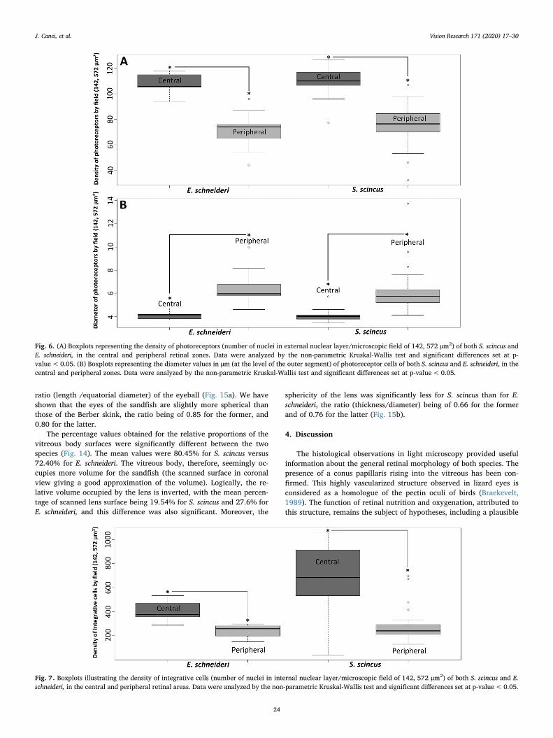
The density of the photoreceptors assessed for both species bymorphometric analysis (Fig. 6A) was significantly different at the intra-specific level, but not at the inter-specific one. For each lizard species,the mean number/0.14 mm2 of photoreceptors was approximately 30%higher at the central retina level. The mean density of photoreceptorsper field was 110 and 76 for S. scincus (central and peripheral, re-spectively), and 107 and 71 for E. schneideri (central and peripherallevel, respectively). Concerning the mean diameter of the inner segmentof photoreceptors (cones and rods included), differences, only intra-specific, were observed in function of the localization, with a significantincrease of the size during the displacement from the central area to-wards the periphery of the retina. As illustrated in Fig. 6b, the meandiameters were 4 and 6.2 µm for S. scincus (central and peripheral level,respectively), and 4.1 and 6.4 µm for E. schneideri (central and per-ipheral level, respectively).
Nuclei density in the inner granular layer of the retina (corre-sponding to bipolar, amacrine and horizontal cells) of both species alsoshowed significant differences between the central versus peripheralretinal area (Fig. 7). The sandfish exhibited a mean of 697 and 287 cellsper field (central and peripheral level, respectively) and the Berberskink exhibited 402 and 242 cells per field (central and peripheral level,respectively). In contrast, no significant differences were observed be-tween the two species for the identical retinal areas.
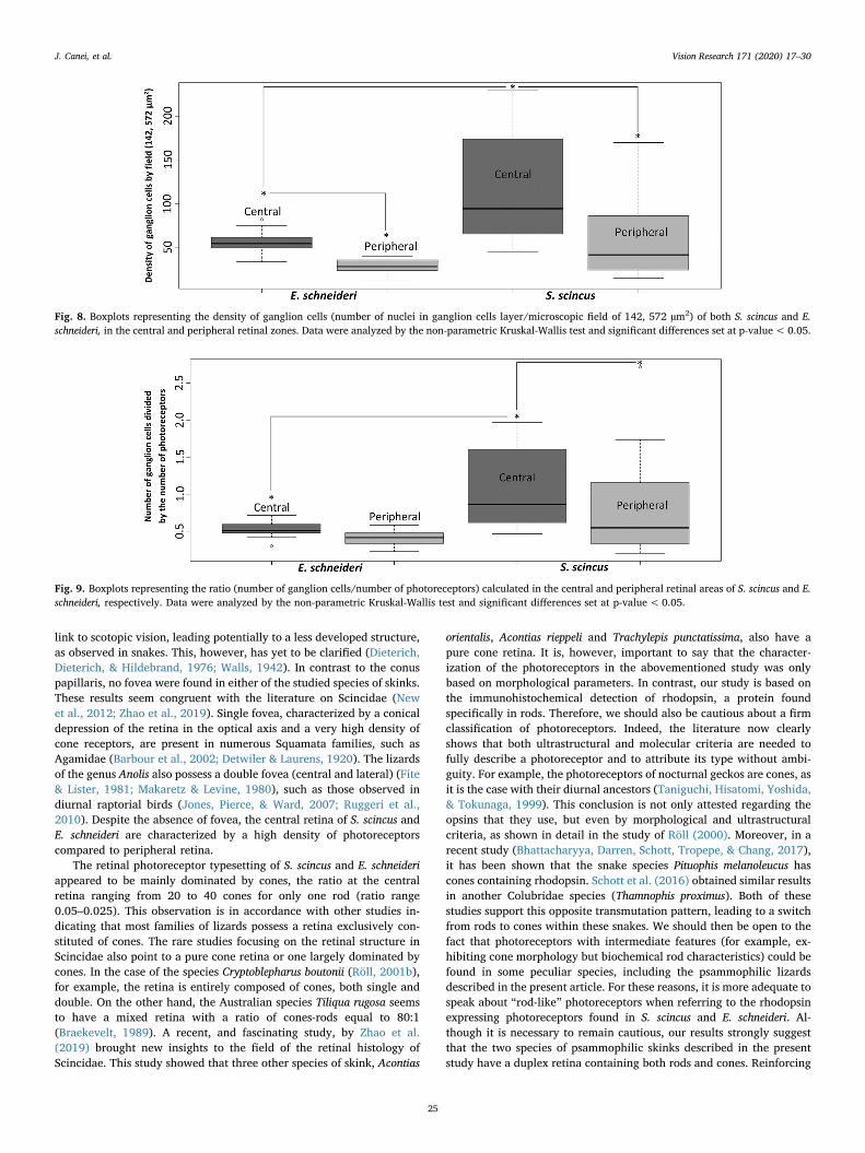
Finally, concerning the ganglion cell densities (Fig. 8), significantdifferences were observed within each species in function of the retinalarea, as well as between both species. The mean numbers of ganglioncells were 120.4 and 58.2 for S. scincus (central and peripheral, re-spectively) and 56.3 and 29 for E. schneideri (central and peripheral,respectively). Thus, the retinal ganglion cell layer of S. scincus presentedapproximately twice density versus E. schneideri.
The retinal ganglion cells (RGC) collect the visual stimuli perceivedby the photoreceptors via intermediate cells (bipolar cells, amacrinecells and horizontal cells) and then transmit action potentials via theoptic nerve to the visual areas of the brain. When one retinal ganglioncell receives visual stimuli from a single, or a small number of photo-receptors, this corresponds to a linear connection in the retina, whichpromotes visual acuity. On the other hand, visual sensitivity is favoredwhen a ganglion cell collects visual stimuli from a large number ofphotoreceptors corresponding to a pyramidal organization of the retina.
J. Canei, et al. Vision Research 171 (2020) 17–30
21
In order to estimate how neurons are connected inside the retina (linearor pyramidal organization), we calculated the ratio between the pho-toreceptor cells of the base and the ganglion cells collecting the signalfrom, and transmitting information to, the brain (Fig. 9). In both spe-cies, this ratio differed in function of the retinal localization (centralversus peripheral). The ratio (ganglion cell number/photoreceptornumber) decreased from the center to the periphery, indicating a shiftfrom a linear connection to a more pyramidal pattern when we moveaway from the central retina. More interestingly, Fig. 9 highlights aninter-specific significant difference between the ratios recorded in the
central retina. Indeed, the mean ratios were 1.09 for S. scincus versus0.52 for E. schneideri, pointing to a more linear connection in S. scincus,whereas the Berber skink showed a more pyramidal organization.
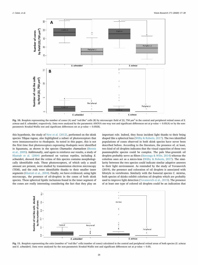
3.5. Respective density of rods and cones
Immunohistochemistry was performed with anti-rhodopsin anti-bodies elicited to distinguish cones (unstained) from rods (labeled inbrown). Morphometric analysis of cone densities pointed to significantdifferences between central and peripheral retinal areas for both species
Fig. 3. High magnifications of the eye structures of S. scincus (A, B, C, D) and E. schneideri (E, F, G, H). Morphological details of the photoreceptor layer in the central(A, E) and peripheral retina (B, F). Pictures illustrating the morphology of the cornea and iris of both species (C, D, G, H). IN: inner nuclear layer (including bipolar,amacrine and horizontal cells), P: photoreceptors, RPE: retinal pigmented epithelium, OD: oil droplets, CE: corneal epithelium, CS: corneal stroma, EC: endotheliumof cornea, L: lens, I: iris, C: cornea, IS: iris stroma, IE: iris anterior myoepithelium (dilatator muscle) and posterior epithelium, CIS muscles of circular iris sphincter.
J. Canei, et al. Vision Research 171 (2020) 17–30
22
(Fig. 10a), which was in agreement with the pre-mentioned globalphotoreceptor counting. The mean numbers of cones/0.22 mm2 for S.scincus were 45.7 and 28.27 (central and peripheral, respectively), and47.2 and 25.93 for E. schneideri (central and peripheral, respectively). Incontrast, the rod densities were significantly lower in both species, butshowed the same intra-specific differences between the central versusperipheral retinal areas (Fig. 10b). The mean numbers of rods for S.scincus were 1.86 and 0.03 (central and peripheral, respectively), and1.1 and 0.21 for E. schneideri (central and peripheral, respectively). Nointer-specific differences were evidenced for either cone nor rod den-sities.
The ratio of photoreceptors (rods/cones), presented in a boxplot inFig. 11, gives a view of the retinal composition. The pattern logicallyfollows the same intra-specific differences previously mentioned, with ahigh domination of cones in both species and their retinal areas. Themean ratios for S. scincus were 0.05 and 0.001 (central and peripheral,respectively), and 0.025 and 0.006 for E. schneideri (central and per-ipheral, respectively). We can note that the mean ratio for the centralretina of the sandfish was approximately twice that of the Berber skink,but this tendency was not statistically significant.
3.6. Cornea morphometric analyses
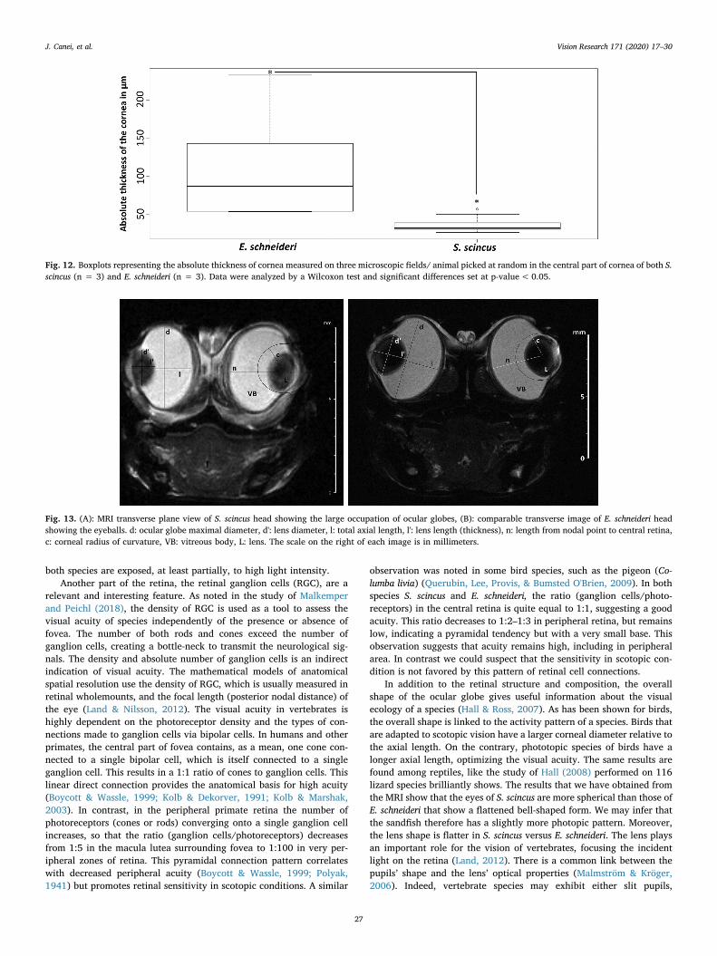
The absolute thickness of the cornea, estimated using morphometricsoftware, was significantly different between our two species of skinks
(Fig. 12). The mean thickness was about 36.88 µm for S. scincus andabout 109.68 µm for E. schneideri.
3.7. Ocular geometry and topography analyzed by MRI
The images obtained by MRI (Fig. 13), were used to obtain precisedimensions of the eyes’ optical parameters in the live animals. Threemain structures of the ocular globes were analyzed: the cornea, thevitreous body and the lens. The images were T2-weighted so the moreaqueous, like the vitreous body, structures appeared light grey, whereasthe harder structures, like the lens, appeared in black. Beneath theocular globes, the bottom half of each image was mostly occupied bythe muscular tongue.
The principal geometrical values (total axial length, maximal dia-meter, corneal radius of curvature, lens length and diameter, lengthfrom nodal point to central retina, relative lens diameter, lens flatteningratio) are summarized in Table 1.
In sandfish (Fig. 13a), the eyeballs occupy a large part of the cranialvolume and the interorbital space is very small. On the other hand, inthe Berber skink (Fig. 13b) the space occupied by the eyes within thecranial volume is smaller, and the internal separation zone between thetwo eyeballs is larger. In both species, the eyes are not spherical, butadopt a flattened bell-shaped structure with a significant protrusion ofthe cornea and lens at the anterior face of the eyeball. The gross ana-tomical size of the eyes of both species was estimated by measuring the
Fig. 4. Histological photographs of the cones con-taining oil droplets of S. scincus (A) and E. schneideri(B). The black arrows indicate the pale blue-greenishoil droplets whereas the white arrows indicate thetransparent colorless oil droplets associated to twocones populations. (For interpretation of the refer-ences to color in this figure legend, the reader isreferred to the web version of this article.)
Fig. 5. Immunohistochemical detection of rho-dopsin in retinal cells of S. scincus (A, B) and E.schneideri (C, D). Photographs of the central (A, C)and peripheral (B, D) retinal areas. PE: pigmentedepithelium, IN: inner nuclear cell layer (includingbipolar, amacrine and horizontal cells), C: cones(rhodopsin-negative photoreceptors), R: rod-likecells (rhodopsin-positive photoreceptors).
J. Canei, et al. Vision Research 171 (2020) 17–30
23
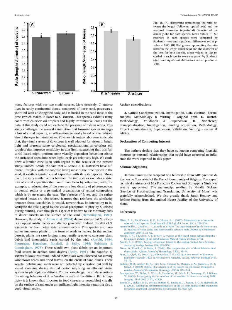
ratio (length /equatorial diameter) of the eyeball (Fig. 15a). We haveshown that the eyes of the sandfish are slightly more spherical thanthose of the Berber skink, the ratio being of 0.85 for the former, and0.80 for the latter.
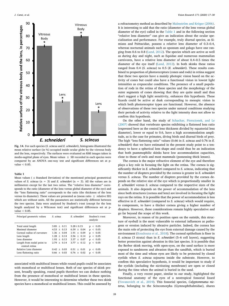
The percentage values obtained for the relative proportions of thevitreous body surfaces were significantly different between the twospecies (Fig. 14). The mean values were 80.45% for S. scincus versus72.40% for E. schneideri. The vitreous body, therefore, seemingly oc-cupies more volume for the sandfish (the scanned surface in coronalview giving a good approximation of the volume). Logically, the re-lative volume occupied by the lens is inverted, with the mean percen-tage of scanned lens surface being 19.54% for S. scincus and 27.6% forE. schneideri, and this difference was also significant. Moreover, the
sphericity of the lens was significantly less for S. scincus than for E.schneideri, the ratio (thickness/diameter) being of 0.66 for the formerand of 0.76 for the latter (Fig. 15b).
4. Discussion
The histological observations in light microscopy provided usefulinformation about the general retinal morphology of both species. Thepresence of a conus papillaris rising into the vitreous has been con-firmed. This highly vascularized structure observed in lizard eyes isconsidered as a homologue of the pectin oculi of birds (Braekevelt,1989). The function of retinal nutrition and oxygenation, attributed tothis structure, remains the subject of hypotheses, including a plausible
Fig. 6. (A) Boxplots representing the density of photoreceptors (number of nuclei in external nuclear layer/microscopic field of 142, 572 µm2) of both S. scincus andE. schneideri, in the central and peripheral retinal zones. Data were analyzed by the non-parametric Kruskal-Wallis test and significant differences set at p-value< 0.05. (B) Boxplots representing the diameter values in µm (at the level of the outer segment) of photoreceptor cells of both S. scincus and E. schneideri, in thecentral and peripheral zones. Data were analyzed by the non-parametric Kruskal-Wallis test and significant differences set at p-value< 0.05.
Fig. 7. Boxplots illustrating the density of integrative cells (number of nuclei in internal nuclear layer/microscopic field of 142, 572 µm2) of both S. scincus and E.schneideri, in the central and peripheral retinal areas. Data were analyzed by the non-parametric Kruskal-Wallis test and significant differences set at p-value< 0.05.
J. Canei, et al. Vision Research 171 (2020) 17–30
24
link to scotopic vision, leading potentially to a less developed structure,as observed in snakes. This, however, has yet to be clarified (Dieterich,Dieterich, & Hildebrand, 1976; Walls, 1942). In contrast to the conuspapillaris, no fovea were found in either of the studied species of skinks.These results seem congruent with the literature on Scincidae (Newet al., 2012; Zhao et al., 2019). Single fovea, characterized by a conicaldepression of the retina in the optical axis and a very high density ofcone receptors, are present in numerous Squamata families, such asAgamidae (Barbour et al., 2002; Detwiler & Laurens, 1920). The lizardsof the genus Anolis also possess a double fovea (central and lateral) (Fite& Lister, 1981; Makaretz & Levine, 1980), such as those observed indiurnal raptorial birds (Jones, Pierce, & Ward, 2007; Ruggeri et al.,2010). Despite the absence of fovea, the central retina of S. scincus andE. schneideri are characterized by a high density of photoreceptorscompared to peripheral retina.
The retinal photoreceptor typesetting of S. scincus and E. schneideriappeared to be mainly dominated by cones, the ratio at the centralretina ranging from 20 to 40 cones for only one rod (ratio range0.05–0.025). This observation is in accordance with other studies in-dicating that most families of lizards possess a retina exclusively con-stituted of cones. The rare studies focusing on the retinal structure inScincidae also point to a pure cone retina or one largely dominated bycones. In the case of the species Cryptoblepharus boutonii (Röll, 2001b),for example, the retina is entirely composed of cones, both single anddouble. On the other hand, the Australian species Tiliqua rugosa seemsto have a mixed retina with a ratio of cones-rods equal to 80:1(Braekevelt, 1989). A recent, and fascinating study, by Zhao et al.(2019) brought new insights to the field of the retinal histology ofScincidae. This study showed that three other species of skink, Acontias
orientalis, Acontias rieppeli and Trachylepis punctatissima, also have apure cone retina. It is, however, important to say that the character-ization of the photoreceptors in the abovementioned study was onlybased on morphological parameters. In contrast, our study is based onthe immunohistochemical detection of rhodopsin, a protein foundspecifically in rods. Therefore, we should also be cautious about a firmclassification of photoreceptors. Indeed, the literature now clearlyshows that both ultrastructural and molecular criteria are needed tofully describe a photoreceptor and to attribute its type without ambi-guity. For example, the photoreceptors of nocturnal geckos are cones, asit is the case with their diurnal ancestors (Taniguchi, Hisatomi, Yoshida,& Tokunaga, 1999). This conclusion is not only attested regarding theopsins that they use, but even by morphological and ultrastructuralcriteria, as shown in detail in the study of Röll (2000). Moreover, in arecent study (Bhattacharyya, Darren, Schott, Tropepe, & Chang, 2017),it has been shown that the snake species Pituophis melanoleucus hascones containing rhodopsin. Schott et al. (2016) obtained similar resultsin another Colubridae species (Thamnophis proximus). Both of thesestudies support this opposite transmutation pattern, leading to a switchfrom rods to cones within these snakes. We should then be open to thefact that photoreceptors with intermediate features (for example, ex-hibiting cone morphology but biochemical rod characteristics) could befound in some peculiar species, including the psammophilic lizardsdescribed in the present article. For these reasons, it is more adequate tospeak about “rod-like” photoreceptors when referring to the rhodopsinexpressing photoreceptors found in S. scincus and E. schneideri. Al-though it is necessary to remain cautious, our results strongly suggestthat the two species of psammophilic skinks described in the presentstudy have a duplex retina containing both rods and cones. Reinforcing
Fig. 8. Boxplots representing the density of ganglion cells (number of nuclei in ganglion cells layer/microscopic field of 142, 572 µm2) of both S. scincus and E.schneideri, in the central and peripheral retinal zones. Data were analyzed by the non-parametric Kruskal-Wallis test and significant differences set at p-value<0.05.
Fig. 9. Boxplots representing the ratio (number of ganglion cells/number of photoreceptors) calculated in the central and peripheral retinal areas of S. scincus and E.schneideri, respectively. Data were analyzed by the non-parametric Kruskal-Wallis test and significant differences set at p-value<0.05.
J. Canei, et al. Vision Research 171 (2020) 17–30
25
this hypothesis, the study of New et al. (2012), performed on the skinkspecies Tiliqua rugosa, also highlighted a subset of photoreceptors thatwere immunoreactive to rhodopsin. As noted in this paper, this is notthe first time that photoreceptors expressing rhodopsin were identifiedin Squamata, as shown in the species Chamaeleo chamaeleon (Benniset al., 2005). Additionally, and again to reinforce our results, a study ofKhattab et al. (2004) performed on various reptiles, including E.schneideri, showed that the retina of this species contains morphologi-cally identifiable rods. These photoreceptors, of which only a smallamount are present, were studied by transmission electron microscopy(TEM), and the rods were identifiable thanks to their smaller innersegments (Khattab et al., 2004). Finally, we have evidenced, using lightmicroscopy, the presence of oil-droplets in the cones of both skinkspecies. These spherical lipidic inclusions found in the inner segment ofthe cones are really interesting considering the fact that they play an
important role. Indeed, they focus incident light thanks to their beingshaped like a spherical lens (Wilby & Roberts, 2017). The two identifiedpopulations of cones observed in both skink species have never beendescribed before. According to the literature, the presence of, at least,two kind of oil droplets indicates that the visual capacities of these twopsammophilic species could be complex. The pale blue-greenish oildroplets probably serve as filters (Stavenga & Wilts, 2014) whereas thecolorless ones act as a micro-lens (Wilby & Roberts, 2017). The simi-larity between the two species could indicate similar adaptive answersto their light environment. As reminded by the study of Yovanovich(2019), the presence and coloration of oil droplets is associated withlifestyle in vertebrates. Similarly with the fossorial species C. nicterus,both species of skinks exhibit colorless oil droplets which are probablyused to improve light detection (Yovanovich et al., 2019). The presenceof at least one type of colored oil droplets could be an indication that
Fig. 10. Boxplots representing the number of cones (A) and “rod-like” cells (B) by microscopic field of 22, 750 µm2 in the central and peripheral retinal zones of S.scincus and E. schneideri, respectively. Data were analyzed by the parametric ANOVA one-way test and significant differences set at p-value<0.05(A) or by the non-parametric Kruskal-Wallis test and significant differences set at p-value<0.05(B).
Fig. 11. Boxplots representing the ratio (number of “rod-like” cells/number of cones) calculated in the central and peripheral retinal areas of both species (S. scincusand E. schneideri). Data were analyzed by the non-parametric Kruskal-Wallis test and significant differences set at p-value< 0.05.
J. Canei, et al. Vision Research 171 (2020) 17–30
26
both species are exposed, at least partially, to high light intensity.Another part of the retina, the retinal ganglion cells (RGC), are a
relevant and interesting feature. As noted in the study of Malkemperand Peichl (2018), the density of RGC is used as a tool to assess thevisual acuity of species independently of the presence or absence offovea. The number of both rods and cones exceed the number ofganglion cells, creating a bottle-neck to transmit the neurological sig-nals. The density and absolute number of ganglion cells is an indirectindication of visual acuity. The mathematical models of anatomicalspatial resolution use the density of RGC, which is usually measured inretinal wholemounts, and the focal length (posterior nodal distance) ofthe eye (Land & Nilsson, 2012). The visual acuity in vertebrates ishighly dependent on the photoreceptor density and the types of con-nections made to ganglion cells via bipolar cells. In humans and otherprimates, the central part of fovea contains, as a mean, one cone con-nected to a single bipolar cell, which is itself connected to a singleganglion cell. This results in a 1:1 ratio of cones to ganglion cells. Thislinear direct connection provides the anatomical basis for high acuity(Boycott & Wassle, 1999; Kolb & Dekorver, 1991; Kolb & Marshak,2003). In contrast, in the peripheral primate retina the number ofphotoreceptors (cones or rods) converging onto a single ganglion cellincreases, so that the ratio (ganglion cells/photoreceptors) decreasesfrom 1:5 in the macula lutea surrounding fovea to 1:100 in very per-ipheral zones of retina. This pyramidal connection pattern correlateswith decreased peripheral acuity (Boycott & Wassle, 1999; Polyak,1941) but promotes retinal sensitivity in scotopic conditions. A similar
observation was noted in some bird species, such as the pigeon (Co-lumba livia) (Querubin, Lee, Provis, & Bumsted O'Brien, 2009). In bothspecies S. scincus and E. schneideri, the ratio (ganglion cells/photo-receptors) in the central retina is quite equal to 1:1, suggesting a goodacuity. This ratio decreases to 1:2–1:3 in peripheral retina, but remainslow, indicating a pyramidal tendency but with a very small base. Thisobservation suggests that acuity remains high, including in peripheralarea. In contrast we could suspect that the sensitivity in scotopic con-dition is not favored by this pattern of retinal cell connections.
In addition to the retinal structure and composition, the overallshape of the ocular globe gives useful information about the visualecology of a species (Hall & Ross, 2007). As has been shown for birds,the overall shape is linked to the activity pattern of a species. Birds thatare adapted to scotopic vision have a larger corneal diameter relative tothe axial length. On the contrary, phototopic species of birds have alonger axial length, optimizing the visual acuity. The same results arefound among reptiles, like the study of Hall (2008) performed on 116lizard species brilliantly shows. The results that we have obtained fromthe MRI show that the eyes of S. scincus are more spherical than those ofE. schneideri that show a flattened bell-shaped form. We may infer thatthe sandfish therefore has a slightly more photopic pattern. Moreover,the lens shape is flatter in S. scincus versus E. schneideri. The lens playsan important role for the vision of vertebrates, focusing the incidentlight on the retina (Land, 2012). There is a common link between thepupils’ shape and the lens’ optical properties (Malmström & Kröger,2006). Indeed, vertebrate species may exhibit either slit pupils,
Fig. 12. Boxplots representing the absolute thickness of cornea measured on three microscopic fields/ animal picked at random in the central part of cornea of both S.scincus (n = 3) and E. schneideri (n = 3). Data were analyzed by a Wilcoxon test and significant differences set at p-value< 0.05.
Fig. 13. (A): MRI transverse plane view of S. scincus head showing the large occupation of ocular globes, (B): comparable transverse image of E. schneideri headshowing the eyeballs. d: ocular globe maximal diameter, d': lens diameter, l: total axial length, l': lens length (thickness), n: length from nodal point to central retina,c: corneal radius of curvature, VB: vitreous body, L: lens. The scale on the right of each image is in millimeters.
J. Canei, et al. Vision Research 171 (2020) 17–30
27
associated with multifocal lenses while round pupils could be associateswith monofocal or multifocal lenses. Both of our species of skink pre-sent, broadly speaking, round pupils therefore we can deduce nothingfrom the presence of monofocal or multifocal lenses in these species.However, it would be interesting to determine whether these two skinkspecies have a monofocal or multifocal lenses. This could be assessed by
a refractometry method as described by Malmström and Kröger (2006).It is interesting to add that the ratio (diameter of the lens versus globaldiameter of the eye) called in the Table 1 and in the following section“relative lens diameter” can give an indication about the ocular spe-cialization and performance. For example, truly diurnal species, as Si-mians and Psittacidae, possess a relative lens diameter of 0.3–0.4,whereas nocturnal animals such as opossum and galago have one ran-ging from 0.6 to 0.8 (Land, 2012). The species which are active as wellas during day and night, such as Equidae and numerous mammaliancarnivores, have a relative lens diameter of about 0.4–0.5 times thediameter of the eye itself (Land, 2012). In both skinks these ratiosranged from 0.4 (S. scincus) to 0.5 (E. schneideri). These results com-bined to proportion of photoreceptors (cones and rods) in retina suggestthat these two species have a mainly photopic vision based on the ac-tivity of cones but could also have a functional vision in lowest lightintensities as crepuscular conditions. The presence of a small popula-tion of rods in the retina of these species and the morphology of theouter segments of cones showing that they are quite small and thusdon't suggest a high light sensitivity, enhances this hypothesis. Theselizards could be active at dusk corresponding to mesopic vision inwhich both photoreceptor types are functional. However, the absenceof observations of these two species under natural conditions studyingtheir periods of activity relative to the light intensity does not allow toconfirm this hypothesis.
On the other hand, the study of Schachar, Pierscionek, and Le(2007) showed that vertebrate species exhibiting a flattened lens ratio(expressed here as the central lens thickness divided by equatorial lensdiameter), lower or equal to 0.6, have a high accommodation ampli-tude, as is the case for primates, diving birds and diurnal birds of prey.The high “lens flattening ratio” values (0.66 in S. scincus and 0.76 in E.schneideri) that we have estimated in the present study point to a ten-dency to have a spherical lens shape and could thus be an indicationthat both psammophilic skinks have low accommodation capacities,close to those of owls and most mammals (possessing thick lenses).
The cornea is the major refractive element of the eye and thereforeplays a key role in focusing the light on the retina. The cornea is sig-nificantly thicker in E. schneideri compared to S. scincus, indicating thatthe number of diopters provided by the cornea is greater in E. schneideriversus S. scincus. The number of diopters provided by the cornea de-pends on the relative size of the eye which is proportionally smaller inE. schneideri versus S. scincus compared to the respective sizes of theanimals. It also depends on the power of accommodation of the lenssince both structures (cornea and lens) are involved in focusing the lightrays on the retina. It is possible that the power of accommodation is lesseffective in E. schneideri (compared to S. scincus) which would require,to compensate, to have a thicker cornea giving a higher number ofdiopters. However, these considerations remain highly speculative andgo far beyond the scope of this work.
Moreover, in reason of its position open on the outside, this struc-ture of eyeball is the most vulnerable to external influences as patho-gens or erosion induced by abrasive particles. Indeed, the cornea hasthe main role of protecting the eye from external damage caused by theenvironment (Koudouna et al., 2018). The corneal epithelium is finer inS. scincus (3 strata) than in E. schneideri (5–6 cell layers), suggestingbetter protection against abrasion in this last species. It is possible thatthe Berber skink moving, with open-eyes, on the sand surface is moreexposed to sandstorm and abrasion than the sandfish, which is buriedmost of the time and whose eyes are likely to be protected by closedeyelids when S. scincus sojourns inside the substrate. However, toconfirm this speculative hypothesis, it would be important to study ifthe eyelids (including the nictitating membrane) are open or closedduring the time when the animal is buried in the sand.
Finally, a very recent paper, similar to our study, highlighted thefunctional anatomy of the eyes of a neotropical fossorial lizard(Yovanovich et al., 2019). This fossorial species, Calyptommatus nic-terus, belonging to the Scincomorpha (Gymnophthalmidae), shares
Fig. 14. For each species (S. scincus and E. schneideri), histograms illustrated themean relative surface (in %) occupied inside ocular globe by the vitreous bodyand the lens, respectively. The surfaces were evaluated on RMI images taken inmedio-sagittal plane of eyes. Mean values ± SD recorded in each species werecompared by an ANOVA one-way test and significant differences set at p-value< 0.05.
Table 1Mean values (± Standard Deviation) of the mentioned principal geometricalvalues of S. scincus (n = 3) and E. schneideri (n = 3). All the values are inmillimeters except for the last two ratios. The “relative lens diameter” corre-sponds to the ratio (diameter of the lens versus global diameter of the eye) andthe “lens flattening ratio” corresponds to the ratio (the thickness of the lensversus its diameter). These values are presented as (mean ratio ± relative SD)which are without units. All the parameters are statistically different betweenthe two species. Data were analyzed by Student’s t-test (except for the lenslength analyzed by a Wilcoxon test) and significant differences set at p-value< 0.05.
Principal geometric values S. scinus E. schneideri Student’s t-testanalysis
Total axial length 3.85 ± 0.11 5.28 t 0.15 p < 0.05Maximal diameter 4.53 ± 0.13 6.59 ± 0.04 p < 0.05Corneal radius of curvature 1.36 ± 0.04 1.91 ± 0.09 p < 0.05Lens length 1.28 ± 0.14 2.56 ± 0.14 p < 0.05Lens diameter 1.93 ± 0.13 3.37 ± 0.07 p < 0.05Length from nodal point to
central retina2.79 ± 0.14 3.77 ± 0.12 p < 0.05
Relative Lens diameter 0.42 ± 0.03 0.51 ± 0.01 p < 0.05Lens flattening ratio 0.66 ± 0.03 0.76 ± 0.02 p < 0.05
J. Canei, et al. Vision Research 171 (2020) 17–30
28
many features with our two model species. More precisely, C. nicteruslives in sandy continental dunes, composed of loose sand, possesses ashort tail with an elongated body, and is buried in the sand most of thetime (which makes it closer to S. scincus). This species exhibits manycones with colorless oil-droplets and highly transmissive lenses but thedata of this study could not exclude the presence of rods in retina. Thisstudy challenges the general assumption that fossorial species undergoa loss of visual capacity, an affirmation generally based on the reducedsize of the eyes in these species. Yovanovich and collaborators concludethat, the visual system of C. nicterus is well adapted for vision in brightlight and presents some cytological specializations as colorless oil-droplets that improve sensitivity in dim light, suggesting that this fos-sorial lizard might perform some visually-dependent behaviour abovethe surface of open dune when light levels are relatively high. We coulddraw a similar conclusion with regard to the results of the presentstudy. Indeed, beside the fact that S. scincus & E. schneideri have dif-ferent lifestyles, with the sandfish living most of the time buried in thesand, it exhibits similar visual capacities with its sister species. More-over, the very similar retina between the two species excludes a strictloss of visual capacities that could have been hypothesized, with, forexample, a reduced size of the eyes or a low density of photoreceptorsin central retina or a pyramidal organization of retinal connectionswhich is by no means the case. The absence of fovea, and the ratherspherical lenses are also shared features that reinforce the similaritybetween these two skinks. It would, nevertheless, be interesting to in-vestigate the role played by the visual perception of prey by S. scincusduring hunting, even though this species is known to use vibratory cuesto detect insects on the surface of the sand (Hetherington, 1989).However, the study of Attum et al. (2004) demonstrates that S. scincusis an opportunistic feeder and dietary generalist. Indeed, the diet of S.scincus is far from being strictly insectivorous. This species also con-sumes numerous plants in the form of seeds or leaves. In the aeoliandeserts, plants are rare forcing many reptile species to consume plantdebris and anemophily seeds carried by the wind (Arnold, 1984;Pietruszka, Hanrahan, Mitchell, & Seely, 1986; Robinson &Cunningham, 1978). These windblown plant debris are an importantfood source in aeolian sand deserts (Seely, 1991). The sandfish S.scincus follows this trend, indeed individuals were observed consumingwindblown seeds and dried leaves, on the crests of sand dunes. Thesevegetal detritus and seeds were not detected by vibration but well byvisual screening during diurnal period requiring an efficient visualsystem in photopic conditions. To our knowledge, no study mentionsthe eating behavior of E. schneideri in natural conditions. But in cap-tivity it is known that it locates its food (insects or vegetables) visuallyon the surface of sand under a significant light intensity requiring also agood visual acuity.
Author contributions
J. Canei: Conceptualization, Investigation, Data curation, Formalanalysis, Methodology & Writing - original draft. C. Burtea:Methodology, Validation & Supervision. D. Nonclercq:Conceptualization, Investigation, Funding acquisition, Methodology,Project administration, Supervision, Validation, Writing - review &editing.
Declaration of Competing Interest
The authors declare that they have no known competing financialinterests or personal relationships that could have appeared to influ-ence the work reported in this paper.
Acknowledgments
Jérôme Canei is the recipient of a fellowship from ARC (Actions deRecherche Concertée) of the French Community of Belgium. The experttechnical assistance of Mrs Françoise Coulon and Giuseppina Ninfa wasgreatly appreciated. The manuscript reading by Natalie Dickson(Service of Proofreading and Translation, University of Mons) wasgratefully acknowledged. We also greatly thank Sarah Hennuy andJérôme Francq from the Animal House Facility of the University ofMons.
References
Allam, A. A., Abo-Eleneen, R. E., & Othman, S. I. (2017). Microstructure of scales inselected lizard species. Saudi Journal of Biological Sciences, 26(1), 129–136.
Ammermüller, J., Muller, J. F., & Kolb, H. (1995). The organization of turtle inner retina.II. Analysis of color-coded and directionally selective cells. Journal of ComparativeNeurology, 358(1), 35–62.
Arnold, E. N., & Leviton, A. E. (1977). A revision of the lizard genus Scincus (Reptilia :Scincidae). Bulletin of the British Museum Natural History Zoology, 31(5).
Arnold, E. N. (1984). Ecology of lowland lizards in the eastern United Arab Emirates.Journal of Zoology London, 204, 329–354.
Attum, O., Covell, C., & Eason, P. (2004). The comparative diet of three Saharan sanddune skinks. African Journal of Herpetology, 53(1), 91–94.
Ayaz, D., Çiçek, K., Tok, C. V., & Dinçaslan, Y. E. (2011). A new record of Eumecesschneideri (Daudin 1802 in Northeastern Anatolia, Turkey. Biharean Biologist, 5(1),78–79.
Barbour, H. R., Archer, M. A., Hart, N. S., Thomas, N., Dunlop, S. A., Beazley, L. D., &Shand, J. (2002). Retinal characteristics of the ornate dragon lizard, Ctenophorusornatus. Journal of Comparative Neurology, 450(4), 334–344.
Baumgartner, W., Fidler, F., Weth, A., Habbecke, M., Jakob, P., Butenweg, C., & Böhme,W. (2008). Investigating the locomotion of the sandfish in desert sand using NMRimaging. PLoS ONE, 3(10), e3309.
Bennis, M., Molday, R. S., Versaux-Botteri, C., Repérant, J., Jeanny, J.-C., & McDevitt, D.S. (2005). Rhodopsin-like immunoreactivity in the 'all cone' retina of the chameleon(Chameleo chameleo). Experimental Eye Research, 80, 623–627.
Fig. 15. (A) Histograms representing the ratio be-tween the length (following optical axis) and themaximal transverse (equatorial) diameter of theocular globe for both species. Mean values ± SDrecorded in each species were compared byStudent’s t-test and significant differences set at p-value<0.05. (B) Histograms representing the ratiobetween the length (thickness) and the diameter ofthe lens for both species. Mean values ± SD re-corded in each species were compared by Student’st-test and significant differences set at p-value<0.05.
J. Canei, et al. Vision Research 171 (2020) 17–30
29
Bhattacharyya, N., Darren, B., Schott, R. K., Tropepe, V., & Chang, B. S. W. (2017). Cone-like rhodopsin expressed in the all-cone retina of the colubrid pine snake as a po-tential adaptation to diurnality. The Journal of Experimental Biology, 220, 2418–2425.https://doi.org/10.1242/jeb.156430.
Boycott, B., & Wassle, H. (1999). Parallel processing in the mammalian retina: TheProctor Lecture. Investigative Ophthalmology & Visual Science, 40(7), 1313–1327.
Braekevelt, C. R. (1989). Photoreceptor fine structure in the bobtail goanna (Tiliqua ru-gosa). Histology and Histopathology, 4, 281–286.
Chen, M., & Goldsmith, T. H. (1986). Four spectral classes of cone in the retinas of birds.Journal of Comparative Physiology, 159, 473–479.
Collin, S. P., & Collin, H. B. (1999). The foveal photoreceptor mosaic in the pipefish,Corythoichthyes paxtoni (Syngnathidae, Teleostei). Histology and Histopathology, 14,369–382.
Detwiler, S. R., & Laurens, H. (1920). Studies on the retina. The structure of the retina ofPhrynosoma cornutum. Journal of Comparative Neurology, 32, 347–356.
Dieterich, C. E., Dieterich, H. J., & Hildebrand, R. (1976). Comparative electron-micro-scopic studies on the conus papillaris and its relationship to the retina in night andday active geckos. Albrecht von Graefes Archiv fur Klinische und ExperimentelleOphthalmologie, 200, 279–292.
Fernald, R. (2004). Evolving eyes. The International Journal of Developmental Biology, 48,701–705.
Easter, S. S. (1992). Retinal growth in foveated teleosts: Nasotemporal asymmetry keepsthe fovea in temporal retina. Journal of Neuroscience, 12, 2381–2392.
Fite, K. V., & Lister, B. C. (1981). Bifoveal vision in Anolis lizards. Brain, Behavior andEvolution, 19, 144–154.
Foureaux, G., Egami, M. I., Jared, C., Antoniazzi, M. M., Gutierre, R. C., & Smith, R. L.(2010). Rudimentary eyes of squamate fossorial reptiles (Amphisbaenia andSerpentes). The Anatomical Record, 293, 351–357.
Gerhing, W. J. (2014). The evolution of vision. WIREs Developmental Biology, 3, 1–40.https://doi.org/10.1002/wdev.96.
Hall, M. I. (2008). Comparative analysis of the size and shape of the lizard eye. Zoology,111, 62–75.
Hall, M. I., & Ross, C. F. (2007). Eye shape and activity pattern in birds. Journal of Zoology,271, 437–444.
Hetherington, T. E. (1989). Use of vibratory cues for detection of insect prey by thesandswimming lizard Scincus scincus. Animal Behaviour, 37, 290–297.
Hunt, D. M., & Collin, S. P. (2014). The Evolution of Photoreceptors and visual photo-pigments in vertebrates. In D. M. Hunt, M. W. Hankins, S. P. Collin, & N. J. Marshall(Eds.). Evolution of Visual and Non-visual Pigments (pp. 163–217). Springer-VerlagLondon Ltd.
Jones, M. P., Pierce, K. E., Jr., & Ward, D. (2007). Avian vision: A review of form andfunction with special consideration to birds of prey. Journal of Exotic Pet Medicine,16(2), 69–87.
Kandilli, C., & Ulgen, K. (2008). Solar illumination and estimating daylight availability ofglobal solar irradiance. Energy Sources, 30(12), 1127–1140.
Kelber, A., Vorobyev, M., & Osorio, D. (2003). Animal colour vision – behavioural testsand physiological concepts. Biological Reviews, 78, 81–118.
Kelber, A., & Lind, O. (2010). Limits of colour vision in dim light. Ophthalmic andPhysiological Optics, 30(5), 454–459.
Kelber, A., Yovanovich, C., & Olsson, P. (2017). Thresholds and noise limitations of colourvision in dim light. Philosophical transactions of the Royal Society of London B,372(1717), 20160065.
Kelber, A. (2019). Bird colour vision – from cones to perception. Current Opinion inBehavioral Sciences, 30, 34–40.
Khattab, F., Khattab, F. I., Fares, N., & Zaki, A. (2004). Retinal Photoreceptor FineStructure in some reptiles. The Egyptian Journal of Hospital Medicine, 17, 167–186.
Kolb, H., & Dekorver, L. (1991). Midget ganglion cells of the parafovea of the humanretina: A study by electron microscopy and serial section reconstructions. Journal ofComparative Neurology, 303, 617–636.
Kolb, H., & Marshak, D. (2003). The midget pathways of the primate retina. DocumentaOphthalmologica, 106(1), 67–81.
Koudouna, E., Winkler, M., Mikula, E., Juhasz, T., Brown, D. J., & Jester, J. V. (2018).Evolution of the vertebrate corneal stroma. Progress in Retinal and Eye Research, 64,65–76.
Land, M. F. (2012). The Evolution of lenses. Ophthalmic & Physiological Optics, 32,449–546.
Land, M. F., & Nilsson, D.-E. (Eds.). (2012). Animal eyes. Oxford University Press.Makaretz, M., & Levine, R. L. (1980). A light microscopic study of the bifoveate retina in
the lizard Anolis carolinensis: General observations and convergence ratios. VisionResearch, 20, 679–686.
Malkemper, E. P., & Peichl, L. (2018). Retinal photoreceptor and ganglion cell types andtopographies in the red fox (Vulpes vulpes) and Arctic fox (Vulpes lagopus). Journal ofComparative Neurology, 526, 2078–2098.
Malmström, T., & Kröger, R. H. H. (2006). Pupil shapesand lens optics in the eyes ofterrestrial vertebrates. The Journal of Experimental Biology, 209, 18–25.
Mohun, S. M., Davies, W. L., Bowmaker, J. K., Pisani, D., Himstedt, W., Gower, D. J., ...Wilkinson, M. (2010). Identification and characterization of visual pigments in cae-cilians, a group of limb-less amphibians with rudimentary eyes from the OrderGymnophiona. Journal of Experimental Biology, 213, 3586–3592.
Moore, B. A., Tyrrell, L. P., Kamilar, J. M., Collin, S. P., Dominy, N. J., Hall, M. I., Heesy,
C. P., Lisney, T. J., Loew, E. R., Moritz, G. L., Nava, S. S., Warrant, E., Yopak, K. E., &Fernández-Juricic, E. (2017). Structure and function of regional specializations in thevertebrate retina. Chapter 1.19. Evolution of nervous systems (pp. 351–372). Academicpress.
Muntz, W. R. A. (1972). Inert absorbing and reflecting pigments. In H. J. A. Dartnall (Ed.).Photochemistry of vision (pp. 529–565). Berlin: Springer-Verlag.
Neumeyer, C. (1992). Tetrachromatic color vision in goldfish: Evidence from color mix-ture experiments. Journal of Comparative Physiology, 171, 639–649.
New, S. T. D., Hemmi, J. M., Kerr, G. D., & Bull, C. M. (2012). Ocular anatomy and retinalphotoreceptors in a skink, the sleepy lizard (Tiliqua rugosa). The Anatomical Record,295, 1727–1735.
Peichl, L., Behrmann, G., & Kroger, R. H. H. (2001). For whales and seals the ocean is notblue: A visual pigment loss in marine mammals. European Journal of Neuroscience, 13,1520–1528.
Pietruszka, R. D., Hanrahan, S. A., Mitchell, D., & Seely, M. K. (1986). Lizard herbivory ina sand dune environment: The diet of Angolosaurus skoogi. Oecologia, 70, 587–591.
Polyak, S. (1941). The retina. Chicago: University of Chigaco Press.Pyron, R. A., Burbrink, F. T., & Wiens, J. J. (2013). A phylogeny and revised classification
of Squamata, including 4161 species of lizards and snakes. BMC Evolutionary Biology,13, 93.
Querubin, A., Lee, H. R., Provis, J. M., & Bumsted O'Brien, K. M. (2009). Photoreceptorand ganglion cell topographies correlate with information convergence and highacuity regions in the adult pigeon (Columba livia) retina. The Journal of ComparativeNeurology, 517, 711–722.
Robinson, M. D., & Cunningham, A. B. (1978). Comparative diet of two Namib desertsand lizards (Lacertidae). Madoqua, 11, 41–53.
Rodieck, R. W. (1998). The First Steps in Seeing. Sinauer Associates Inc Edition.Röll, B. (2000). Gecko vision—visual cells, evolution, and ecological constraints. Journal
of Neurocytology, 29, 471–484.Röll, B. (2001a). Gecko vision—retinal organization, foveae and implications for bino-
cular vision. Vision Research, 41, 2043–2056.Röll, B. (2001b). Retina of Bouton’s Skink (Reptilia, Scincidae): Visual Cells, Fovea, and
Ecological Constraints. The Journal of Comparative Neurology, 436, 487–496.Ruggeri, M., Major, J. C., Jr., McKeown, C., Knighton, R. W., Puliafito, C. A., & Jiao, S.
(2010). Retinal structure of birds of prey revealed by ultra-high resolution spectral-domain optical coherence tomography. Investigative Ophthalmology & Visual Science,51(11), 5789–5795.
Saleh, M. A. (1997). Amphibians and reptiles of Egypt. Publication of the NationalBiodiversity Unit, 6, 1–234.
Schachar, R. A., Pierscionek, B. K., & Le, T. (2007). The relationship between accom-modative amplitude and the ratio of central lens thickness to its equatorial diameterin vertebrates eyes. British Journal of Ophthalmology, 91, 812–817.
Schott, R. K., Müller, J., Yang, C. G. Y., Bhattacharyya, N., Chan, N., Xu, M., ... Chang, B.S. W. (2016). Evolutionary transformation of rod photoreceptors in the all-cone re-tina of a diurnal garter snake. Proceedings of the National Academy of Sciences, 113(2),356–361.
Seely, M. K. (1991). Sand dune communities. In G. A. Polis (Ed.). The ecology of desertcommunities (pp. 348–382). Tuscan: University of Arizona Press.
Stadler, A. T., Vihar, B., Günther, M., Huemer, M., Riedl, M., Shamiyeh, S., ...Baumgartner, W. (2016). Adaptation to life in aeolian sand: How the sandfish lizard,Scincus scincus, prevents sand particles from entering its lungs. Journal of ExperimentalBiology, 219, 3597–3604.
Stavenga, D. G., & Wilts, B. D. (2014). Oil droplets of bird eyes: Microlenses acting asspectral filters. Philosophical Transactions of the Royal Society of London B, 369,20130041. https://doi.org/10.1098/rstb.2013.0041.
Taniguchi, Y., Hisatomi, O., Yoshida, M., & Tokunaga, F. (1999). Evolution of visualpigments in geckos. Federation of European Biochemical Societies Letters, 445, 36–40.
Vihar, B., Hanisch, F. G., & Baumgartner, W. (2016). Neutral glycans from sandfish skincan reduce friction of polymers. Journal of the Royal Society Interface, 13, 20160103.https://doi.org/10.1098/rsif.2016.0103.
Vorobyev, M. (2003). Coloured oil droplets enhance colour discrimination. Proceedings ofthe Royal Society B London, 270, 1255–1261.
Walls, G. (1942). The vertebrate eye and its adaptive radiation. Bloomfield Hills: CranbrookInstitute of Science.
Wilby, D., & Roberts, N. (2017). Optical influence of oil droplets on cone photoreceptorsensitivity. The Journal of Experimental Biology, 220, 1997–2004.
Yovanovich, C. A. M., Koskela, S. M., Nevala, N., Kondrashev, S. L., Kelber, A., & Donner,K. (2017). The dual rod system of amphibians supports colour discrimination at theabsolute visual threshold. Philosophical Transactions of the Royal Society of London B,372, 20160066.
Yovanovich, C. A. M., Pierotti, M. E. R., Rodrigues, M. T., & Grant, T. (2019). A dune witha view: The eyes of a neotropical fossorial lizard. Frontiers in Zoology, 16, 17. https://doi.org/10.1186/s12983-019-0320-2.
Zeigler, H. P., & Bischof, H. J. (1993). Vision, brain and behavior in birds. A Bradford book.Massachusetts Institute of Technology (MIT) Press.
Zhao, Z., Goedhals, J., Verdú-Ricoy, J., Jordaan, A., & Heideman, N. (2019). Comparativeanalysis of the eye anatomy in fossorial and surface- living skink species (Reptilia:Scincidae), with special reference to the structure of the retina. Acta Zoologica, 00,1–13. https://doi.org/10.1111/azo.12297.
J. Canei, et al. Vision Research 171 (2020) 17–30
30