establishment of streptococcus mutans in infants induces decrease in the proportion of salivary...
TRANSCRIPT

This document is downloaded at: 2013-03-06T02:09:53Z
Title Establishment of Streptococcus mutans in infants induces decrease in theproportion of salivary α-haemolytic bacteria.
Author(s) Kawaguchi, Mamoru; Hoshino, Tomonori; Ooshima, Takashi; Fujiwara,Taku
Citation International Journal of Paediatric Dentistry, 22(2), pp.139-145; 2012
Issue Date 2011-09-19
URL http://hdl.handle.net/10069/27435
Right© 2011 The Authors. International Journal of Paediatric Dentistry © 2011BSPD, IAPD and Blackwell Publishing Ltd; The definitive version isavailable at www.wileyonlinelibrary.com.
NAOSITE: Nagasaki University's Academic Output SITE
http://naosite.lb.nagasaki-u.ac.jp

1
Establishment of Streptococcus mutans in infants induces decrease in the proportion of
salivary -haemolytic bacteria
Mamoru Kawaguchi2, Tomonori Hoshino1, Takashi Ooshima2, Taku Fujiwara1
1Department of Pediatric Dentistry, Nagasaki University Graduate School of Biomedical
Sciences, Nagasaki, Japan
2Department of Pediatric Dentistry, Osaka University Graduate School of Dentistry, Osaka,
Japan
Running title: Observation of infant oral flora succession
Key words: longitudinal study, cross-sectional study, -haemolytic bacteria
Correspondence address: Dr. Tomonori Hoshino
Department of Pediatric Dentistry, Nagasaki University Graduate
School of Biomedical Sciences, 1-7-1 Sakamoto, Nagasaki,
852-8588, Japan
e-mail: [email protected]
Tel: +81(Japan)-95-819-7672
Fax: +81(Japan)-95-819-7675
2
Summary
Objective: For paediatric dentists, an indicator to assess caries risk of infants is very important.
Conventionally, the number and/or proportions of Streptococcus mutans have been employed
as risk indicator. However, since such figures reflect the existing situation, they are not
suitable for assessing caries risk of infants that have not yet been infected with S. mutans.
Thus, we searched for an indicator for the establishment of S. mutans.
Methods: To evaluate the changes caused by the establishment of S. mutans in the microbiota
of the infant oral cavity, we monitored changes in the oral microbiota of two pre-dentate
infants over a 3-year period and in a cross-sectional study of 40 nursery school-aged children
by cultivation of saliva on non-selective blood agar, Mitis-Salivarius agar, and
Mitis-Salivarius agar supplemented with bacitracin combined with identification of selected
isolates.
Results: Two longitudinal observations suggested that the establishment of S. mutans would
induce a decrease of -haemolytic bacteria in the microbial population of the oral cavity. This
suggestion was compensated with the results of cross-sectional study and it was revealed that
the establishment of 103 CFU/mL of mutans streptococci in saliva might be predicted by a
microbiota comprising less than approximately 55% of -haemolytic.

3
Conclusion: Decrease of the proportion of -haemolytic bacteria in saliva of infant was found
to be applicable as an indicator to predict the establishment of S. mutans and to assess dental
caries risk as a background for planning of dental care and treatment in the infants before
infection with S. mutans.

4
Introduction
For dentists, especially paediatric dentists, an indicator that allows assessment of caries risk is
very important for planning dental care and prevention of dental caries in infants. As the
development of human dental caries and the condition of oral hygiene are associated with
social and economical factors such as ethnicity, income, and the application of fluoride, such
factors were used as indicators to discuss epidemiologically the timing of oral risk assessment
and the establishment of dental care [1-4]. As mutans streptococci, especially Streptococcus
mutans, are closely related to the development of human dental caries [5] and early
colonization of S. mutans is a major risk factor for early childhood caries and/or future dental
caries [6], the quantity of S. mutans in saliva or plaque has been employed as the principal
indicator to assess the dental caries risk [7-10]. However, no studies focused on parameters in
the oral flora influenced by the establishment of S. mutans. Theoretically, phenomena in the
oral microbiota occurring before or after infection with S. mutans might be a potential
predictor of infection with S. mutans and/or the occurrence of future caries.
Streptococcal colonization patterns in the oral cavity have been reported in some studies. In
pre-dentate infants, Streptococcus salivarius, Streptococcus mitis and Streptococcus oralis are
considered to be the predominant streptococcal species. Soon after the eruption of teeth,

5
Streptococcus sanguinis and later mutans streptococci become established in the mouth
[11-14]. It was also reported that early colonizers such as S. oralis play an important role in
the establishment of S. mutans on tooth surfaces [15]. In addition, a longitudinal study of up
to 2 years revealed that streptococci that produce IgA1 protease, including S. mitis biovar 1
and S. oralis predominate in the mouth and nasopharynx, conceivably because of the presence
of secretory immunoglobulin A as the main protective antibody on mucosal surfaces [16].
Accordingly, the initially colonizing streptococci may facilitate establishment of S. mutans,
which does not possess IgA1 protease. On the other hand, it has also been reported that S.
mutans produces the anti-bacterial peptide mutacin, which inhibitis other Gram-positive
bacteria and mainly closely related species [17]. These findings may appear contradictory.
However, it was reported that many streptococci use quorum-sensing systems to govern
biofilm formation as well as expresssion of several physiological properties by horizontal
gene transfer [18]. In any case, as the establishment of S. mutans would affect the other parts
of the oral microbiota, we hypothesized that such changes might be a predictor of future
caries risks of infants.
In this study, we investigated changes in the oral microbiota that occurred before and after
colonization by S. mutans. We focused on the relationship between the proportions of

6
-haemolytic bacteria in saliva and colonization by S. mutans, investigated in a longitudinal
study of two pre-dentate infants over a 3-year period and by a cross-sectional study of 40
nursery school-aged children.
Materials and methods
Subjects and saliva sampling
For a longitudinal observation of the oral microbiota, we studied two pre-dentate infants
(Subject A, female; Subject B, male) who had been referred to the Osaka University Dental
Hospital. For the parents of these infants, we informed the purpose of this study and explained
that caries preventive treatments such as application of fluoride and fissure sealant were not
carried out to show the natural establishment of S. mutans before the experiment. We could
obtain only two volunteers and these were selected as the subjects of this study. After
informed consent was obtained from the respective parents, saliva samples were collected
from the two infants on dentistry cotton rolls each month during the age 4 to 36 months and 3
to 43 months, respectively. The samples were kept on ice and processed within 2 hours of
collection.
For the cross-sectional study, saliva samples were collected from 40 nursery school

7
students (aged 1 to 4 years old) during a routine dental checkup by spitting unstimulated
whole saliva into a sterile tube, after receiving informed consent from their parents. The
samples were kept on ice and processed within 2 hours of collection.
The study protocols were approved by the Ethics Board of the Institute of Dentistry, Osaka
University.
Culture conditions and quantitation of bacteria in saliva samples
The saliva samples were serially diluted with sterilized saline, then inoculated onto Trypticase
Soy agar (Difco Laboratories, Detroit, Mich.) supplemented with 5% defibrinated sheep blood
(blood agar) and onto Mitis-Salivarius (MS agar; Difco), or MS agar supplemented with 0.2
U/mL of bacitracin (Difco) (MSB agar). The agar plates were incubated at 37°C for 2 days
under a 5% CO2-enriched atmosphere. Thereafter, the number of colony forming units per
milliliter (CFU/mL) was determined by counting the number of colonies growing on each
type of culture medium yielding a countable number of colonies. For this study, the number of
CFU/mL on blood agar, MS agar, and MSB agar was defined as the number of total bacteria,
streptococci, and mutans streptococci, respectively. Further, the number of -haemolytic
bacterial colonies on blood agar was counted and the proportion of the total number of

8
bacteria in saliva was calculated.
Identification of oral streptococci in saliva samples
In the longitudinal study, we identified selected bacteria growing in the saliva cultures. We
isolated at least 10 colonies based on different colony morphologies from the MS agar plate
cultures. The isolates were then grown on a blood agar plate for 24 hours at 37°C in a 5%
CO2-enriched atmosphere and checked for purity. These strains were examined for hemolysis
and Gram staining reaction, and then characterized biochemically using an API 20 STREP
(bioMériux, Marcy-I’Étoile, France), according to the directions of the manufacturer [19].
Statistical examination
For the cross-sectional examination of the oral microbiota, correlation analysis was performed
using the age of the tested nursery school children ([age]) and proportion of -haemolytic
bacteria ([-ratio]), as well as the common logarithms of total number of bacteria (log [BA]),
streptococci (log [MS]), and mutans streptococci (log [MSB]). In addition, regression analysis
of any 2 variables that showed a correlation was applied by making one value into a
subordinate variable and the other into an independent variable. For these statistical analyses,

9
statistics processing software, StatView® (SAS Institute Inc.) was used.
Results
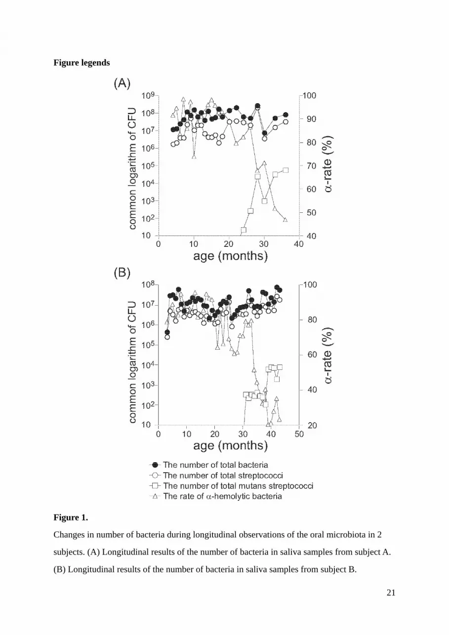
Longitudinal observation of changes of microbiota in saliva
Both of the two infants became colonized with S. mutans during the observation period. The
transition of the microbiota in saliva was monitored for approximately 1 year after S. mutans
was detected to confirm the sustained establishment of mutans streptococci and to study
subsequent changes in the microbiota. The transition of the salivary microbiota is shown in
Figure 1 and Table 1. During the examination period, the total number of cultivable bacteria
and streptococci in saliva ranged from 107 to 109 CFU/mL and 106 to 108 CFU/mL,
respectively. Thus, no noticeable change in the total number of bacteria occurred, in spite of
environmental changes due to tooth eruption and the establishment of S. mutans in the oral
cavity.
In Subject A, mutans streptococci were first detected at 24 months after birth, which was
the time when the first primary molar erupted. On the other hand, in Subject B, mutans
streptococci were first detected at 36 months after birth when the second primary molar
erupted. In both subjects, the proportion of -haemolytic bacteria began to decrease rapidly

10
from the time when mutans streptococci were detected in saliva. However, this result would
contain limitation since the subjects were only two.
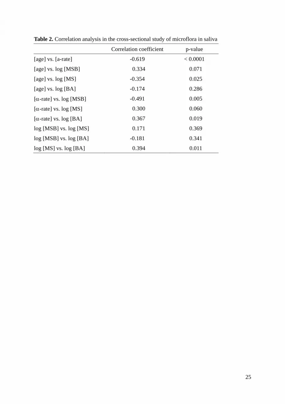
Cross-sectional study of changes of the microbiota in saliva
To statistically compensate the limitation in the result of the longitudinal observation, the
cross-sectional study of changes of the microbiota in saliva was carried out and analysed with
correlation analysis. In our correlation analysis of the results of the cross-sectional study of
the microbiota in saliva, the following 2 combinations of 2 variables showed a correlation at a
p-value of less than 0.01: [age] and [-ratio], and [-ratio] and log [MSB] (Table 2). As for
the correlation between [age] and [-ratio], the regression line was expressed as [-ratio] =
96.989 – 11.082 × [age]; R2 = 0.383 (Fig. 2A). From this expression, it was suggested that the
ratio of -haemolytic bacteria in saliva decreases with increasing age of the children.
Regarding the correlation between [-ratio] and log [MSB], the regression line was expressed
as log [MSB] = 4.328 – 0.024 × [ -ratio]; R2 = 0.241 (Fig. 2B). From this, it was revealed
that the number of mutans streptococci was high when the ratio of -haemolytic bacteria in
saliva was low.

11
Discussion
For paediatric dentists, it is very important to know the changes of the oral microbiota relative
to pre-dentate to dentate phase, from bottle- or breast-feeding to eating phase. In particular,
the transition from before to after the establishment of mutans streptococci has received much
focus because this change is considered a predictor of future dental caries. In this respect, the
recent comprehensive mapping of the oral microbiota by metagenomic approaches is very
interesting [20, 21]. However, due to significant costs, these analyses are not realistic as a tool
to assess the caries risk of an individual basis in the clinical setting. The goal of the present
study was to develop a inexpensive, easy and effective procedure to monitor changes of the
oral microbiota in infants that may predict the future risk of dental caries activity. Although it
has been generally assumed that development of a “good oral microbiota” may be conducive
to oral health, it is less clear what characterizes such a microbiota.
Our longitudinal observations on bacterial changes in saliva from two infants revealed that
the early members of the oral microbiota till 12 months after birth were bacteria acquired
from the maternal intestines or vagina, that the succeeding members up till 18 months after
birth were early colonizing streptococci such as S. mitis, S. oralis and S. salivarius, and that
the establishment of S. mutans occurred after the eruption of the primary molars. These

12
findings are in accordance with previous reports [16, 22, 23]. Although dramatic changes in
the oral environment such as the changes of food intake and the eruption of teeth occurred
during this period, it had no apparent effect on the total number of bacteria in saliva. This
finding suggests a limited capacity of the oral cavity for bacteria. This may explain why
introduction of S. mutans induced a rapid decrease of -haemolytic bacteria in saliva.
Cross-sectional observation showed that both increasing age and the increase of mutans
streptococci were associated with a decrease of proportions of -haemolytic bacterial in saliva,
in full accord with the result of the longitudinal observations. In the infants, the predominant
streptococcal species are the Mitis group streptococci, which are -haemolytic [11-14]. It was
previously reported that an inverse relationship between the occurrence of S. mutans and S.
sanguinis was observed during drastic carbohydrate dietary restriction [24]. With regard to an
increase of S. mutans associated with decrease of -haemolytic bacteria including S.
sanguinis, this previous finding is accordance with ours. It is conceivable that the change
from bottle- or breast-feeding to a more varied food intake, which correlates with increasing
age, and the change from sucrose-free to sucrose-containing food, which reflects
establishment of S. mutans, provide the background for the observed increase of S. mutans
and ensuing decrease of -haemolytic bacteria.
The present results of both the longitudinal and cross-sectional studies showed that
establishment of S. mutans was associated with decreasing proportions of -haemolytic
bacteria. We consider this finding important, because the observed decrease in proportions of

13
-haemolytic bacteria in saliva may be useful as an indicator of the establishment of mutans
streptococci by clinical laboratories. Conventionally, MSB agar, a selective medium for
mutans streptococci, has been used to detect mutans streptococci in saliva samples [25].
However, it is difficult to detect fewer than 103 CFU of mutans streptococci /mL saliva using
that medium. Our findings suggest that the establishment of small numbers of mutans
streptococci in saliva can be predicted by the demonstration of proportions of -haemolytic
bacteria less than approximately 55%. Although highly sensitive and reliable detection
methods of mutans streptococci by species-specific PCR were reported [26-29], such methods
require specialized and expensive equipment. In contrast, we consider the marker
demonstrated in this study a convenient method to determine the establishment of mutans
streptococci by simple bacteriological procedures. If paediatric dentists can obtain the
cooperation of infant patients and their parents, this phenomenon may provide a basis for
planning their need for dental prophylaxis and treatment.
In conclusion, our study demonstrates that oral establishment of S. mutans is predicted by a
decrease of the proportions of -haemolytic bacteria in saliva. Thus, the relative decrease of
-haemolytic bacteria may be applicable as an indicator to assess dental caries risk and
provide a basis of planning dental care and treatment in infants. In addition, that the results

14
suggest that maintenance and growth of -haemolytic bacteria in the infant oral cavity may be
associated with the prevention of infection with S. mutans.
Bullet point:
What this paper adds
• The total number of total bacteria in saliva of infant is not significantly changed as a
result of the eruption of primary teeth and the early changes of eating habits.
• Establishment of S. mutans in the infant oral cavity induces a decrease in proportions of
-haemolytic bacteria in saliva.
• The establishment of small numbers (103 CFU/mL) of mutans streptococci is predicted
by proportions of -haemolytic in infant saliva less than approximately 55%.
Why this paper is important to paediatric dentists
• The result of this paper suggested that the decrease in salivary proportions of
-haemolytic bacteria may be applicable as a predictor of the establishment of S. mutans
and thereby of dental caries risk, and may be used as a basis for planning of dental care
and treatment.

15
• It was suggested that the maintenance and growth of -haemolytic bacteria in the infant
oral cavity might be associated with the prevention of infection with S. mutans.
Acknowledgement
This work was supported by KAKENHI (Grant-in-Aid for Scientific Research) from the
Japan Society for the Promotion of Science (18592243 and 21659477).

16
References
1. Leverett DH, Featherstone JD, Proskin HM et al. Caries risk assessment by a
cross-sectional discrimination model. J Dent Res. 1993;72: 529-37.
2. Hale KJ. Oral health risk assessment timing and establishment of the dental home.
Pediatrics. 2003;111: 1113-6.
3. Grembowski D, Spiekerman C, Milgrom P. Linking mother and child access to
dental care. Pediatrics. 2008;122: e805-14.
4. Grembowski D, Spiekerman C, Milgrom P. Linking mother access to dental care and
child oral health. Community Dent Oral Epidemiol. 2009;37: 381-90.
5. Hamada S, Slade HD. Biology, immunology, and cariogenicity of Streptococcus
mutans. Microbiol Rev. 1980;44: 331-84.
6. Berkowitz RJ. Causes, treatment and prevention of early childhood caries: a
microbiologic perspective. J Can Dent Assoc. 2003;69: 304-7.
7. Zickert I, Emilson CG, Krasse B. Effect of caries preventive measures in children
highly infected with the bacterium Streptococcus mutans. Arch Oral Biol. 1982;27:
861-8.
8. Yano A, Kaneko N, Ida H, Yamaguchi T, Hanada N. Real-time PCR for

17
quantification of Streptococcus mutans. FEMS Microbiol Lett. 2002;217: 23-30.
9. Fazilat S, Sauerwein R, McLeod J et al. Application of adenosine
triphosphate-driven bioluminescence for quantification of plaque bacteria and
assessment of oral hygiene in children. Pediatr Dent. 2010;32: 195-204.
10. Nurelhuda NM, Al-Haroni M, Trovik TA, Bakken V. Caries experience and
quantification of Streptococcus mutans and Streptococcus sobrinus in saliva of
Sudanese schoolchildren. Caries Res. 2010;44: 402-7.
11. Caufield PW, Cutter GR, Dasanayake AP. Initial acquisition of mutans streptococci
by infants: evidence for a discrete window of infectivity. J Dent Res. 1993;72:
37-45.
12. Caufield PW, Dasanayake AP, Li Y et al. Natural history of Streptococcus sanguinis
in the oral cavity of infants: evidence for a discrete window of infectivity. Infect
Immun. 2000;68: 4018-23.
13. Pearce C, Bowden GH, Evans M et al. Identification of pioneer viridans streptococci
in the oral cavity of human neonates. J Med Microbiol. 1995;42: 67-72.
14. Smith DJ, Anderson JM, King WF, van Houte J, Taubman MA. Oral streptococcal
colonization of infants. Oral Microbiol Immunol. 1993;8: 1-4.

18
15. Fujiwara T, Hoshino T, Ooshima T, Sobue S, Hamada S. Purification,
characterization, and molecular analysis of the gene encoding glucosyltransferase
from Streptococcus oralis. Infect Immun. 2000;68: 2475-83.
16. Kononen E, Jousimies-Somer H, Bryk A, Kilp T, Kilian M. Establishment of
streptococci in the upper respiratory tract: longitudinal changes in the mouth and
nasopharynx up to 2 years of age. J Med Microbiol. 2002;51: 723-30.
17. Novak J, Caufield PW, Miller EJ. Isolation and biochemical characterization of a
novel lantibiotic mutacin from Streptococcus mutans. J Bacteriol. 1994;176:
4316-20.
18. Cvitkovitch DG, Li YH, Ellen RP. Quorum sensing and biofilm formation in
Streptococcal infections. J Clin Invest. 2003;112: 1626-32.
19. Poutrel B, Ryniewicz HZ. Evaluation of the API 20 Strep system for species
identification of streptococci isolated from bovine mastitis. J Clin Microbiol.
1984;19: 213-4.
20. Lazarevic V, Whiteson K, Huse S et al. Metagenomic study of the oral microbiota by
Illumina high-throughput sequencing. J Microbiol Methods. 2009;79: 266-71.
21. Parahitiyawa NB, Scully C, Leung WK et al. Exploring the oral bacterial flora:

19
current status and future directions. Oral Dis. 2010;16: 136-45.
22. Rotimi VO, Olowe SA, Ahmed I. The development of bacterial flora of premature
neonates. J Hyg (Lond). 1985;94: 309-18.
23. Fujiwara T, Sasada E, Mima N, Ooshima T. Caries prevalence and salivary mutans
streptococci in 0-2-year-old children of Japan. Community Dent Oral Epidemiol.
1991;19: 151-4.
24. De Stoppelaar JD, Van Houte J, Backer DO. The effect of carbohydrate restriction
on the presence of Streptococcus mutans, Streptococcus sanguis and iodophilic
polysaccharide-producing bacteria in human dental plaque. Caries Res. 1970;4:
114-23.
25. van Houte J, Gibbs G, Butera C. Oral flora of children with "nursing bottle caries". J
Dent Res. 1982;61: 382-5.
26. Igarashi T, Ichikawa K, Yamamoto A, Goto N. Identification of mutans streptococcal
species by the PCR products of the dex genes. J Microbiol Methods. 2001;46:
99-105.
27. Okada M, Soda Y, Hayashi F et al. PCR detection of Streptococcus mutans and S.
sobrinus in dental plaque samples from Japanese pre-school children. J Med

20
Microbiol. 2002;51: 443-7.
28. Yoshida A, Suzuki N, Nakano Y et al. Development of a 5' nuclease-based real-time
PCR assay for quantitative detection of cariogenic dental pathogens Streptococcus
mutans and Streptococcus sobrinus. J Clin Microbiol. 2003;41: 4438-41.
29. Hoshino T, Kawaguchi M, Shimizu N et al. PCR detection and identification of oral
streptococci in saliva samples using gtf genes. Diagn Microbiol Infect Dis. 2004;48:
195-9.
21
Figure legends
Figure 1.
Changes in number of bacteria during longitudinal observations of the oral microbiota in 2
subjects. (A) Longitudinal results of the number of bacteria in saliva samples from subject A.
(B) Longitudinal results of the number of bacteria in saliva samples from subject B.

22
Figure 2.
Regression analyses of variables with a correlation. (A) Correlation between [age]and
[-ratio]. (B) Correlation between [-ratio] and log [MSB].

23
Table 1A. The detected bacteria in the saliva of Subject A identified by API 20 Strep
age (months)
Detected cocci 4 6 12 18 24 30 36
Aerococcus viridans 3 1
Gardnerella vaginalis 3 2 1
Gemella morbillorum 4 1 1
Gemella haemolysans 1 1
Enterococcus faecalis 1 1
Enterococcus avium 1
Lactococcus lactis 1 1
S. acidominimus 1
S. mitis 2 1 3 2 3 2
S. sanguinis 2 1 1 1
S. oralis 2 1 1 2 2 2
S. salivarius 1 2 2 1 1 1
S. anginosus 1
S. mutans 1 1 1
S. pneumoniae 1
Not identified 4 1 2 3 2 2 1
Number tested (n = ) 10 20 10 10 10 10 10

24
Table 1B. The detected bacteria from the saliva of Subject B identified by API 20 Strep
age (months)
Detected cocci 4 6 12 18 24 30 36
Gardnerella vaginalis 3
Gemella morbillorum 3 2
Gemella haemolysans
Enterococcus faecalis 1 1 1
Enterococcus faecium 1
Lactococcus lactis 1 3 1 1
S. acidominimus 4 4 4 3 2
S. mitis 1 1
S. sanguinis 1 1 1 3 2 3 1
S. oralis 2 1 1 1
S. salivarius 1 1
S. anginosus 1
S. mutans 1
S. canis 1
Not identified 5 2 2 1 1 1 1
Number tested (n = ) 10 10 10 12 10 10 10
25
Table 2. Correlation analysis in the cross-sectional study of microflora in saliva
Correlation coefficient p-value
[age] vs. [a-rate] -0.619 < 0.0001
[age] vs. log [MSB] 0.334 0.071
[age] vs. log [MS] -0.354 0.025
[age] vs. log [BA] -0.174 0.286
[-rate] vs. log [MSB] -0.491 0.005
[-rate] vs. log [MS] 0.300 0.060
[-rate] vs. log [BA] 0.367 0.019
log [MSB] vs. log [MS] 0.171 0.369
log [MSB] vs. log [BA] -0.181 0.341
log [MS] vs. log [BA] 0.394 0.011