epigenetic regulation of phosphodiesterases 2a and 3a underlies compromised β-adrenergic signaling...
TRANSCRIPT

Article
Epigenetic Regulation of P
hosphodiesterases 2Aand3A Underlies Compromised b-Adrenergic Signaling inan iPSCModel of Dilated CardiomyopathyGraphical Abstract
Highlights
d b-AR signaling switch from b-2 AR to b-1/2 AR mode during
human iPSC-CM maturation
d Upregulation of PDE2/3 leads to compromised b-adrenergic
regulation in DCM iPSC-CMs
d Epigenetic activation of PDE2/3 is a key molecular event
during pathogenesis of DCM
d Nuclear localization of mutated TNNT2 contributes to
epigenetic modification in DCM
Wu et al., 2015, Cell Stem Cell 17, 89–100July 2, 2015 ª2015 Elsevier Inc.http://dx.doi.org/10.1016/j.stem.2015.04.020
Authors
Haodi Wu, Jaecheol Lee,
Ludovic G. Vincent, ..., Yang K. Xiang,
Donald M. Bers, Joseph C. Wu
In Brief
In this paper, Wu et al. profiled the
b-adrenergic signaling properties in
human iPSC-CMs and demonstrated
novel epigenetic mechanisms that
underlie the compromised b-adrenergic
signaling in DCM, a common cause of
heart failure and cardiac transplantation.
These results enhance our understanding
of DCM pathogenesis and may uncover
new therapeutic targets.
Cell Stem Cell
Article
Epigenetic Regulation of Phosphodiesterases 2Aand3AUnderliesCompromisedb-AdrenergicSignalingin an iPSCModel of Dilated CardiomyopathyHaodi Wu,1,2,3 Jaecheol Lee,1,2,3 Ludovic G. Vincent,4 Qingtong Wang,5 Mingxia Gu,1,2,3 Feng Lan,1,2,3
Jared M. Churko,1,2,3 Karim I. Sallam,1,2,3 Elena Matsa,1,2,3 Arun Sharma,1,2,3 Joseph D. Gold,1 Adam J. Engler,4,6
Yang K. Xiang,5 Donald M. Bers,5 and Joseph C. Wu1,2,3,*1Stanford Cardiovascular Institute, Stanford University School of Medicine, Stanford, CA 94305, USA2Division of Cardiology, Department of Medicine, Stanford University School of Medicine, Stanford, CA 94305, USA3Institute for Stem Cell Biology and Regenerative Medicine, Stanford University School of Medicine, Stanford, CA 94305, USA4Department of Bioengineering, University of California, San Diego, La Jolla, CA 92093, USA5Department of Pharmacology, University of California, Davis, Davis, CA 95616, USA6Sanford Consortium for Regenerative Medicine, La Jolla, CA 92037, USA
*Correspondence: [email protected]
http://dx.doi.org/10.1016/j.stem.2015.04.020
SUMMARY
b-adrenergicsignalingpathwaysmediatekeyaspectsof cardiac function. Its dysregulation is associatedwith a range of cardiac diseases, including dilatedcardiomyopathy (DCM). Previously, we establishedan iPSC model of familial DCM from patients with amutation in TNNT2, a sarcomeric protein. Here, wefound that the b-adrenergic agonist isoproterenolinduced mature b-adrenergic signaling in iPSC-derived cardiomyocytes (iPSC-CMs) but that thispathway was blunted in DCM iPSC-CMs. Althoughexpression levels of several b-adrenergic signalingcomponents were unaltered between control andDCM iPSC-CMs, we found that phosphodiesterases(PDEs) 2A and PDE3A were upregulated in DCMiPSC-CMs and that PDE2A was also upregulated inDCM patient tissue. We further discovered increasednuclear localization of mutant TNNT2 and epigeneticmodifications of PDE genes in both DCM iPSC-CMsand patient tissue. Notably, pharmacologic inhibitionof PDE2A and PDE3A restored cAMP levels andameliorated the impaired b-adrenergic signaling ofDCM iPSC-CMs, suggesting therapeutic potential.
INTRODUCTION
Dilated cardiomyopathy (DCM) is a common myocardial disor-
der characterized by ventricular chamber enlargement and sys-
tolic dysfunction (Maron et al., 2006). DCM gives rise to sudden
cardiac death, hypertension, and heart failure, and contributes
significantly to health care costs. Recent studies have shown
that more than 40% of DCM is caused by mutations in genes
that encode sarcomeric, cytoskeletal, mitochondrial, calcium
handling, or nuclear membrane proteins (Burkett and Hersh-
berger, 2005; Morita et al., 2005). Accordingly, multiple molecu-
lar mechanisms, including loosened mechanical linkage of the
extracellular matrix to the cytoskeleton (Lapidos et al., 2004),
disarrangement of Z-disc protein elements (Knoll et al., 2002),
decreased myofilament calcium sensitivity (Kamisago et al.,
2000), ion channel abnormalities (Bienengraeber et al., 2004),
and remodeled intracellular calcium handling (Schmitt et al.,
2003) have been reported to underlie the decreased systolic
contractile function of cardiac muscle in DCM. However, the
heterogeneous etiologies underlying DCM also have limited
our understanding of the respective roles of such factors in the
long-term pathogenesis of DCM.
Ever since the discovery of the four key reprogramming factors
by Takahashi and Yamanaka (2006), significant strides have
been made in deriving cardiomyocytes from human originated
stem cells (Burridge et al., 2012; Takahashi et al., 2007; Yu
et al., 2007). These advances have enabled disease modeling
and development of regenerative medicine approaches for car-
diac diseases (Chong et al., 2014; Lan et al., 2013; Liang et al.,
2013; Sun et al., 2012;Wang et al., 2014). Human induced plurip-
otent stem cell (iPSC)-derived cardiomyocytes (iPSC-CMs) have
been shown to recapitulate morphological and functional prop-
erties of native cardiomyocytes. However, few studies have eval-
uated the platform’s ability to recapitulate signaling pathways,
molecular pathophysiology, and underlying transcriptional regu-
lation in diseased cardiomyocytes. The ability to generate iPSC-
CMs from patients carrying known or novel mutations, coupled
with the feasibility of introducing specific modifications to their
genome, presents an unprecedented opportunity to investigate
pathogenic mutations and identify new treatments for the dis-
eases they cause. Thus, uncovering the novel mechanism of
DCM in stem cell-derived cardiomyocyte models will greatly
contribute to our understanding of the application of stem cell
based disease models in both basic scientific and translational
research.
It is well known that b-adrenergic signaling pathways mediate
the inotropic and chronotropic regulation of cardiac function
and release reserved pumping power to meet the increased
demand for heart output under stress (Rockman et al., 2002;
Xiang and Kobilka, 2003). Moreover, abnormalities in b-adren-
ergic signaling are associated with certain cardiomyopathies
such as DCM (Cho et al., 1999), cardiac hypertrophy (Engelhardt
Cell Stem Cell 17, 89–100, July 2, 2015 ª2015 Elsevier Inc. 89
et al., 1999), and heart failure (Lohse et al., 2003; Post et al.,
1999). Clinically, b-blockers are commonly prescribed for hyper-
tension, arrhythmia, and heart failure. Therefore, improving the
understanding of b-adrenergic signaling in iPSC-CMs and its
regulation in DCM is scientifically and clinically significant
because it can elucidate the pathophysiologic mechanism of
the disorder and identify new treatments for DCM.
In the present study, we focused on b-adrenergic signaling
pathway development in iPSC-CMs, measured their responses
to b-adrenergic activation, and investigated their receptor
subtype dependence at different maturation stages. Then, by
comparing control (Ctrl) and DCM iPSC-CMs, we demonstrated
impaired b-adrenergic signaling and contractile function in DCM
iPSC-CMs. Expression profiles showed a significant upregula-
tion of phosphodiesterases (PDE) subtypes in DCM iPSC-CMs,
which could restrict cyclic (c)AMP signaling evoked by b-adren-
ergic activation. Further functional assays confirmed that DCM
iPSC-CMs regain their reactivity to b-agonist stimulation after
subtype-specific blockade of PDE 2A and 3A. Finally, chromatin
immunoprecipitation (ChIP) studies suggest nuclear TNNT2 may
contribute to novel epigenetic mechanisms that underlie DCM
pathogenesis.
RESULTS
Differentiation of iPSC-CMs Was Accompanied bySpecific Regulation of b-Adrenergic Signaling RelatedProteinsTo investigate the maturation of b-adrenergic signaling path-
ways in iPSC-CMs, we used iPSC lines from three healthy volun-
teers (Table S1). Genetic screening showed no knownmutations
related to familial heart diseases. All of the iPSC lines were iden-
tified by positive immunostaining for multiple ESC-like markers
such as SSEA-4, TRA-1-81, Oct4, Sox2, Nanog, and Klf4 (Fig-
ure S1A). The pluripotent nature of iPSC lines was further
demonstrated by their potential to form all three germ layers
in vivo (Figure S1B). Beating iPSC-CMs were differentiated and
purified as described (Lian et al., 2012) (Movie S1). Fluores-
cence-activated cell sorting (FACS) and immunostaining illus-
trated typical properties of cardiomyocyte lineages in these
iPSC-CMs (Figures S1C–S1E).
For the expression profiling of genes related to b-adrenergic
signaling during maturation, total RNA was extracted from
iPSC lines and at days 12, 30, and 60 of differentiation. The
cDNA libraries were constructed and then subjected to RNA
sequencing (seq) analysis. Most components of the b-adren-
ergic signaling apparatus such as b2 adrenergic receptor
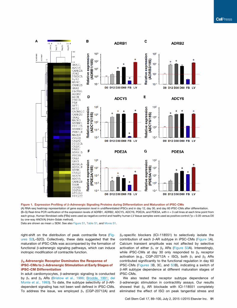
(ADRB2), adenylate cyclase (ADCY) 5 and 6, and PDE4D and
PDE5A (Figure 1A) were sharply upregulated in iPSC-CMs
compared to iPSC lines. RNA-seq results were further verified
by quantitative (q)PCR. Although the expression of ADRB2 was
increased by 7.37 ± 0.74-fold at day 12 of differentiation, no sig-
nificant changes in b1 adrenergic receptors (ADRB1) expression
were seen prior to day 30 (Figures 1B and 1C). Expression of
alpha adrenergic receptors varied: ADRA1A was mildly upregu-
lated by 1.81 ± 0.21-fold at day 60 of differentiation. ADRA1B
was significantly upregulated by 15.08 ± 1.31-fold at day 12
of differentiation, while ADRA1D expression was negligible
(Figures S1F–S1H). The expression of adenylyl cyclase subtypes
(ADCY5/6) increased significantly to a level comparable to that
of adult human left ventricle (LV) tissues after 12–30 days of
differentiation (Figures 1D and 1E). Throughout differentiation,
PDE subtypes underwent distinct alterations in expression:
PDE2A expression was decreased sharply to 1.85 ± 0.06% of
its original level after 12 days of differentiation and remained
low (Figure 1F), while the levels of both PDE4D and PDE5A
rose more than 40-fold by day 60 of maturation (Figures 1G
andS1I). No significant upregulation of PDE3Awas observed un-
til day 60 of differentiation (Figure S1J). The expression levels of
key calcium-handling proteins (e.g., PLN, CASQ2, LCC, RyR2,
and SERCA2a) and other b-adrenergic signaling related proteins
(e.g., PRKACA, CAMIIA, and CAMIID) showed significant upre-
gulation during differentiation (Figures S1K–S1R). No change
was observed for the Gi subunit expression (Figure S1S), while
Gs subunit expression level decreased throughout differentiation
(Figure S1T).
b-Adrenergic Stimulation Induced ChronotropicResponses in iPSC-CMsIn order to test the effects of b-adrenergic stimulation, iPSC-CMs
were next treated with 1 mM isoproterenol (ISO) and sponta-
neous calcium transients were analyzed (Figures 2A and 2B). Hu-
man iPSC-CMs at day 30 and day 60 of differentiation exhibited
similar beating rates (35.7 ± 1.4 and 34.5 ± 0.7 bpm) at basal
level, increasing to 84.2 ± 1.9 and 58.7 ± 3.0 bpm after ISO treat-
ment (Figure 2C). The transient decay Tau in day 30 and day 60
iPSC-CMs were curtailed by 50.5% ± 2.6% and 19.7% ± 4.7%,
respectively, by b-adrenergic activation, suggesting a possible
b-adrenergic signaling-dependent activation of calcium recy-
cling in iPSC-CMs (Figure 2D). Likewise, time to peak in both
groups decreased by 47.4% ± 2.4% and 19.8% ± 2.8%, respec-
tively (Figure 2E). Interestingly, transient amplitude was not
increased after ISO treatment at different stages of differentia-
tion (Figure S2A), yet other temporal parameters such as tran-
sient duration 90 and 50 were accelerated (Figures S2B and
S2C). We also observed that pretreatment with the non-selective
b-blocker propranolol eliminated the effect of ISO in iPSC-CMs,
indicating that the ISO signaling in these cells was indeed con-
ducted via b-adrenergic receptors (ARs) (data not shown).
b-Adrenergic Stimulation Induced Inotropic Responsesin iPSC-CMsAs b-adrenergic activation accelerated the calcium handling
in iPSC-CMs, we wondered if it can also increase contractile
force in these cells. Using a hydrogel-based traction forcemicro-
scopy (TFM) imaging assay (Movie S2), we next measured the
contractile force of iPSC-CMs with or without ISO treatment at
different stages of maturation (Figures 2F and 2G). ISO treatment
increased the peak contractile force and the maximum contrac-
tile rate by 35.0%± 3.5%and 44.9%± 5.0%, respectively, in day
30 iPSC-CMs and by 59.3% ± 12.9% and 120.6% ± 19.0%,
respectively, in day 60 iPSC-CMs (Figures 2H and 2I). Further-
more, ISO-treated iPSC-CMs demonstrated positive functional
regulation in other contractile parameters such as time to
peak, contractile duration 90 and 50, half rising and decay
time, beating rate, and maximum decay rate at both maturation
stages (Figures S2D–S2K). By analyzing all the readouts in the
contractility assay, we found that ISO treatment induced a
90 Cell Stem Cell 17, 89–100, July 2, 2015 ª2015 Elsevier Inc.
right-shift on the distribution of peak contractile force (Fig-
ures S2L–S2O). Collectively, these data suggested that the
maturation of iPSC-CMs was accompanied by the formation of
functional b-adrenergic signaling pathways, which can induce
inotropic modification of contractile function.
b2 Adrenergic Receptor Dominates the Response ofiPSC-CMs tob-Adrenergic Stimulation at Early Stages ofiPSC-CM DifferentiationIn adult cardiomyocytes, b-adrenergic signaling is conducted
by b1 and b2 ARs (Bristow et al., 1986; Brodde, 1991; del
Monte et al., 1993). To date, the subtype selectivity of b-AR-
dependent signaling has not been well defined in iPSC-CMs.
To address the issue, we employed b1 (CGP-20712A) and
b2-specific blockers (ICI-118551) to selectively isolate the
contribution of each b-AR subtype in iPSC-CMs (Figure 3A).
Calcium transient amplitude was not affected by selective
activation of either b1 or b2 ARs (Figure S3A). Interestingly,
while iPSC-CMs at day 30 only responded to b2 receptor
activation (e.g., CGP-20712A + ISO), both b1 and b2 ARs
contributed significantly to the functional regulation in day 60
iPSC-CMs (Figures 3B, 3C, and S3B), indicating a switch of
b-AR subtype dependence at different maturation stages of
iPSC-CMs.
We also tested the receptor subtype dependence of
b-adrenergic stimulation in contractility assays. Our results
showed that b2 AR blockade with ICI-118551 completely
eliminated the effect of ISO on peak tangential stress and
Figure 1. Expression Profiling of b-Adrenergic Signaling Proteins during Differentiation and Maturation of iPSC-CMs
(A) RNA-seq heatmap representation of gene expression level in undifferentiated iPSCs and in day 12, day 30, and day 60 iPSC-CMs after differentiation.
(B–G) Real-time PCR verification of the expression levels of ADRB1, ADRB2, ADCY5, ADCY6, PDE2A, and PDE5A, with n = 3 cell lines at each time point from
each group. Human fibroblast cells (FBs) were used as negative control and healthy human LV tissue samples were used as positive control (*p < 0.05 versus D0
by one-way ANOVA) (Holm-Sidak method).
Data are shown as mean ± SEM. See also Figure S1, Table S1, and Movie S1.
Cell Stem Cell 17, 89–100, July 2, 2015 ª2015 Elsevier Inc. 91

beating rate in the day 30 group, whereas in the day 60 group,
neither b1 nor b2 AR blockers inhibited responses to b-adren-
ergic stimulation (Figures S3C and S3D). In further support of
this observation, an ELISA-based cAMP assay also showed
that at day 30, b2 activation (e.g., CGP-20712A + ISO), but
not b1 activation (e.g., ICI-118551 + ISO) induced significant
cAMP elevation. By contrast, abundant cAMP was generated
by both b1 and b2 activation in day 60 iPSC-CMs (Figures 3D
and 3E). These functional results are in line with our b-AR
expression profiles, with b2 AR showing a relatively higher
expression at early stages of maturation. These findings also
independently suggest a dynamic regulation of the receptor
dependence of b-adrenergic signaling pathways during the
maturation of iPSC-CMs.
Figure 2. Functional Regulation by
b-Adrenergic Signaling in iPSC-CMs
(A and B) Representative calcium imaging
recording traces in wild-type (WT) iPSC-CMs
before and after ISO treatment.
(C–E) Statistics of calcium handling properties
such as beating rate (C), decay tau (D), and time to
peak (E) with and without ISO at different matu-
ration stages with n > 35 iPSC-CMs from three
lines in each group (*p < 0.05 versus baseline
group in each time point by two-way ANOVA)
(Holm-Sidak method).
(F) Representative temporal series of contractility
distribution pattern throughout one contraction
cycle of WT iPSC-CMs.
(G) Representative contractility profiling before
and after ISO treatment, Pascal (Pa).
(H and I) Measurements on contraction parame-
ters showing increased peak tangential force (H)
and maximum contract rate (I) with n > 25 iPSC-
CMs from three lines in each group (*p < 0.05 and
**p < 0.01 versus baseline untreated group by
Student’s t test).
Data are shown as mean ± SEM. See also Fig-
ure S2, Table S5, and Movie S2.
DCM iPSC-CMs Exhibit ImpairedResponse to b-AdrenergicStimulationAs abnormal b-adrenergic regulation in
cardiac diseases has been well docu-
mented (Lohse et al., 2003; Post et al.,
1999), we next examined whether DCM
iPSC-CMs can also recapitulate this
dysfunction. To address this question,
we differentiated iPSC-CMs from both
familial Ctrl and DCM (TNNT2 R173W)
groups as described (Sun et al., 2012).
Flow cytometry assays indicated that
the efficiency of iPSC-CM differentiation
in n = 3 Ctrl and n = 3 DCM patients
was >90% (Figures S4A–S4C). Success-
ful differentiation was further confirmed
by immunostaining of cardiac-specific
markers such as TNNT2 and a-actinin
(Figures S4D and S4F). Furthermore,
detailed analysis of myofilament protein arrangement revealed
an abnormal pattern of sarcomere structure in DCM iPSC-
CMs, but not in Ctrl iPSC-CMs (Figures S4E, S4G, and S4H–
S4K). According to our baseline data, most of the experiments
in the DCM study were carried out using day 60 iPSC-CMs while
they developed more matured beta-adrenergic signaling.
Day 60 iPSC-CMs from both groups were challenged with ISO
(Figures 4A–4D). We found that the same dose of ISO induced
larger effects on transient decay Tau in Ctrl iPSC-CMs than in
DCM iPSC-CMs (Figure 4E). ISO boosted the spontaneous
beating rate of Ctrl iPSC-CMs by 70.3% ± 6.8% (p < 0.001),
whereas an increase of only 18.6% ± 3.9% was observed in
DCM cells (p = 0.035) (Figure 4F). In addition, fluorescence reso-
nance energy transfer (FRET)-based protein kinase A (PKA)
92 Cell Stem Cell 17, 89–100, July 2, 2015 ª2015 Elsevier Inc.

activity imaging confirmed that ISO stimulation induced lower
levels of PKA activity in DCM iPSC-CMs in comparison to Ctrl
iPSC-CMs (Figures 5A–5C). Moreover, contractility assays (Fig-
ures 5D and 5E) confirmed that b-adrenergic signaling-induced
inotropic and chronotropic augmentation in DCM iPSC-CMs
was greatly impaired compared to that seen in Ctrl iPSC-CMs.
While ISO induced a 59.4% ± 7.7% increase of peak tangential
stress and 83.1% ± 9.1% increase of maximum contraction
rate in the Ctrl group, ISO failed to improve the contractile force
of DCM iPSC-CMs and only induced a 36.7% ± 4.5% increase in
their maximum contraction rate (Figures 5F and 5G). Other read-
outs for contractile function also showed the impaired respon-
siveness to b-adrenergic activation in DCM iPSC-CMs (Figures
S5A–S5F). These results taken en masse suggest that DCM
iPSC-CMs can recapitulate the disease-like b-adrenergic regu-
lation phenotypes seen in the intact diseased heart (Cho et al.,
1999). Interestingly, a similar development process of b-adren-
ergic signaling is at work in both Ctrl and DCM iPSC-CMs, since
the b2-AR dominance at day 30 also shift more toward b1-AR and
b2-AR codominance at day 60 in the DCM as we observed in Ctrl
cells (Figures S5G–S5J; Table S2).
Figure 3. Subtype Dependence of b-Adren-
ergic Signaling in Different Maturation
Stages of iPSC-CMs
(A) Representative calcium handling traces of day
30 and day 60 iPSC-CMs at baseline after specific
b2 AR activation (CGP-20712A + ISO) and after
specific b1 AR activation (ICI-118551 + ISO).
(B and C) Statistics of calcium handling properties
of day 30 and day 60 iPSC-CMs in baseline, ISO
treated, CGP-20712A, CGP-20712A + ISO, ICI-
118551, and ICI-118551 + ISO groups with n > 30
iPSC-CMs from three lines in each time point from
each group (*p < 0.05 versus baseline group in
each time point by two-way ANOVA) (Holm-Sidak
method).
(D) Standard curve for ELISA-based cAMP assay,
with final results presented in the form of per-
centage activity, as shown in the formula.
(E) cAMP assay assessment of cAMP generation
in iPSC-CMs upon different drug treatment at day
30 and day 60 after differentiation. The data were
from six independent experiments using three
lines of iPSC-CMs (*p < 0.05 and **p < 0.01 versus
baseline group by one-way ANOVA) (Holm-Sidak
method).
Data are shown as mean ± SEM. See also
Figure S3.
PDE2A and PDE3A Expressions AreUpregulated in DCM iPSC-CMsIn order to uncover the molecular basis of
the ‘‘desensitized’’ b-adrenergic signaling
pattern in DCM iPSC-CMs, we next
utilized microarray analysis to examine
the whole transcriptomes of both DCM
and Ctrl groups. The mRNA levels of
the main components of the b-adren-
ergic signaling protein apparatus were
compared (Figure 6A). Interestingly, while
the expression levels of b1 and b2 ARs
were comparable in day 60 DCM and Ctrl groups, several
members from the PDE family showed subtype-specific alter-
ations in DCM iPSC-CMs. In cardiomyocytes, the PDEs hydro-
lyze phosphodiester bonds of cyclic nucleotides and regulate
the distribution, duration, and amplitude of cyclic nucleotide
signaling (Jeonet al., 2005). Real-timePCRshowednosignificant
differences between PDE levels in undifferentiated DCM versus
Ctrl iPSCs, while the expression levels of PDE2A, PDE3A, and
PDE5A were increased by 680% ± 129%, 77% ± 12%, and
76% ± 13% in day 30 and increased by 1,681% ± 95%,
367% ± 14%, and 97% ± 20% in day 60 DCM iPSC-CMs
compared to Ctrl iPSC-CMs. By contrast, PDE4D showed a
48% ± 5% decrease in day 60 DCM iPSC-CMs (Figures 6B–6D
and S6A). Expression levels of ADRB1 and ADRB2 were compa-
rable between DCM and Ctrl iPSC lines and in day 30 and day 60
DCM and Ctrl iPSC-CMs (Figures S6B and S6C). Expression
profile comparison of other key maturation related genes from
Ctrl andDCM iPSC-CMsat bothmaturation stageswere summa-
rized in Table S3. Taken together, these results clearly demon-
strated subtype-specific expression regulation of PDEs in DCM
iPSC-CMs.
Cell Stem Cell 17, 89–100, July 2, 2015 ª2015 Elsevier Inc. 93

Subtype Specific Inhibition of PDEs Rescues theb-Adrenergic Signaling Response in DCM iPSC-CMsSince both PDE2A and PDE3A hydrolyze cAMP (Baillie, 2009),
we hypothesized that the elevated expression of PDE2A and
PDE3A in DCM iPSC-CMs might contribute to lower cAMP
production and weakened b-adrenergic responses upon ISO
treatment. To test this hypothesis, DCM and Ctrl iPSC-CMs
were pretreated with PDE blockers (PDE2A: 100 nM Bay-60-
7550; PDE3A: 10 mM milrinone; and PDE5A: 10 mM sildenafil)
for 15 min before the challenge with 1 mM ISO and the resultant
levels of cAMPweremeasured. Readingswere normalized to the
positive controls (10 mM IBMX + 10 mM Forskolin) in Ctrl group.
We noticed that PDE2A or PDE3A inhibition significantly
enhanced the response to b-adrenergic stimulation in both Ctrl
and DCM iPSC-CMs (Figure 6E). Interestingly, although PDE5A
is cGMP specific, the treatment by PDE5A blocker is able to
enhance cAMP generation in DCM iPSC-CMs upon ISO chal-
lenge (Figure 6E). Also, FRET-based imaging of PKA activity
showed that blocking either PDE2A or PDE3A could recover
the PKA FRET signal in DCM iPSC-CMs (Figure S6D).
We then examined if treatment by PDE blockers could relieve
the impaired functional outputs in DCM iPSC-CMs. By calcium
imaging and TFM study, our results showed preinhibition of
PDE2A or PDE3A slightly improved calcium cycling in DCM
iPSC-CMs at baseline and induced a much more robust func-
tional enhancement in DCM iPSC-CMs compared to Ctrl groups
(Figures S6E–S6G). Thus, PDE2A/3A selective inhibition
restored the impaired b-adrenergic signaling in DCM iPSC-
CMs (Figures 6F, 6G, and S6H–S6J). These results suggest
that the functional impairment of b-adrenergic signaling in
DCM iPSC-CMs could be ameliorated by subtype selective
repression of the observed overexpressed PDE2A and PDE3A.
Epigenetic Modifications Contribute to Overexpressionof PDE2/3a during Maturation of DCM iPSC-CMsTo uncover the underlying reasons for the upregulated expres-
sion of PDE2A/3A in DCM iPSC-CMs, we next examined epige-
netic modulation of PDE family members by ChIP (Figure 7A)
(Paige et al., 2012). We observed no significant differences in
the histone markers for activation (H3K4me3) and repression
(H3K27me3) in the PDE2A gene in Ctrl and DCM iPSC cells (Fig-
ures 7B and 7C). However, compared to Ctrl cells, the level of the
activation marker H3K4me3 in the regions of PDE2A-R1 was
increased by 200% ± 35% and 284% ± 29% in day 30 and
day 60 DCM iPSC-CMs, respectively. In PDE2A-R2, the
H3K4me3 marker was increased by 186% ± 28% and 484% ±
66% in day 30 and day 60 DCM iPSC-CMs, respectively. By
contrast, repression marker H3K27me3 was decreased by
54%± 6%and 67%± 6% in PDE2A-R1 and PDE2A-R2, respec-
tively, in day 60 DCM iPSC-CMs compared to Ctrl cells. How-
ever, in day 30 DCM iPSC-CMs, the H3K27me3 marker was
unchanged in PDE2A-R1 and decreased by 42% ± 2% in
PDE2A-R2 (Figures 7B and 7C). Measurement of additional his-
tone markers such as H3K36me3 and H3K27AC in multiple re-
gions of PDE2A also shows general epigenetic activation of
PDE2A expression (Figures S7A–S7D), which was supported
by the observation of increased PDE2Aprotein levels (Figure 7D).
We also examined histone marker modifications in the PDE3A
and PDE5A genes (Figures S7E and S7F), and again, no differ-
ences were detected between Ctrl and DCM iPSCs. However,
in accordance with the increased protein expression of PDE3A
in DCM iPSC-CMs and tissues (Figure S7G), the activation
marker in the PDE3A-R1 was increased by 298% ± 9.5% and
448% ± 75% in day 30 and 60 DCM iPSC-CMs, respectively,
whereas the repressive marker was decreased by 68% ± 8.1%
and 69% ± 6.2%, in day 30 and 60 DCM iPSC-CMs, respec-
tively, compared to Ctrl group (Figure S7H). Measurement of
additional histone markers in other PDE3A gene regions showed
similar increases in activation markers and decreases in repres-
sive markers (Figures S7H–S7J). For PDE5A, both the activation
marker H3K4me3 and repressive marker H3K27me3 were
downregulated in DCM iPSC-CMs (Figure S7K); no significant
difference was detected in marker H3K36me3, while H3K27AC
showed a slight increase in DCM cells. These results suggest a
much more complex picture on regulation of the PDE5A gene.
We next examined the epigenetic status of the PDE2A/3A/5A
gene in human cardiac tissues obtained from five healthy indi-
viduals and four DCM patients undergoing cardiac surgeries.
Figure 4. Impaired b-Adrenergic Signaling
Response of Calcium Handling Properties
in DCM iPSC-CMs
(A and B) Representative recording of sponta-
neous calcium transient of Ctrl (A) and DCM iPSC-
CMs (B) at baseline.
(C and D) Representative recording of calcium
transient in Ctrl (C) and DCM iPSC-CMs (D) after
1 mM ISO treatment.
(E and F) Statistics of calcium handling parameters
(E, decay Tau and F, beating rate) in Ctrl and
DCM iPSC-CMs before and after ISO treatment
with n > 30 iPSC-CMs in each group (**p < 0.01
versus baseline and significant statistical differ-
ence yp < 0.05 versus WT in each group by two-
way ANOVA) (Holm-Sidak method).
Data are shown as mean ± SEM. See also Fig-
ure S4 and Table S2.
94 Cell Stem Cell 17, 89–100, July 2, 2015 ª2015 Elsevier Inc.

Western blot confirmed PDE2A upregulation in DCM patients
(Figure 7D). Interestingly, PDE2A-R1 and PDE2A-R2 in DCM
hearts showed 446% ± 38% and 160% ± 16% of increase in
activation marks and 42% ± 6.7% and 25% ± 4.8% of decrease
in repressive marks compared to healthy tissues (Figures 7E and
7F). The PDE3A gene in DCM patient tissues demonstrated
similar patterns as PDE2A (Figure S7L). For PDE5A, there was
no significant change in the activation marker, but the repressive
histone marker level was decreased (Figure S7M). These find-
ings are congruent with our observations in DCM iPSC-CMs
and support the importance of the underlying epigenetic regula-
tory mechanisms that lead to altered expression levels of PDE
subtypes in DCM iPSC-CMs.
A question that remains unclear is how the mutation in TNNT2
affects the epigenetic modification of histones. It has been re-
ported that a fraction of TNNT2was localized in the nuclei of adult
cardiomyocytes (Bergmann et al., 2009), and TNNT2 is predicted
to contain a strong nuclear localization signal (NLS), which the
R173W mutation may alter (Figure S7N). The function of this nu-
clear TNNT2 is unknown. In our experiments, both western blot
(Figure 7G) and immunostaining (Figures S7O and S7P) show
that mutated TNNT2 is more likely to be located in the nuclei of
Figure 5. DCM iPSC-CMs Exhibit Smaller Increases in PKA Signaling as well as Impaired Inotropic and Chronotropic Functional Regulationupon b-Adrenergic Stimulation
(A) Representative tracing of living cell PKA activity imaging based on FRET; arrow indicates application of ISO.
(B) YFP/CFP FRET ratio profile shows an increase in signaling ratio after ISO treatment; inserted panels show original false color recording of a single iPSC-CM in
both YFP and CFP channels.
(C) DCM iPSC-CMs show compromised responsiveness in cAMP generation upon ISO treatment compared to Ctrl iPSC-CMs with n > 20 iPSC-CMs in each
group (**p < 0.01 versus Ctrl group by Student’s t test).
(D and E) Representative recording of tangential stress generated by spontaneous contraction of both Ctrl (D) and DCM iPSC-CMs (E) at baseline.
(F and G) Statistics of peak tangential stress (F) and maximum contract rate (G) in both Ctrl and DCM iPSC-CMs before and after ISO treatment with n > 25 iPSC-
CMs in each group (*p < 0.05 versus baseline and yp < 0.05 versus Ctrl line in each group by two-way ANOVA) (Holm-Sidak method).
Data are shown as mean ± SEM. See also Figure S5 and Table S5.
Cell Stem Cell 17, 89–100, July 2, 2015 ª2015 Elsevier Inc. 95

iPSC-CMs. We used a TNNT2-specific antibody to isolate po-
tential TNNT2-interacting proteins from cardiomyocyte nuclear
extracts. These samples were subjected to mass spectrometry
analysis (Table S4). Among these proteins, several of them were
related to our current study: histone H3, KDM1A, and KDM5A.
KDM1A (alsoknownasLSD1)andKDM5A (alsoknownasJARID1)
arebothhistonedemethylases,which could lead todemethylation
of H3K4 (Chaturvedi et al., 2012; Schenk et al., 2012). Our results
indicate that in the nucleus of cardiomyocyte, TNNT2may interact
with these key histone demethylases and affect epigenetic modi-
fication. In DCM iPSC-CMs, increased nuclear TNNT2 content
may induce stronger interactions with KDM1A and KDM5A and
lead to abnormal distribution of these key enzymes (or influence
their enzymatic activities), resulting in enhanced active epigenetic
markers in thePDE2AandPDE3Agenes. Indeed, further coimmu-
noprecipitation (coIP) experiments confirmed the increased inter-
action of TNNT2 with KDM1A, KDM5A, and histone H3 in DCM
iPSC-CMs compared to the Ctrl group (Figure 7H).
Figure 6. Subtype-Specific Blockade of PDE Rescue Impaired Beta Signaling and Contractility in DCM iPSC-CMs
(A) Heatmap profiling of expression level of b-adrenergic related genes in Ctrl and DCM iPSC-CMs by microarray data.
(B–D) Real-time PCR analysis of mRNA expression level of PDE2A (B), PDE3A (C), PDE5A (D) in iPSCs, and day 30 and day 60 iPSC-CMs from both Ctrl and DCM
groups (*p < 0.05 versus Ctrl group and yp < 0.05 versus iPSC group by two-way ANOVA) (Holm-Sidak method) (n = 3 different lines for each bar).
(E) ELISA-based cAMP assay assessment of b-adrenergic responsiveness in Ctrl and DCM iPSC-CMswith or without PDE blocker treatments. The data from six
independent experiments is shown (*p < 0.05 versus baseline group and yp < 0.05 versus ISO group by one-way ANOVA) (Holm-Sidak method).
(F) Spontaneous calcium transient decay Tau of Ctrl and DCM iPSC-CMs following different PDE blocker treatments with n > 25 iPSC-CMs in each group
(*p < 0.05 versus DCM group by one-way ANOVA) (Holm-Sidak method).
(G) Assessment of the peak tangential contractile force regulation by ISO in Ctrl and DCM iPSC-CMs pretreated by different PDE blockers with n > 25 iPSC-CMs
in each group (*p < 0.05 versus DCM group by one-way ANOVA) (Holm-Sidak method).
Data are shown as mean ± SEM. See also Figure S6 and Table S3.
96 Cell Stem Cell 17, 89–100, July 2, 2015 ª2015 Elsevier Inc.

Figure 7. Epigenetic Regulation Underlying the PDE Expression Pattern in DCM iPSC-CMs and DCM Heart Tissues
(A) Designing of ChIP primers in PDE2A gene structure based on key active and repressive histone marker regions of ESCs and ESC-CMs (Paige et al., 2012).
(B and C) ChIP-qPCRmeasurement of histone marker modification levels at region 1 (B) and region 2 (C) of PDE2A gene in iPSC and iPSC-CM cells from both Ctrl
and DCM group (*p < 0.05 in two-way ANOVA) (Holm-Sidak method).
(D)Western blot assessment of PDE2A protein expression in iPSCs, iPSC-CMs, and LV tissue samples from both Ctrl and DCMgroups (n = 3 different lines for cell
lines and n = 4 in both healthy and DCM patients) (**p < 0.01 versus Ctrl in two-way ANOVA) (Holm-Sidak method).
(E and F) Quantification of active and repressive histonemarker of PDE2A-R1 (E) and -R2 (F) in LV tissue of healthy individuals (n = 3) and DCMpatients (n = 3) (*p <
0.05 and **p < 0.01 versus healthy group in two-way ANOVA) (Holm-Sidak method).
(G)Western blot analysis of the subcellular distribution of TNNT2 in Ctrl andDCM iPSC-CMs (**p < 0.01 versus Ctrl group by Student’s t test) (n = 3 cell lines in both
Ctrl and DCM groups).
(H) CoIP analysis of the TNNT2-interacting proteins in nucleus from both Ctrl and DCM iPSC-CMs (*p < 0.05 versus Ctrl TNNT2 IP group in two-way ANOVA)
(Holm-Sidak method).
Data are shown as mean ± SEM. See also Figure S7 and Tables S1 and S4.
Cell Stem Cell 17, 89–100, July 2, 2015 ª2015 Elsevier Inc. 97
Collectively, our results demonstrate that undifferentiated iPSC
lines from both DCM and Ctrl groups showed no differences in
epigenetic modifications on key genes. However, similar epige-
netic activation of PDE2A and 3A was observed in both mature
DCM iPSC-CMs and DCM cardiac tissues. Mechanistic studies
indicate that the mutated TNNT2 contribute to the ‘‘acquired
epigenetic pattern’’ of DCM iPSC-CMs, which leads to the dysre-
gulation of PDE subtypes in pathogenesis.
DISCUSSION
In summary, the current study focused on the development of
b-adrenergic signaling in the Ctrl and DCM iPSC-CMs. Human
iPSC-CMs are utilized for cardiovascular disease modeling and
drug screening (Itzhaki et al., 2011; Lan et al., 2013; Liang
et al., 2013; Sun et al., 2012). However, the degree of conserva-
tion of b-adrenergic signaling in iPSC-CMs compared to in vivo
cardiomyocytes was previously unclear. To our knowledge,
this is the first study to investigate b-adrenergic signaling during
the differentiation and maturation of iPSC-CMs. Our results
show that despite some immature features, b-adrenergic
signaling can induce inotropic and chronotropic regulation of
contractile function in iPSC-CMs.
b1 and b2 ARs have been found to coexist in isolated single hu-
man LV cardiomyocytes (del Monte et al., 1993), where the ratio
of b1/b2 ARs is around 70%–80%/30%–20% in human ventricles
(Brodde, 1991; Engelhardt et al., 1996). Activation of both b1 and
b2 ARs leads to the enhancement of contractile function,
whereas the effect of b1 AR is overwhelmingly dominant in
normal adult cardiomyocytes. In iPSC-CMs, however, b1 AR
showed a late-onset expression pattern compared to b2 AR.
Accordingly, adrenergic regulation of calcium homeostasis and
contractile force was dominated by b2 AR in iPSC-CMs at early
stages and then gradually switched to b1 AR during maturation
in vitro. In day 60 iPSC-CMs, the contractility was actively regu-
lated by both b1 and b2 adrenergic activation. Interestingly, Khan
et al. have also showed dominant expression of b2 AR in mouse
and human cardiac progenitor cells (CPCs), while cardiac
commitment lead to acquisition of b1 AR expression in CPCs,
indicating a similar pattern of b1/b2 AR expression regulation in
CPCs and CPC-derived cardiomyocytes (Khan et al., 2013).
b-adrenergic stimulation leads to enhanced systolic and dia-
stolic function through the activation of downstream effectors
by PKA (Kaumann et al., 1999). In adult ventricular cardiomyo-
cytes, b2 AR activation is not coupled to calcium dynamics and
contractility (Xiao et al., 1994), probably due to the compartmen-
talized regulation (Baillie, 2009; Xiang, 2011). However, in iPSC-
CMs, b2 signaling could go beyond compartmentalization to
directly enhance contractile function. The regulatory role of the
b2 AR became less important at later stages of iPSC-CM matu-
ration, probably due to loss of coupling of b2 AR to Gs proteins,
as the expression level of Gs dramatically decreased in differen-
tiated iPSC-CMs, whereas the expression of Gi slightly
increased throughout maturation (Figures S3S and S3T). On
the other hand, the increased expression level of many PDE fam-
ily proteins inmorematured iPSC-CMs greatly contributed to the
localized control of intracellular cAMP signaling, suggesting a
general suppression of cAMP signaling in more mature iPSC-
CMs that may lead to the milder responses to b-adrenergic stim-
ulation in iPSC-CMs at later maturation stages (Figures 2C–2E,
3E, and S3). Another intriguing phenomenon is that b-adrenergic
activation cannot further increase calcium transient amplitude,
even if the calcium recycling was activated. This might be due
to the relatively immature sarcoplasmic reticulum system and
calcium handling in iPSC-CMs. As shown in Figures S3N and
S3O, the expression levels of RyR2 and CASQ2 in iPSC-CMs
were �100-fold lower than in adult ventricle tissue, indicating
very limited space for the b-adrenergic signaling-induced func-
tional enhancement of SR calcium load and release.
Abnormalities of b-adrenergic signaling have been well docu-
mented in various cardiomyopathies (Ahmet et al., 2008; Lohse
et al., 2003; Lowes et al., 2002). Our observations demonstrate
that PDE2A, PDE3A, and PDE5A are upregulated in diseased
cardiomyocytes from both DCM iPSC-CMs and DCM heart tis-
sues. Increased levels of PDE2A and PDE3A could contribute
to cAMP hydrolysis activity in DCM iPSC-CMs and thus blunt
the b-AR responsiveness. Interestingly, a recent report showed
that upregulated PDE2A in human heart failure suppresses the
response of cardiomyocytes to b-adrenergic signaling (Mehel
et al., 2013). This result was also confirmed by our findings, sug-
gesting that subtype-specific PDE expression regulation might
be a key mechanism in DCM. However, it is worth noticing that
unlike prior results reported in DCM and heart failure patients,
we found that the expression of b-AR subtypes was not signifi-
cantly changed between DCM and Ctrl iPSC-CMs. Interestingly,
even without the change of extracellular adrenaline level and
the desensitization/downregulation of b-ARs, the auto-onset of
epigenetic regulation in DCM iPSC-CMs could still lead to re-
modeled b-adrenergic signaling and impaired contractility, indi-
cating a novel mechanism that underlies the early stage of DCM
pathogenesis.
In recent years, epigenetic regulation has been shown to be
related to cardiomyopathies, including HCM, DCM, and diabetic
cardiomyopathy (Asrih and Steffens, 2013; Haas et al., 2013).
Here, we demonstrate a novel epigenetic mechanism of patho-
genesis in DCM iPSC-CM models. Interestingly, in undifferen-
tiated Ctrl and DCM iPSCs, the histone markers showed no
difference in the PDE2A and PDE3A gene. However, the histone
markers pattern was discordant in iPSC-CMs from both groups
duringdifferentiation, asweobserved increasedactivationmarks
and decreased repressive marks on the PDE2A and PDE3A
genes in DCM iPSC-CMs. Moreover, measurement of different
histone markers at various gene locations shows temporal and
regional specific regulation in epigeneticmodification throughout
the maturation of iPSC-CMs and the development of DCM path-
ogenesis. Similar trendswere found in the cardiac tissue samples
fromDCMpatients and healthy individuals. Such results suggest
that the epigenetic modifications on PDE2A and PDE3A
contribute to the impaired b-adrenergic signaling in DCM, which
was recapitulated during the maturation of our DCM iPSC-CMs.
TNNT2 is well known as a thin filament component that
anchors tropomyosin with the troponin complex. Although
TNNT2 was shown to contain a NLS, the functional implication
of nuclear TNNT2 is not understood (Bergmann et al., 2009).
Our current study has identified potential TNNT2-interacting nu-
clear proteins, such as KDM1A and KDM5A. We also showed
that increased nuclear localization of mutated TNNT2 might
enhance the interaction of TNNT2 with these key epigenetic
98 Cell Stem Cell 17, 89–100, July 2, 2015 ª2015 Elsevier Inc.
enzymes and contribute to the altered epigenetic regulation of
key b-adrenergic signaling genes in DCM iPSC-CMs. These find-
ings cast new light on the novel mechanism of pathogenesis of
familial DCM with a single gene mutation in TNNT2.
Collectively, the current study characterized the properties of
b-adrenergic signaling during in vitro differentiation and matura-
tion of iPSC-CMs and confirmed active inotropic and chrono-
tropic regulation in these models. It also demonstrated that in
DCM iPSC-CMs, the subtype-specific epigenetic modifications
lead to upregulation of PDE2A and PDE3A, resulting in compro-
mised b-adrenergic signaling and contractile function. Selective
blockade of PDE2A, PDE3A, and PDE5A rescued the calcium
handling and contractile force in DCM iPSC-CMs and potenti-
ated the responsiveness of these cells to b-agonist stimulation,
which may be a new clinical target in the treatment of DCM.
Moreover, our study indicates that DCM iPSC-CM modeling
can recapitulate not only the disease phenotype, but also the
pathogenesis process, which will greatly facilitate and deepen
our understanding of the underlying mechanisms of DCM.
EXPERIMENTAL PROCEDURES
Cell Culture and Cardiac Differentiation of Human iPSCs
All of the protocols for this study were approved by the Stanford University
Human Subjects Research Institutional Review Board (IRB). Human iPSC lines
were maintained on Matrigel-coated plates (BD Biosciences) in Essential
8 Medium (GIBCO, Life Technology). Human iPSC-CMs were generated as
described previously (Lian et al., 2012). Briefly, pluripotent stem cells were
treated with 6 mM CHIR99021 (http://www.selleckchem.com) for 2 days,
recovered in insulin-minus RPMI+B27 for 24 hr, treated with 5 mM IWR-1
(Sigma) for 2 days, then insulin minus medium for another 2 days, and finally
switched to RPMI+B27 plus insulin medium. Beating cells were observed at
day 9–11 after differentiation. iPSC-CMs were re-plated and purified with
glucose-free medium treatment for two-three rounds. Typically, cultures
were more than 90%pure by FACS assessment of TNNT2+ cells after purifica-
tion. Cultures were maintained in a 5% CO2/air environment.
Immunofluorescence Staining
For characterization of differentiated iPSC-CMs, immunofluorescent stainswere
performed using cardiac troponin T (cTnT, Thermo Scientific), sarcomeric a-ac-
tinin (CloneEA-53,Sigma), andDAPI (Molecular Probes) aspreviously described
(Sun et al., 2012). Labeled cells were examined and imaged by confocal micro-
scope (Carl Zeiss, LSM 510 Meta) at 203 to 633 objectives as appropriate.
Statistical Analysis
For statistical analysis, Student’s t test was used to compare two normally
distributed data sets. A one-way or two-way ANOVA was used, where appro-
priate, to compare multiple data sets and Holm-Sidak or Tukey after-tests
were used for all pairwise comparisons, depending on the properties of the
data sets. p < 0.05 was considered to be statistically significant. All data
were shown as mean ± SEM.
A complete description of methods is available in the Supplemental Informa-
tion section.
SUPPLEMENTAL INFORMATION
Supplemental Information includes Supplemental Experimental Procedures,
seven figures, five tables, and two movies and can be found with this article
online at http://dx.doi.org/10.1016/j.stem.2015.04.020.
AUTHOR CONTRIBUTIONS
H.W. conceived, performed, and interpreted the experiments and wrote the
manuscript; J.L. performed epigenetic analysis; L.G.V. and A.J.E. provided
advice and performed contractility assay; Q.W. and Y.K.X. provided advice
and performed the cAMP imaging experiment; F.L. andM.G. performed immu-
nostaining and FACS sorting; E.M. prepared cells; J.C. analyzed RNA-seq and
microarray data; K.S. provided healthy and diseased cardiac tissue; A.S. and
J.D.G. provided experimental advice; D.M.B. provided advice on data inter-
pretation and editing of the manuscript; and J.C.W. conceived the idea and
provided experimental advice, manuscript writing, and funding support.
ACKNOWLEDGMENTS
We would like to thank Andrew Olson from Neuroscience Microscopy Service
(NMS) and Jon Mulholland from Cell Sciences Imaging Facility (CSIF) for their
help with confocal imaging. We thank Dr. Bhagat Patlolla from the Stanford
Cardiovascular Institute for his help with human tissue sampling. We would
like to thank Chris Adams and Ryan Leib at the Vincent Coates Foundation
Stanford University Mass Spectrometry facility for their assistance in mass
spectrometry data collection and analysis. We would like to thank Yonglu
Che and Tianying Su for their help with MATLAB programming and data anal-
ysis. This work was supported by the American Heart Association Established
Investigator Award 14420025 and the NIH U01 HL099776, R01 HL113006,
R01 HL123968, and R24 HL117756 (J.C.W.). J.C.W. is a co-founder of Stem
Cell Theranostics.
Received: October 9, 2014
Revised: March 1, 2015
Accepted: April 28, 2015
Published: June 18, 2015
REFERENCES
Ahmet, I., Krawczyk, M., Zhu, W., Woo, A.Y., Morrell, C., Poosala, S., Xiao,
R.P., Lakatta, E.G., and Talan, M.I. (2008). Cardioprotective and survival ben-
efits of long-term combined therapy with beta2 adrenoreceptor (AR) agonist
and beta1 AR blocker in dilated cardiomyopathy postmyocardial infarction.
J. Pharmacol. Exp. Ther. 325, 491–499.
Asrih, M., and Steffens, S. (2013). Emerging role of epigenetics and miRNA in
diabetic cardiomyopathy. Cardiovasc. Pathol. 22, 117–125.
Baillie, G.S. (2009). Compartmentalized signalling: spatial regulation of cAMP
by the action of compartmentalized phosphodiesterases. FEBS J. 276, 1790–
1799.
Bergmann, O., Bhardwaj, R.D., Bernard, S., Zdunek, S., Barnabe-Heider, F.,
Walsh, S., Zupicich, J., Alkass, K., Buchholz, B.A., Druid, H., et al. (2009).
Evidence for cardiomyocyte renewal in humans. Science 324, 98–102.
Bienengraeber, M., Olson, T.M., Selivanov, V.A., Kathmann, E.C., O’Cochlain,
F., Gao, F., Karger, A.B., Ballew, J.D., Hodgson, D.M., Zingman, L.V., et al.
(2004). ABCC9 mutations identified in human dilated cardiomyopathy disrupt
catalytic KATP channel gating. Nat. Genet. 36, 382–387.
Bristow, M.R., Ginsburg, R., Umans, V., Fowler, M., Minobe, W., Rasmussen,
R., Zera, P., Menlove, R., Shah, P., Jamieson, S., et al. (1986). Beta 1- and beta
2-adrenergic-receptor subpopulations in nonfailing and failing human ventric-
ular myocardium: coupling of both receptor subtypes to muscle contraction
and selective beta 1-receptor down-regulation in heart failure. Circ. Res. 59,
297–309.
Brodde, O.E. (1991). Beta 1- and beta 2-adrenoceptors in the human heart:
properties, function, and alterations in chronic heart failure. Pharmacol. Rev.
43, 203–242.
Burkett, E.L., and Hershberger, R.E. (2005). Clinical and genetic issues in famil-
ial dilated cardiomyopathy. J. Am. Coll. Cardiol. 45, 969–981.
Burridge, P.W., Keller, G., Gold, J.D., and Wu, J.C. (2012). Production of de
novo cardiomyocytes: human pluripotent stem cell differentiation and direct
reprogramming. Cell Stem Cell 10, 16–28.
Chaturvedi, C.P., Somasundaram, B., Singh, K., Carpenedo, R.L., Stanford,
W.L., Dilworth, F.J., and Brand, M. (2012). Maintenance of gene silencing by
the coordinate action of the H3K9 methyltransferase G9a/KMT1C and the
H3K4 demethylase Jarid1a/KDM5A. Proc. Natl. Acad. Sci. USA 109, 18845–
18850.
Cell Stem Cell 17, 89–100, July 2, 2015 ª2015 Elsevier Inc. 99
Cho, M.C., Rapacciuolo, A., Koch, W.J., Kobayashi, Y., Jones, L.R., and
Rockman, H.A. (1999). Defective beta-adrenergic receptor signaling precedes
the development of dilated cardiomyopathy in transgenic mice with calse-
questrin overexpression. J. Biol. Chem. 274, 22251–22256.
Chong, J.J., Yang, X., Don, C.W., Minami, E., Liu, Y.W., Weyers, J.J.,
Mahoney, W.M., Van Biber, B., Cook, S.M., Palpant, N.J., et al. (2014).
Human embryonic-stem-cell-derived cardiomyocytes regenerate non-human
primate hearts. Nature 510, 273–277.
del Monte, F., Kaumann, A.J., Poole-Wilson, P.A., Wynne, D.G., Pepper, J.,
and Harding, S.E. (1993). Coexistence of functioning beta 1- and beta 2-adre-
noceptors in single myocytes from human ventricle. Circulation 88, 854–863.
Engelhardt, S., Bohm, M., Erdmann, E., and Lohse, M.J. (1996). Analysis of
beta-adrenergic receptor mRNA levels in human ventricular biopsy specimens
by quantitative polymerase chain reactions: progressive reduction of beta
1-adrenergic receptor mRNA in heart failure. J. Am. Coll. Cardiol. 27, 146–154.
Engelhardt, S., Hein, L., Wiesmann, F., and Lohse, M.J. (1999). Progressive
hypertrophy and heart failure in beta1-adrenergic receptor transgenic mice.
Proc. Natl. Acad. Sci. USA 96, 7059–7064.
Haas, J.,Frese,K.S.,Park,Y.J.,Keller,A.,Vogel,B., Lindroth,A.M.,Weichenhan,
D., Franke, J., Fischer, S., Bauer, A., et al. (2013). Alterations in cardiac DNA
methylation in human dilated cardiomyopathy. EMBOMol. Med. 5, 413–429.
Itzhaki, I., Maizels, L., Huber, I., Zwi-Dantsis, L., Caspi, O., Winterstern, A.,
Feldman, O., Gepstein, A., Arbel, G., Hammerman, H., et al. (2011). Modelling
the longQTsyndromewith inducedpluripotent stemcells.Nature471, 225–229.
Jeon, Y.H., Heo, Y.S., Kim, C.M., Hyun, Y.L., Lee, T.G., Ro, S., and Cho, J.M.
(2005). Phosphodiesterase: overview of protein structures, potential therapeu-
tic applications and recent progress in drug development. Cell. Mol. Life Sci.
62, 1198–1220.
Kamisago, M., Sharma, S.D., DePalma, S.R., Solomon, S., Sharma, P.,
McDonough, B., Smoot, L., Mullen, M.P., Woolf, P.K., Wigle, E.D., et al.
(2000). Mutations in sarcomere protein genes as a cause of dilated cardiomy-
opathy. N. Engl. J. Med. 343, 1688–1696.
Kaumann, A., Bartel, S., Molenaar, P., Sanders, L., Burrell, K., Vetter, D.,
Hempel, P., Karczewski, P., and Krause, E.G. (1999). Activation of beta2-
adrenergic receptors hastens relaxation and mediates phosphorylation of
phospholamban, troponin I, and C-protein in ventricular myocardium from
patients with terminal heart failure. Circulation 99, 65–72.
Khan, M., Mohsin, S., Avitabile, D., Siddiqi, S., Nguyen, J., Wallach, K.,
Quijada, P., McGregor, M., Gude, N., Alvarez, R., et al. (2013). b-Adrenergic
regulation of cardiac progenitor cell death versus survival and proliferation.
Circ. Res. 112, 476–486.
Knoll, R., Hoshijima,M.,Hoffman,H.M., Person,V., Lorenzen-Schmidt, I., Bang,
M.L., Hayashi, T., Shiga, N., Yasukawa, H., Schaper, W., et al. (2002). The car-
diac mechanical stretch sensor machinery involves a Z disc complex that is
defective in a subset of human dilated cardiomyopathy. Cell 111, 943–955.
Lan, F., Lee, A.S., Liang, P., Sanchez-Freire, V., Nguyen, P.K., Wang, L., Han,
L., Yen, M., Wang, Y., Sun, N., et al. (2013). Abnormal calcium handling prop-
erties underlie familial hypertrophic cardiomyopathy pathology in patient-
specific induced pluripotent stem cells. Cell Stem Cell 12, 101–113.
Lapidos, K.A., Kakkar, R., and McNally, E.M. (2004). The dystrophin glycopro-
tein complex: signaling strength and integrity for the sarcolemma. Circ. Res.
94, 1023–1031.
Lian, X., Hsiao, C., Wilson, G., Zhu, K., Hazeltine, L.B., Azarin, S.M., Raval,
K.K., Zhang, J., Kamp, T.J., and Palecek, S.P. (2012). Robust cardiomyocyte
differentiation from human pluripotent stem cells via temporal modulation of
canonical Wnt signaling. Proc. Natl. Acad. Sci. USA 109, E1848–E1857.
Liang, P., Lan, F., Lee, A.S., Gong, T., Sanchez-Freire, V., Wang, Y., Diecke, S.,
Sallam, K., Knowles, J.W., Wang, P.J., et al. (2013). Drug screening using a
library of human induced pluripotent stem cell-derived cardiomyocytes reveals
disease-specific patterns of cardiotoxicity. Circulation 127, 1677–1691.
Lohse, M.J., Engelhardt, S., and Eschenhagen, T. (2003). What is the role of
beta-adrenergic signaling in heart failure? Circ. Res. 93, 896–906.
Lowes, B.D., Gilbert, E.M., Abraham, W.T., Minobe, W.A., Larrabee, P.,
Ferguson, D., Wolfel, E.E., Lindenfeld, J., Tsvetkova, T., Robertson, A.D.,
et al. (2002). Myocardial gene expression in dilated cardiomyopathy treated
with beta-blocking agents. N. Engl. J. Med. 346, 1357–1365.
Maron, B.J., Towbin, J.A., Thiene, G., Antzelevitch, C., Corrado, D., Arnett, D.,
Moss, A.J., Seidman, C.E., and Young, J.B.; American Heart Association;
Council on Clinical Cardiology, Heart Failure and Transplantation
Committee; Quality of Care and Outcomes Research and Functional
Genomics and Translational Biology Interdisciplinary Working Groups;
Council on Epidemiology and Prevention (2006). Contemporary definitions
and classification of the cardiomyopathies: an American Heart Association
Scientific Statement from the Council on Clinical Cardiology, Heart Failure
and Transplantation Committee; Quality of Care and Outcomes Research
and Functional Genomics and Translational Biology Interdisciplinary
Working Groups; and Council on Epidemiology and Prevention. Circulation
113, 1807–1816.
Mehel,H., Emons, J., Vettel,C.,Wittkopper, K., Seppelt, D.,Dewenter,M., Lutz,
S., Sossalla, S., Maier, L.S., Lechene, P., et al. (2013). Phosphodiesterase-2
is up-regulated in human failing hearts and blunts b-adrenergic responses in
cardiomyocytes. J. Am. Coll. Cardiol. 62, 1596–1606.
Morita, H., Seidman, J., and Seidman, C.E. (2005). Genetic causes of human
heart failure. J. Clin. Invest. 115, 518–526.
Paige, S.L., Thomas, S., Stoick-Cooper, C.L., Wang, H., Maves, L.,
Sandstrom, R., Pabon, L., Reinecke, H., Pratt, G., Keller, G., et al. (2012). A
temporal chromatin signature in human embryonic stem cells identifies regu-
lators of cardiac development. Cell 151, 221–232.
Post, S.R., Hammond, H.K., and Insel, P.A. (1999). Beta-adrenergic receptors
and receptor signaling in heart failure. Annu. Rev. Pharmacol. Toxicol. 39,
343–360.
Rockman, H.A., Koch, W.J., and Lefkowitz, R.J. (2002). Seven-transmem-
brane-spanning receptors and heart function. Nature 415, 206–212.
Schenk, T., Chen, W.C., Gollner, S., Howell, L., Jin, L., Hebestreit, K., Klein,
H.U., Popescu, A.C., Burnett, A., Mills, K., et al. (2012). Inhibition of the
LSD1 (KDM1A) demethylase reactivates the all-trans-retinoic acid differentia-
tion pathway in acute myeloid leukemia. Nat. Med. 18, 605–611.
Schmitt, J.P., Kamisago, M., Asahi, M., Li, G.H., Ahmad, F., Mende, U.,
Kranias, E.G., MacLennan, D.H., Seidman, J.G., and Seidman, C.E. (2003).
Dilated cardiomyopathy and heart failure caused by a mutation in phospho-
lamban. Science 299, 1410–1413.
Sun, N., Yazawa, M., Liu, J., Han, L., Sanchez-Freire, V., Abilez, O.J.,
Navarrete, E.G., Hu, S., Wang, L., Lee, A., et al. (2012). Patient-specific
induced pluripotent stem cells as a model for familial dilated cardiomyopathy.
Sci. Transl. Med. 4, 130ra147.
Takahashi, K., and Yamanaka, S. (2006). Induction of pluripotent stem cells
from mouse embryonic and adult fibroblast cultures by defined factors. Cell
126, 663–676.
Takahashi, K., Tanabe, K., Ohnuki, M., Narita, M., Ichisaka, T., Tomoda, K.,
and Yamanaka, S. (2007). Induction of pluripotent stem cells from adult human
fibroblasts by defined factors. Cell 131, 861–872.
Wang, G., McCain, M.L., Yang, L., He, A., Pasqualini, F.S., Agarwal, A., Yuan,
H., Jiang, D., Zhang, D., Zangi, L., et al. (2014). Modeling the mitochondrial
cardiomyopathy of Barth syndrome with induced pluripotent stem cell and
heart-on-chip technologies. Nat. Med. 20, 616–623.
Xiang, Y.K. (2011). Compartmentalization of beta-adrenergic signals in cardi-
omyocytes. Circ. Res. 109, 231–244.
Xiang, Y., and Kobilka, B.K. (2003). Myocyte adrenoceptor signaling path-
ways. Science 300, 1530–1532.
Xiao, R.P., Hohl, C., Altschuld, R., Jones, L., Livingston, B., Ziman, B., Tantini,
B., and Lakatta, E.G. (1994). Beta 2-adrenergic receptor-stimulated increase
in cAMP in rat heart cells is not coupled to changes in Ca2+ dynamics,
contractility, or phospholamban phosphorylation. J. Biol. Chem. 269,
19151–19156.
Yu, J., Vodyanik, M.A., Smuga-Otto, K., Antosiewicz-Bourget, J., Frane, J.L.,
Tian, S., Nie, J., Jonsdottir, G.A., Ruotti, V., Stewart, R., et al. (2007). Induced
pluripotent stem cell lines derived from human somatic cells. Science 318,
1917–1920.
100 Cell Stem Cell 17, 89–100, July 2, 2015 ª2015 Elsevier Inc.